Targeting
PPM1D
by lentivirus-mediated
RNA interference inhibits the
tumorigenicity of bladder cancer cells
W. Wang
1,3, H. Zhu
3, H. Zhang
2, L. Zhang
2, Q. Ding
1,2and H. Jiang
1,21Institute of Urology, Huashan Hospital, Fudan University, Shanghai, China 2Department of Urology, Huashan Hospital, Fudan University, Shanghai, China 3Department of the Intensive Care Unit, Huashan Hospital, Fudan University, Shanghai, China
Abstract
Protein phosphatase magnesium/manganese-dependent 1D (PPM1D) is a p53-induced phosphatase that functions as a negative regulator of stress response pathways and has oncogenic properties. However, the functional role ofPPM1Din bladder cancer (BC) remains largely unknown. In the present study, lentivirus vectors carrying small hairpin RNA (shRNA) targetingPPM1Dwere used to explore the effects ofPPM1Dknockdown on BC cell proliferation and tumorigenesis. shRNA-mediated knockdown ofPPM1Dsignificantly inhibited cell growth and colony forming ability in the BC cell lines 5637 and T24. Flow cytometric analysis showed thatPPM1Dsilencing increased the proportion of cells in the G0/G1 phase. Downregulation ofPPM1Dalso inhibited 5637 cell tumorigenicity in nude mice. The results of the present study suggest thatPPM1Dplays a potentially important role in BC tumorigenicity, and lentivirus-mediated delivery of shRNA againstPPM1Dmight be a promising therapeutic strategy for the treatment of BC.
Key words: Protein phosphatase magnesium/manganese-dependent 1D; Bladder cancer; Gene silencing; RNA interference; Proliferation
Introduction
In 2012, bladder cancer (BC) was the fourth most common cancer in males and the eighth most common in females in the United States (1). Approximately 20% of tumor node metastasis (TNM) stage T1 primary tumors that undergo re-resection progress to invasive BC (2,3). In addition, overexpression of p53, p21, and p16 is associated with increased risk of recurrence and poor long-term survival in BC, suggesting that targeted treatment in the early stages of the disease could be a useful strategy (4,5).
Protein phosphatase magnesium/manganese-dependent 1D (PPM1D), also called wild-type p53-induced phospha-tase (Wip1), is a member of the magnesium-dependent serine/threonine protein phosphatase (PPM) family (6,7). It was first identified as a phosphatase induced by p53 in response to ultraviolet and ionizing radiation (8). ThePPM1D
gene is located on chromosome 17q23.2 and is a negative regulator of stress response pathways. PPM1D plays a variety of roles in cellular processes, including abrogation of cell cycle checkpoints and inhibition of senescence, apop-tosis, and DNA repair (9). Studies have shown thatPPM1D
possesses oncogenic properties (10,11). Amplified levels of the PPM1Dgene have been found in several cancer cell lines including neuroblastoma and lung, breast, pancreatic, bladder, and liver cancers (11,12). Moreover, PPM1D is overexpressed in a number of human primary tumors, such as breast cancer (13), ovarian cancer (14,15), neuroblas-toma (16), hepatocellular cancer (17) and lung cancer (18), and is associated with poor prognosis.
RNA interference (RNAi) is an endogenous protein suppression mechanism by which short double-stranded RNA (dsRNA) mediates sequence-specific degradation of mRNA, thereby preventing translation of the protein en-coded by the target mRNA (19,20). RNAi can be used to specifically target mutant genes, cancer-associated genes or receptors involved in oncogenic pathways, thereby opening new avenues in anticancer therapy (21,22). RNAi has been successfully used to control cell proliferation and the invasive ability of BC cells (23).
To elucidate the role ofPPM1Din BC, we used lentivirus-delivered shRNA to knock downPPM1Dexpression. This
Correspondence: H. Jiang, Department of Urology, Huashan Hospital, Fudan University, No. 12, WuLuMuQi Middle Road, Shanghai 200040, China. Fax: 86-21-6249-5490. E-mail: drjianghaowen@126.com
model system was used to examine the effect ofPPM1D
silencing on BC cell proliferation and growth and the antitumor potential ofPPM1DshRNAin vivoandin vitro.
Material and Methods
Cells lines and cell culture
The human urinary BC cell lines 5637 and T24 and the human renal epithelial cell line HEK293T were purchased from the American Type Culture Collection (USA) and maintained at 376C and 5% CO2. The HEK293T and T24 cell lines were cultured in Dulbecco’s modified Eagle’s medium (DMEM; Invitrogen, USA) supplemented with 10% fetal bovine serum (FBS, Invitrogen), and the 5637 cell line was cultured in RPMI-1640 (Invitrogen) supplemented with 10% FBS.
Lentiviral plasmid construction, lentivirus production, and cell infection
The human PPM1D (Gen-Bank accession no. NM_003620.3) specific small interfering RNA (siRNA) sequence, which was designed with online software from Invitrogen, was 59-CCCTTCTCGTGTTTGCTTAAA-39. The nonsilencing (NS) sequence (59-TTCTCCGAACGTGTCAC GT-39) was used as a scrambled control (24). Pairs of complementary oligonucleotides with these sequences were synthesized, annealed, and ligated into a linearized pGCSIL-GFP plasmid vector. These plasmids were ampli-fied inE. coliDH5 and purified using a QIAGEN Plasmid Maxi Kit (Qiagen, The Netherlands). Lentivirus was gener-ated in 293T cells by cotransfection of the recombinant pGCSIL-GFP vector, together with pHelper 1.0 and pHelper 2.0 plasmids using Lipofectamine 2000 (Invitrogen). The lentiviral particles were harvested 48 h after transfection and purified by ultracentrifugation (2 h at 50,000g) (25), and are hereafter referred to as Lv-si-PPM1D(a specific interference construct for PPM1D) or Lv-si-CTRL (ne-gative control). For cell infection, 30% confluent 5637 and T24 cells were incubated with lentiviruses for 48 h, and the medium, which contained puromycin (10mg/mL; Sigma-Aldrich, USA), was replaced to select stable clones. Each cell line was divided into two experimental groups, the si-CTRL group (cells infected with Lv-si-CTRL) and the si-PPM1Dgroup (cells infected with
Lv-si-PPM1D).
Quantitative real-time polymerase chain reaction (PCR) and Western blotting
Total RNA was extracted and reverse-transcribed as described previously (26). Quantitative real-time PCR reactions were carried out with an ABI Prism 7900 Sequence Detection System (PE Applied Biosystems, USA) using 25mL of a reaction mixture that consisted of 0.1mM primers, 10mL 26SYBR Premix Ex Taq (Takara,
Japan), and 20-100 ng cDNA sample. The following primers were used:PPM1D, 59-AGAGAATGTCCAAGGTGTAGTC
-39 and 59-TCGTCTATGCTTCTTCATCAGG-39; b-actin, 59-GTGGACATCCGCAAAGAC-39and 59-TCGTCTATGCT TCTTCATCAGG-39. An initial denaturation/activation step (15 s, 956C) was followed by 45 cycles (5 s at 956C, 30 s at 606C). The relative expression ofPPM1D mRNA was calculated with the 2-DDCt method, and b-actin mRNA expression was used for normalization.
Western blot analysis was performed to detectPPM1D
protein expression. Cells were scraped and homogenized in radioimmunoprecipitation assay (RIPA) lysis buffer. Proteins extracted from cellular lysates were separated on 12% sodium dodecyl (SDS)-polyacrylamide gels and transferred onto polyvinylidene fluoride (PVDF) mem-branes (Millipore, USA). After blocking, the memmem-branes were incubated with mouse anti-PPM1Dand anti-GAPDH
monoclonal antibodies (1:200 and 1:5000, respectively, Santa Cruz Biotechnology, USA) overnight at 46C. After washing with Tris-buffered saline/Tween-20 solution, the membranes were incubated with horseradish peroxidase-conjugated goat anti-mouse IgG (1:5000, Santa Cruz Biotechnology) at room temperature for 1 h. Bands were detected using an enhanced chemiluminescence system (Amersham, USA).
Cell proliferation and colony formation assay
Cells were trypsinized, resuspended, seeded onto 96-well plates in 100mL (56103cells) per well, and incubated
at 376C. The number of viable cells was measured daily using 3-(4,5-dimethylthiazol-2-yl)-2,5-diphenyltetrazolium bromide (MTT) as described previously (26). For the cell colony formation assay, cells were seeded onto 6-well plates at a density of 200 cells per well and cultured at 376C for 14 days. After fixing with paraformaldehyde, cells were stained with Giemsa (Sigma) for 10 min, and washed with double distilled H2O three times. The plates were photo-graphed with a digital camera. Each experiment was per-formed in triplicate and repeated three times.
Flow cytometric assay
Cells were harvested and fixed with cold 70% ethanol for 1 h. The cells were sequentially centrifuged (5 min at 100g) and resuspended with phosphate-buffered saline (PBS). Cells were stained with propidium iodide (Sigma-Aldrich) at 46C for 30 min in the dark and analyzed using flow cytometry. Each experiment was conducted in triplicate.
Animal experiments
Five-week-old male BALB/c mice were purchased from Shanghai Slac Laboratory Animal Co. Ltd. (China) and received humane care in compliance with the Guidelines for the Care and Use of Experimental Animals in Research. Mice were divided into 2 groups of 10 mice each, referred to as the si-CTRL and si-PPM1Dgroups. A total of 56106
corresponding group. The tumor diameter was measured, and the volume was calculated using the formula V= 0.46ab2 (V=volume, a=largest diameter, b=smallest
diameter) on days 10, 14, 18, and 24. Mice were photo-graphed and humanely killed on day 24, and the tumors were dissected and weighed.
Statistical analysis
All data are reported as means±SE. Statistical analysis was performed using the Student two-tailed unpairedt-test for comparisons between two groups. In all cases, P,0.05 was considered to be statistically significant.
Results
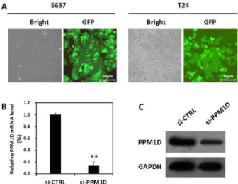
Lentivirus-mediated shRNA inhibited the expression of PPM1D in BC cells
The 5637 and T24 cell lines were infected with Lv-si-PPM1D; the highest infection efficiency was .90%, as determined by detecting the expression of green fluorescent protein (GFP) 96 h after infection (Figure 1A). Quantitative real-time PCR analysis showed that the PPM1D mRNA level was significantly lower in the si-PPM1Dgroup com-pared to the si-CTRL group (Figure 1B). The protein level of
PPM1Din the si-PPM1Dgroup was also strongly decreased compared with the si-CTRL group (Figure 1C).
Knockdown of PPM1D inhibited BC cell growth
To examine the effect ofPPM1Dknockdown on BC cell
growth, Lv-si-PPM1D- or Lv-si-CTRL-infected 5637 and T24 cells were subjected to MTT and colony formation assays. As shown in Figure 2, cell proliferation in the
si-PPM1D group was significantly inhibited compared with that in the si-CTRL group. Colony formation ability was significantly lower in the si-PPM1Dgroup compared to the si-CTRL group (Figure 3). Similar results were obtained in 5637 and T24 cells.
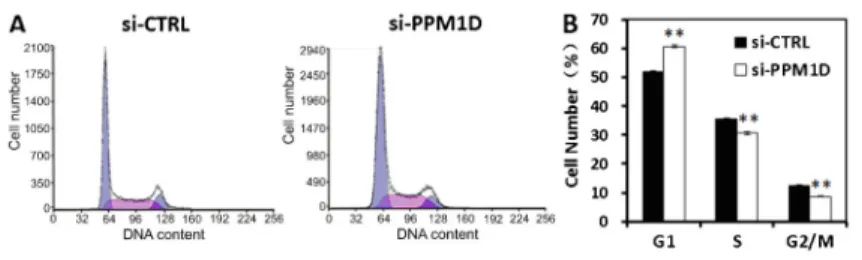
Flow cytometric analysis showed that the proportion of cells in the G1 phase was markedly increased in the
si-PPM1Dgroup compared with the si-CTRL group (Figure 4), partly explaining the growth suppression mediated by
Lv-si-PPM1D.
Knockdown of PPM1D inhibited BC tumorigenicity
in vivo
The 5637 cells infected with Lv-si-CTRL or Lv-si-PPM1D
were subcutaneously implanted into nude mice to examine
Figure 1. Knockdown of PPM1D in bladder cancer cells by lentivirus-mediated shRNA. A, Detection of lentiviral infection efficiency. The 5637 and T24 cells were infected with Lv-si-PPM1D, and phase contrast (left) or GFP (right) images were obtained 96 h after infection. Magnification: 2006.B, Analyses of
PPM1DmRNA expression in 5637 cells by quantitative real-time PCR.C, Western blot analysis ofPPM1Dprotein expression in 5637 cells. Data are reported as means±SD of three indepen-dent experiments. **P,0.01,t-test.
Figure 2.Knockdown ofPPM1Dattenuated the growth potential of bladder cancer cellsin vitro. The proliferation of 5637 (A) and T24 (B) cells was assessed by MTT assay after infection with Lv-si-CTRL or Lv-si-PPM1D. Data are reported as means±SD of three independent experiments. Lv: lentiviral; d: day. **P,0.01, compared to Lv-si-CTRL (t-test).
the effect of PPM1D knockdown on BC tumorigenicity
in vivo. All of the mice in the si-CTRL group displayed steadily and progressively growing tumors, whereas the BC cells in the si-PPM1Dgroup showed weaker tumorigenicity, and the mice developed smaller tumors (Figure 5).
Discussion
Gene dysregulation is frequently observed in cancer, and gene expression profiles vary among different cancers.
PPM1Dor Wip1 is a serine/threonine phosphatase that is overexpressed and shows oncogenic activity in multiple human cancers (10). However, the role of thePPM1Dgene in BC has not been investigated to date. To elucidate the function of PPM1D in BC, we used lentivirus-mediated RNAi to inhibitPPM1Dexpression in T24 and 5637 cells and investigated the effects ofPPM1Dknockdown in these BC cell lines. A lentiviral vector carryingPPM1DshRNA and a GFP reporter gene was constructed, which showed high infection efficiency in 5637 and T24 cells and effectively silenced PPM1D expression. These results indicated the successful construction of an effective shRNA vector targeting thePPM1Dgene.
Lentivirus-mediatedPPM1Dsilencing strongly inhibited the growth and proliferation of T24 and 5637 BC cellsin vitro, as demonstrated by MTT and colony formation
assays. The data showed that the role of PPM1Din BC was consistent with that in other cancers (16,27-29). Flow cytometric cell cycle analysis showed thatPPM1D knock-down increased the proportion of T24 and 5637 BC cells in the G0/G1 phase, indicating thatPPM1Ddownregulation blocked cell cycle progression. This could be a mechanism by whichPPM1Dsilencing suppresses proliferation.
To determine the therapeutic value of lentivirus-mediated RNAi ofPPM1Dfor BC treatment, we analyzed its effect in a xenograft model. The results showed that sh-PPM1D lentivirus inhibited 5637 BC cell proliferation and suppressed their tumorigenic potential, indicating that targetingPPM1Dmay be a potential therapeutic strategy for the treatment of BC.
Previous studies have shown that PPM1D promotes tumorigenesis in a p53-dependent manner. PPM1D is induced by p53 in response to various environmental stresses and facilitates the return of cells to the pre-stress state (6). In addition,PPM1Dinhibits p53 activity by directly dephosphorylating p53 or its regulators such as ATM,
Chk1, andChk2, which indirectly inhibit p53 activity (10,30). However, T24 and 5637 BC cells have p53 mutations, which implies that PPM1D may function in a p53-independent manner in BC cells. PPM1D is a target of p53 and other transcription factors, including the estrogen receptor-a and nuclear factor-kB (NF-kB) (9). The p38
Figure 4. Knockdown of PPM1D increases the proportion of 5637 cells in G1 phase. Cell cycle distribution was analyzed by flow cytometry. A, Representative images of three independent FACS analyses are shown.B, Proportion of cells in the different cell cycle phases. Data are reported as means±SD of three independent experiments. **P,0.01, compared to si-CTRL (t-test).
mitogen-activated protein kinase (MAPK) (31) and Akt (28) signaling pathways may be downstream mediators of
PPM1D activity in BC. Further studies are required to distinguish these mechanisms.
In conclusion, the results of the present study provided evidence thatPPM1D plays a potentially important role in BC tumorigenicity and could be a promising target for therapeutic intervention.
Acknowledgments
This research was supported by grants from the National Natural Science Foundation of China (Project No. 81272835), the Shanghai Science and Technology Commission and international cooperation projects (Project No. 11410708200), and the Shanghai Natural Science Foundation (Project No. 13ZR1405600).
References
1. Siegel R, Naishadham D, Jemal A. Cancer statistics, 2013. CA Cancer J Clin2013; 63: 11-30, doi: 10.3322/caac.21166. 2. Vianello A, Costantini E, Del Zingaro M, Bini V, Herr HW, Porena M. Repeated white light transurethral resection of the bladder in nonmuscle-invasive urothelial bladder can-cers: systematic review and meta-analysis. J Endourol 2011; 25: 1703-1712, doi: 10.1089/end.2011.0081. 3. Ku JH, Lerner SP. Strategies to prevent progression of
high-risk bladder cancer at initial diagnosis.Curr Opin Urol2012; 22: 405-414, doi: 10.1097/MOU.0b013e328356adff. 4. Vishnu P, Mathew J, Tan WW. Current therapeutic strategies
for invasive and metastatic bladder cancer. Onco Targets Ther2011; 4: 97-113.
5. Gakis G, Schwentner C, Todenhofer T, Stenzl A. Current status of molecular markers for prognostication and out-come in invasive bladder cancer.BJU Int2012; 110: 233-237, doi: 10.1111/j.1464-410X.2011.10839.x.
6. Rossi M, Demidov ON, Anderson CW, Appella E, Mazur SJ. Induction of PPM1D following DNA-damaging treatments through a conserved p53 response element coincides with a shift in the use of transcription initiation sites.Nucleic Acids Res2008; 36: 7168-7180, doi: 10.1093/nar/gkn888. 7. Douarre C, Mergui X, Sidibe A, Gomez D, Alberti P, Mailliet
P, et al. DNA damage signaling induced by the G-quadruplex ligand 12459 is modulated byPPM1D/WIP1 phosphatase. Nucleic Acids Res2013; 41: 3588-3599, doi: 10.1093/nar/ gkt073.
8. Fiscella M, Zhang H, Fan S, Sakaguchi K, Shen S, Mercer WE, et al. Wip1, a novel human protein phosphatase that is induced in response to ionizing radiation in a p53-dependent manner.Proc Natl Acad Sci U S A1997; 94: 6048-6053, doi: 10.1073/pnas.94.12.6048.
9. Lowe J, Cha H, Lee MO, Mazur SJ, Appella E, Fornace AJ Jr. Regulation of the Wip1 phosphatase and its effects on the stress response.Front Biosci2012; 17: 1480-1498, doi: 10.2741/3999.
10. Lu X, Nguyen TA, Moon SH, Darlington Y, Sommer M, Donehower LA. The type 2C phosphatase Wip1: an oncogenic regulator of tumor suppressor and DNA damage response pathways.Cancer Metastasis Rev2008; 27: 123-135, doi: 10.1007/s10555-008-9127-x.
11. Bulavin DV, Phillips C, Nannenga B, Timofeev O, Donehower LA, Anderson CW, et al. Inactivation of the Wip1 phospha-tase inhibits mammary tumorigenesis through p38 MAPK-mediated activation of the p16(Ink4a)-p19(Arf) pathway.Nat Genet2004; 36: 343-350, doi: 10.1038/ng1317.
12. Bulavin DV, Demidov ON, Saito S, Kauraniemi P, Phillips C, Amundson SA, et al. Amplification of PPM1D in human tumors abrogates p53 tumor-suppressor activity.Nat Genet
2002; 31: 210-215, doi: 10.1038/ng894.
13. Ruark E, Snape K, Humburg P, Loveday C, Bajrami I, Brough R, et al. MosaicPPM1Dmutations are associated with predisposition to breast and ovarian cancer. Nature 2013; 493: 406-410, doi: 10.1038/nature11725.
14. Tan DS, Lambros MB, Rayter S, Natrajan R, Vatcheva R, Gao Q, et al. PPM1D is a potential therapeutic target in ovarian clear cell carcinomas.Clin Cancer Res2009; 15: 2269-2280, doi: 10.1158/1078-0432.CCR-08-2403. 15. Ali AY, Abedini MR, Tsang BK. The oncogenic phosphatase
PPM1D confers cisplatin resistance in ovarian carcinoma cells by attenuating checkpoint kinase 1 and p53 activation. Oncogene2012; 31: 2175-2186, doi: 10.1038/onc.2011.399. 16. Saito-Ohara F, Imoto I, Inoue J, Hosoi H, Nakagawara A, Sugimoto T, et al.PPM1Dis a potential target for 17q gain in neuroblastoma.Cancer Res2003; 63: 1876-1883. 17. Li GB, Zhang XL, Yuan L, Jiao QQ, Liu DJ, Liu J. Protein
phosphatase magnesium-dependent 1delta (PPM1D) mRNA expression is a prognosis marker for hepatocellular carci-noma. PLoS One 2013; 8: e60775, doi: 10.1371/journal. pone.0060775.
18. Satoh N, Maniwa Y, Bermudez VP, Nishimura K, Nishio W, Yoshimura M, et al. Oncogenic phosphatase Wip1 is a novel prognostic marker for lung adenocarcinoma patient survival. Cancer Sci2011; 102: 1101-1106, doi: 10.1111/j.1349-7006. 2011.01898.x.
19. Castanotto D, Rossi JJ. The promises and pitfalls of RNA-interference-based therapeutics. Nature 2009; 457: 426-433, doi: 10.1038/nature07758.
20. Mohr SE, Perrimon N. RNAi screening: new approaches, understandings, and organisms.Wiley Interdiscip Rev RNA 2012; 3: 145-158, doi: 10.1002/wrna.110.
21. Bora RS, Gupta D, Mukkur TK, Saini KS. RNA interference therapeutics for cancer: challenges and opportunities (review).Mol Med Rep2012; 6: 9-15.
22. Seth S, Johns R, Templin MV. Delivery and biodistribution of siRNA for cancer therapy: challenges and future prospects. Ther Deliv2012; 3: 245-261, doi: 10.4155/tde.11.155. 23. Zhang H, Jiang H, Wang W, Gong J, Zhang L, Chen Z, et al.
Expression of Med19 in bladder cancer tissues and its role on bladder cancer cell growth.Urol Oncol2012; 30: 920-927, doi: 10.1016/j.urolonc.2010.10.003.
24. Pullmann R Jr, Juhaszova M, Lopez dS, I, Kawai T, Mazan-Mamczarz K, Halushka MK, et al. Enhanced proliferation of cultured human vascular smooth muscle cells linked to increased function of RNA-binding protein HuR.J Biol Chem 2005; 280: 22819-22826, doi: 10.1074/jbc.M501106200. 25. Sakoda T, Kasahara N, Hamamori Y, Kedes L. A high-titer
of differentiated cells including beating cardiac myocytes. J Mol Cell Cardiol1999; 31: 2037-2047, doi: 10.1006/jmcc. 1999.1035.
26. Zhang H, Jiang H, Wang W, Gong J, Zhang L, Chen Z, et al. Expression of Med19 in bladder cancer tissues and its role on bladder cancer cell growth.Urol Oncol2012; 30: 920-927, doi: 10.1016/j.urolonc.2010.10.003.
27. Zhang X, Wan G, Mlotshwa S, Vance V, Berger FG, Chen H, et al. Oncogenic Wip1 phosphatase is inhibited by miR-16 in the DNA damage signaling pathway.Cancer Res2010; 70: 7176-7186, doi: 10.1158/0008-5472.CAN-10-0697. 28. Yin H, Yan Z, Liang Y, Liu B, Su Q. Knockdown of protein
phosphatase magnesium-dependent 1 (PPM1D) through lentivirus-mediated RNA silencing inhibits colorectal carcinoma
cell proliferation.Technol Cancer Res Treat2013; 12: 537-543. 29. Parssinen J, Alarmo EL, Karhu R, Kallioniemi A.PPM1D silencing by RNA interference inhibits proliferation and induces apoptosis in breast cancer cell lines with wild-type p53.Cancer Genet Cytogenet2008; 182: 33-39, doi: 10.1016/ j.cancergencyto.2007.12.013.
30. Zhu YH, Bulavin DV. Wip1-dependent signaling pathways in health and diseases.Prog Mol Biol Transl Sci2012; 106: 307-325, doi: 10.1016/B978-0-12-396456-4.00001-8. 31. Demidov ON, Kek C, Shreeram S, Timofeev O, Fornace AJ,