ISSN 0100-879X
BIOMEDICAL SCIENCES
AND
CLINICAL INVESTIGATION
www.bjournal.com.br
www.bjournal.com.br
Volume 45 (3) 179-290 March 2012
Braz J Med Biol Res, March 2012, Volume 45(3) 197-204
doi:
10.1590/S0100-879X2012007500015
Cultivation and identification of colon cancer stem cell-derived
spheres from the Colo205 cell line
Ying-fei Li, Bing Xiao, San-fang Tu, Yuan-yuan Wang and Xiao-lang Zhang
Institutional Sponsors
The Brazilian Journal of Medical and Biological Research is partially financed by
Faculdade de Medicina de Ribeirão Preto Campus
Ribeirão Preto
Explore High - Performance MS Orbitrap Technology In Proteomics & Metabolomics
Cultivation and identification of colon cancer
stem cell-derived spheres from
the Colo205 cell line
Ying-fei Li
1,2, Bing Xiao
1, San-fang Tu
3, Yuan-yuan Wang
1and Xiao-lang Zhang
11Department of Gastroenterology and Institute of Gastroenterology, Nanfang Hospital,
Southern Medical University, Guangzhou, China
2Department of Gastroenterology and Hepatology, First Municipal People’s Hospital of Guangzhou,
Guangzhou Medical College, Guangzhou, China
3Hematology Department, Zhujiang Hospital, Southern Medical University, Guangzhou, China
Abstract
Our group established a method to culture spheres under serum-free culture condition. However, the biological characteristics and the tumorigenicity of spheres are unknown. Here, we demonstrate that sphere cells expressed high levels of the putative colorectal cancer stem cell markers CD133 and CD44. The CD133-positive rates were 13.27 ± 5.62, 52.71 ± 16.97 and 16.47 ± 2.45% in sphere cells, regular Colo205 cells and differentiated sphere cells, respectively, while the CD44-positive rates were 62.92 ± 8.38, 79.06 ± 12.10 and 47.80 ± 2.5%, respectively, and the CD133/CD44-double-positive rates were 10.77 ± 4.96, 46.89 ± 19.17 and 12.41 ± 2.27%, respectively (P < 0.05). Cancer sphere cells formed crypt-like structures in 3-D culture. Moreover, cells from cancer spheres exhibited more tumorigenicity than regular Colo205 cells in a xenograft assay. The cancer sphere cells displayed much higher oncogenicity than regular Colo205 cells to initiate neoplasms, as assayed by H&E stain-ing, Musashi-1 staining and electron microscopy. Our findings indicated that the sphere cells were enriched with cancer stem cells (CSCs), and exhibited more proliferation capacity, more differentiation potential and especially more tumorigenicity than regular Colo205 cells in vitro and in vivo. Further isolation and characterization of these CSCs may provide new insights for novel therapeutic targets and prognostic markers.
Key words: Colon cancer stem cells; Cancer sphere; Self-renew; Differentiation; Tumorigenicity
Introduction
Correspondence: Bing Xiao, Department of Gastroenterology and Institute of Gastroenterology, Nanfang Hospital, Southern Medical University, 510515 Guangzhou, China. E-mail: [email protected]
Received April 29, 2011. Accepted January 25, 2012. Available online February 10, 2012. Published March 19, 2012. Recent studies have shown that a small population of
undifferentiated cancer cells, identifiable by the expression
of special cell surface markers, possesses high differentia-tion potential and constant self-renewal capacity. These cancer stem cells (CSCs) are preserved and regulated by microenvironments such as tumor-associated stromal and
vascular endothelial cells (1). In vivo xenotransplantations
demonstrate the high tumorigenic potential of CSCs (2). In addition, CSCs express multiple drug-resistant proteins,
and their greater efficiency at repairing DNA reverts damage
from chemotherapy and radiation. CSCs are considered to be the root of cancer, and are responsible for cancer metastasis, recurrence and drug resistance (3). Previous studies have demonstrated CSCs in blood (4), breast (5), brain (6), pancreas (7), colon (8), liver (9), prostate (10), and
skin (11) cancers. They have also been identified in several
lines, such as glioma and breast cancer cell lines (12). The Colo205 cell line was established in 1975 by Dr.
Semple (13)from ascitic fluid obtained from a 70-year-old
Caucasian male with colon carcinoma. In the previous study, we established a method for culturing spheres under serum-free conditions (14). However, little is known about the biology and oncogenicity of cancer spheres. In the present study, we further investigated the biological characteristics and the tumorigenicity of spheres. MTT [3-(4,5-dimethylthi-azol-2-yl)-2,5-diphenyltetrazolium bromide] cell proliferation
assay and 3-D culture were performed in vitro to evaluate
198 Ying-fei Li et al.
was done by H&E staining, Musashi-1 staining and electron microscopy.
Material and Methods
Culture of colon cancer spheres and differentiation assay
Colo205 colon cancer cell lines were supplied by the
American Type Culture Collection. The serum-supplemented
medium (SSM) consisted of RPMI-1640 supplemented with 10% fetal calf serum. The serum-free medium (SFM)
was prepared from 1:1 (v/v) Dulbecco’s modified Eagle’s
medium and Ham’s F-12 nutrient mixture (DMEM/F12;
HyClone, USA), B27 supplement (1:50; Gibco, USA), 20 ng/mL epidermal growth factor (EGF; PeproTech, USA), 10 ng/mL basic fibroblast growth factor (bFGF; PeproTech), 10 ng/mL leukemia inhibitory factor (Chemicon, USA), and 2
mM L-glutamine. Colo205 cells were subcultured in SSM. Cells at the exponential growth phase were washed with PBS and digested with trypsin, followed by resuspension in SFM. Living cells were counted by Trypan blue
exclu-sion and subcultured in SFM at a concentration of 5 x 10⁵/
mL. After cancer spheres were generated, EGF and bFGF
were removed from the culture medium and 10% serum was added to induce differentiation. Cell morphology was observed with a light microscope.
Cell proliferation assays
Undifferentiated sphere cells, differentiated sphere cells and regular Colo205 cells were plated in 0.1 mL volumes of SFM, SSM, and SSM, respectively, at a density of 1000 cells/well in 96-well microwell plates. Cell proliferation as-says were performed on days 0, 2, 4, 6, and 8 using the
MTT method (Sigma, USA). Quantification of viable cells
by measuring absorption spectra at 575 nm was performed with a Versamax microplate reader.
Detection of the surface marker of cancer spheres
Cells were collected separately from colon cancer spheres, post-differentiated sphere cells and regular Colo205 cells by trypsin digestion, followed by washing and
resuspending in PBS at a concentration of 5 x 10⁶/mL. Cells
were incubated with fluorescein isothyocyanate
(FITC)-conjugated anti-CD44 and phycoerythrin (PE)-(FITC)-conjugated anti-CD133 monoclonal antibodies for 30 min at 4°C. The
fluorescence intensity was measured by flow cytometry.
3-D culture
When typical colon cancer spheres were generated, cells were mechanically and enzymatically dissociated and resuspended in SSM, and the concentration was adjusted
to 6 x 10⁴/mL. The regular Colo205 cells and differentiated
sphere cells at the same concentrations were used as controls. 3-D culture was conducted using Matrigel matrix
(BD, USA) to observe crypt-like structures. Briefly, cells
were mixed 1:1 (v/v) with Matrigel and 0.5-mL aliquots were subsequently plated onto 24-well plates. The Matrigel was incubated at 37°C for 30 min, followed by the addition of 1 mL SFM. The medium was changed every 3 days by replacing 0.5 mL spent medium with fresh medium.
Subcutaneous xenograft assay in NOD/SCID mice
All animal experiments were approved by the Animal
Ethics Committee of Southern Medical University and the protocol of animal treatment was approved by the Institutional
Animal Care and Use Committee. To study the tumorigenic
potential of CSCs isolated from Colo205 cells, cancer spheres were mechanically and enzymatically dissociated into
single-cell suspensions in PBS at concentrations of 1 x 10⁵, 5 x
105, 1 x 10⁶, and 1 x 10⁷/mL. Cells were mixed 1:1 (v/v) with
Matrigel and injected subcutaneously into one side of the
flanks of NOD/SCID mice (Shanghai Si Laike Experimental Animals, China). Regular Colo205 cells were injected into the
other side as a control. Fifteen days after the injection, mice
were sacrificed, tumor nodules were immediately removed, fixed in 10% phosphate-buffered formalin, and embedded in paraffin. Four micrometer-thick sections were cut, mounted
on poly-L-lysine-coated slides (Sigma), dried overnight at 37°C, dewaxed in xylene, rehydrated according to standard histopathological procedure, and stained with H&E.
To determine Musashi-1 immunoreactivity, we used heat-induced epitope retrieval methods. The antigen was
retrieved with 1.0 mM EDTA repair fluid, pH 8.0, via micro -wave heating (high power for 5 min, followed by moderate power for 15 min). Sections were incubated overnight at 4°C with anti-human Musashi-1 antibody (1:200; Chemicon). Immunodetection was performed using an egg protein-biotin-horseradish peroxidase kit (Sigma).
For transmission electron microscopy, the fresh tumor
was cut into 1 x 1 x 1-mm3 cubes, fixed with fresh pre-cooled
2.5% glutaraldehyde for 72 h at 4°C, followed by incubation
in 1% osmium acid for 2 h in the dark. After dehydration with
acetone, immersion and embedding, thin sections were cut and stained with uranyl acetate and lead citrate.
Results
Culture of colon cancer spheres and differentiation assay
A small proportion of Colo205 cells (~5%) survived in
SFM containing a growth factor after 7 days as measured by Trypan blue exclusion, and formed suspended colon cancer spheres. The refractile sphere cells were intensely stained, and cells were closely connected. New spheres were generated next to existing spheres, demonstrating their self-renewal capacity. Cancer spheres were passaged
through at least eight generations in SFM.When the SFM
Cell proliferation assays
To evaluate the proliferative capacity of cancer sphere cells, cells were plated at 1000 cells/well, and the number of
viable cells was quantified on days 0, 2, 4, 6, and 8 by the MTT
assay. Undifferentiated sphere cells in SFM demonstrated increased proliferative capacity compared to differentiated sphere cells and regular Colo205 cells in SSM (Figure 2).
Detection of the surface marker of cancer spheres
Cells in cancer spheres expressed high levels of CD133 and CD44 in SFM (Figure 3). However, when the SFM was replaced with SSM for 10 days, the proportion of CD133
and CD44 decreased significantly (Figure 4).
3-D culture
The 3-D in vitro cell cultivation system provides the
physiological signals necessary for colon cancer morpho-genesis, i.e., the differentiation potential of sphere cells
to recapitulate in vivo spatial orientation and crypt-like
architecture. Differences between cancer spheres and the
control groups were not notable on the first day. Typical
crypt-like structures emerged from cancer spheres on day
18. A tubular ring interconnected structure and overlapping
upward growth layers were formed in the colon cancer spheres, while the control groups did not form any crypt-like structure. These results demonstrated that colon cancer
spheres had a stronger proliferation and differentiation potential than regular Colo205 cells (Figure 5).
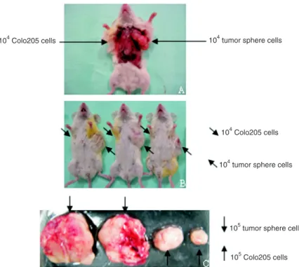
In vivo injection of cancer sphere cells
To assess the tumorigenicity of cancer sphere cells,
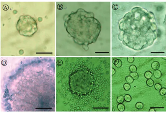
Figure 1. Cell morphology observed with an inverted microscope. A, Cancer sphere at first passage; B, cancer sphere at passage 3; C, cancer sphere at passage 8; D, cells are closely connected within spheres; E, one day after supplementation with fetal bovine serum (FBS); F, ten days after supplementa-tion with FBS. Magnificasupplementa-tion bars: 50 μm (A,D); 25 μm (B,C,F); 100 μm (E).
200 Ying-fei Li et al.
we injected the cells into NOD/SCID mice. The xenograft
transplantation assay showed that 104 colon cancer sphere
cells were sufficient to induce tumorigenesis, while the
same amount of regular Colo205 cells failed to induce vis-ible tumors 15 days after injection. The volume of tumors
generated by 105 colon cancer sphere cells was significantly
greater than the tumor volume of the control group, indi-cating that colon cancer spheres had high tumorigenicity (Table 1 and Figure 6). The histochemical assay showed
that the tumors generated from sphere cells and regular Colo205 cells had a similar differentiation structure. The intestinal Musashi-1-positive stem cells were similar in the two types of tumor. Similar malignant phenotypes were also observed in transmission electron micrographs of both xenografts (Figure 7).
Discussion
A long-term study (15) reported that EGF and bFGF
have a synergistic effect on proliferation of neural stem
cells in vitro. Martens et al. (15) found that FGF-responsive
neural stem cells cultured in SFM containing EGF and bFGF divided asymmetrically to reproduce themselves and produced EGF-responsive progenitor stem cells. The EGF-responsive population increased in size by asym-metric divisions of FGF-responsive cells and by symasym-metric divisions of EGF-responsive stem cells. Thus, with certain
growth factors, neural stem cells could proliferate in vitro and
maintain their pluripotency potential. Similar findings have
been obtained in brain tumors and epithelial cancer, such as
glioma(6) and colon cancer (8), respectively. Once cancer
cells were cultured in SFM containing EGF and bFGF, most of the differentiated cells could not survive. However, a few undifferentiated cells, i.e., CSCs, were able to form cancer spheres and to maintain an undifferentiated state.
In the present study, only a small fraction of cells sur-vived in SFM with EGF and bFGF, proliferated and formed suspended cancer spheres. Over a prolonged culture pe-riod, cancer spheres became larger and larger, and could
Figure 3. Flow cytometry dot plotsshow that cancer sphere cells expressed high levels of CD133 and CD44 in serum-free medium. Left, Isotype control. Right, Cancer sphere cells incubated with FITC-conju-gated anti-CD44 and PE-conjuFITC-conju-gated anti-CD133 monoclonal antibodies. FITC = fluorescein isothyocya -nate; PE = phycoerythrin.
Figure 4. Expression of CD133 and CD44 in regular Colo205 cells cultured in serum-supplemented medium (SSM), undifferen-tiated cells from cancer spheres cultured in serum-free medium (SFM), and differentiated cells from cancer spheres cultured in SSM. Data are reported as means ± SD. #P < 0.05, differentiated
passage into at least eight generations, evidence of their self-renewal capacity and differentiation potential.
A phenotype study was performed to analyze the
expression of CD133 and CD44 on cells from cancer
spheres. CD133 is a 120-kDa five-transmembrane domain
glycoprotein expressed on normal primitive hematopoi-etic, endothelial, neural, and epithelial cells. This molecule shows a highly homologous structure from nematodes to
Homo sapiens. The specific
function of CD133 is unclear (16). Recently, it has been considered to be a CSC sur-face marker for brain tumors
(6),and pancreatic (7), colon
(8), liver (9), and prostate (10) cancers.
CD44 is the receptor for hyaluronic acid and belongs to a type of adhesion molecule. CD44 is involved in a variety of signaling pathways, medi-ated cells and cell connections
(17). CD44+ cancer cells are
regarded as more primitive, and also as a stem cell marker for cancers of the breast (5), pancreas (7), prostate (10), head and neck squamous cell carcinoma (18), and
colon (19,20). CD44+ colon
polyp cells have been found to express high levels of the intestinal stem cell marker Musashi-1 (21).
In the present study, the expression of CD133 and CD44 in colon cancer spheres
was significantly higher than
in regular Colo205 cells cul-tured in SSM. When SFM was replaced with SSM to induce differentiation, the expression of CD133 and CD44 in cells from cancer spheres was
significantly decreased, sug -gesting that CD133 and CD44 were primarily expressed on undifferentiated rather than differentiated cancer cells. We concluded that there was a small population of CSCs that persisted in the Colo205 cell line. These cells formed cancer spheres in SFM and could maintain an undifferenti-ated stage.
202 Ying-fei Li et al.
Two groups have recently reported the biology of colon cancer sphere cells in a 3-D culture system (22,23). We also studied the biological characteristics of cancer spheres
cultured in vitro using a Matrigel matrix. The Matrigel matrix
is a solubilized basement membrane preparation extracted from mouse sarcoma, which plays a role in adhesion and differentiation of epithelial cells. Cancer spheres survived in the 3-D culture and formed crypt-like structures, while the regular Colo205 cells and post-differentiated cancer cells failed to proliferate. This suggests that cancer spheres
have a high differentiation potential and strong proliferation capacity. It is noteworthy that in this study the basement membrane analogues of the Matrigel matrix induced cancer differentiation, indicating the importance of the microenviron-ment in maintaining an undifferentiated state and the high capacity of CSCs for self-renewal. Thus, drugs targeted to a microenvironment may also be effective in killing or inducing differentiation of CSCs.
Tumorigenicity is considered to be an indispensable
prerequisite for fulfilling the definition of CSCs, which should
also possess self-renewal and differentiation capacities (24). The xenograft transplantation assay showed that
cancer spheres are of high tumorigenicity. As few as 104
colon cancer sphere cells induced evident cancer, while the same number of regular Colo205 cells did not show any tumorigenicity. This indicates that cancer spheres were enriched with colon CSCs, which had strong proliferative capacity. In contrast, most of the regular Colo205 cells are differentiated, with only limited proliferative capacity and therefore displayed less tumorigenicity.
H&E analysis showed a similar differentiation structure between the two types of xenografts. Both types expressed the intestinal stem cell marker Musashi-1 at similar levels. Similar malignant phenotypes were also found by transmis-sion electron microscopy in both xenografts. Thus, although the degree of tumorigenicity varied greatly between the
Figure 6. Immunodeficient mouse transplantation assays. A and B, Colon cancer sphere cells (104) induced neoplasms, while the same number of regular Colo205
cells did not show any tumorigenicity. C, Cancer sphere cells (104) generated a
larger neoplasm than the same number of regular cancer cells.
Table 1.In vivo tumorigenicity of cancer sphere cells and regular Colo205 cells in NOD/SCID mice.
Cell No.
1 x 10⁴ 5 x 10⁴ 1 x 10⁵ 1 x 10⁶
Cancer sphere cells 6/8 8/8 8/8 8/8 Regular Colo205 cells 0/8 0/8 6/8 8/8
Cells were assayed for the ability to form tumors after subcutane-ous injection into the flank of NOD/SCID mice at 1 x 10⁴, 5 x 10⁴,
1 x 10⁵, and 1 x 10⁶ cells per injection. The analysis was
cancer spheres and regular Colo205 cells, the phenotypes of tumors generated from these two groups of cells were the same.
In summary, the sphere cells exhibited more
prolifera-tion capacity and more differentiaprolifera-tion potential in in vitro
cell proliferation assays and 3-D culture assays. Moreover, cancer sphere cells displayed more tumorigenicity than regular Colo205 cells in xenograft transplantation assays.
Our findings may provide new insights into the tumorigenesis
of colon cancer and offer a potential target for anti-cancer therapy.
Acknowledgments
The authors thank Medjaden Bioscience Limited for
assisting with the preparation of this manuscript.
204 Ying-fei Li et al.
References
1. Gilbert CA, Ross AH. Cancer stem cells: cell culture, markers, and targets for new therapies. J Cell Biochem 2009; 108: 1031-1038.
2. Ferrand A, Sandrin MS, Shulkes A, Baldwin GS. Expression of gastrin precursors by CD133-positive colorectal cancer cells is crucial for tumour growth. Biochim Biophys Acta
2009; 1793: 477-488.
3. Ward RJ, Dirks PB. Cancer stem cells: at the headwaters of tumor development. Annu Rev Pathol 2007; 2: 175-189. 4. Lapidot T, Sirard C, Vormoor J, Murdoch B, Hoang T,
Caceres-Cortes J, et al. A cell initiating human acute my -eloid leukaemia after transplantation into SCID mice. Nature
1994; 367: 645-648.
5. Al-Hajj M, Wicha MS, Benito-Hernandez A, Morrison SJ, Clarke MF. Prospective identification of tumorigenic breast cancer cells. Proc Natl Acad Sci U S A 2003; 100: 3983-3988.
6. Singh SK, Clarke ID, Terasaki M, Bonn VE, Hawkins C, Squire J, et al. Identification of a cancer stem cell in human brain tumors. Cancer Res 2003; 63: 5821-5828.
7. Lee CJ, Dosch J, Simeone DM. Pancreatic cancer stem cells. J Clin Oncol 2008; 26: 2806-2812.
8. Ricci-Vitiani L, Lombardi DG, Pilozzi E, Biffoni M, Todaro M, Peschle C, et al. Identification and expansion of human colon-cancer-initiating cells. Nature 2007; 445: 111-115. 9. Suetsugu A, Nagaki M, Aoki H, Motohashi T, Kunisada T,
Moriwaki H. Characterization of CD133+ hepatocellular carcinoma cells as cancer stem/progenitor cells. Biochem Biophys Res Commun 2006; 351: 820-824.
10. Collins AT, Berry PA, Hyde C, Stower MJ, Maitland NJ. Pro -spective identification of tumorigenic prostate cancer stem cells. Cancer Res 2005; 65: 10946-10951.
11. Schatton T, Murphy GF, Frank NY, Yamaura K, Waaga-Gasser AM, Waaga-Gasser M, et al. Identification of cells initiating human melanomas. Nature 2008; 451: 345-349.
12. Setoguchi T, Taga T, Kondo T. Cancer stem cells persist in many cancer cell lines. Cell Cycle 2004; 3: 414-415. 13. Semple TU, Quinn LA, Woods LK, Moore GE. Tumor and
lymphoid cell lines from a patient with carcinoma of the colon for a cytotoxicity model. Cancer Res 1978; 38: 1345-1355. 14. Li YF, Xiao B, Lai ZS, Tu SF, Wang YY, Zhang XL. [Spheres
isolated from Colo205 cell line possess cancer stem-like cells under serum-free culture condition]. Nan Fang Yi Ke Da Xue Xue Bao 2008; 28: 236-240.
15. Martens DJ, Tropepe V, van Der Kooy D. Separate prolif-eration kinetics of fibroblast growth factor-responsive and epidermal growth factor-responsive neural stem cells within the embryonic forebrain germinal zone. J Neurosci 2000; 20: 1085-1095.
16. Mizrak D, Brittan M, Alison MR. CD133: molecule of the mo -ment. J Pathol 2008; 214: 3-9.
17. Schmidt DS, Klingbeil P, Schnolzer M, Zoller M. CD44 vari-ant isoforms associate with tetraspanins and EpCAM. Exp Cell Res 2004; 297: 329-347.
18. Prince ME, Sivanandan R, Kaczorowski A, Wolf GT, Kaplan MJ, Dalerba P, et al. Identification of a subpopulation of cells with cancer stem cell properties in head and neck squamous cell carcinoma. Proc Natl Acad Sci U S A 2007; 104: 973-978.
19. Dalerba P, Dylla SJ, Park IK, Liu R, Wang X, Cho RW, et al. Phenotypic characterization of human colorectal cancer stem cells. Proc Natl Acad Sci U S A 2007; 104: 10158-10163.
20. Horst D, Kriegl L, Engel J, Kirchner T, Jung A. Prognostic significance of the cancer stem cell markers CD133, CD44, and CD166 in colorectal cancer. Cancer Invest 2009; 27: 844-850.
21. Schulenburg A, Cech P, Herbacek I, Marian B, Wrba F, Va -lent P, et al. CD44-positive colorectal adenoma cells express the potential stem cell markers musashi antigen (msi1) and ephrin B2 receptor (EphB2). J Pathol 2007; 213: 152-160. 22. Vermeulen L, Todaro M, de Sousa Mello F, Sprick MR,
Kem-per K, Perez Alea M, et al. Single-cell cloning of colon cancer stem cells reveals a multi-lineage differentiation capacity.
Proc Natl Acad Sci U S A 2008; 105: 13427-13432. 23. Yeung TM, Gandhi SC, Wilding JL, Muschel R, Bodmer WF.
Cancer stem cells from colorectal cancer-derived cell lines.
Proc Natl Acad Sci U S A 2010; 107: 3722-3727.