Faculdade de Medicina de Ribeirão Preto Campus
Ribeirão Preto
Institutional Sponsors
The Brazilian Journal of Medical and Biological Research is partially financed by
Braz J Med Biol Res, December 2009, Volume 42(12) 1242-1247
Pattern of functional antibody activity against Haemophilus
influenzae type b (Hib) in infants immunized with
diphtheria-tetanus-pertussis/Hib Brazilian combination vaccine
D.C.S. Matos, A.M.V. Silva, P.C.C. Neves, R.M. Martins, A. Homma and R. Marcovistz
ISSN 0100-879X
BIOMEDICAL SCIENCES
AND
CLINICAL INVESTIGATION
www.bjournal.com.br
www.bjournal.com.br
Pattern of functional antibody activity against
Haemophilus influenzae
type b (Hib) in infants
immunized with diphtheria-tetanus-pertussis/Hib
Brazilian combination vaccine
D.C.S. Matos
1, A.M.V. Silva
1, P.C.C. Neves
1, R.M. Martins
2,
A. Homma
3and R. Marcovistz
11Laboratório de Tecnologia Imunológica, 2Assessoria Clínica, Bio-Manguinhos, 3Diretoria de Bio-Manguinhos, Fiocruz, Rio de Janeiro, RJ, Brasil
Abstract
We evaluated the functional activity of Haemophilus influenzae B (Hib) antibodies elicited in a group of infants immunized with the diphtheria-tetanus-pertussis vaccine combined with an Hib vaccine produced totally in Brazil after technological transfer of Hib vaccine production from Glaxo SmithKline, Belgium. Blood samples from immunized infants (N = 985) were collected for the determination of Hib antibodies. Total Ig and IgM and IgG subclasses of antibodies against polyribosyl ribitol phosphate (PRP)
were analyzed by ELISA. Almost all vaccinees (97.56%, 961/985) developed a strong anti-PRP IgG antibody response (≥1.0 μg/mL), while an anti-PRP IgM response was observed in 64.24% (634/985) of them (≥0.15 μg/mL). Only 18.88% (186/985) of the infants in the group with high PRP antibody IgG concentrations (≥1.0 μg/mL) developed a high IgM antibody response. Anti-PRP IgG antibody levels were significantly higher than anti-PRP IgM. These results demonstrate the predominance of
IgG antibodies over IgM antibodies in response to PRP, with a ratio of 17:1. IgG antibodies were predominantly of the IgG1 subclass. An increase in IgG avidity was also observed during the course of immunization.
Key words: DTP-Hib vaccine; Haemophilus influenzae type b; Antibody avidity; PRP immune response
Introduction
Correspondence: R. Marcovistz, Bio-Manguinhos, Fiocruz, Av. Brasil, 4365, Pavilhão Rocha Lima, 21040-360 Rio de Janeiro, RJ,
Brasil. Fax: +55-21-2260-4727. E-mail: rugimar@bio.fiocruz.br
Research supported by FINEP, Ministério da Ciência e Tecnologia, Bio-Manguinhos-Fiocruz.
Received May 20, 2009. Accepted August 19, 2009. Available online November 6, 2009. Published December 4, 2009. Since 1998 the WHO has recommended that
Haemo-philus influenzae type b (Hib) vaccination be included in routine infant immunization programs, according to country capacities, preferably as a diphtheria-tetanus-pertussis (DTP)-based combination (1). However, the immunologi-cal response, which insures the improved immunogenicity and priming induced by conjugated vaccines, is still only partially understood. It is known that IgG and IgM antibod-ies against the capsular polysaccharide polyribosyl ribitol phosphate (PRP) are very important for the protective im-munity to Hib, and that the covalent linkage of PRP to any protein is also important for the enhanced immunogenicity of glycoconjugated vaccines (2-4).
The response to conjugated vaccine is known to be thy-mus dependent (T cell dependent), thus permitting memory responses even in infants. In contrast, molecules such as
PRP, a thymus-independent class 2 antigen (TI-2 antigen), can activate only mature B cells, preventing infants from producing antibodies against polysaccharide epitopes in an
efficient manner. The immune systems of young children are
immature and do not respond effectively to carbohydrate
antigens before about 5 years of age. On the other hand,
most physiological TI-2 responses occur in the spleen, the marginal zone B cells, which are rare at birth and which accumulate with age (5-11). Thus, the development of vac-cines with PRP conjugated with a protein carrier was very important and has reduced the recommended minimum age of 5 years to 6 months for this vaccination (5).
vac-Activity of anti-PRP antibody induced by DTP-Hib vaccine 1243
www.bjournal.com.br Braz J Med Biol Res 42(12) 2009
cines are available, according to the carrier protein: conjugation with diphtheria toxoid (PRP-D), or an avirulent mutant of the diphtheric toxin (PRP-CRM), PRP conjugated with tetanus toxoid (PRP-T), and PRP conjugated with the outer membrane protein of Neisseria meningitidis (PRP-OMP) (12-14).
Conjugated vaccines have not only been able to induce an immune response in young infants, but have also been able to induce a memory immune response and to promote a secondary boost response with either conjugated or unconju-gated PRP vaccines. Vaccine trials have shown the ability of the conjugated vaccines to activate and prime the previously immature, non-responsive immunity of infants (15,16).
In 2006, a randomized, double-blind study was designed to evaluate the immunogenicity, safety and consistency of production of a combined DTP-Hib vaccine totally produced in Brazil (DTP/Hib-BM) after a technological transfer of Hib production from Glaxo SmithKline (GSK), Belgium.
Overall, 1000 infants were recruited and received vac -cine doses at 2, 4, and 6 months of age. Regarding the Hib
component, there was 100% seroprotection (≥0.15 μg/mL),
and there was no clinically relevant difference in reactogenicity between the 3 lots of Brazilian DTP/Hib studied and the refer-ence vaccine lot with Hib produced by GSK (17).
The objective of the present study was to analyze the classes and subclasses as well as the functional activity of antibodies to Hib elicited in infants immunized with DTP/Hib-BM or DTP/Hib-GSK vaccines.
Subjects and Methods
Subjects and vaccines
This study is a follow-up of a previously reported clinical trial in which the immunogenicity, reactivity and consistency
of production of DTP/Hib-BM was assessed (17). One thou -sand infants were immunized according to the immunization program of the Brazilian Health Ministry in a double-blind and randomized study approved by the National Research
Eth-ics Committee, the EthEth-ics Committee of Fundação Oswaldo
Cruz and of the Municipal Health Secretary of Rio de Janeiro, and by the National Regulatory Agency. The protocol was registered under Standard Randomized Clinical Trial Number 67676353 (17).
Serum samples
Blood samples from 985 infants were collected for evalu-ation of Ig, IgG, and IgM PRP antibodies. In a subset of 300 infants, randomly chosen sera collected before and 1 month after the 3rd vaccine dose were used in comparative stud-ies. Serum samples from the same infant were tested in parallel.
Antibody assay
IgG, IgM, and IgG subclasses of antibodies against
PRP were analyzed by ELISA (3-6). Briefly, each well of a
microplate (Maxisorb; Nunc, USA) was coated with 0.4 µg/
mL PRP-tyramine antigen in 0.1 mL 0.05 M carbonate buf-fer, pH 9.6, overnight at 4°C. Plates were washed with PBS plus 0.05% Tween 20 (PBS-Tween), and blocked with 0.1 mL PBS containing 0.5% bovine serum albumin (BSA) for 1 h at 37°C. Sera were serially diluted 2-fold in 0.5% PBS-Tween-BSA starting at 1:10 dilution and up to 1:1280 dilution and the plates were incubated for 1 h at 37°C. After washing three times with PBS-Tween, 0.1 mL/well of a 1/500 dilution of mouse anti-human IgG conjugated with horseradish per-oxidase (HRP, BD-Pharmingen, USA) or a 1/30,000 dilution of biotinylated mouse anti-human IgM (BD-Pharmingen) in PBS-Tween-BSA was added and the plates were incubated again for 1 h at 37°C. For IgM antibody detection, a strepta-vidin HRP-conjugate (BD-Pharmingen) was added to the plates for 1 h at 37°C. IgG1 and IgG2 anti-PRP antibody
titers were determined using specific monoclonal antibodies
(BD-Pharmingen). Next, the tetramethylbenzidine substrate (Sigma, USA) was added and the reaction stopped with 2 M NaOH 15 min later. Absorbance was measured at 450 nm with an ELISA microreader (Sunrise; Tecan, Germany). Standard curves were generated using human IgG, IgG1, and IgG2 and IgM immunoglobulins. The ELISA antibody titer curves for each serum specimen were analyzed by a
four-parameter logistic curve-fitting technique (Softmax Pro
version 5, Molecular Devices, USA). The minimum level of detection of this assay was estimated to be 0.10 µg/mL Ig at a 1:10 dilution of the tested serum.
IgG antibody avidity assay
Anti-PRP IgG avidity was measured by ELISA by the
method of Agbarakwe et al. (16) with few modifications (6). Briefly, plates were coated with PRP and saturated as ex -plained above, duplicate serum samples were then diluted to 1:20 in PBS-Tween-BSA and plates were incubated at 37°C for 1 h followed by three washes with PBS-Tween. Next, 100 µL 3.0 M urea was added to only one of the serum dilution rows, while 100 µL PBS-Tween was added to the other serum dilutions, corresponding to the control without urea. After 15 min of incubation, plates were washed with PBS-Tween for removal of both the unbound antibodies and the chaotropic agent by washing. The remaining bound IgG antibodies were detected with peroxidase-conjugated anti-human IgG in a manner similar to that described above.
The avidity index (AI) was calculated as the mean absor-bance of reactions in which the antibodies were exposed to urea divided by the mean absorbance of reactions in which the antibodies were not exposed to urea, and expressed as percentage. The avidity assay could not be evaluated for sera with <0.5 µg/mL IgG anti-PRP antibody. Samples with an AI below 49% were considered to have low avidity and samples with an AI of 50 to 79% and of more than 80% were considered to have medium and high avidity, respectively.
Statistical analyses
compare anti-PRP antibody titers and AI between vaccina-tion time points (before and 1 month after the 3rd dose). A P
value of <0.05 was considered to be statistically significant. Spearman’s correlation coefficient was used to evaluate the
relationship between IgG subclasses and IgG avidity.
Results
Anti-PRP classes and subclass antibodies
Anti-PRP Ig total, IgG and IgM antibody titers were evaluated in serum samples obtained from children 1 month after the 3rd dose of DTP-Hib vaccine and are summarized in Table 1. All infants were seroprotected against PRP-T after the complete vaccination schedule, and 97.25% (958/985) of them presented high concentrations of total
anti-PRP Ig (≥1.0 μg/mL). Almost all vaccinees (97.56%,
961/985) developed a strong IgG anti-PRP antibody
re-sponse (≥1.0 μg/mL), while an anti-PRP IgM rere-sponse was observed in 64.24% (634/985) of them (≥0.15 μg/mL). Only 18.88% (186/985) of the infants developed high levels
of IgM antibody in the group with high PRP antibody IgG
concentrations (≥1.0 μg/mL). The levels of anti-PRP IgG antibodies were significantly higher than the levels of
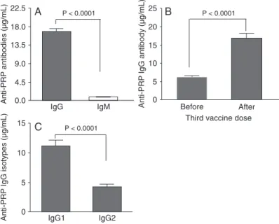
anti-PRP IgM antibodies (P < 0.0001), with IgG levels ranging from 0.17 to 163 µg/mL (mean = 16.91 ± 0.62) and IgM levels ranging from 0.10 to 18 µg/mL (mean = 0.88 ± 0.05; Figure 1A). These results demonstrate the predominance of IgG antibodies over IgM antibodies in response to PRP, with a ratio of 17:1.
Figure 1B shows the geometric means (GM) of specific
IgG levels before and one month after the 3rd vaccine dose
in all volunteers. As expected, a significant increase (P <
0.0001) of PRP IgG levels was observed one month after the 3rd vaccine dose (GM = 10.16 µg/mL, 95%CI= 15.6-18.12
µg/mL) compared to PRP IgG levels before the last dose of vaccine (GM = 5.03 µg/mL, 95%CI = 5.87-6.63 µg/mL).
The DTP-Hib vaccine induced a definitely predominant
IgG1 response since 90% of IgG antibodies belonged to
the IgG1 subclass. Anti-PRP IgG1 levels were significantly
higher than anti-PRP IgG2 levels (11.13 ± 0.51 vs 4.23 ± 0.24 µg/mL, P < 0.0001) as shown in Figure 1C.
IgG antibody avidity
There was an increase in IgG antibody avidity during the course of immunization. In fact, the IgG AI were
sig-nificantly higher in sera obtained from infants one month
after the complete schedule of vaccination than in sera obtained before the 3rd vaccine dose (P < 0.0001), as shown in Figure 2A. The avidity percentage of absorbance values in sera, following 2 doses of DTP-Hib vaccine was 70% (95%CI= 68-73%). After the 3rd dose, the mean IgG AI in the group was 80% (95%CI= 78-83%), indicating a
significant increase (P < 0.0001).
The pattern antibody avidity distribution induced against Hib during the immunization process presented different
Figure 1. Concentration of anti-PRP antibodies in
sera of immunized children. The bars indicate the
geometric mean of antibody levels and 95% confi -dence intervals. Statistical comparisons were made with the Wilcoxon test. A, Comparison of anti-PRP IgG and IgM concentrations in the sera one month after the 3rd DTP/Hib vaccine dose. B, Comparison of anti-PRP IgG concentration before and one month after the 3rd DTP/Hib vaccine dose. C, IgG subclass-es one month after the 3rd DTP/Hib vaccine dose. PRP = polyribosyl ribitol phosphate; DTP/Hib = Diph-theria-tetanus-pertussis/Haemophilus influenzae B.
Table 1. Anti-PRP antibodies in infants one month after the third
DTP/Hib vaccine dose.
Anti-PRP antibody concentration (µg/mL)
Ig (%)
IgG (%)
IgM (%)
≤0.14 0.00 0.20 16.75
0.15-0.99 2.75 2.24 64.37
≥1.00 97.25 97.56 18.88
Activity of anti-PRP antibody induced by DTP-Hib vaccine 1245
www.bjournal.com.br Braz J Med Biol Res 42(12) 2009
profiles of avidity intensity before and after the 3rd dose
(Figure 2B).
Discussion
In this study, we quantitatively and qualitatively evaluated the specific anti-Hib response induced by the Hib PRP-T
conjugated vaccine during a clinical trial of the DTP-Hib vaccine produced by Bio-Manguinhos/Fiocruz, Brazil, after technological transfer from Glaxo-SmithKline.
High-affinity antibodies are required to neutralize the
infectivity of most pathogens. Therefore, effective vaccines
against these microorganisms must induce affinity matura -tion and memory B cell forma-tion, and both these events will occur only if the vaccines are able to activate T helper cells. This concept has been applied to the design of vac-cines against some bacterial infections in which the target antigen is a capsular polysaccharide, which is incapable of stimulating T cells. This is also the case of the polysac-charide from Hib which, when covalently linked to tetanus
toxoid, forms the equivalent of an hapten-carrier conjugate,
which does activate T helper cells. Thus, the antibody
response induced by this conjugate vaccine requires the participation of T cells that may consequently stimulate the subclass switching, affinity maturation and memory
with long-lived protective immunity in B lymphocytes. In contrast, the antibodies produced in the absence of T helper cells by polysaccharide vaccines not conjugated to
proteins are generally of low affinity and consist mainly of
IgM and IgG2 (18).
The humoral immune response to Hib conjugated vac-cines is usually evaluated by measuring serum anti-PRP antibodies and by the AI. Avidity of anti-PRP IgG has been tested by ELISA-based elution assays (4,19-24), which have demonstrated that PRP conjugates elicit high-avidity antibodies, with increased functional activity (4,18).
We observed here that 97.25% of the infants immunized with DTP-Hib produced high levels
(≥1.0 µg/mL) of PRP IgG antibodies, considered
to be a serological surrogate of long-term protec-tion. Some researchers have already established that protection from Hib disease is correlated with the presence of antibodies to PRP, and have considered 0.15 µg/mL of anti-PRP to be the minimum level of antibodies for short-term protec-tion against Hib disease and 1 µg/mL to be the minimum level for long-term protection (25,26). Regarding the IgM antibody, our results showed that almost all vaccinees produced anti-PRP IgM, but at low levels compared to anti-IgG antibodies. The production of IgM antibodies against PRP induced by DTP-Hib, although small, plays an important role in opsonization and phagocytosis by activation of the classical complement path-way, an important defense mechanism against bacteria with polysaccharide-rich capsules, such as Haemophilus. The IgM antibody, due to its pentameric
structure, is a more efficient complement-binding antibody
than IgG.
When we assessed the subclasses of IgG we observed that the vaccinees responded with IgG anti-PRP of both
IgG1 and IgG2 subclasses, but with a significant predomi -nance of anti-PRP IgG1. We observed a good relationship between IgG and IgG1 antibody levels, but no correlation
was found with IgG2 levels. Our results agree with those
obtained with different PRP conjugates by Granoff et al. (27),
who also reported that infants immunized with PRP-OMP
respond predominantly with anti-PRP IgG1, while adults immunized with unconjugated PRP or PRP-D respond with anti-PRP of both IgG1 and IgG2 subclasses, as is the case for the DTP-Hib vaccine. Hetherington et al. (28) have also suggested that the IgG subclass response to PRP protein
conjugate is age independent. Other investigators have also
reported that IgG1 antibodies are more effective than IgG2 antibodies for Hib in an opsonization test and in activating complement-mediated bacteriolysis (29-32).
The present results demonstrate that antibody avidity was increased after the third immunization with the DTP-Hib vaccine. The increased AI between the 4th and 7th month of age indicates that memory responses must have occurred during DTP-Hib vaccination in Brazilian infants. These results might be due to the vaccine reactivation of earlier induced memory B cells, as reported by Hougs et al. (33). Goldblatt et al. (18) have demonstrated that antibody avidity was lower in infants with anti-PRP antibody concentrations below 1.0 µg/mL, indicating the absence of priming when antibody concentrations are low. Amir et al. (29) also evalu-ated the functional antibody activity of vaccines induced by Hib and found the avidity of antibodies to be an essential factor for serum bactericidal activities.
The increase in avidity reflects the process of somatic
Figure 2. Avidity index and distribution of PRP IgG antibodies in serum
samples from children receiving DTP-Hib vaccination. A. Avidity index before and 1 month after the 3rd DTP-Hib vaccine dose in 300 infants. Statistical comparison was made with the Wilcoxon test. B. Distribution of the avidity of anti-PRP IgG antibodies in a group of 300 infants immunized with DTP/Hib.
Open and filled bars represent blood sampling before and 1 month after the
hypermutation of immunoglobulin genes and the
subse-quent selection resulting in high affinity B cells that occurs
in the germinal center following a T cell-dependent response (34). Thus, the AI has been used as a surrogate marker of immunological priming since it also relies on a T cell-dependent response (18).
The present study shows that the Brazilian DTP/Hib vaccine demonstrated strong immunogenicity after 2 doses, with most infants producing high levels of IgG antibodies, but the results were even better one month after the 3rd vaccine dose. When analyzing the antibody AI, we were able to demonstrate that high avidity PRP antibodies were found after the 2nd DTP/Hib vaccine dose, and avidity had improved after the third dose. Although two vaccine doses induce antibodies with a protection level above 1 µg/mL,
the 3rd dose significantly improves the functional activity of
antibodies against Haemophilus influenzae, thus ensuring effective long-term protection against Hib.
Finally, we emphasize that the combined DTP/Hib
vac-cine allowed a reduction in the number of required injections,
thus improving compliance with the vaccination schedule and higher vaccination coverage. Moreover, the combined vaccine reduced the logistic costs related to factors such as number of visits to health care centers.
Acknowledgments
The authors would like to thank the mothers, guardians and all partners inside and outside of Bio-Manguinhos who participated in the clinical study of Brazilian DTP-Hib vaccine in 2007.
References
1. Global Programme for Vaccines and Immunization (GPV).
The WHO position paper on Haemophilus influenzae type b conjugate vaccines. Wkly Epidemiol Rec 1998; 73: 64-68. 2. Kelly DF, Moxon ER, Pollard AJ. Haemophilus influenzae
type b conjugate vaccines. Immunology 2004; 113: 163-174.
3. Anderson P, Pichichero M, Insel R, Farsad P, Santosham M. Capsular antigens noncovalently or covalently associated with protein as vaccines to Haemophilus influenzae type b: comparison in two-year-old children. J Infect Dis 1985; 152: 634-636.
4. Romero-Steiner S, Holder PF, Gomez de Leon P, Spear W, Hennessy TW, Carlone GM. Avidity determinations for Hae-mophilus influenzae type b anti-polyribosylribitol phosphate antibodies. Clin Diagn Lab Immunol 2005; 12: 1029-1035. 5. McCool TL, Harding CV, Greenspan NS, Schreiber JR. B-
and T-cell immune responses to pneumococcal conjugate vaccines: divergence between carrier- and
polysaccharide-specific immunogenicity. Infect Immun 1999; 67: 4862-4869.
6. Phan TG, Gardam S, Basten A, Brink R. Altered migration, recruitment, and somatic hypermutation in the early re-sponse of marginal zone B cells to T cell-dependent antigen.
J Immunol 2005; 174: 4567-4578.
7. Martin F, Kearney JF. Marginal-zone B cells. Nat Rev Im-munol 2002; 2: 323-335.
8. Delemarre FG, Claassen E, Van Rooijen N. Primary in situ
immune response in popliteal lymph nodes and spleen of mice after subcutaneous immunization with thymus-depen-dent or thymus-indepenthymus-depen-dent (type 1 and 2) antigens. Anat Rec 1989; 223: 152-157.
9. Lentz VM, Manser T. Cutting edge: germinal centers can be induced in the absence of T cells. J Immunol 2001; 167: 15-20.
10. Willenbrock K, Jungnickel B, Hansmann ML, Kuppers R. Human splenic marginal zone B cells lack expression of activation-induced cytidine deaminase. Eur J Immunol 2005; 35: 3002-3007.
11. Wu ZQ, Khan AQ, Shen Y, Schartman J, Peach R, Lees A, et
al. B7 requirements for primary and secondary protein- and polysaccharide-specific Ig isotype responses to Streptococ-cus pneumoniae. J Immunol 2000; 165: 6840-6848. 12. Peltola H. Worldwide Haemophilus influenzae type b disease
at the beginning of the 21st century: global analysis of the disease burden 25 years after the use of the polysaccharide vaccine and a decade after the advent of conjugates. Clin Microbiol Rev 2000; 13: 302-317.
13. Mulholland K, Hilton S, Adegbola R, Usen S, Oparaugo A, Omosigho C, et al. Randomised trial of Haemophilus influen -zae type-b tetanus protein conjugate vaccine [corrected] for prevention of pneumonia and meningitis in Gambian infants.
Lancet 1997; 349: 1191-1197.
14. Lagos R, Horwitz I, Toro J, San Martin O, Abrego P, Busta -mante C, et al. Large scale, postlicensure, selective vaccina-tion of Chilean infants with PRP-T conjugate vaccine: practi-cality and effectiveness in preventing invasive Haemophilus influenzae type b infections. Pediatr Infect Dis J 1996; 15: 216-222.
15. Claesson BA, Trollfors B, Lagergard T, Taranger J, Bryla D,
Otterman G, et al. Clinical and immunologic responses to the
capsular polysaccharide of Haemophilus influenzae type b alone or conjugated to tetanus toxoid in 18- to 23-month-old children. J Pediatr 1988; 112: 695-702.
16. Agbarakwe AE, Griffiths H, Begg N, Chapel HM. Avidity of specific IgG antibodies elicited by immunisation against Haemophilus influenzae type b. J Clin Pathol 1995; 48: 206-209.
17. Martins Rde M, Camacho LA, Marcovistz R, Noronha TG, Maia Mde L, dos Santos EM, et al. Immunogenicity, reactogenicity and consistency of production of a Brazilian combined vaccine against diphtheria, tetanus, pertussis and
Haemophilus influenzae type b. Mem Inst Oswaldo Cruz 2008; 103: 711-718.
18. Goldblatt D, Vaz AR, Miller E. Antibody avidity as a surrogate marker of successful priming by Haemophilus influenzae type b conjugate vaccines following infant immunization. J Infect Dis 1998; 177: 1112-1115.
Activity of anti-PRP antibody induced by DTP-Hib vaccine 1247
www.bjournal.com.br Braz J Med Biol Res 42(12) 2009
maturation of antibody to Haemophilus influenzae type b (Hib) after immunization with diphtheria-tetanus-acellular pertussis-hib-hepatitis B combined vaccine in infants. J Infect Dis 1999; 180: 1390-1393.
20. Plikaytis BD, Holder PF, Pais LB, Maslanka SE, Gheesling LL, Carlone GM. Determination of parallelism and nonparal-lelism in bioassay dilution curves. J Clin Microbiol 1994; 32: 2441-2447.
21. Poolman J, Kaufhold A, De Grave D, Goldblatt D. Clinical relevance of lower Hib response in DTPa-based combina-tion vaccines. Vaccine 2001; 19: 2280-2285.
22. Romero-Steiner S, Fernandez J, Biltoft C, Wohl ME, San-chez J, Feris J, et al. Functional antibody activity elicited by fractional doses of Haemophilus influenzae type b conjugate vaccine (polyribosylribitol phosphate-tetanus toxoid conju-gate). Clin Diagn Lab Immunol 2001; 8: 1115-1119. 23. Schlesinger Y, Granoff DM. Avidity and bactericidal activity of
antibody elicited by different Haemophilus influenzae type b conjugate vaccines. The Vaccine Study Group. JAMA 1992; 267: 1489-1494.
24. Singleton R, Hammitt L, Hennessy T, Bulkow L, DeByle C, Parkinson A, et al. The Alaska Haemophilus influenzae type b experience: lessons in controlling a vaccine-preventable disease. Pediatrics 2006; 118: e421-e429.
25. Kayhty H, Peltola H, Karanko V, Makela PH. The protective level of serum antibodies to the capsular polysaccharide of Haemophilus influenzae type b. J Infect Dis 1983; 147: 1100.
26. Santosham M, Reid R, Ambrosino DM, Wolff MC, Ameido-Hill J, Priehs C, et al. Prevention of Haemophilus influenzae type b infections in high-risk infants treated with bacterial polysaccharide immune globulin. N Engl J Med 1987; 317: 923-929.
27. Granoff DM, Weinberg GA, Shackelford PG. IgG subclass response to immunization with Haemophilus influenzae type b polysaccharide-outer membrane protein conjugate
vaccine. Pediatr Res 1988; 24: 180-185.
28. Hetherington SV, Rutkowski AF. Antibody affinity in infants
after immunization with conjugated capsular polysaccharide from Haemophilus influenzae type b. J Infect Dis 1990; 162: 1185-1188.
29. Amir J, Scott MG, Nahm MH, Granoff DM. Bactericidal and opsonic activity of IgG1 and IgG2 anticapsular antibodies to Haemophilus influenzae type b. J Infect Dis 1990; 162: 163-171.
30. Bredius RG, de Vries CE, Troelstra A, van Alphen L, Weening RS, van de Winkel JG, et al. Phagocytosis of Staphylococ-cus aureus and Haemophilus influenzae type B opsonized with polyclonal human IgG1 and IgG2 antibodies. Functional hFc gamma RIIa polymorphism to IgG2. J Immunol 1993; 151: 1463-1472.
31. Bredius RG, Driedijk PC, Schouten MF, Weening RS, Out
TA. Complement activation by polyclonal immunoglobulin G1 and G2 antibodies against Staphylococcus aureus,
Haemophilus influenzae type b, and tetanus toxoid. Infect Immun 1992; 60: 4838-4847.
32. Breukels MA, Jol-van der Zijde E, van Tol MJ, Rijkers GT. Concentration and avidity of anti-Haemophilus influenzae type b (Hib) antibodies in serum samples obtained from pa-tients for whom Hib vaccination failed. Clin Infect Dis 2002; 34: 191-197.
33. Hougs L, Juul L, Ditzel HJ, Heilmann C, Svejgaard A,
Bar-ington T. The first dose of a Haemophilus influenzae type b conjugate vaccine reactivates memory B cells: evidence for
extensive clonal selection, intraclonal affinity maturation,
and multiple isotype switches to IgA2. J Immunol 1999; 162: 224-237.
34. Abbas AK, Lichtman AH, Pillai S. Cellular and Molecular Immunology. In: Abbas AK, Lichtman AH, Pillai S (Editors),