Somatic cytogenetic and azoospermia
factor gene microdeletion studies in
infertile men
1Departamento de Genética, 2Divisão de Urologia, Departamento de Cirurgia,
Faculdade de Medicina de Ribeirão Preto, Universidade de São Paulo, Ribeirão Preto, SP, Brasil
3Centro de Biologia Molecular e Engenharia Genética,
Universidade Estadual de Campinas, Campinas, SP, Brasil J.M. Pina-Neto1,
R.C.V. Carrara1,
R. Bisinella1,
L.F. Mazzucatto1,
M.D. Martins1
E. Sartoratto3
and R. Yamasaki2*
Abstract
The objective of the present study was to determine the frequency of somatic chromosomal anomalies and Y chromosomal microdeletions (azoospermia factor genes, AZF) in infertile males who seek assisted reproduction. These studies are very important because the assisted reproduction techniques (mainly intracytoplasmic sperm injection) bypass the natural selection process and some classical chromosomal abnormalities, microdeletions of AZF genes or some deleterious genic mutations could pass through generations. These genetic abnormali-ties can cause in the offspring of these patients male infertility, ambiguous external genitalia, mental retardation, and other birth defects. We studied 165 infertile men whose infertility was attribut-able to testicular problems (60 were azoospermic, 100 were oligosper-mic and 5 were asthenosperoligosper-mic). We studied 100 metaphases per patient with GTG banding obtained from temporary lymphocyte culture for chromosomal abnormality detection and performed a genomic DNA analysis using 28 Y chromosome-specific sequence-tagged sites for Y AZF microdeletion detection. Karyotyping revealed somatic anomalies in 16 subjects (16/165 = 9.6%). Of these 16, 12 were in the azoospermic group (12/60 = 20%) and 4 were in the oligospermic group (4/100 = 4%). The most common chromosomal anomaly was Klinefelter syndrome (10/165 = 6%). Microdeletions of AZF genes were detected in 12 subjects (12/160 = 7.5%). The frequen-cies detected are similar to those described previously. These results show the importance of genetic evaluation of infertile males prior to assisted reproduction. Such evaluation can lead to genetic counseling and, consequently, to primary and secondary prevention of mental retardation and birth defects.
Correspondence
J.M. Pina-Neto
Departamento de Genética FMRP, USP
Av. Bandeirantes, 3900 14049-900 Ribeirão Preto, SP Brasil
Fax: +55-16-3633-0069 E-mail: [email protected]
*In memoriam.
Research supported by FAPESP (No. 99/00943-3).
Received January 24, 2005 Accepted January 2, 2006
Key words
•Cytogenetics •Y microdeletions •Male infertility •Karyotype
Introduction
The genetic factors most frequently re-lated to male infertility are somatic chromo-somal anomalies and Y chromochromo-somal mi-crodeletions within the Yq11 region, where the genes that control spermatogenesis, known as azoospermia factor genes (AZF), are located (1).
The incidence of somatic chromosomal anomalies in the infertile male population is approximately 10% and this frequency in-creases as the sperm concentration in ejacu-late decreases (2). These anomalies may be numerical or structural and involve sex chro-mosomes (e.g., 47,XXY) or autosomes (e.g., balanced Robertsonian translocations).
The reported incidence of AZF microde-letions in non-obstructive azoospermia or severe idiopathic oligospermia varies widely due to the selection criteria used. The inci-dence of such microdeletions is higher in azoospermic than in oligospermic men and, consequently, the frequency of deletion found in different laboratories may vary from 2 to 10% (or higher), reflecting the composition of the study sample (3). After the Klinefelter syndrome, Y chromosomal microdeletions are the most frequent genetic cause of male infertility (3). Analysis of these deletions demonstrates that at least three loci (AZFa, AZFb, and AZFc) are required for normal spermatogenesis (4). Microdeletions of the AZF genes are caused by intrachromosomal recombination events between large homolo-gous repetitive sequence blocks, and it is currently accepted that AZFb contains eight protein-coding genes (CDY2, EIF1AY, HSFY, PRY, RBMYL1, RPS4YS, SMCY, and XKRY) and AZFc contains five such genes (BPY2, CDY1, CSPG4LY, DAZ, and GOLGA2LY), which are all transcribed in testicular tissue and, therefore, are all candi-dates for some function in human spermato-genesis (5).
In recent years, the importance of genetic studies of male infertility has been
high-lighted by advances in assisted reproduction techniques, particularly intracytoplasmic sperm injection (ICSI). This procedure has raised important questions regarding the po-tential transmission of genetic abnormalities to the offspring, since the natural selection process of sperm cells is bypassed. There-fore, we evaluated somatic cytogenetic and AZF microdeletions in a sample of 165 Bra-zilian infertile men whose infertility was related to testicular problems. The aim of the present study was to determine the frequen-cies and the characteristics of these disor-ders, in order to indicate appropriate genetic counseling of individuals referred for as-sisted reproduction.
Patients and Methods
Patients
The study involved 165 infertile men whose infertility was related to testicular problems. All subjects were evaluated clini-cally at the Male Infertility Outpatient Clinic of the University Hospital, Faculdade de Medicina de Ribeirão Preto, Ribeirão Preto, SP, Brazil. Individuals presenting pre- or post-testicular problems were excluded from the study. The sample consisted of 60 pa-tients (60/165 = 36.3%) with azoospermia, 100 patients (100/165 = 60.6%) with oli-gospermia (sperm count below 20 x 106
sperms/mL), and 5 patients (5/165 = 3.0%) with asthenospermia.
This investigation was approved by the Hospital das Clínicas de Ribeirão Preto Eth-ics Committee (No. 3308/97) and informed written consent was obtained from all pa-tients.
Somatic cytogenetic analysis
A minimum of 100 metaphases were ana-lyzed per patient, with the aim of detecting chromosomal mosaicism equal to or greater than 3% with a 90% confidence interval or equal to or greater than 5% with a 99% confidence interval, as described by Hook (7). The chromosomal abnormalities were described according to the norms established by the International System for Human Cy-togenetic Nomenclature (8).
Analysis of the AZF genes (Yq11) by PCR-sequence-tagged sites
Analysis of microdeletions in the AZF (Yq11) region was performed in only 160 patients. In the remaining 5 patients, DNA samples were not available.
Genomic DNA was extracted from the peripheral blood of patients and control sub-jects using standard techniques. The DNA was amplified by multiplex PCR using 28 Y chromosome-specific sequence-tagged sites (STS) according to the method described by Henegariu et al. (9). Reaction products were separated on 3% agarose gel (Metaphor, FMC Bioproducts, Rocklands, ME, USA) and stained with ethidium bromide. The de-letion of one or more PCR fragments was confirmed by analyzing the corresponding patient genomic DNA in single-primer pair PCRs, using the same experimental condi-tions and primer pairs from the missing am-plification products of the multiplex PCR. This analysis was performed at least three times for each microdeleted sample.
Results
Somatic cytogenetic analysis
Karyotyping revealed chromosomal anom-alies in 16 subjects (16/165 patients = 9.6%). Twelve chromosomal anomalies were de-tected in the azoospermic patients (12/60 = 20%) and four in the oligospermic patients (4/100 = 4%). The most frequent
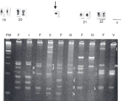
chromoso-mal anochromoso-maly was Klinefelter syndrome (10/ 165 = 6%). All of these cases were in the azoospermic group (10/60 = 15.3.%), seven in the non-mosaic 47,XXY form and three in the mosaic form - 46,XY (52)/47,XXY (48) - 46,XY (97)/47,XXY (3) - 46,XY (94)/ 47,XXY (6). The other two patients with chromosomal anomalies in the azoospermic group were one case of 46,XX male and one case of a mosaic 45,X (11)/46,X,del(Y) pre-senting 13 deleted STS in AZF analysis (Figure 1). In the oligospermic group, 4 cases of chromosomal anomalies were iden-tified - 3 cases of balanced Robertsonian translocations - 45,XY,der (13q,14q); 45, XY,der (13q,14q); 45,XY,der (14q,21q) -and one case of 46,X,del(Y) presenting only one deleted STS in the AZF analysis (Figure 2). Another patient in the oligospermic group presented the 46,XY (90)/47,XY, + i 22 p (10) karyotype. This was considered to be an anomaly with no clinical significance, mainly because the infertility of this patient was attributed to five microdeletions detected in
Figure 1. Top: Male infertility studies by classical cytogenetics and multiplex PCR for microdeletions of AZF gene detection. Partial karyotype of patient with 46,X,del(Y). The arrow indicates the Y deleted chromosome. Bottom: Multiplex PCR revealed deletion of 13 sequence-tagged sites indicated by letters a to m. PM = ladder; F = fertile man; I to V = mix I to V; a = sY117; b = sY102; c = sY143; d= sY157; e = sY105; f = Y6HPc54pr; g = sY153; h = sY97; i = sY127; j = sY109; k = sY149; l = Y6BaH34pr; m = Fr15Hpr.
19 20
21 22 Y
the AZFc region (case 12; Table 1).
Microdeletions in the Yq11 region
Microdeletions were detected in 12 pa-tients (12/160 papa-tients = 7.5%) and were most frequently limited to the AZFc region alone (7 patients: cases 1, 2, 4, 8, 9, 10, and 11 = 58.3%), followed by microdeletions in the combined AZFb and AZFc regions (3 patients: cases 5, 6, and 7 = 25%), in the AZFa region alone (1 patient: case 3 = 8.3%) and in the combined AZFa, AZFb, and AZFc regions (1 patient: case 12 = 8.3%). The position, extension and distribution of mi-crodeletions detected in the present sample are shown in Figure 3. The microdeletion frequency was 7% (7/100) in the oligosper-mic group, 6.6% (4/60) in the azoosperoligosper-mic group and 20% (1/5) in the asthenospermic group. There was an association between the number and position of microdeletions and the sperm counts. When one STS microdele-tion was detected in AZFa, oligospermia was mild (13 x 106 sperms/mL), whereas
one such microdeletion in the AZFc corre-sponded to moderate oligospermia (6 x 10⁶
or 1.3 x 10⁶ sperms/mL). When there were
two such microdeletions (in AZFb and AZFc) we observed only asthenospermia or moder-ate oligospermia (4.5 x 10⁶ sperms/mL).
However, when both microdeletions were in
Figure 2. Male infertility studies by classical cytogenetics and multiplex PCR for microdele-tions of AZF gene detection. Partial karyotype of 46,XY,del(Y). Multiplex PCR revealed deletion of one sequence-tagged site (sY147). PM = ladder; F = fertile man; II = mix II showing the sY147 deleted.
21 22 Y
PM F II
sY147
AZFc, oligospermia was severe (1 x 10⁵
sperms/mL). When there were four or more microdeletions, regardless of the region, oli-gospermia was always severe (0 to 2 x 10⁵
sperms/mL; Table 1). We performed a tes-ticular biopsy and histopathological analy-sis in 5 of the 12 patients with AZF letions. In 1 of the 5 patients, four microde-letions (one in AZFb and three in AFZc) and severe oligospermia (1 x 10⁵ sperms/mL)
were detected and histopathologic evalua-tion revealed severe hypospermatogenesis. In another patient there were five microdele-tions in the AZFc region and azoospermia, and the evaluation revealed tubular atrophy and maturation arrest in spermatocyte I. Another one of the 5 patients presented five microdeletions in the AZFc region and azoospermia, as well as germinal aplasia in 90% of tubules and maturation arrest in 10%. There was also 1 patient with five microde-letions in the AZFc, severe oligospermia (2 x 10⁵ sperms/mL), germinal aplasia in 75%
of the tubules and spermatocyte I maturation arrest in 25% of the tubules. Finally, in the 46,X,del(Y) patient with 13 microdeletions and azoospermia, we observed tubular fi-brosis, tubular hyalinization and germinal maturation arrest in spermatocytes I (Table 1).
Discussion
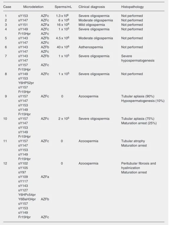
Table 1. Genotype-phenotype association in azoospermia factor gene (AZF) microdeletions.
Case Microdeletion Sperms/mL Clinical diagnosis Histopathology
1 sY153 AZFc 1.3 x 10⁶ Severe oligospermia Not performed
2 sY147 AZFc 6 x 10⁶ Moderate oligospermia Not performed
3 sY151 AZFa 16 x 106 Mild oligospermia Not performed
4 sY149 AZFc 1 x 10⁵ Severe oligospermia Not performed
Fr15Hpr AZFc
5 sY143 AZFb 4.5 x 10⁶ Moderate oligospermia Not performed
sY147 AZFc
6 sY143 AZFb 40 x 106 Asthenospermia Not performed
sY147 AZFc
7 sY143 AZFb 1 x 10⁵ Severe oligospermia Severe
sY147 hypospermatogenesis
sY157 AZFc
Fr15Hpr
8 sY149 AZFc 1 x 10⁵ Severe oligospermia Not performed
sY153 Y6HP52pr sY157 Fr15Hpr
9 sY157 AZFc 0 Azoospermia Tubular aplasia (90%)
sY147 Hypospermatogenesis (10%)
sY153 sY149 Fr15Hpr
10 sY157 AZFc 2 x 10⁵ Severe oligospermia Tubular aplasia (75%)
sY147 Maturation arrest (25%)
sY153 sY149 Fr15Hpr
11 sY157 AZFc 0 Azoospermia Tubular atrophy
sY147 Maturation arrest
sY153 sY149 Fr15Hpr
12 sY102 0 Azoospermia Peritubular fibrosis and
sY105 hyalinization
sY97 Maturation arrest
sY109 AZFa
sY117 sY143 sY127 Y6HPc54pr Y6BaH34pr AZFb sY157
sY153 sY149
Fr15Hpr AZFc
Klinefelter syndrome (46,XY - 94/47,XXY - 6 and, 46,XY - 97/47,XXY - 3) that would probably not have been detected in an anal-ysis involving fewer metaphases.
The frequency of AZF microdeletions detected in our sample was 7.5% (12/160
6.6% in the azoospermic group, comparable to the 7% seen in the oligospermic group. This could be an effect of the small size and slightly unbalanced nature of our sample (oligospermic patients = 100, azoospermic patients = 60). The distribution of microde-letions detected in our sample showed that the AZFc region was the most prevalent (58.3%), followed by the AZFb/AZFc com-bination (25%), by AZFa (8.3%), and by the AZFa/AZFb/AZFc (also 8.3%) combination. These results differ from those reported by Simoni et al. (3) only in the fact that we detected no microdeletions exclusively in the AZFb region, while they found this to be the second most frequent region in their sample of 34 patients (AZFc = 79%; AZFb = 9%; AZFb/AZFc = 6%; AZFa = 3%, and AZFa/AZFb/AZFc = 3%). The same inves-tigators found that deletions of isolated genes in the AZFa region were related to a variable
testicular phenotype and that complete dele-tion of the AZFa region makes it virtually impossible to retrieve testicular sperm for ICSI. Similarly, in our sample, the size of the deleted segment was associated with sperm counts, with a clear relationship between the size (number of STS deleted) and the level of sperm count reduction (Table 1). Unfortu-nately, we only had access to testicular bi-opsy samples from those patients with 4 or more deleted STS. Nevertheless, in the pa-tient with 4 STS deletions, there was severe hypospermatogenesis, whereas the patient with 5 STS deletions presented more severe testicular involvement with tubular atrophy or aplasia, and the patient with 13 STS dele-tions presented maturation arrest, as well as peritubular fibrosis and hyalinization. How-ever, the AZFc deletion phenotype is less severe because, in some cases, these dele-tions are compatible with residual spermato-genesis and, in rare cases, can even be trans-mitted naturally to the male offspring (14, 16,17). Our case 2, with 1 deleted STS (sY147) in the AZFc, presented a sperm count of 6 x 10⁶ sperms/mL, whereas case 1,
with a different STS deleted (sY153) in the same AZFc region, had only 1.3 x 10⁶ sperms/
mL. An explanation for the difference in phenotype between these two patients re-quires a more complete study of gene ex-pression in these cases.
In view of the genetic risks for the next generation, the importance of careful evalu-ation of karyotypes and AZF microdeletions in male infertility prior to assisted reproduc-tion by ICSI is evident. In our evaluareproduc-tion, we found that some subjects presented a higher risk to transmit genetic diseases to their off-spring. For example, the patient with the balanced Robertsonian translocation 45,XY, der (14q, 21q) has a high risk of fathering a child with Down syndrome, just as the pa-tient with the 46,XX karyotype has a high risk of fathering another male with the same pathology, the patient with the balanced Robertsonian translocation 13q, 14q has a
high risk of fathering a child with trisomy 13 syndrome, the patients with Klinefelter syn-drome have a high risk of fathering children with complete Klinefelter syndrome, and the patients with AZF microdeletions have a high risk of fathering infertile males (16) or offspring with severe clinical consequences other than male infertility, such as the devel-opment of sexual ambiguities and Turner stigmata (17). Patsalis et al. (17) screened 12 patients who had a 45,X/46,XY karyotype and presented Turner stigmata or sexual ambiguities, or both, for Y chromosomal microdeletion and detected that one third of these patients had Y chromosome microde-letions. Vogt (5) stated that AZF
microdele-tions can also become pre-mutamicrodele-tions for a subsequent complete loss of the Y chromo-some in the sperm of AZF-deleted patients, increasing the risk for XO cells and sexual ambiguities since the 45,XO/46,XY mosaic karyotype has been associated with ambigu-ous genitalia.
Acknowledgments
We would like to thank Mariluce Riegel and Albert Schinzel from Institut fuer Medi-cine Genetik, Univ. Zurich, for conducting the FISH studies of the case with the 47,XY, + i 22p/46,XY karyotype.
References
1. Vogt P, Chandley AC, Hargreave TB et al. (1992). Microdeletions in interval 6 of the Y chromosome of males with idiopathic sterility point to disruption of AZF, a human spermatogenesis gene. Human Ge-netics, 89: 491-496.
2. Chandley AC, Edmond P, Christie S et al. (1975). Cytogenetics and infertility in man. I. Karyotype and seminal analysis. Annals of Hu-man Genetics, 39: 231-254.
3. Simoni M, Bakker E & Krausz C (2004). EAA/EMQN best practice guidelines for molecular diagnosis of Y-chromosomal microdele-tions. State of art 2004. International Journal of Andrology, 27: 240-249.
4. Vogt PH, Edelmann A, Kirsch S et al. (1996). Human Y chromosome azoospermia factors (AZF) mapped to different subregions in Yq11. Human Molecular Genetics, 5: 933-943.
5. Vogt PH (2004). Genomic heterogeneity and instability of the AZF locus on the human Y chromosome. Molecular and Cellular Endocri-nology, 224: 1-9.
6. Moorhead PS, Nowell PC, Mellman WJ et al. (1960). Chromosome preparations of leukocytes cultured from human peripheral blood. Experimental Cell Research, 20: 613-616.
7. Hook EB (1977). Exclusion of chromosomal mosaicism - Tables of 90%, 95%, and 99% confidence limits and comments on use. Ameri-can Journal of Human Genetics, 29: 94-97.
8. ISCN (1995). An International System for Human Cytogenetic No-menclature. Karger/Cytogenetics and Cell Genetics, Basel, Switzer-land.
9. Henegariu O, Hirschmann P, Kilian K et al. (1994). Rapid screening of the Y chromosome in idiopathic sterile men, diagnostic for dele-tions in AZF, a genetic Y factor expressed during spermatogenesis. Andrologia, 26: 97-106.
10. Chandley AC (1979). The chromosomal basis of human infertility. British Medical Bulletin, 35: 181-186.
11. De Braekeleer M & Dao TN (1991). Cytogenetics studies in male infertility: a review. Human Reproduction, 6: 245-250.
12. Van Assche E, Banduelle M, Touenaye H et al. (1996). Cytogenet-ics of infertile men. Human Reproduction, 11: 1-23.
13. Tuerlings JHAH & Kremer JAM (2000). Genetics of male subfertility. European Journal of Obstetrics, Gynecology, and Reproductive Biology, 89: 117-121.
14. Rolf C, Gromoll J, Simoni M et al. (2002). Natural transmission of a partial AZF b deletion of the Y chromosome over three generations. Human Reproduction, 17: 2267-2271.
15. Foresta C, Moro E & Ferlin AY (2001). Y chromosome microdele-tions and alteramicrodele-tions of spermatogenesis. Endocrine Reviews, 22: 226-239.
16. Silber SJ & Repping S (2002). Transmission of male infertility to future generations: lessons from the Y chromosome. Human Repro-duction Update, 8: 217-229.