Changes in Sperm Motility and Capacitation
Induce Chromosomal Aberration of the
Bovine Embryo following Intracytoplasmic
Sperm Injection
Yoku Kato1,2, Yoshikazu Nagao1,2*
1Department of Animal Production Science, United Graduate School of Agricultural Science, Tokyo University of Agriculture and Technology, Fuchu, 183-8509, Japan,2University Farm, Faculty of Agriculture, Utsunomiya University, Mohka, Tochigi 321-4415, Japan
Abstract
Intracytoplasmic sperm injection (ICSI) has become the method of choice to treat human male infertility. One of the outstanding problems associated with this technique is our cur-rent lack of knowledge concerning the effect of sperm capacitation and motility upon the subsequent development of oocytes following ICSI. In the present study, we first examined the capacitation state of sperm exhibiting normal motility, along with sperm that had been activated, and examined the effect of reactive oxygen species (ROS) produced by these sperm types upon embryogenesis following bovinein vitrofertilization (IVF) and ICSI. Data
showed that activated sperm reduced the chromosomal integrity of IVF/ICSI embryos at the blastocyst stage, while capacitated sperm produced ROS in capacitation media. Secondly, we treated sperm with carbonyl cyanide m-chlorophenyl hydrazine (CCCP), a chemical known to uncouple cell respiration within the mitochondria, and investigated the effect of this treatment upon blastocyst formation and chromosomal integrity at the blastocyst stage. Activated sperm in which the mitochondria had been treated with CCCP reduced levels of chromosomal aberration at the blastocyst stage following ICSI, by reducing mitochondrial activity in activated sperm. In conclusion, these findings suggest that capacitated sperm ex-hibiting activated motility induced chromosomal aberration during development to the blas-tocyst stage following ICSI. The injection of sperm exhibiting normal motility, or activated sperm in which mitochondrial activity had been reduced, improved the quality of ICSI-derived embryos. Therefore, the selection of sperm exhibiting progressive motility may not always be better for early embryo development and fetal growth following human ICSI, and that the use of a bovine model may contribute to a deeper understanding of sperm selection for human ICSI embryo development.
a11111
OPEN ACCESS
Citation:Kato Y, Nagao Y (2015) Changes in Sperm Motility and Capacitation Induce Chromosomal Aberration of the Bovine Embryo following Intracytoplasmic Sperm Injection. PLoS ONE 10(6): e0129285. doi:10.1371/journal.pone.0129285
Academic Editor:Xiuchun (Cindy) Tian, USA, UNITED STATES
Received:August 4, 2014
Accepted:May 6, 2015
Published:June 10, 2015
Copyright:© 2015 Kato, Nagao. This is an open access article distributed under the terms of the
Creative Commons Attribution License, which permits unrestricted use, distribution, and reproduction in any medium, provided the original author and source are credited.
Funding:These authors have no support or funding to report.
Mammalian capacitation has previously been correlated with changes in sperm plasma membrane fluidity, intracellular ion concentration, reactive oxygen species (ROS), metabolism and motility [12,13]. Capacitating sperm are also known to produce controlled amounts of ROS [14]. We previously demonstrated that capacitated bovine sperm exhibited activated mo-tility in capacitation medium [15]. These reports highlight the possibility that capacitated sperm exhibiting activated motility could be associated with the production of ROS by the plas-ma membrane and mitochondria [14,16]. On the other hand, increased ROS generation from sperm would inevitably lead to oxidative stress, including sperm DNA fragmentation and re-duced motility [17,18]. ROS derived from oxygen within the culture environment is also known to impair the development ofin vitromatured and fertilized bovine oocytes [19,20]. Ca-pacitating mouse sperm has been shown to affect embryo development and the chromosomal integrity of embryos duringin vitroculture following mouse ICSI [10]. In mouse sperm, capac-itation also increased the levels of asynchronous remodeling and resulted in delayed pronuclei formation and DNA synthesis following sperm injection [11]. Moreover, rhesus macaque em-bryos arising from ICSI carried out with sperm exhibiting ROS damage, resulted in impair-ments to mitosis, DNA integrity, and development to the blastocyst stagein vitro[21].
However, one of the outstanding problems associated with ICSI is our current lack of knowledge concerning the effect of sperm capacitation, motility, and mitochondrial status upon blastocyst formation and chromosomal integrity following ICSI. In the present study, we carried out a systematic analysis of these important sperm parameters with regard to embryo development following ICSI. In order to accomplish this, we examined sperm capacitation, motility, mitochondrial activity and ROS production, upon the development of embryos creat-ed by ICSI with sperm exhibiting inactivatcreat-ed/activatcreat-ed motility and hyper-activation.
Materials & Methods
Oocyte collection and
in vitro
maturation
(m-TCM199) for 22–25 h at 39.0°C under 5% CO2in air with high humidity. m-TCM199
con-sisted of hepes-buffered medium 199 (Gibco) supplemented with 0.1% (w/v) polyvinyl alcohol (PVA;Sigma Chemical Co, St. Louis,MO.), 0.5 mM sodium pyruvate (Nacalai Tesque Inc., Kyoto, Japan), 1% AB, 0.02 AU/ml FSH (Antrin, Denka), and 1μg/ml estradiol-17β(Sigma).
The rates ofin vitromaturation after 22–24 hrs were more than 95% in all experiments.
Washing, pre-incubation of sperm and insemination
Sperm washing and pre-incubation was carried out as previously described [25]. The motility ratios (%) of frozen-thawed semen were greater than 70% before sperm washing with percoll. Fresh semen were frozen at Genetics Hokkaido Co., Ltd. In brief, frozen-thawed sperm were washed with a discontinuous percoll solution. Washed sperm (2.0 x 10⁸cells/ml) were incubat-ed in Brackett and Oliphant mincubat-edium (BO) supplementincubat-ed with 0.05% (w/v) PVA, 1% AB (BOPVA: non-capacitation media: control) for 0, 2 and 4 hrs at 39.0°C under 5% CO2in air
with high humidity. Washed sperm were then cultured in BOPVA with methyl-β-cyclodextrin (MBCD: BOMBCD: capacitation media) for 0, 2 and 4 h. Following culture in capacitation media for 4 hrs, sperm were introduced into BO supplemented with progesterone (P) or car-bonyl cyanide m-chlorophenyl hydrazone (CCCP), a chemical known to uncouple cell respira-tion within the mitochondria for 0.5–1 hour. Matured cumulus-oocyte complex (COCs) or denuded oocytes (10 oocytes/100μL droplet) were inseminated in the presence of the sperm
(2.0 ×10⁶cells/ml) which were activated immediately after pre-incubation for 2-4hrs. Oocytes and spermatozoa were co-incubated at 39.0°C under 5% CO2in air with high humidity.
Analysis of sperm motility
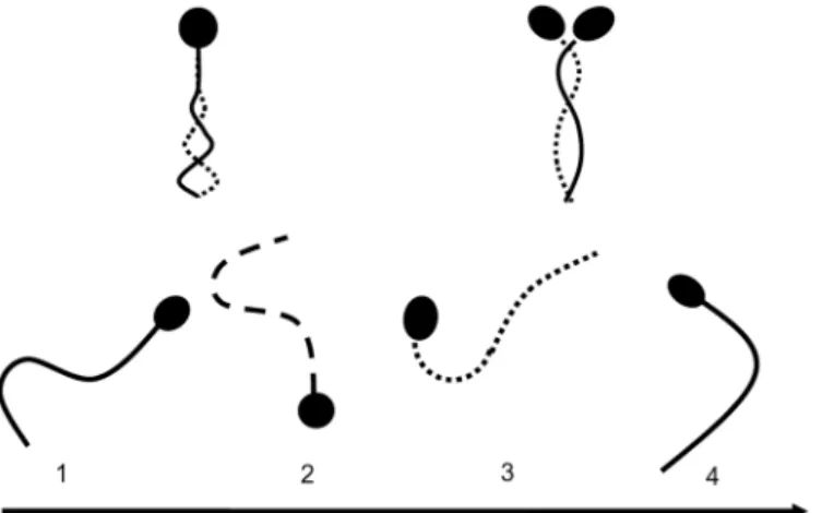
Sperm motility was categorized according to motility patterns as previously described [15,26]. Over 100 sperm were examined in PBS containing 10% polyvinylpyrrolidone (PVP) solution in order to reduce and consider sperm motility on an interference contrast microscope. The ra-tios (%) of normal motility, activated, and hyper-activated motility sperm were determined as follows. Normal motile sperm exhibited a motile tail (Fig 1, Top left). Following the activation of motility, sperm head and flagella beat in an almost symmetrical fashion, propelling the sperm in almost linear trajectories [27–29] (Fig 1, Top right). Hyper-activated motility was characterized by asymmetrical beating of the flagellum resulting in a circular or vigorous‘‘ fig-ure of 8”trajectory [27–29] (Fig 1, below). The proportion (%) of motile sperm refers to the number of motile sperm divided by the number of total sperm, including all motile and immo-tile sperm in each media, respectively. The proportion (%) of activated sperm refers to the number of such sperm divided by the number of motile sperm in each media, respectively. Fi-nally, the proportion (%) of hyper-activated motility sperm refers to the number of such sperm divided by the number of motile sperm in each media, respectively.
Normal, activated and hyper-activated motility of individual sperm and
the chlortetracycline fluorescence assay
Single sperm were collected and examined after pre-incubation in capacitation media or non-capacitation media. Single sperm were collected using an injection pipette immediately prior to CTC staining as previously described [15]. Ten to twenty sperm exhibiting normal motility, ac-tivated or hyper-acac-tivated motility were collected in PBS containing 10% PVP. Sperm were placed into non-capacitation media on a glass slide (Matsunami, Japan). The chlortetracycline (CTC) stain was used to evaluate capacitation status as previously described [15,30]. Precipi-tated sperm were mixed thoroughly with 750μM CTC solution (Sigma Chemical Co.), 5 mM
pH 7.8), fixed with 12.5% (w/v) paraformaldehyde in 0.5 M Tris-HCl (pH 7.4), and finally placed on a glass slide and covered with a coverslip. Sperm were assessed for CTC-staining using a 400 to 440-nm filter and a DM 455 dichroic mirror fitted to a fluorescent microscope. CTC-staining patterns were classified into three types according to the method described by Fraser et al [30]: F (uncapacitated)-pattern, bright fluorescence detected over the whole region of the sperm head; B (capacitated)-pattern, fluorescence detected on the sperm head but not over the post-acrosomal region; and AR (acrosome-reacted)-pattern, weak fluorescence ob-served over the sperm head with a bright band sometimes present in the equatorial region (Fig 2).
Immunolabeling of tyrosine phosphorylation
Tyrosine phosphorylation was carried out in bovine sperm by immunofluorescence using 4G10 (4G10: Millipore corporation. Billerica, MA) and anti-mouse IgG conjugated with phy-coerythrin (EI-2000: Vector Labratory,Inc. Burlingame, CA) [31]. For immunofluorescence microscopy, washed sperm were stained in suspension, dried onto poly-lysine-coated glass slides and then fixed. Bovine sperm were fixed in PBS containing 2% paraformaldehyde for 30 min at room temperature (RT). Sperm were then washed once in PBS and blocked with PBS
Fig 1. Sperm motility patterns of bovine spermatozoa cultured in non-capacitation media and capacitation media.Top left; normal motile sperm, right; sperm exhibiting activated motility, bottom; hyper-activated motility.
doi:10.1371/journal.pone.0129285.g001
Fig 2. Sperm patterns arising from the chlortetracycline fluorescence assay.Left; F (uncapacitated)-pattern, central; B (capacitated)-(uncapacitated)-pattern, right; AR (acrosome-reacted)-pattern. Bovine normal motile and activated sperm were incubated in the three types of media and analysed by CTC staining. Bar = 20μm (B).
containing 0.1% bovine serum albumin (BSA: PBSBSA) for 30 min before the addition of pri-mary antibodies. The monoclonal antibody 4G10 was used at a dilution of 1:100 in PBSBSA. Incubations were performed for 60 min at RT with three washes in PBSBSA between incuba-tions. The sperm pellet was then incubated with anti-mouse IgG conjugated with phycoery-thrin (diluted by 1:100) in PBS supplemented with 3 mg/ml BSA for 1h at RT. The sperm suspension was then centrifuged at 300×g for 3 min. After removal of the supernatant, the sperm were washed twice, as described above, to remove unbound secondary antibody. Sam-ples were then mounted, kept for up to one day at 4°C in the dark, and finally examined by fluorescent microscopy (Olympus).
Evaluation of individual sperm mitochondria by a JC-1 probe
To evaluate changes in mitochondrial membrane potential, sperm were labeled with potential-sensitive 5,5’,6,6’-tetrachloro-1,1’,3,3’-tetraethylbenzimidazolylcarbocyanine iodide (JC-1; Mo-lecular Probes, Eugene, OR) for 10 min [32]. Live mitochondria were labeled in PVP solution supplemented with JC-1 for 10 min at 39.0°C. Sperm exhibiting normal motility or activated motility were collected in PBS containing 10% PVP on a glass slide within 5 minutes as previ-ously described [15]. Those sperm in PVP solution were placed onto a glass slide, fixed in PBS containing 2% paraformaldehyde for 30 min at RT and washed three times in PBSBSA. After that, sperm were covered with PBSBSA and a cover slip. Samples were kept for up to one day at 4°C in the dark and finally examined by fluorescent microscopy. The percentage of sperm with green (inactive mitochondria, no membrane potential,Fig 3B, above), bright orange or faint orange (active mitochondria, high membrane potentialFig 3B, below) were evaluated vi-sually by fluorescence microscopy.
Intracytoplasmic sperm injection and embryo culture
Intracytoplasmic sperm injection was performed as described previously [33] with some modi-fications. Briefly, after maturation, cumulus cells were thoroughly dispersed with 0.1% hyal-uronidase (Sigma) and removed by vortexing. After removal of the cumulus cells, only oocytes with a visible first polar body were selected for experimental use. Selected oocytes were washed three times in m-TCM199 without FSH and estradiol-17β. Before sperm injection, motile sperm were immobilized by applying several piezo pulses (M-701, Suruga Seiki, Shizuoka city, Japan). The immobilized spermatozoon was drawn into the injection pipette tail first and the zona pellucida drilled by applying several piezo pulses. Then, the pipette was advanced me-chanically into the ooplasm. ICSI was performed after 3–4 hrs of sperm culture in capacitation media or non-capacitation media. After injection with sperm, oocytes were immediately trans-ferred to modified synthetic oviduct fluid with BSA [34] (SOFBSA) and cultured for 3 h. After culture, oocytes injected with sperm were treated for 3 min with 7% ethanol in TCM 199 con-taining 1 mg/mL PVP. Then, 15 to 50 of these ICSI embryos were placed in SOFBSA and cul-tured at 39.0°C under 5% CO2, 5% O2and 90% N2at high humidity for 7–8 days [34].
Assessment of fertilization and embryo development
In Experiment 2, the ratio (%) of blastocyst formation refers to the numbers of blastocysts di-vided by the number of cleaved embryos. In Experiment 3 and 4, the ratio (%) of blastocyst for-mation refers to the number of blastocysts divided by the number of all cultured oocytes.
Evaluation of chromosomal integrity at the blastocyst stage
Chromosomes were counted as previously described [35]. Briefly, some blastocysts were washed and placed in 1% sodium citrate solution for 15 min, and fixed by pouring 0.02 to 0.03 ml of acetic-alcohol fixative (acetic acid 1: ethanol 1) into 0.4 ml of sodium citrate solution. Blastocysts were placed onto a glass slide, immediately covered with a very small droplet of ace-tic acid to separate each blastomere, and then immediately re-fixed with several drops of the initial fixative. After drying completely, chromosome samples from the blastocysts were stained
Fig 3. Mitochondrial activity of sperm by JC-1 staining.Top; Sperm annotated with green staining represent inactive mitochondria with no membrane potential (left: differential interference contrast: DIC, right: JC-1), Bottom; sperm annotated with bright orange staining represent active mitochondria with high membrane potential (left: DIC, right: JC-1). Bar = 20μm.
with 2% Giemsa solution for 10 min. The number of chromosomes were counted as‘normal’, 60 (= 2n), or‘abnormal’, 30 (= n) and 90 (= 3n) (Fig 4).
Evaluation of DNA fragmentation by TUNEL staining during the
pronuclear stage
Pronuclear stage embryos were assessed by the TUNEL stain as previously described [36]. All samples were washed in PBS containing 1 mg/ml PVP (PBSPVP), then fixed in PBSPVP with 4% paraformaldehyde solution for 1h at RT. Afterwards, samples were rinsed with PBSPVP and incubated in PBSPVP including 0.5% Triton X-100 and 0.1% sodium citrate for 1 h at 39.0°C. 1 h post-incubation, TUNEL staining was performed, following the manufacturer’s in-structions (Roche, Mannheim, Germany) at RT. Subsequently, PBS containing 2 mg/ml Hoechst 33342 was employed to stain samples and controls for 15 min at RT. Samples were then mounted on glass slides and examined under a fluorescent microscope as described above. The fragmented nucleus was stained yellow-green by fluorescein as a result of the TUNEL assay (Fig 5A, below). The non fragmented nucleus did not stain yellow-green (Fig 5A, above).
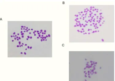
Fig 4. Chromosomal pattern of the bovine embryo at the blastocyst stage.(A) Typical example in which the number of chromosomes were classified as‘normal’, 60 (= 2n). (B) Typical example in which the number of chromosomes were classified as‘Abnormal’, 90 (= 3n). (C) Typical example in which the number of chromosomes were classified as‘Abnormal’, 30 (= n).
doi:10.1371/journal.pone.0129285.g004
Fig 5. DNA fragmentation at the pronuclear stage following the injection of activated and hyper-activated sperm.(A) Positive (below) and negative (above) pattern following TUNEL staining. Differential interference contrast (DIC: left panel), Hoechst staining (central panel), and TUNEL staining (right panel). (B) Rate of DNA fragmentation in the three experimental groups. n = number of embryos. Different letters indicate significant differences as determined by the Chi Square test (P<0.01). Letters indicate significant differences (a-b). This experiment was repeated 3 times. Bar = 20μm.
Measurement of ROS produced by sperm
ROS levels were estimated using the luminol assay [16]. Luminol (5-amino-2, 3-dihydro-1,4-phthalazinedione; also, 3- aminophthalic hydrazide) is used to estimate the combined levels of total ROS. The assay was sensitized by the addition of horseradish peroxidase to the sperm suspension, so as to increase the intensity of the spontaneous luminescence levels. In brief, 2μl
of 5mM luminol was added to 75μl aliquot of sperm (15 million sperm) and the reaction
initi-ated by adding 23μl of 2.5 units of horseradish peroxidase at RT. Results were monitored using
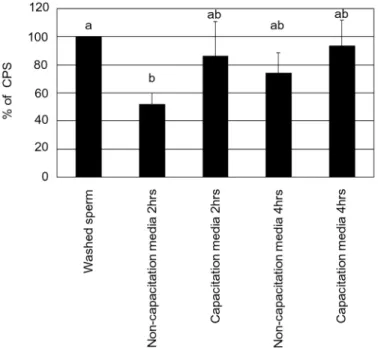
a luminometer (Wallac 1420 VICTOR 2 T Multilabel Counter; PerkinElmer, Inc., Wellesley, MA) for 30 minutes at RT. InFig 6, ROS production was expressed as the % of counted pho-tons per second (CPS). InFig 7, ROS production (CPS) was divided by the number of motile sperm in capacitation media and non-capacitation media, then ROS production by one indi-vidual sperm was expressed as a % of CPS.
Experimental design
In experiment 1, we examined the capacitation state of normal motile, activated, and hyper-ac-tivated sperm and the ROS produced by these sperm. In experiment 2, we examined the effect of insemination with activated sperm and denuded oocytes upon embryo development. In ex-periment 3, we examined the effect of these sperm motile types upon blastocyst formation and chromosomal integrity at the blastocyst stage following ICSI. In experiment 4, we investigated the effect of sperm treated with CCCP, a chemical known to uncouple cell respiration within the mitochondria, upon blastocyst formation and chromosomal integrity at the blastocyst stage following ICSI.
Fig 6. ROS production of sperm (CPS) in non-capacitation media and capacitation media.Different letters within columns indicate significant differences as determined by one-way ANOVA with Tukey-Kramer analysis (P<0.05). This experiment was repeated 3–4 times. Statistical significance compared to control: a
vs b.
Statistical analyses
All experiments were replicated over 3 times. Sperm samples from at least three different bulls were used for each set of experiments. The rates of normal sperm and sperm with activated motility were evaluated for significance using one-way factorial analysis of variance (ANOVA) with Tukey-Kramer analysis. These rates were evaluated for significant differences between 0–4 hrs or between capacitation media and non-capacitation media. In Experiment 1, the means of ROS concentration, as measured by a luminometer, were evaluated between capacita-tion media and non-capacitacapacita-tion media for significance using the one-way factorial ANOVA with Tukey-Kramer analysis. In Experiment 1, 2, 3 and 4, the rates of sperm capacitation, fertil-ization, cleavage and blastocyst formation, along with chromosomal integrity and the rate of DNA fragmentation, were evaluated for significance using the Chi square test. In Experiment 3 and 4, the rate of fertilization, cleavage and blastocyst formation were evaluated for significant differences using the Chi square test. In all experiments, values were considered significantly different when P<0.05.
Results
Methyl-
β
-cyclodextrin (MBCD) induced the activation and capacitation of
motile sperm
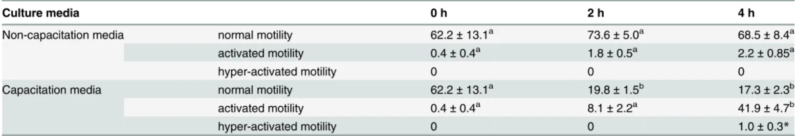
The proportion of activated sperm at 4 hrs was higher than that at 0 and 2 hrs in capacitation media (P<0.05,Table 1). The proportion of sperm with normal motility at 2 and 4 hrs was lower than that at 0 h in capacitation media (P<0.05,Table 1). Conversely, the proportion of activated and normal motile sperm did not change significantly with the duration of time cul-tured in non-capacitation media for 4 hrs (P>0.05,Table 1).
Fig 7. ROS production of one individual motile sperm in non-capacitation media and capacitation media.Different letters within columns indicate significant differences as determined by one-way ANOVA with Tukey-Kramer analysis (P<0.01). This experiment was repeated 3–4 times. Statistical significance compared to control: a vs b vs c.
In this study, the average proportion of whole live sperm displaying the B (capacitated) pat-tern at 0, 2 and 4 hrs of incubation were 34.2, 53.8 and 32.1% in non-capacitation media and 34.2, 71.7 and 61.6% in capacitation media, respectively. There were no differences between the present results and those reported previously [22]. The proportion of capacitation in normal motile, activated and hyper-activated sperm were 40.9 (n = 44), 78.8 (n = 33) and 29.6% (n = 24), respectively (Fig 8) after sperm were cultured for 2–4 hrs. The proportion of capacita-tion in activated sperm was higher than sperm exhibiting normal motility (P<0.05).
Sperm cultured in capacitation media exhibited a positive pattern of tyrosine phosphoryla-tion (Fig 9). HHHowever, sperm cultured in non-capacitation media exhibited a negative pat-tern of tyrosine phosphorylation (Fig 9).
Sperm generated ROS during culture
Mean values of ROS production were 141, 74 and 104 in non-capacitation media and 141, 122 and 132 CPS in capacitation media after culture for 0, 2 and 4 hrs, respectively. Following sperm culture, there was no significant difference between the values of ROS in capacitation media and non-capacitation media at 2 and 4 hrs (Fig 6, P>0.05). There was a significant re-duction between 0 h and 2hrs in non-capacitation media (Fig 6, P<0.05). There was no signifi-cant reduction between 0–4 hrs in capacitation media (Fig 6, P>0.05). The number of motile sperm in capacitation media was significantly lower than that in non-capacitation media at 2 and 4 hrs (Table 1, P<0.05). Mean values of ROS production (CPS) were divided by the
Fig 8. The proportion of capacitation and acrosome-reacted patterns in bovine sperm.n = number of sperm. This experiment was repeated 3 times.
number of motile sperm in each media at 2 and 4 hrs. Therefore, the mean values of ROS pro-duction by one individual motile sperm in capacitation media (% of CPS) were higher than that in non-capacitation media at 2 and 4hrs (Fig 7, P<0.05).
Blastocyst formation and chromosome integrity at the blastocyst stage
following IVF of denuded oocytes with activated sperm
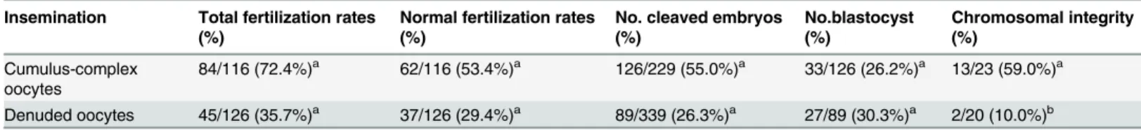
There were no differences in the blastocyst ratio of cleaved embryos between the two groups (Table 2, P>0.05). However, chromosomal integrity at the blastocyst stage arising from the in-semination of denuded oocytes in the presence of activated sperm was lower than the insemi-nation of COCs with activated sperm (Table 2, P<0.05).
Blastocyst formation and chromosome integrity at the blastocyst stage
following ICSI with activated sperm
There were no differences in the ratio of blastocyst formation among the four groups tested (Table 3, P>0.05). However, chromosomal integrity of the blastocyst stage arising from the in-jection of activated and hyper-activated sperm was lower than that with normal motile sperm and IVF (Table 3, P<0.05).
Fig 9. Tyrosine phosphorylation localization in sperm.Top and bottom images represent sperm cultured in non-capacitation media and capacitation media for 6hrs. Left) differential interference contrast (DIC) and right) immunofluorescence images. This experiment was repeated 3 times. Bar = 100μm.
doi:10.1371/journal.pone.0129285.g009
Table 2. The direct effect of motile sperm in denuded oocytes on embryo developments and chromosomal aberration (n = 8).
Insemination Total fertilization rates (%)
Normal fertilization rates (%)
No. cleaved embryos (%)
No.blastocyst (%)
Chromosomal integrity (%)
Cumulus-complex oocytes
84/116 (72.4%)a 62/116 (53.4%)a 126/229 (55.0%)a 33/126 (26.2%)a 13/23 (59.0%)a
Denuded oocytes 45/126 (35.7%)a 37/126 (29.4%)a 89/339 (26.3%)a 27/89 (30.3%)a 2/20 (10.0%)b
n = number of replicated experiments.
Letters within vertical columns indicate significant differences (a-b: Chi Square test: P<0.01).
activated and hyper-activated sperm
The ratios of DNA fragmentation at the pronuclear stage following IVF and ICSI were 17.9 (n = 39) in IVF, 11.5 (n = 26) in normal motile sperm, 50.0 (n = 12) in activated motile sperm and 50.0% (n = 18) in hyper-activated sperm, respectively. The ratios following the injection of activated and hyper-activated sperm were higher than that with sperm showing normal motili-ty and IVF (Fig 5B, P<0.05).
The effect of CCCP treatment upon capacitated sperm
The proportions of sperm exhibiting the characteristic pattern B of capacitation, as determined by chlortetracycline staining, were 58% (n = 24), 71% (n = 35) and 71% (n = 21) in normal mo-tile sperm, activated sperm and mitochondria reduced activated sperm, respectively (Fig 10).
The percentage of sperm with bright orange (active mitochondria, high membrane poten-tial,Fig 3, right-bottom) as indicated by JC-1 were 82% (n = 23), 83% (n = 37) and 33% (n = 23) in normal motile sperm, activated sperm and mitochondria reduced activated sperm, respectively. This proportion of mitochondria reduced activated sperm was lower than the other treatment groups (P<0.05)
Effect of CCCP upon the developmental competence of sperm
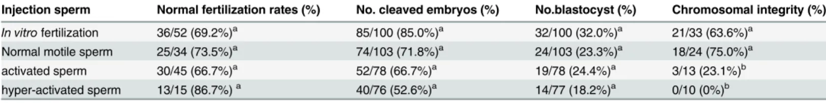
There were no significant differences in the rate of embryo development to the blastocyst stage among the three groups (P>0.05,Table 4). Chromosomal integrity of the blastocyst stage of
Fig 10. The effect of CCCP treatment upon capacitated sperm.The rate of capacitation and acrosome-reacted patterns of sperm as determined by CTC staining. This experiment was repeated 3 times.
both mitochondria reduced activated sperm and normal motile sperm were higher than that of activated sperm (P<0.05,Table 4).
Discussion
ICSI has become the method of choice to treat human male infertility. One of the outstanding problems associated with this technique is our current lack of knowledge concerning the effect of sperm capacitation and motility upon the subsequent development of embryos. Most sperm used for ICSI are artificially capacitated. However, some reports claim that sperm capacitation is not necessary for ICSI and could in fact, reduce embryo development.
In experiment 1, over 30% of sperm had been activated, resulting in an increase in the pro-duction of reactive oxygen species (ROS) in capacitation media while ROS propro-duction in non-capacitation media remained steady in the presence of less than 5% of sperm which was acti-vated. The resulting capacitation and ROS are most likely to be related to protein phosphoryla-tion events known to occur in mammalian sperm [13]. In fact, the present study shows that capacitation in the bovine model was associated with the appearance of a tyrosine phosphopro-tein complex in Experiment 1 (Fig 9). There are two reasons underlying the observed increase in ROS production. Firstly, increased production of ROS has been demonstrated from the sperm membrane during the capacitation of human and cryopreserved bovine sperm
[13,14,37]. Secondly, motility causes the sperm flagellum to beat vigorously with asymmetrical beats of high amplitude [27–29]. While changes in motility would require significant ATP pro-duction by the mitochondria, these organelles can produce large levels of ROS [38,39]. There-fore, we infer that increased mitochondrial activity and increased numbers of capacitated sperm may be associated with the increased production of ROS. We further suggest that the ac-tivation of sperm contributes to the production of ROS.
In Experiment 2, the insemination of denuded oocytes with activated sperm increased the ratio of chromosomal aberration at the blastocyst stage while high levels of ROS by producing capacitated sperm was detected in Experiment 1. When sperm were incorporated into COCs during IVF, the cumulus cells protected the oocyte against oxidative stress in the media [40]. Moreover, during fertilization, sperm cells generate ROS, which not only facilitate certain sperm functions such as capacitation, hyper-activation and the acrosome reaction [41], but could also cause damage to cells [20,42]. Therefore, we speculate that the insemination of de-nuded oocytes with activated sperm is related to chromosomal aberration of the blastocyst due to the destroy of the defense system of mature oocytes against ROS by the removal of cumulus cells and the increase of ROS production by capacitated sperm in capacitation media.
In Experiment 3, those embryos produced by the injection of activated and hyper-activated sperm resulted in a lower percentage of normal chromosomal integrity and DNA fragmenta-tion while capacitated sperm could produce high levels of ROS in Experiment 1. During IVF, the sperm nucleus gradually moves away from the site of penetration and slowly becomes
Table 4. The effect of reduced mitochondria activity upon on embryo developments and chromosomal aberration in bovine embryos after ICSI (n = 11).
Injection sperm No. cleaved embryos (%) No.blastocyst (%) Chromosomal integrity (%)
Normal motile sperm 103/140 (73.5%)a 29/140 (20.7%)a 11/14 (78.6%)a
Activated sperm 44/80 (55.0%)a 20/80 (25.0%)a 1/14 (7.1%)b
mitochondria reduced activated sperm 54/86 (62.8%)a 27/86 (31.4%)a 13/15 (86.7%)a
n = number of replicated experiments.
Letters within vertical columns indicate significant differences in chromosomal integrity (a-b: Chi square test P<0.01).
sperm did not induce chromosomal aberration at the blastocyst stage, while the uncoupling agent carbonyl cyanide m-chlorophenyl hydrazine (CCCP) reduced the mitochondrial activity of sperm and stop the mitochondrial membrane proton gradient and reduce ROS production [46]. To support the result of Experiment 4, we put forward two hypotheses. Firstly, during natural fertilization or IVF, sperm mitochondria activity would be reduced following incorpo-ration of the sperm into the oocyte while such mitochondrial activity was not impeded in oo-cytes subject to ICSI. In fact, sperm mitochondria activity was regulated by progesterone produced by the cumulus cells [47]. Secondly, literature appears to suggest that the ubiquitin-dependent proteolysis of paternal mitochondria may be delayed in oocytes after ICSI and that sperm mitochondria are degraded earlier after IVF than ICSI [48], suggesting that the mito-chondrial activity of sperm was not inhibited following ICSI and ROS generated by the sperm mitochondria in injected oocytes. Hence, we speculate that the reduction of mitochondrial ac-tivity of sperm exhibiting activated motility improved chromosomal integrity at the blastocyst stage following ICSI.
Interestingly, ICSI represents approximately one-half of all assisted reproductive techniques used in Europe [49]. In human ICSI, sperm exhibiting high speed motility are always selected as being the most likely to fertilize, and are thus injected into oocytes in human ICSI. However, from the view of a clinical embryologist, it was thought that the mitochondria and membrane of selected sperm are likely to be damaged by the micropipette during sperm immobilization for human ICSI. On the other hand, there is little evidence that the results of this present paper applies to humans, and a large number of healthy babies have been delivered by ICSI. There-fore, we think that our current findings are specific to bovine species and that sperm selection methodology should be investigated more thoroughly and future experiments should also as-sess the relationship between motility and genetic integrity, including chromosomal aberration and DNA fragmentation in mammalian species, notably mice and humans.
In conclusion, capacitated sperm showing activated and hyper-activated motility did not af-fect the frequency of blastocyst development, but did induce chromosomal aberration at the blastocyst stage. It is feasible that ROS produced by the sperm is a potentially dangerous factor when ICSI is carried out in humans and domestic animals. Hence, these findings suggest that the injection of sperm in which ROS production was eliminated could improve the quality of ICSI-derived bovine embryos and contribute to a deeper understanding of sperm motility and selection for human ICSI embryo development.
Acknowledgments
Author Contributions
Conceived and designed the experiments: YK YN. Performed the experiments: YK. Analyzed the data: YK YN. Contributed reagents/materials/analysis tools: YK YN. Wrote the paper: YK YN.
References
1. Palermo GD, Neri QV, Monahan D, Kocent J, Rosenwaks Z. (2012) Development and current applica-tions of assisted fertilization. Fertil Steril 97: 248–259. doi:10.1016/j.fertnstert.2011.12.037PMID: 22289284
2. Kimura Y, Yanagimachi R. (1995) Intracytoplasmic sperm injection in the mouse. Biol Reprod 52: 709–720. PMID:7779992
3. Oikawa T, Takada N, Kikuchi T, Numabe T, Takenaka M, Horiuchi T. (2005) Evaluation of activation treatments for blastocyst production and birth of viable calves following bovine intracytoplasmic sperm injection. Anim Reprod Sci 86: 187–194. PMID:15766799
4. Martin MJ. (2000) Development of in vivo-matured porcine oocytes following intracytoplasmic sperm in-jection. Biol Reprod 63: 109–112. PMID:10859248
5. Palermo G, Joris H, Devroey P, Van Steirteghem AC. (1992) Pregnancies after intracytoplasmic injec-tion of single spermatozoon into an oocyte. Lancet 340: 17–18. PMID:1351601
6. Van Steirteghem A. (2012) Fare well, Human Reproduction. Hum Reprod 27: 3363–3364. doi:10. 1093/humrep/des414PMID:23166158
7. Morozumi K, Yanagimachi R. (2005) Incorporation of the acrosome into the oocyte during intracytoplas-mic sperm injection could be potentially hazardous to embryo development. Proc Natl Acad Sci 102: 14209–14214. PMID:16183738
8. Morozumi K, Shikano T, Miyazaki S, Yanagimachi R. (2006) Simultaneous removal of sperm plasma membrane and acrosome before intracytoplasmic sperm injection improves oocyte activation/embryon-ic development. Proc Natl Acad Sci 103: 17661–17666. PMID:17090673
9. Gianaroli L, Magli MC, Ferraretti AP, Crippa A, Lappi M, Capitani S, et al. (2010) Birefringence charac-teristics in sperm heads allow for the selection of reacted spermatozoa for intracytoplasmic sperm injec-tion. Fertil Steril 93: 807–813. doi:10.1016/j.fertnstert.2008.10.024PMID:19064263
10. Tateno H, Kamiguchi Y. (2007) Evaluation of chromosomal risk following intracytoplasmic sperm injec-tion in the mouse. Biol Reprod 77: 336–342. PMID:17409376
11. Ajduk A, Yamauchi Y, Ward MA. (2006) Sperm chromatin remodeling after intracytoplasmic sperm in-jection differs from that of in vitro fertilization. Biology of Reproduction 75: 442–451. PMID:16775225
12. Bailey JL. (2010) Factors regulating sperm capacitation. Syst Biol Reprod Med 56: 334–348. doi:10. 3109/19396368.2010.512377PMID:20849222
13. O'Flaherty C, de Lamirande E, Gagnon C. (2006) Reactive oxygen species modulate independent pro-tein phosphorylation pathways during human sperm capacitation. Free Radic Biol Med 40: 1045–1055. PMID:16540400
14. de Lamirande E, O’Flaherty C. (2008) Sperm activation: Role of reactive oxygen species and kinases. Biochim Biophys Acta 1784: 106–115. PMID:17920343
15. Kato Y, Sugita S, Nagao Y. (2011) Capacitation status of activated bovine sperm cultured in media con-taining methyl-β-cyclodextrin affects the acrosome reaction and fertility. Zygote 19: 21–30. doi:10. 1017/S0967199410000304PMID:20727245
16. Koppers AJ, De Iuliis GN, Finnie JM, McLaughlin EA, Aitken RJ. (2008) Significance of mitochondrial reactive oxygen species in the generation of oxidative stress in spermatozoa. J Clin Endocrinol Metab 93: 3199–3207. doi:10.1210/jc.2007-2616PMID:18492763
17. Chen SJ, Allam JP, Duan YG, Haidl G. (2013) Influence of reactive oxygen species on human sperm functions and fertilizing capacity including therapeutical approaches. Arch Gynecol Obstet 288: 191–199. doi:10.1007/s00404-013-2801-4PMID:23543240
18. Aitken RJ, Koppers AJ. (2011) Apoptosis and DNA damage in human spermatozoa. Asian J Androl 13: 36–42. doi:10.1038/aja.2010.68PMID:20802502
19. Betts DH, Madan P. (2008) Permanent embryo arrest: molecular and cellular concepts. Mol Hum Reprod 14: 445–453. doi:10.1093/molehr/gan035PMID:18511487
neme by Ca2+ and not cAMP. Dev Biol 250: 208–217. PMID:12297107
28. Kumar V, Rangaraj N, Shivaji S. (2006) Activity of pyruvate dehydrogenase A (PDHA) in hamster sper-matozoa correlates positively with hyperactivation and is associated with sperm capacitation. Biol Reprod 75: 767–777. PMID:16855207
29. Suarez SS. (2008) Control of hyperactivation in sperm. Hum Reprod Update 14: 647–657. doi:10. 1093/humupd/dmn029PMID:18653675
30. Fraser LR, Abeydeera LR, Niwa K. (1995) Ca2+-regulating mechanisms that modulate bull sperm capacitaion and acrossomal exocytosisi as determined by chlortetracycline analysis. Mol Reprod Dev 40: 233–241. PMID:7766417
31. Mariappa D, Aladakatti RH, Dasari SK, Sreekumar A, Wolkowicz M, van der Hoorn F, et al. (2010) Inhi-bition of tyrosine phosphorylation of sperm flagellar proteins, outer dense fiber protein-2 and tektin-2, is associated with impaired motility during capacitation of hamster spermatozoa. Mol Reprod Dev 77: 182–193. doi:10.1002/mrd.21131PMID:19953638
32. Hung PH, Miller MG, Meyers SA, VandeVoort CA. (2008) Sperm mitochondrial integrity is not required for hyperactivated motility, zona binding, or acrosome reaction in rhesus macaque. Biol Reprod 79: 367–375. doi:10.1095/biolreprod.107.066357PMID:18480469
33. Emuta C, Horiuchi T. (2001) Effect of timing of activation and aging of bovine oocytes fertilized by intra-cytoplasmic sperm injection (ICSI) on cleavage and subsequent embryonic development in vitro. J Reprod Dev 47: 399–405.
34. Nagao Y, Iijima R, Saeki K. (2008) Interaction between embryos and culture conditions during in vitro development of bovine early embryos. Zygote 16: 127–133. doi:10.1017/S0967199408004644PMID: 18405433
35. Yoshizawa M, Konno H, Zhu S, Kageyama S, Fukui E, Muramatsu S, et al. (1999) Chromosomal diag-nosis in each individual blastomere of 5- to 10-cell bovine embryos derived from in vitro fertilization. Theriogenology 51: 1239–1250 PMID:10729088
36. Sagirkaya H, Misirlioglu M, Kaya A, First NL, Parrish JJ, Memili E. (2007) Developmental potential of bovine oocytes cultured in different maturation and culture conditions. Anim Reprod Sci 101: 225–240. PMID:17052869
37. O'Flaherty C, Beorlegui N, Beconi MT. (2003) Participation of superoxide anion in the capacitation of cryopreserved bovine sperm. Int J Androl 26: 109–114. PMID:12641829
38. Ferramosca A, Focarelli R, Piomboni P, Coppola L, Zara V. (2008) Oxygen uptake by mitochondria in demembranated human spermatozoa: a reliable tool for the evaluation of sperm respiratory efficiency. Int J Androl 31: 337–345. PMID:17573845
39. Piomboni P, Focarelli R, Stendardi A, Ferramosca A, Zara V. (2012) The role of mitochondria in energy production for human sperm motility. Int J Androl 35: 109–124. doi:10.1111/j.1365-2605.2011.01218.x PMID:21950496
40. Fatehi AN, Roelen AJB. (2005) Presence of cumulus cells during in vitro fertilization protects the bovine oocyte against oxidative stress and improves first cleavage but does not affect further development. Zy-gote 13: 177–185. PMID:16128413
41. Kothari S, Thompson A, Agarwal A, du Plessis SS. (2010) Free radicals: their beneficial and detrimental effects on sperm function. Indian J Exp Biol 48: 425–435. PMID:20795359
43. Caglar GS, Hammadeh M, Asimakopoulos B, Nikolettos N, Diedrich K, Al-Hassani S. (2005) In vivo and in vitro decondensation of human sperm and assisted reproduction technologies. In Vivo. 19: 623–630. PMID:15875785
44. Dozortsev D, De Sutter P, Dhont M. (1994) Behaviour of spermatozoa in human oocytes displaying no or one pronucleus after intracytoplasmic sperm injection. Hum Reprod. 9: 2139–2144. PMID:7868687
45. Nagy ZP, Liu J, Joris H, Bocken G, Desmet B, Van Ranst H, et al. (1995) The influence of the site of sperm deposition and mode of oolemma breakage at intracytoplasmic sperm injection on fertilization and embryo development rates. Hum Reprod 10: 3171–3177. PMID:8822437
46. Nishikawa T, Edelstein D, Du XL, Yamagishi S, Matsumura T, Kaneda Y, et al. (2000) Normalizing mito-chondrial superoxide production blocks three pathways of hyperglycaemic damage. Nature 404: 787–790. PMID:10783895
47. Tantibhedhyangkul J, Hawkins KC, Dai Q, Mu K, Dunn CN, Miller SE, et al. (2014) Expression of a mito-chondrial progesterone receptor in human spermatozoa correlates with a progestdependent in-crease in mitochondrial membrane potential. Andrology 6: 875–883. doi:10.1111/j.2047-2927.2014. 00263.xPMID:25187426
48. Sutovsky P, Van Leyen K, McCauley T, Day BN, Sutovsky M. (2004) Degradation of paternal mitochon-dria after fertilization: implications for heteroplasmy, assisted reproductive technologies and mtDNA in-heritance. Reprod Biomed Online 8: 24–33. PMID:14759284