Functio ns o f the e xtrace llular
m atrix and m atrix de grading
pro te ase s during tum o r
pro gre ssio n
1Center for Molecular Medicine, Maine Medical Center Research Institute,
South Portland, ME, USA
2Department of Cell Biology, Vanderbilt University, Nashville, TN, USA
L. Liaw1 and
H.C. Crawford2
Abstract
Cell interactions with extracellular matrices are important to patho-logical changes that occur during cell transformation and tumorigen-esis. Several extracellular matrix proteins including fibronectin, throm-bospondin-1, laminin, SPARC, and osteopontin have been suggested to modulate tumor phenotype by affecting cell migration, survival, or angiogenesis. Likewise, proteases including the matrix metallopro-teinases (MMPs) are understood to not only facilitate migration of cells by degradation of matrices, but also to affect tumor formation and growth. We have recently demonstrated an in vivo role for the
RGD-containing protein, osteopontin, during tumor progression, and found evidence for distinct functions in the host versus the tumor cells. Because of the compartmentalization and temporal regulation of MMP expression, it is likely that MMPs may also function dually in host stroma and the tumor cell. In addition, an important function of proteases appears to be not only degradation, but also cleavage of matrix proteins to generate functionally distinct fragments based on receptor binding, biological activity, or regulation of growth factors.
Co rre spo nde nce
L. Liaw
Center for Molecular Medicine Maine Medical Center Research Institute
125 John Roberts Rd. # 12 South Portland, ME 04106 USA
Fax: + 1-207-828-8071 E-mail: liawl@ poa.mmc.org Presented at the I International Symposium on “Signal Transduction and Gene Expression in Cell Proliferation and Differentiation”, São Paulo, SP, Brasil,
August 31-September 2, 1998.
Received November 26, 1998 Accepted January 27, 1999
Ke y wo rds
·Matrix metalloproteinase ·O steopontin
·Carcinoma ·Protease
Intro ductio n
The ultimate fate of a tumor is deter-mined by its ability to productively interact with its host. The alterations in gene expres-sion that occur in a tumor due to cumulative genetic mutations would be inconsequential if they did not provide a means to exploit the supportive host responses while escaping the destructive ones. The host responds to the presence of the tumor initially through changes in gene expression found in the
migration as well as a barrier to tumor inva-sion and metastasis, and the matrix metallo-proteinases (MMPs) which, by degrading the ECM, can theoretically nullify its ef-fects, whether supportive or obstructive.
It has long been recognized that follow-ing cell transformation and the initiation of a tumor, the environment of the stroma sur-rounding the tumor changes. A parallel be-tween the tumor stroma and a wound envi-ronment has been proposed (1) due to in-creases in fibrinogen, increased permeabil-ity of vessels, and an inflammatory response in both cases (2,3). Indeed, the extracellular milieu is further altered in various tumors with changes in ECM proteins including fibronectin, thrombospondin, osteopontin (OPN), laminin, SPARC, and hyaluronan proteoglycans, as well as MMPs such as collagenase, stromelysins and gelatinases. The ability of the tumor cell to survive, migrate, invade, and eventually colonize a secondary site is dependent on its interac-tions with ECM proteins and the ability to modify its extracellular environment either by the expression of ECM proteins or ma-trix-degrading proteases. Much of the inter-action with the surrounding environment can be understood by the interaction of cell sur-face receptors such as integrins with extra-cellular proteins. The modulation of integrins during tumor progression has been the sub-ject of several recent reviews (4) and will not be discussed comprehensively here.
ECM pro te ins during tum o r fo rmatio n and gro wth
The altered nature of the tumor stroma has suggested that proteins that are either suppressed or induced may function during tumor growth or metastasis. Molecules in-cluding thrombospondin-1, laminin, fibro-nectin, proteoglycans, SPARC, and OPN have been implicated. Analyses of the func-tions of these proteins have been compli-cated, and in some instances the activities of
a particular protein are dependent on the cell lines studied. Evidence suggests a positive role in tumor progression for laminin and proteoglycans and their receptors (5,6). An-tisense reduction of thrombospondin reduced the growth rate of a carcinoma line both in vitro and in vivo (7). In contrast,
subcutane-ous growth of tumor cells expressing high levels of thrombospondin-1, or injection of purified thrombospondin-1, has been shown to inhibit growth of experimental lung me-tastases in the same animal (8). SPARC over-expression in carcinoma cells was shown to suppress tumorigenesis (9), although in mela-noma lines, antisense inhibition of SPARC had a similar effect of abolishing tumorige-nicity (10). Additionally, a positive role for SPARC in the process of angiogenesis has been indicated(11).Some of the difficulties in interpreting these data may lie in the fact that, as mentioned, these matrix proteins are often host stromal cell products as well as tumor products, and thus may affect primary tumor cell growth and migration, as well as host-derived properties such as angiogene-sis and inflammation.
In the case of fibronectin, tumor growth has been associated with reduced levels of the protein or its receptor. Both transformed cells and tumors have been shown to have reduced levels of fibronectin (12), and the restoration of fibronectin, or its receptor, the
We have focused our studies on OPN, a multifunctional secreted protein whose over-expression is associated with cell transfor-mation. Recent analyses of a variety of hu-man tumor specimens demonstrated that OPN expression is present in tumor cells and/or stromal cells in human carcinomas of the colon, duodenum, stomach, breast, lung, prostate, melanoma, bladder, ovary, thyroid, and pancreas (15). Evidence for the func-tional consequences of OPN in tumors has been obtained using antisense OPN con-structs designed to eliminate secretion of OPN in transformed cells. Gardner et al. (16) have expressed antisense OPN in transformed malignant Rat 1 fibroblasts and shown that the reduction in OPN protein secretion cor-relates with a decrease in tumor growth in the lung as well as growth in soft agar. Su et al. (17) reported that antisense OPN con-structs in epidermal cells could inhibit the induction of OPN following tetradecanoyl-phorbol acetate treatment, and clones stably expressing antisense OPN failed to grow in an anchorage-independent manner in soft agar. Consistent results were also obtained by Feng et al. (18) who found that OPN-targeted ribozymes in H-ras-transformed 3T3 cells had reduced tumorigenicity, perhaps due to a greater sensitivity to the cytotoxic activity of macrophage-like cells. Finally, overexpression of OPN in a benign mam-mary epithelial cell line was sufficient to cause significant metastases of the injected transfectants (19). These results support a causal role for OPN in the ability of tumor cells to survive and metastasize to secondary sites, and suggest that initial OPN induction at stages as early as cell transformation may be critical to the tumor cell phenotype.
One hypothesis as to why OPN-produc-ing tumors are more successful is that the protein provides an adhesive matrix suitable for tumor cell survival and invasion. As mentioned, in comparison to normal tissues, the tumor stroma is of unique composition. As a parallel to this, many transformed cells
alter their complements of receptors for ex-tracellular matrix, including modulating cell surface integrins (20). Expression of any of a number of OPN receptors (see below) may facilitate interaction of the tumor cell with the tumor stroma. Denhardt and Chambers (21) have also demonstrated that production of OPN by tumor cells promotes survival by inhibiting cytotoxic attack from host cells via regulation of genes such as nitric oxide synthase, which decreases the ability of the host cell to target the tumor cell.
We have addressed the roles of OPN in vivo in a murine model of squamous cell
carcinoma using OPN null mutant mice (22). In this system, the carcinogen causes devel-opment of benign papillomas, which progress to invasive carcinomas, metastatic tumors, and frequently form secondary tumors in the lungs (23). We have shown that in papillo-mas, OPN expression is limited to the stroma surrounding the tumor, and it is not until the tumor becomes invasive that the tumor cells produce OPN (24). The extent of expression also correlates with progression state in that tumors graded as metastatic spindle cell car-cinomas express high levels of OPN.
Our studies demonstrated that on an OPN null background, chemically induced squa-mous cell carcinomas grow faster, appar-ently progress faster, and have more, albeit smaller, lung metastases compared to wild type animals. Tumor lines were derived from
carcinomas of wild type and OPN null ani-mals, and characterized in vivo and in vitro
with an activated phenotype. Levels of mac-rophage mannose receptor are decreased fol-lowing activation with interferons, lipopoly-saccharide, and antigen challenge, and in-versely correlate with the generation of su-peroxide radicals and production of plas-minogen activator (25,26). Finally, OPN-producing versus OPN null tumor lines also behaved differently in vitro, where survival
of cells at low density was compromised in the absence of OPN.
Taken together, our findings support a model where OPN produced by the host and OPN produced by the tumor cells have dif-ferent functions during tumorigenesis. Dur-ing the early papilloma stage, we propose that the OPN produced by the stroma sur-rounding the tumor functions as a chemoat-tractant for macrophages as a host response. The presence of macrophages at the tumor site can function to inhibit tumor growth. Therefore, on the OPN null background, this host response would be abolished, and the tumors would be able to grow at a faster rate. However, once the invasive/metastatic tu-mor begins to produce OPN, this tutu-mor- tumor-derived protein inhibits the activation of cells including macrophages, allowing greater tu-mor survival. This concept is consistent with previous findings by Denhardt and Cham-bers (21) suggesting that tumor-derived OPN provides a survival advantage by inhibiting cells that would cytotoxically attack tumor cells.
Evasion of macrophages may account for the growth differences of the primary tumor, but cannot explain the increased survival in vitro. For a metastasis to be successful, the
tumor cell must not only reach the secondary site, but should also exhibit growth from clonal density. In vitro, this property was
reflected in the fact that OPN-producing cells were able to form colonies at clonal densities at which OPN null cells did not survive. The observation that the lung metastases in the OPN mutant mice were significantly smaller than in wild type animals supports the notion that OPN provides a growth advantage un-der these conditions in vivo as well. These
results are consistent with the previously discussed antisense experiments in cell lines, where the predominant result of OPN inhibi-tion was reduced clonal growth in soft agar, and reduced experimental lung metastasis.
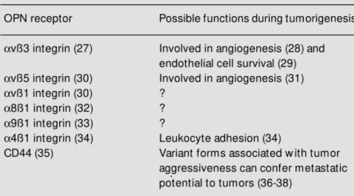
Our studies of OPN during tumor pro-gression point out that cell compartmentali-zation (host versus tumor) is very important in determining the overall effects of this protein in vivo, and in fact, the effects may be antagonistic. We postulate that this diver-sity may be explained in part by the presence of multiple cellular receptors, or different activities of multiple forms of the protein. Table 1 indicates the identified OPN recep-tors, many which have been described re-cently (27-38). Several of these receptors have been implicated in some stage of tumor growth or progression, including the integrins
avß3, avß5, and avß1, and the glycoprotein CD44. Cell adhesion to the matrix is critical for the ability of a tumor cell to migrate and invade. On the other hand, expression of matrix receptors by stromal components such as angiogenic endothelium also is vital to tumor survival. Increasing evidence suggests that both avß3 and avß5are critically in-volved in the angiogenesis process (28,31). CD44 is a recently identified OPN receptor corresponding to a family of proteins gener-ated by alternate splicing of a single gene. CD44 has been of interest in tumor progres-Table 1 - Osteopontin (OPN) receptors.
OPN receptor Possible functions during tumorigenesis
avß3 integrin (27) Involved in angiogenesis (28) and endothelial cell survival (29)
avß5 integrin (30) Involved in angiogenesis (31)
avß1 integrin (30) ?
a8ß1 integrin (32) ?
a9ß1 integrin (33) ?
a4ß1 integrin (34) Leukocyte adhesion (34)
CD44(35) Variant forms associated w ith tumor
sion since variant forms of the protein corre-late with progression and metastatic spread of malignant cells. One explanation for this could be CD44 interaction with hyaluronan, a glycosaminoglycan that is enriched in the stroma of carcinomas of the esophagus, stom-ach, and colon(39), and another possibility is an interaction with OPN, also produced in both tumor and stromal cells during malig-nant progression. A possible mechanism for the diverse effects of OPN on various cells is that the receptors utilized are different and have distinct signaling cascades. In the case of the CD44 receptor, OPN has been shown to stimulate cell migration, whereas another ligand, hyaluronan, induced cell aggregation (35). We have also shown that OPN is a chemotactic stimulus for avß3-bearing cells, and did not induce migration even if the adhesive receptors avß5 and avß1 were pres-ent (40).
Secondly, modified forms of OPN may account for different activities. Biochemical studies of the protein show extensive post-translational modification including phos-phorylation, glycosylation, sialylation, and transglutaminase-mediated crosslinking. Sev-eral lines of evidence indicate that these post-translational modifications can alter the ability of OPN to bind to other proteins (41,42) or bind to cellular receptors (43). OPN is also a substrate for proteolytic cleav-age, and fragments of the protein have dif-ferent adhesive properties, effects on migra-tion, and receptor-binding capabilities (44, 45). Importantly, proteolytic fragments of OPN occur naturally in vivo (45), and throm-bin is one known protease that cleaves intact OPN. As discussed further below, proteolytic cleavage of ECM proteins may be one im-portant step regulating their activities.
Matrix m e tallo pro te inase s in tum o rs
MMPs have had a long history associated with tumor progression. The consistent ex-pression of MMPs in invasive metastatic
tumor cells (46) has pigeonholed this large family of proteases into the generic role of clearing ECM components from the path of a migrating tumor cell. However, just as the expression in invasive tumors led to this model, closer examination of MMP expres-sion in vivo has forced us to consider more
complex functions for these enzymes in tu-mor progression. In the majority of epithelial tumors, expression of most MMPs is found initially in the surrounding tumor stroma. It is not until the latest stages of tumor progres-sion that these MMPs become widely ex-pressed by the tumor cells. Representative exceptions to this expression pattern range from stromelysin-3, which is virtually never expressed by the tumor cells at any stage of progression, but is highly expressed in the tumor stroma (47), to matrilysin, which is highly expressed in benign epithelial tumors, but not in the tumor stroma (48). Overall, the expression patterns of MMPs are more com-plicated than simply being associated with metastatic tumors and thus suggest a multi-functional role for MMPs beyond simple invasion and metastasis.
By examining animals with targeted in-activating mutations in MMP genes, the com-plexity of MMP functions in tumors is just beginning to be unraveled. In gelatinase A (MMP-2) null mice, tumor angiogenesis and progression of injected tumor cell lines is inhibited (49). Chemically induced skin tu-morigenesis is inhibited in the stromelysin-3 (MMP-11) null mouse, and stromelysin-3 null fibroblasts fail to support the growth of injected breast tumor cells (50). Multiple intestinal neoplasia (Min) mice on a
mammary gland enhances tumorigenesis (52,53). Similarly, collagenase overexpres-sion in the skin of mice increases tumorigen-esis of chemically induced tumors (54).
Though there have been no reports of MMPs whose activities inhibit tumor growth, preliminary studies with the stromelysin-1 null mouse (55) indicate that, in the very earliest stages of skin tumor growth, such a function appears to exist. When skin tumors are chemically induced in the stromelysin-1 null mouse, we see a higher rate of initial tumor growth as determined by tumor size. This accelerated growth is completely lim-ited to the first 7 weeks after the appearance of the tumor, a time consistent with the stromal expression of stromelysin-1. How-ever, once the tumors progress beyond 7 weeks, there are no apparent differences in tumor growth, invasion or metastasis. The explanation for this phenotype may lie in the observation that stromelysin-1 is one of the very few MMPs that appears to have a role in normal connective tissue as exemplified by its high fibroblast expression during the cu-taneous wound healing process. In fact, the stromelysin null mouse is deficient in wound contraction, a process that we are exploring as having an effect on tumor growth.
Pe rspe ctive s
Though it is possible to hypothesize that the effects of ECM proteins on tumor behav-ior are due merely to their altered expres-sion, it is unlikely that this is the case for MMPs. A more likely possibility is that MMPs exert their effects by proteolyzing available substrates, whether matrix compo-nents or other effector molecules (Table 2; 56-62). For instance, the tumor growth-en-hancing effect of stromelysin-3-producing fibroblasts requires the presence of growth factors bound to the matrix, implying that stromelysin-3 processes matrix components in such a way that growth factors become newly bioavailable to the tumor (50). MMP processing of ECM components has also been shown to create fragments of matrix proteins that were not present in the intact molecule, such as in the case of gelatinase A cleavage of laminin 5 inducing cell migra-tion (56). Conversely, proteolytic process-ing may also inactivate matrix protein func-tion. MMPs have also been shown to be capable of processing integrin receptors for ECM components (60), another mechanism by which the cellular response to matrix can be modified.
The seemingly diverse and even contra-dictory activities of particular matrix pro-teins during tumor progression will likely be reconciled by a more extensive consider-ation of the specific extracellular environ-ment. Expression and localization of cell surface receptors, expression of activating and inactivating proteases and their inhibi-tors, and alterations in expression of the matrix components themselves will all inte-grate to determine the behavioral responses of the tumor cells and the selective pressures that determine tumor progression.
Table 2 - Consequences of protein cleavage by matrix metalloproteinases (M M Ps).
Protein Protease Functional consequence
of proteolysis
Laminin-5 M M P-2 Induce cell migration (56)
Decorin M M P-2, -3, -7 Release of TGFß1 (57)
Entactin Str-1 (M M P-3) Cell apoptosis (58)
Fibronectin M M P-2? M odulate cell proliferation and
migration (59)
Beta 4 integrin M atrilysin Regulate cell surface beta
4 levels? (60)
Collagen XVIII ? Generation of endostatin
Re fe re nce s
1. Dvorak HF (1986). Tumors: w ounds that do not heal. Similarities betw een tumor stroma generation and w ound healing.
New England Journal of M edicine, 315: 1650-1659.
2. Yeo TK, Brow n L & Dvorak HF (1991). Alterations in proteoglycan synthesis common to healing w ounds and tumors.
Am erican Journal of Pat hology, 138: 1437-1450.
3. Brow n LF, Van de Water L, Harvey VS & Dvorak HF (1988). Fibrinogen influx and accumulation of cross-linked fibrin in heal-ing w ounds and in tumor stroma. Ameri-can Journal of Pathology, 130: 455-465. 4. Keely P, Parise L & Juliano R (1998).
Inte-grins and GTPases in tumour cell grow th, motility and invasion. Trends in Cell Biol-ogy, 8: 101-106.
5. Tuszynski GP, W ang TN & Berger D (1993). Altered proteoglycan gene expres-sion and the tumor stroma. Experientia, 49: 447-455.
6. Ziober BL, Lin CS & Kramer RH (1996). Laminin-binding integrins in tumor pro-gression and metastasis. Seminars in Can-cer Biology, 7: 119-128.
7. Castle V, Varani J, Fligiel S, Prochow nik EV & Dixit V (1991). Antisense-mediated reduction in thrombospondin reverses the malignant phenotype of a human squa-mous carcinoma. Journal of Clinical Inves-tigation, 87: 1883-1888.
8. Volpert OV, Law ler J & Bouck NP (1998). A human fibrosarcoma inhibits systemic angiogenesis and the grow th of experi-mental metastases via thrombospondin-1. Proceedings of the National Academy of Sciences, USA, 95: 6343-6348. 9. M ok SC, Chan WY, Wong KK, M uto M G
& Berkow itz RS (1996). SPARC, an extra-cellular matrix protein w ith tumor-sup-pressing activity in human ovarian epithe-lial cells. Oncogene, 12: 1895-1901. 10. Ledda M F, Adris S, Bravo AI, Kairiyama C,
Bover L, Chernajovsky Y, M ordoh J & Podhajcer OL (1997). Suppression of SPARC expression by antisense RNA ab-rogates the tum origenicity of hum an melanoma cells. Nature M edicine, 3: 171-176.
11. Sage EH (1997). Terms of attachment: SPARC and tumorigenesis [new s]. Nature M edicine, 3: 144-146.
12. Akiyama SK, Olden K & Yamada KM (1995). Fibronectin and integrins in inva-sion and metastasis. Cancer and M etasta-sis Review s, 14: 173-189.
13. Hynes RO & Plantefaber LC (1991).
Inte-grin receptors for extracellular matrix and their involvement in oncogenic transfor-mation. In: Brugge J, Curran T, Harlow E & M cCormick F (Editors), Origins of Hu-man Cancer: A Comprehensive Review. Cold Spring Harbor Laborat ory, Cold Spring Harbor.
14. Taverna D, Ullman-Cullere M , Rayburn H, Bronson RT & Hynes RO (1998). A test of the role of a5 integrin/fibronectin interac-tions in tumorigenesis. Cancer Research, 58: 848-853.
15. Brow n LF, Papadopoulos-Sergiou A, Berse B, M anseau EJ, Tognazzi K, Perruzzi CA, Dvorak HF & Senger DR (1994). Osteopontin expression and dis-tribution in human carcinomas. American Journal of Pathology, 145: 610-623. 16. Gardner HA, Berse B & Senger DR (1994).
Specific reduction in osteopontin synthe-sis by antisense RNA inhibits the tumori-genicity of transformed Rat1 fibroblasts.
Oncogene, 9: 2321-2326.
17. Su L, M ukherjee AB & M ukherjee BB (1995). Expression of antisense osteopon-tin RNA inhibits tumor promoter-induced neoplastic transformation of mouse JB6 epidermal cells. Oncogene, 10: 2163-2169.
18. Feng B, Rollo EE & Denhardt DT (1995). Osteopontin (OPN) may facilitate metas-tasis by protecting cells from macrophage NO-mediated cytotoxicity: evidence from cell lines dow n-regulated for OPN expres-sion by a targeted ribozyme. Clinical and Experimental M etastasis, 13: 453-462. 19. Chen H, Ke Y, Oates AJ, Barraclough R &
Rudland PS (1997). Isolation of and effec-tor for metastasis-inducing DNAs from a human metastatic carcinoma cell line. On-cogene, 14: 1581-1588.
20. Sanders RJ, M ainiero F & Giancotti FG (1998). The role of integrins in tumorigen-esis and metastasis. Cancer Investigation, 16: 329-344.
21. Denhardt DT & Chambers AF (1994). Overcoming obstacles to metastasis - de-fenses against host dede-fenses: osteopon-tin (OPN) as a shield against attack by cytotoxic host cells. Journal of Cellular Biochemistry, 56: 48-51.
22. Liaw L, Birk DE, Ballas CB, Whitsitt JS, Davidson JM & Hogan BL (1998). Altered w ound healing in mice lacking a functional osteopontin gene. Journal of Clinical In-vestigation, 101 (Suppl 1): 1468-1478. 23. Balmain A & Brow n K (1988). Oncogene
activation in chemical carcinogenesis. Ad-vances in Cancer Research, 51: 147-182.
24. Craw ford HC, M atrisian LM & Liaw L (1998). Distinct roles of osteopontin in host defense activity and tumor survival during squamous cell carcinoma progres-sion in vivo. Cancer Research,58: 5206-5215.
25. M okoena T & Gordon S (1995). Human macrophage activation. M odulation of mannosyl, fucosyl receptor activity in vi-tro by lymphokines, gamma and alpha interferons, and dexamethasone. Journal of Clinical Investigation, 75: 624-631. 26. Chroneos Z & Shepherd VL (1995).
Differ-ential regulation of the mannose and SP-A receptors on macrophages. American Journal of Physiology, 269: L721-L726. 27. Hu DD, Hoyer JR & Smith JW (1995).
Ca2+ suppresses cell adhesion to osteo-pontin by attenuating binding affinity for integrin alpha v beta 3. Journal of Biologi-cal Chemistry, 270: 9917-9925. 28. Brooks PC, M ontgomery AM , Rosenfeld
M , Reisfeld RA, Hu T, Klier G & Cheresh DA (1994). Integrin alpha v beta 3 antago-nists promote tumor regression by induc-ing apoptosis of angiogenic blood ves-sels. Cell, 79: 1157-1164.
29. Scatena M , Almeida M , Chaisson M L, Fausto N, Nicosia RF & Giachelli CM (1998). NF-kappaB mediates alphavbeta3 integrin-induced endothelial cell survival.
Journal of Cell Biology, 141: 1083-1093. 30. Hu DD, Lin EC, Kovach NL, Hoyer JR &
Smith JW (1995). A biochemical charac-terization of the binding of osteopontin to integrins alpha v beta 1 and alpha v beta 5. Journal of Biological Chemistry, 270: 26232-26238.
31. Friedlander M , Brooks PC, Shaffer RW, Kincaid CM , Varner JA & Cheresh DA (1995). Definition of tw o angiogenic path-w ays by distinct alpha v integrins. Sci-ence, 270: 1500-1502.
32. Denda S, Reichardt LF & M uller U (1998). Identification of osteopontin as a novel ligand for the integrin alpha8 beta1 and potential roles for this integrligand in-teraction in kidney morphogenesis. M o-lecular Biology of the Cell, 9: 1425-1435. 33. Smith LL, Cheung HK, Ling LE, Chen J, Sheppard D, Pytela R & Giachelli CM (1996). Osteopontin N-terminal domain contains a cryptic adhesive sequence rec-ognized by alpha9beta1 integrin. Journal of Biological Chem istry, 271: 28485-28491.
Cell Science, 111: 1165-1174.
35. Weber GF, Ashkar S, Glimcher M J & Can-tor H (1996). RecepCan-tor-ligand interaction betw een CD44 and osteopontin (Eta-1).
Science, 271: 509-512.
36. Gunthert U, Hofmann M , Rudy W, Reber S, Zoller M , Haussm an I, M atzku S, Wenzel A, Ponta H & Herrlich P (1991). A new variant of glycoprotein CD44 confers metastatic potential to rat carcinoma cells.
Cell, 65: 13-24.
37. Seiter R, Arch R, Reber S, Komitow ski D, Hofmann M , Ponta H, Herrlich P, M atzku S & Zoller M (1993). Prevention of tumor m et ast asis f orm at ion by ant i-variant CD44. Journal of Experimental M edicine, 177: 443-445.
38. Naot D, Sionov RV & Ish-Shalom D (1997). CD44: structure, function, and associa-tion w ith the m alignant process. Ad-vances in Cancer Research, 71: 241-319. 39. Wang C, Tammi M , Guo H & Tammi R (1996). Hyaluronan distribution in the nor-mal epithelium of esophagus, stomach, and colon and their cancers. American Journal of Pathology, 148: 1861-1869. 40. Liaw L, Skinner M P, Raines EW, Ross R,
Cheresh DA, Schw artz SM & Giachelli CM (1995). The adhesive and migratory ef-fects of osteopontin are mediated via dis-tinct cell surface integrins. Role of alpha v beta 3 in smooth muscle cell migration to osteopontin in vitro. Journal of Clinical Investigation, 95: 713-724.
41. Beninati S, Senger DR, Cordella-M iele E, M ukherjee AB, Chackalaparam pil I, Shanmugam V, Singh K & M ukherjee BB (1994). Osteopontin: its transglutaminase-catalyzed posttranslational modifications and cross-linking to fibronectin. Journal of Biochemistry, 115: 675-682.
42. Singh K, M ukherjee AB, De Vouge M W & M ukherjee BB (1992). Differential pro-cessing of osteopontin transcripts in rat kidney- and osteoblast-derived cell lines.
Journal of Biological Chem istry, 267: 23847-23851.
43. Shanmugam V, Chackalaparampil I, Kundu GC, M ukherjee AB & M ukherjee BB (1997). Altered sialylation of osteopontin prevents its receptor-mediated binding on the surface of oncogenically transformed tsB77 cells. Biochemistry, 36: 5729-5738. 44. Senger DR, Perruzzi CA,
Papadopoulos-Sergiou A & Van de Water L (1994). Adhe-sive properties of osteopontin: regulation by a naturally occurring thrombin-cleav-age in close proximity to the GRGDS cell-binding domain. M olecular Biology of the Cell, 5: 565-574.
45. Senger DR, Perruzzi CA, Gracey CF, Papadopoulos A & Tenen DG (1988). Se-creted phosphoproteins associated w ith neoplastic transformation: close homol-ogy w ith plasma proteins cleaved during blood coagulation. Cancer Research, 48: 5770-5774.
46. Pow ell WC & M atrisian LM (1996). Com-plex roles of matrix metalloproteinases in tumor progression. Current Topics in M i-crobiology and Immunology, 213: 1-21. 47. Basset P, Wolf C & Chambon P (1993).
Expression of the stromelysin-3 gene in fibroblastic cells of invasive carcinomas of the breast and other human tissues: a review . Breast Cancer Research and Treatment, 24: 185-193.
48. Wilson CL & M atrisian LM (1998). M atrily-sin. In: Parks WC & M echam RP (Editors),
M at rix M et alloprot einases. Academ ic Press, San Diego, 149-184.
49. Itoh T, Tanioka M , Yoshida H, Yoshioka T, Nishimoto H & Itohara S (1998). Reduced angiogenesis and tumor progression in gelatinase A-deficient mice. Cancer Re-search, 58: 1048-1051.
50. M asson R, Lefebvre O, Noel A, Fahime M E, Chenard M P, Wendling C, Kebers F, LeM eur M , Dierich A, Foidart JM , Basset P & Rio M C (1998). In vivo evidence that the stromelysin-3 metalloproteinase con-tributes in a paracrine manner to epithelial cell malignancy. Journal of Cell Biology, 140: 1535-1541.
51. Wilson CL, Heppner KJ, Labosky PA, Hogan BL & M atrisian LM (1997). Intesti-nal tumorigenesis is suppressed in mice lacking the metalloproteinase matrilysin.
Proceedings of the National Academy of Sciences, USA, 94: 1402-1407.
52. Sympson CJ, Bissell M J & Werb Z (1995). M ammary gland tumor formation in trans-genic mice overexpressing stromelysin-1.
Seminars in Cell Biology, 6: 159-163. 53. Rudolph-Ow en LA, Chan R, M uller WJ &
M atrisian LM (1998). The matrix metallo-proteinase matrilysin influences early-stage mammary tumorigenesis. Cancer
Research, 58: 5500-5506.
54. D’Armiento J, DiColandrea T, Dalal SS, Okada Y, Huang M T, Conney AH & Chada K (1995). Collagenase expression in trans-genic mouse skin causes hyperkeratosis and acanthosis and increases susceptibil-ity to tumorigenesis. M olecular and Cellu-lar Biology, 15: 5732-5739.
55. M udgett JS, Hutchinson NI, Chartrain NA, Forsyth AJ, M cDonnell J, Singer II, Bayne EK, Flanagan J, Kaw ka D, Shen CF, Stevens K, Chen H, Trumbauer M & Visco DM (1998). Susceptibility of stromelysin 1-deficient mice to collagen-induced ar-thritis and cartilage destruction. Arthritis and Rheumatism, 41: 110-121.
56. Giannelli G, Falk-M arzillier J, Schiraldi O, St et ler-St evenson W G & Quarant a V (1997). Induction of cell migration by ma-trix metalloprotease-2 cleavage of lami-nin-5. Science, 277: 225-228.
57. Im ai K, Hiram at su A, Fukushim a D, Pierschbacher M D & Okada Y (1997). Degradation of decorin by matrix metallo-proteinases: identification of the cleavage sites, kinetic analyses and transforming grow th factor-beta1 release. Biochemical Journal, 322: 809-814.
58. Alexander CM , How ard EW, Bissell M J & Werb Z (1996). Rescue of mammary epi-thelial cell apoptosis and entactin degra-dation by a tissue inhibitor of metallopro-teinases-1 transgene. Journal of Cell Biol-ogy, 135: 1669-1677.
59. Grant M B, Caballero S, Bush DM & Spoerri PE (1998). Fibronectin fragments modulate human retinal capillary cell pro-liferation and migration. Diabetes, 47: 1335-1340.
60. von Bredow DC, Nagle RB, Bow den GT & Cress AE (1997). Cleavage of beta 4 inte-grin by matrilysin. Experimental Cell Re-search, 236: 341-345.
61. Lijnen HR, Ugw u F, Bini A & Collen D (1998). Generation of an angiostatin-like fragment from plasminogen by stromely-sin-1 (M M P-3). Biochemistry, 37: 4699-4702.
62. Pat t erson BC & Sang QA (1997). Angiostatin-converting enzyme activities of human matrilysin (M M P-7) and gelati-nase B/type IV collagegelati-nase (M M P-9).