Adhe sio n o f the hum an patho ge n
Sp orothrix schenckii
to se ve ral
e xtrace llular m atrix pro te ins
Departamentos de 1Biologia Celular e Genética and 2Bioquímica, Instituto de Biologia Roberto Alcântara Gomes,
Universidade do Estado do Rio de Janeiro, Rio de Janeiro, RJ, Brasil
3Fundação O swaldo Cruz, FIO CRUZ, Rio de Janeiro, RJ, Brasil
O .C. Lima1,3,
C.C. Figueiredo1,
B.A.S. Pereira1,
M.G.P. Coelho2,
V. Morandi1 and
L.M. Lopes-Bezerra1
Abstract
The pathogenic fungus Sporothrix schenckii is the causative agent of sporotrichosis. This subcutaneous mycosis may disseminate in immu-nocompromised individuals and also affect several internal organs and tissues, most commonly the bone, joints and lung. Since adhesion is the first step involved with the dissemination of pathogens in the host, we have studied the interaction between S. schenckii and several extracellular matrix (ECM) proteins. The binding of two morphologi-cal phases of S. schenckii, yeast cells and conidia, to immobilized type II collagen, laminin, fibronectin, fibrinogen and thrombospondin was investigated. Poly (2-hydroxyethyl methacrylate) (poly-HEMA) was used as the negative control. Cell adhesion was assessed by ELISA with a rabbit anti-S. schenckii antiserum. The results indicate that both morphological phases of this fungus can bind significantly to type II collagen, fibronectin and laminin in comparison to the binding ob-served with BSA (used as blocking agent). The adhesion rate obob-served with the ECM proteins (type II collagen, fibronectin and laminin) was statistically significant (P<0.05) when compared to the adhesion obtained with BSA. No significant binding of conidia was observed to either fibrinogen or thrombospondin, but yeast cells did bind to the fibrinogen. Our results indicate that S. schenckii can bind to fibronec-tin, laminin and type II collagen and also show differences in binding capacity according to the morphological form of the fungus. Co rre spo nde nce
L.M. Lopes-Bezerra
Departamento de Biologia Celular e Genética
Instituto de Biologia, UERJ Rua São Francisco Xavier, 524 (PHLC. 205)
20550-900 Rio de Janeiro, RJ Brasil
Fax: + 55-21-587-7377 E-mail: leila@ uerj.br.
Presented at the 5th Brazilian Symposium on Extracellular Matrix - SIMEC, Angra dos Reis, RJ, Brasil, September 7-10, 1998.
Research supported by FAPERJ (No. E-26/170.634/96) and CNPq (No. 521443/95-4).
Received December 2, 1998 Accepted January 11, 1999
Ke y wo rds
·Sporothrix schenckii
·Fibronectin
·Laminin
·Type II collagen
·Adhesion
Intro ductio n
Sporotrichosis is a deep mycosis caused by the traumatic implantation of the dimor-phic pathogenic fungus, Sporothrix schenckii.
This disease has several clinical forms, the most frequent being the cutaneous/subcuta-neous one, with associated lymphangitis and
spo-rotrichosis has been reported to be an impor-tant mycosis in HIV+ individuals (5). In these patients, osteoarthritis and the disseminated form of sporotrichosis are frequently ob-served (3,4).
The dimorphism of S. schenckii is deter-mined by temperature. At 37o
C this fungus differentiates to the yeast form. This mor-phology is present in cutaneous lesions. The mycelial phase is the saprophytic form of S. schenckii and is obtained invitro by cultiva-tion at 28o
C.
The adherence of a pathogen to host tissues is believed to be an essential step in infectious diseases leading to the dissemi-nation of the microorganism. The most im-portant barriers to be overcome are the endothelium and basement membranes. Little is known about the adhesion and dis-semination of pathogenic fungi inside the host. Entactin, fibronectin (FN), laminin, type I collagen and type IV collagen were re-ported to be involved in the adhesion of some pathogenic fungi to the extracellular matrix (ECM) (7-9). Differences in the binding capacity of Candida albicans were correlated with growth conditions and with the fungal isolate (10). Virulent strains have enhanced binding capacity to fibronectin when compared to avirulent strains (10). An association between binding capacity to ECM proteins and fungus morphological phase was also reported. Conidia of Aspergil-lus fumigatus bind to fibronectin but the mycelia do not (11). On the other hand, no differences in the ability to bind to entactin were observed between the yeast and hyphal morphologies of Candida albicans
(7).
Fungal cell wall componentsinvolved in the adhesion process of these pathogens such as cell wall proteins (integrin-like proteins) and mannoproteins were described (12). S. schenckii expresses on its cell wall a glyco-peptide component, peptido-rhamnomannan (13). This glycopeptide possesses the main antigenic epitopes present on the cell surface
of S. schenckii and also binds to the lectin Concanavalin A by means of the O-glyco-sidically linked oligosaccharides (14,15). Differences in cell wall composition of both conidia and yeast cells were observed in S. schenckii and are related to the virulence and pathogenesis of this microorganism (16). It is not known whether the cell surface of S. schenckii contains adhesins that could medi-ate host attachment and invasion.
The present investigation is a prelimi-nary study carried out to determine the bind-ing capacity of S. schenckii to several extra-cellular matrix proteins.
Mate rial and Me tho ds
Mate rial
Poly (2-hydroxyethyl methacrylate) (poly-HEMA), and mouse and human laminin were from Sigma Chemical Co., St. Louis, MO, USA; goat anti-rabbit peroxidase conjugate was from Gibco Life Technologies do Brazil (São Paulo, SP, Brazil); an antiserum against yeast cells of S. schenckii was raised in rabbits as previously described (15).
Micro o rganism and gro wth co nditio ns
Sporothrix schenckii strain 1099-18 was used throughout this study. This strain was originally obtained from the Mycology Sec-tion, Department of Dermatology, Columbia University, New York, USA. Yeast forms of
S. schenckii were grown in brain heart infu-sion broth (BHI, Difco, Detroit, MI, USA) at 37o
C in rotary shaker (150 rpm). After 7 days in culture the cells were harvested and washed with sterile 50 mM phosphate buff-ered saline (PBS), pH 7.2. The mycelial phase of S. schenckii was grown in Sabouraud broth (2% glucose, 0.5% yeast extract and 1% peptone) at 25o
chamber. The cell suspension was adjusted to 108 cells/ml.
Iso latio n o f plasma fibro ne ctin and human
plate le t thro m bo spo ndin
Plasma fibronectin was freshly prepared by gelatin-Sepharose chromatography ac-cording to Vuento and Vaheri (17). Throm-bospondin (TSP) was obtained from throm-bin-activated platelet supernatants as previ-ously described (18). Briefly, supernatants were submitted to heparin-Sepharose chro-matography and bound proteins were eluted with growing NaCl concentrations in the running buffer. The fraction eluted with 0.55 M NaCl was concentrated and applied to a Sepharose CL4B column. TSP was eluted in the void volume.
Cartilage co llage n purificatio n
Type II collagen was purified from xi-phoid cartilage of chicks by a modification of the method of Trenthan et al. (19). The cartilage was pulverized with liquid nitrogen and incubated with 20 volumes (w/v) of a 50 mM Tris/2 M MgCl2 solution, pH 7.4, at 4
o C for 18 h with constant shaking. The extract was then centrifuged at 25,000 g for 15 min and the precipitate was washed 3 times with distilled water. The residue was then sus-pended in 3 volumes of 0.5 M acetic acid and the pH was adjusted to 2.5 with formic acid. Pepsin was added to the suspension (1 g/30 g cartilage) followed by incubation at 4oC for 48 h, with constant gentle shaking. After this period, the undigested residue was separated by centrifugation at 25,000 g for 15 min and washed 3 times with 0.5 M acetic acid under the same conditions. The supernatants from the extraction and washes were pooled and dialyzed twice, first against distilled water for 12 h and then against 50 mM Tris/0.2 M NaCl, pH 7.6, for 24 h, followed by centrifu-gation. The extract was diluted 5-fold in dialysis buffer and then passed through a
DEAE-cellulose column previously equili-brated with the same buffer. Non-binding protein (collagen) was eluted and precipi-tated by the addition of NaCl to a final concentration of 3 M. After centrifugation at 20,000 g for 10 min, the precipitate was dissolved in 0.5 M acetic acid and exhaus-tively dialyzed against 10 mM Na2HPO4. The precipitate isolated by centrifugation was dissolved in acetic acid and then submit-ted to another precipitation with 1 M NaCl. Finally, the collagen was solubilized and dialyzed against acetic acid at 4oC and lyo-philized.
Po lyacrylam ide ge l e le ctro pho re sis
Purity of type II collagen, FN and TSP preparations was characterized by SDS-poly-acrylamide gel discontinuous electrophore-sis (SDS-PAGE) using the method of Laem-mli (20). The proteins were silver stained by the method of Morrissey (21).
ELISA
The adhesion of S. schenckii cells to the ECM proteins was evaluated by ELISA with the immobilized proteins. Wells of polysty-rene microtiter plates (Maxisorp, Nunc, Roskilde, Denmark) were coated with 100 µl of TSP, FN, laminin, fibrinogen or type II collagen (CII) (10 µg/ml in 0.2 M bicarbo-nate buffer, pH 9.4) by passive adsorption overnight at 4o
C. The plates were then washed with PBS containing 0.05% Tween 20 (PBS-Tween). Non-specific binding was blocked by incubating the plates for 2 h at 37o
C with 1% BSA in 50 mM PBS, pH 7.4. After a further PBS-Tween washing step, 107
yeast cells or conidia were added per well fol-lowed by incubation for 1 h at 37o
goat anti-rabbit IgG peroxidase-linked conjugate (1:4000 in PBS-Tween). The plates were washed with PBS-Tween and the reaction was developed with the sub-strate o-phenylenediamine (OPD) (0.5 mg/ ml and 0.005% H2O2 in 0.01 M sodium cit-rate buffer, pH 5.6). The reaction was stopped after 5 min with 0.2 M H2SO4, and the A490 was measured using an automated reader (BioRad ELISA Reader, Hercules, CA, USA). Each experiment was done in tripli-cate and the results correspond to a typical experiment from at least three independent repeats.
Adhe sio n inde x
The differential binding capacity between the yeast phase and conidia of S. schenckii
was determined by the application of the formula presented below, in order to
estab-lish the relative adhesion index for FN and laminin: A490 ECM protein/A490 BSA - A490 poly-HEMA
Statistical analysis
Values are reported as mean ± standard deviation. The unpaired Student t-test was used to compare differences between the binding capacity to extracellular matrix pro-teins and the binding to BSA, as well as to determine differences associated with the morphological phase of the fungus. P<0.05 was considered significant.
Re sults
Adhe sio n o f S. schenckii to FN, TSP and fibrino ge n
In order to evaluate the adhesion of S. schenckii yeast cells and conidia to the extra-cellular matrix proteins, fibronectin, throm-bospondin and collagen, these proteins were immobilized on ELISA plates and incubated with 107
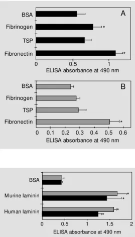
cells of each morphological phase of this fungus. The binding was measured as described in Methods and expressed by the absorbance at 490 nm. Figure 1 (A and B) represents a typical experiment from at least three independent experiments. As observed in Figure 1, both morphological phases sig-nificantly bound to fibronectin, as compared to the binding observed with BSA (P<0.05). No significant adhesion was observed for conidia assayed with TSP or fibrinogen (Fig-ure 1B). On the other hand, S. schenckii
yeast cells showed a significant extent of adhesion to fibrinogen (P<0.05) but not to TSP (Figure 1A).
Adhe sio n o f S. schenckii to murine and human laminin
Both the yeast phase and conidia of S. schenckii bound to laminin from different sources (Figure 2). No differences were ob-Figure 1 - Adhesion of S.
schen-ckii yeast cells (A) and conidia (B) t o im m obilized f ibronect in, thrombospondin and fibrinogen at 10 µg/ml compared to adhe-sion to BSA. Values w ere sub-tracted from the negative con-trol (poly-HEM A). S. schenckii
adhesion (107 cells/w ell) w as
as-sessed by ELISA w ith an anti-S. schenckii ant iserum * P< 0.05 com pared t o BSA (St udent
t-test).
Figure 2 - Binding of S. schenckii
conidia (gray bars) and yeast cells (black bars) to immobilized murine and human laminins at 10 µg/ml compared to adhesion to BSA. Values w ere subtracted from the negative control (poly-HEM A). S. schenckii adhesion (107 cells/w ell) w as assessed by
ELISA w ith an anti-S. schenckii
antiserum. * P<0.05 compared to BSA (Student t-test).
BSA
M urine laminin
Human laminin
ELISA absorbance at 490 nm
0.5 1 1.5 2
0
* *
* *
BSA
Fibrinogen
TSP
Fibronectin
BSA
Fibrinogen
TSP
Fibronectin
0 0.5 1
ELISA absorbance at 490 nm
0.1
ELISA absorbance at 490 nm 0.2 0.3 0.4 0.5 0.6 0
*
*
*
A
Laminin
Fibronectin
3 4 5 6
0
*
2
1 7
served in the rate of adhesion to human and murine laminins.
Co mpariso n o f the adhe sio n inde x fo r
laminin and FN
Since the absolute adhesion rates varied between experiments, although adhesion to fibronectin and laminin was always signifi-cant, we determined the relative adhesion index as described in Methods. Figure 3 shows that the adhesion rate to laminin and FN is almost similar. However, differences according to the fungal morphological phase were observed only for FN.
Binding o f S. schenckii to type II co llage n
Type II collagen was purified as described in Methods. The purification of chick type II collagen by this procedure yielded 35.5 ± 16 mg CII/g cartilage. The degree of purifica-tion of the CII preparapurifica-tion was evaluated by SDS-PAGE (Figure 4). A main 156-kDa protein band corresponding to the alpha chains of chick type II collagen was ob-served. The purified CII showed a high de-gree of purity in comparison to the commer-cial patterns. S. schenckii conidia and yeast cells were able to significantly attach to type II collagen (Figure 5). Conidia displayed an increased adhesion to CII compared to yeast cells (P<0.05).
D iscussio n
Sporotrichosis is a deep mycosis which may evolve to a systemic pathology in im-munocompromised individuals. The yeast phase of S. schenckii is the morphology present in lesions and is thought to dissemi-nate in the systemic disease. However, myce-lial elements have already been described in histopathological specimens of lung from sporotrichosis patients (22). Conidia are also thought to infect humans by inhalation (6).
Primary adhesion of endothelial and/or
Figure 3 - Relative adhesion in-dex of S. schenckii yeast phase (black bars) and conidia (gray bars) to immobilized fibronectin and laminin. The adhesion index w as calculated using the for-mula: A490 to ECM protein/A490
to BSA - A490 w ith poly-HEM A
(negative control). S. schenckii
binding (107 cells/w ell) w as
as-sessed by ELISA w ith an anti-S. schenckii antiserum. * P< 0.05 compared to yeast cells (Student
t-test).
Figure 4 - Analysis of chick type II collagen by 7% SDS-polyacryl-amide gel electrophoresis, w ith silver staining. Chicken CII: lane 1, 14 ng; lane 2, 20 ng; lane 3, 30 ng; lane 4, 60 ng and type VII collagen (Sigma); lane 5, 50 ng, and lane 6, 100 ng.
Figure 5 - Binding of S. schenckii
conidia (gray bars) and yeast cells (black bars) to immobilized type II collagen at 10 µg/ml in com-parison w ith the adhesion ob-served to BSA. Values w ere sub-tracted from the negative con-trol (poly-HEM A). S. schenckii
adhesion (107 cells/w ell) w as
as-sessed by ELISA w ith an anti-S. schenckii antiserum. * P< 0.05 com pared t o BSA (St udent
t-test). BSA
Adhesion index
1 1.5 2
0
*
0.5 Type II
collagen *
kDa
-120 -194
- 87 - 64
- 52
1 2 3 4 5 6
epithelial cells, as well as extracellular ma-trix components seem to be essential for successful host invasion by pathogens. In the present report we have investigated the binding capacity of two infective forms of S. schenckii (yeast form and conidia) to FN, laminin, type II collagen, TSP and fibrino-gen. The binding to BSA observed here has already been described for other pathogenic fungi such as Candida albicans (23).
Our results show that S. schenckii cells can recognize three important ECM glyco-proteins: fibronectin, laminin and type II
collagen. ECM-binding adhesins have been described for several microorganisms, in-cluding bacteria, protozoa and fungi. Most common adhesins recognize fibronectin and collagenous proteins (24). Type II collagen is present in hyaline cartilage, as well as in vitreous humor (25). In immunocompromised individuals, mainly in HIV+
patients and alcoholics, the pulmonary and articular forms of sporotrichosis are the most frequent clini-cal manifestations (5,26). This fungus was also recently isolated from aqueous humor (27). Since this apparent tropism of S. schenckii may be associated with the ability to recognize specific ECM components, we investigated binding of this pathogen to type II collagen. Our results showed that conidia and, less markedly, yeast cells bind to type II collagen significantly more than to BSA. Interestingly, S. schenckii cells bound only slightly to type I and III collagens (data not shown). Fibronectin is a ubiquitous glyco-protein found in high concentrations in the extracellular matrix and body fluid of verte-brates. Binding to fibronectin may play a role in the invading ability of microorganisms.
In the present study, S. schenckii cells strongly recognized laminin from different sources. Laminin is a major component of basement membranes, which, together with endothelial and epithelial monolayers, con-stitute the main physiological barriers to be surmounted by invading organisms. How-ever, data concerning the involvement of laminin as a target of fungal adhesins are
scarce. It has been suggested that the ability to bind basement membrane components is correlated with high virulence phenotypes. Vicentini et al. (28) have reported that the 43-kDa antigen binds to laminin, which leads to the enhancement of Paracoccidioides bra-siliensis pathogenesis, by means of laminin recognition. Whether the laminin-binding capacity of S. schenckii is critical for the onset of systemic infections remains to be established.
Taken together, our data suggest that S. schenckii presents an adhesive behavior com-parable to that of other opportunistic patho-gens, since it is able to bind both to the basement membrane and to connective ECM proteins. Indeed, this is supported by the fact that this fungus also interacts with specific molecules on human endothelial cells in vi-tro (Figueiredo CC, Lima OC, Lopes-Bezerra LM and Morandi V, unpublished data).
The present report contributes new data to the understanding of the pathogenesis of sporotrichosis. The identification of S. schen-ckii cell surface adhesin(s), as already de-scribed for other pathogenic fungi (12,29), constitutes a future challenge that could lead to new antifungal therapies.
Ackno wle dgm e nts
The authors acknowledge the skillful tech-nical assistance of Mr. Heclair Rodrigues Pimentel Filho and Mrs. Maria Cristina da Costa e Silva.
Re fe re nce s
1. Travassos LR (1985). Sporothrix schenckii.
In: Szaniszlo PJ (Editor), Fungal Dimor-phism, w ith Emphasis on Fungi Patho-genic for Humans. Plenum Press, New York.
2. Castrejón OV, Robles M & Arroyo OEZ (1995). Fatal fungemia due to Sporothrix schenckii. M ycoses,38: 373-376. 3. Heller HM & Fuhrer J (1991).
Dissemi-nated sporotrichosis in patients w ith
AIDS: case report and review of the litera-ture. AIDS, 5: 1243-1246.
4. Durden FM & Elew ski B (1997). Fungal infections in HIV-infected patients. Semi-nars in Cutaneous M edicine and Surgery, 16: 200-212.
5. How ell SJ & Toohey JS (1998). Sporotri-chal arthritis in south central Kansas. Clini-cal Orthopedic, 346: 207-214.
6. Farley M L, Fagan M F, M abry LC &
W allace RJ (1991). Present at ion of
Sporothrix schenckii in pulmonary cytol-ogy specimens. Acta Cytologica, 35: 389-395.
7. López-Ribot JL & Chaffin WLJ (1994). Binding of extracellular matrix component entactin to Candida albicans. Infection and Immunity, 62: 4564-4571.
albi-cans to extracellular matrix proteins. M i-crobiology, 141: 2681-2684.
9. Gil M L, Peñalter M C, Lopez-Ribot JL, O’Connor JE & M artinez JP (1996). Bind-ing of extracellular matrix proteins to As-pergillus fumigatus conidia. Infection and Immunity, 64: 5239-5247.
10. Nègret E, Vogel T, Levanon A, Guy R, Walsh TJ & Roberts DD (1994). The col-lagen binding domain of fibronectin con-tains a high affinity binding site for Can-dida albicans.Journal of Biological Chem-istry, 269: 22039-22045.
11. Peñalter M C, O’Connor JE, M artinez JP & Gil M L (1996). Binding of human fibronec-tin to Aspergillus fumigatus conidia. In-fection and Immunity, 64: 1146-1153. 12. Hostetter M K (1996). Adherence
mol-ecules in pathogenic fungi. Current Opin-ion in Infectious Diseases, 9: 141-145. 13. Lloyd KO & Bittoon M A (1971). Isolation
and purification of a peptido-rhamnoman-nan from the yeast form of Sporothrix schenckii. Structural and immunochemi-cal studies. Journal of Immunology, 107: 663-671.
14. Lopes LM , M endonça-Previato L, Fournet B, Degand P & Previato JO (1992). O-glycosidically linked oligosaccharides from peptidorham nom annans of Sporothrix schenckii. Glycoconjugate Journal, 9: 75-81.
15. Lopes LM , Travassos LR, Previato JO & M endonça-Previato L (1994). Novel anti-genic determinants from peptidorham-nomannans of Sporothrix schenckii.
Gly-cobiology,4: 281-288.
16. Fernandes KSS (1998). Estudo da virulên-cia do pat ógeno hum ano Sporot hrix schenckii em modelo murino in vivo e in vitro. M aster’s thesis, Universidade do Estado do Rio de Janeiro, Rio de Janeiro, Brasil.
17. Vuento M & Vaheri A (1979). Purification of fibronectin from human plasma by af-f init y chrom at ography under non-denat urat ing condit ions. Biochem ical Journal, 183: 331-337.
18. Dubernard V & Legrand C (1991). Charac-terization of the binding of thrombospon-din to human platelets and its association w ith the platelet cytoskeleton. Journal of Laboratory and Clinical M edicine, 118: 446-457.
19. Trenthan DE, Tow nes AS & Kang AH (1977). Autoimmunity to type II collagen: an experimental model of arthritis. Jour-nal of Experimental M edicine, 146: 857-868.
20. Laemmli UK (1970). Cleavage of struc-tural proteins during the assembly of the head of bacteriophage T4. Nature, 227: 680-685.
21. M orrissey JH (1981). Silver stain for pro-teins in polyacrylamide gels: A modified procedure w ith enhanced uniform sensi-tivity. Analytical Biochemistry, 117: 307-310.
22. Gori S, Lupetti A, M oscato G, Parenti M & Lofaro A (1997). Pulmonary sporotricho-sis w ith hyphae in a human immunodefi-ciency virus-infected patient. A case
re-port. Acta Cytologica, 41: 519-521. 23. Santoni G, Gismondi A, Liu JH, Punturieri
A, Santoni A, Frati L, Piccoli M & Djeu JY (1994). Candida albicans expresses a fi-bronectin receptor antigenically related to a5ß1 integrin. M icrobiology, 140: 2971-2979.
24. Patti JM & Höök M (1994). M icrobial ad-hesins recognizing extracellular matrix macromolecules. Current Opinion in Cell Biology, 6: 752-758.
25. Rest M VD & Garrone R (1991). Collagen family of proteins. FASEB Journal, 5: 2814-2823.
26. Kauffman CA, Pappas PG, M cKinsey DS, Grenfield RA, Perfect JR, Cloud GA, Tho-mas CJ & Dismukes WE (1996). Treat-ment of lymphocutaneous and visceral sporotrichosis w ith fluconazole. Clinical Infectious Diseases, 22: 46-50.
27. Vieira Dias D, Sena CM , Oréfice F, Tanure M A & Hamdan JS (1997). Ocular and con-comitant cutaneous sporotrichosis. M y-coses, 40: 5-6.
28. Vicentini AP, Geszetesi JL, Franco M F, Souza W, M oraes JZ, Travassos LR & Lopes JD (1994). Binding of Paracoccidi-oides brasiliensis to laminin through sur-face glycoprotein gp43 leads to enhance-ment of fungal pathogenesis. Infection and Immunity, 62: 1465-1469.