Prevalence of
Chlamydia trachomatis
infection among
women seen at the lower genital tract pathology clinic,
Jundiaí School of Medicine, Brazil
Prevalência de infecção por
Chlamydia trachomatis
em mulheres assistidas no ambulatório de
patologia do trato genital inferior da Faculdade de Medicina de Jundiaí, Brasil
João Bosco Ramos Borges1, Ana Carolina Marchesini1, Luciana Francine Bocchi De Stefani2, Marcus Vinícius
Gonçalves Belintani2, Thaís Andrea dos Santos2
ABSTRACT
Objective: To estimate the prevalence of Chlamydia trachomatis in a population with a high risk of sexually transmitted diseases and to compare data of the literature and the relationship of infection with the presence of human papilloma virus induced lesions. Methods: A total of 28 hybrid capture tests for C. trachomatis were collected from patients referred to the Municipal Health Division of the city of Jundiaí (SP) for the lower genital tract pathology. The results were compared with findings in the literature, and with the test results from a general population of the city of Jundiaí. Results: Of the 28 tests, 3 (10.7%) were positive. We did not find a positive association between C. trachomatis infection and the presence or aggravation of intraepithelial cervical cancer. Conclusion: Our findings showed a high prevalence of C. trachomatis infection in the population studied, but no association with human papilloma virus infection. Because the number of patients assessed was small, it is difficult to generalize from our findings. We suggest there is a need to expand screening programs for C. trachomatis, mainly in symptomatic patients and in those patients with cervical changes.
Keywords: Chlamydia trachomatis; Prevalence; Papilomavirus infections; Cervix uteri
RESUMO
Objetivo: Estimar a prevalência de Chlamydia trachomatis numa população de alto risco para doenças sexualmente transmissíveis, comparar com os dados da literatura e da população geral do próprio município e verificar sua associação com as alterações citopatológicas induzidas pelo papilomavírus humano. Métodos: Foram colhidos 28 exames de captura híbrida para C. trachomatis entre as pacientes
referendadas ao Ambulatório de Patologia do Trato Genital Inferior da Secretaria Municipal de Saúde de Jundiaí e os dados foram comparados aos da literatura e com os resultados do mesmo exame aplicado à população geral da cidade de Jundiaí (SP). Resultados: Três (10,7%) exames foram positivos na amostra das pacientes referendadas e não houve associação positiva entre a infecção por
C. trachomatis e a presença ou o agravo de neoplasia intraepitelial cervical. Conclusão: O presente estudo mostra uma prevalência elevada de infecção por C. trachomatis na população referendada, mas não consegue associá-la à infecção por papilomavírus humano, provavelmente devido ao número reduzido de pacientes da amostra. Além disso, reforça-se a necessidade de programas de detecção de C. trachomatis, sobretudo em pacientes sintomáticas ou com alterações no colo uterino.
Descritores: Chlamydia trachomatis; Prevalência; Infecções por papilomavírus; Colo do útero
INTRODUCTION
Sexually transmitted infections are frequent and constitute a serious public health problem in almost all countries. The Centers for Disease Control and Prevention (CDC), in the United States, underline
that there are more new cases each year of Chlamydia
trachomatis (CT) than other sexually transmitted
diseases (STD)(1,2).
CT, a member of Chlamydia family, is a single genus
of Chlamydiacea that is Gram-negative and obligatorily
intracellular. It is in the same genus as Chlamydia
Study carried out at Faculdade de Medicina de Jundiaí – FMJ, Jundiaí (SP), Brazil.
1 Discipline of Gynecology, Faculdade de Medicina de Jundiaí – FMJ, Jundiaí (SP), Brazil. 2 Undergraduate Medical Course, Faculdade de Medicina de Jundiaí – FMJ, Jundiaí (SP), Brazil.
Corresponding author: João Bosco Ramos Borges – Rua Francisco Telles 250 – Vila Arens – CEP 13202-550 – Jundiaí (SP), Brasil – Tel.: 11 4587-1095 – E-mail: [email protected] Received on: March 1, 2011 – Accepted on: Jun 20, 2011
psittaci and Chlamydia pneumoniae and is classified into serotypes. Serotypes D-K are responsible for causing urethritis and sexually transmitted cervicitis; serotypes A, B, Ba and C are the agents of trachoma; and serotypes L1, L2 and L3 are responsible for lymphogranuloma
venereum(3,4). In adults such agents can cause eye
diseases such as trachoma and conjunctivitis. Children whose mothers had genital infection with CT may
develop conjunctivitis and pneumonia(5).
CT is the leading agent of urogenital tract disease in both sexes. However, it has a higher impact on women’s reproductive systems. It is estimated that 10 to 15% of women of reproductive age will have at least one symptomatic or asymptomatic episode of pelvic
inflammatory disease (PID) caused by CT(6). Even if an
episode is asymptomatic, it can cause salpingitis which is an important cause of infertility and is highly associated
with ectopic pregnancy(2).
In the United States, the prevalence of CT infection among users of family planning clinics ranges from 4.5 to
12.4%(2). However, because in most cases, CT infection
is asymptomatic and diagnosis is delayed and/or difficult, the precise number of young adults infected by CT
remains unknown(7). The World Health Organization
(WHO) estimates that approximately 90 million new
cases of infection by Chlamydia appear in the world every
year, being 4 million cases in the United States(8), which
cost more than US$ 4.2 billion yearly(2). In Brazil, there is
no such an official measure on this infection(9).
The high prevalence of STD among women in developing countries has suggested the need to
expand programs to detect CT infection(10,11). The
epidemiologic data suggest a prevalence of CT infection in about 5% of the asymptomatic population whereas this frequency increases to almost 20 to 40%
in patients with STD(3). In a study done in the city
of Porto Alegre (RS), Brazil, from 1987 to 1990, CT infection was found in 25.5% in 235 patients with
pelvic inflammatory disease(12).
The Brazilian National Coordination of STD/ AIDS estimates a prevalence of 3.5% of infection by
Chlamydia in sexually active women(13). In Brazil, the growing transmission of Human immunodeficiency virus/ Acquired immune deficiency syndrome (HIV/ AIDS) among heterosexuals suggests the necessity of obtaining epidemiologic data in the population to
establish strategies for more adequate interventions(14).
Codes et al.(7) recommended an investigation to
determine the presence of STD in even asymptomatic women, which diverges from the current practice in Brazil and in other developing countries that look for STD in people who are symptomatic or have risk factors.
Detecting CT infection is important because if this infection is not treated, it can cause severe complications
such as pelvic inflammatory disease that leads to chronic pelvic pain and infertility. In the pregnant-puerperal cycle, CT infection is associated with abortion, premature labor, premature rupture of membranes and
fetal lung prematurity(15,16) and it also can be transmitted
neonatally(15).Therefore family planning services in the
United States routinely test for CT infection(7).
The use of CT screening test that aims to identify even asymptomatic patients reduces the incidence of
PID by approximately 56%(16-18). Recurrent episodes
of infection increase the risk of complications and the
chance of acquiring the HIV infection(11).
In addition to the primary infection, recurrence occur more often in people infected under 20 years old because the evolved immunity is only partially protective against the 15 or more serotypes of CT.
CT may play a role in the development of other infections. It may increase the oncogenic potential of human papilomavirus (HPV) with regard to cervical
cancer(19-23). The high incidence and the long duration of
HPV latency period suggest, however, that other factors or infections are involved, increasing the risk of CIN throughout several mechanisms including modulation of
the carrier’s immunity(3,14). There are also evidences of
the association of CT and the herpes simplex virus in the induction of cervical intraepithelial neoplasia (CIN).
The higher frequency of CT infection in women with abnormal Pap smear (high-and low-grade lesions) suggests that this co-infection initiates the carcinogenic
action of HPV(24). There is evidence that CT infection
can worsen a neoplastic lesion and speed the progression
of the cervical cancer(25). In addition to the synergy
with HPV to promote cancer, the presence of CT is considered a marker for exposure to HPV because of
the similar risk factors for both these infections(6,25).
OBJECTIVE
To determine the prevalence of CT infection in patients referred to a lower genital tract pathology clinic and to verify possible association with HPV virus induced lesions.
METHODS
A cross-sectional study of patients referred to the Municipal Health Division, city of Jundiaí (SP), lower genital track pathology clinic was performed. The patients were invited to participate, and those who agreed had a sample of cervical secretion collected and tested by captured hybrid test to detect CT infection.
Kits from QIAGEN® that are based on amplification and detection of CTs DNA were used for the CT detection. These kits had analytic sensibility of 1 pg/mL equivalent to 0.1 bacterial copy for each cell. The results from the test were later compared to those of women who underwent collection during oncotic routine cytology collection at the basic health units of the city of Jundiaí during the same period.
The collection of the secretion to detect CT obeyed the following instructions: patients were required to abstain from sex for 3 days, and they could not be menstruating. The collection was performed before any cytological or colposcopy tests. First, excess vaginal secretions were removed, then a cytologic brush was introduced 1 to 1.5 cm into the endocervical canal until its larger bristles reached the ectocervix, after which it was rotated 5 times clockwise. The brush was then removed and placed in the tube with the kit solution, and the shank was broken off.
The presence of induced lesions by HPV was established by a cervix-vaginal cytology (vaginal smears) or by histological study of a cervical biopsy.
Statistical analysis
Median, standard deviation, absolute (n) and relative (%) frequencies were determined. The association
between Chlamydia alterations and the lesions was
assessed using the χ2 test or the Fisher’s exact test –
when 25% or more of the cells in the table reached an expected value fewer than 5 – ALTMAN.
Age, gestation and parity were studied regarding normal distribution using the Kolmogorov-Smirnov test and by normal plot graph. Because the distribution of points was found not to be normal, the values were compared to check the presence or changes in lesions by using a Mann-Whitney test.
Calculations were carried out with SAS, version 9.02. A p value of < 0.05 was considered significant.
RESULTS
A total of 28 patients were studied and hybrid captured tests were performed during August 2009 through June 2010 to detect CT in the studied women.
Patients’ ages ranged from 17 to 83 years, mean
31.2 ± 14.4 years. The average number of gestation was
1.8 ± 2.3 and parity was 1.6 ± 2.1 (Table 1).
In the gynecologic examinations test 25% of the patients showed lesions, being condylomatous lesions in the vulva and/or acetowhite lesions (Table 2). Some patients actually had both condylomatous lesions of the vulva and acetowhite lesions.
Table 1. Descriptions of the sample by age, parity and gestation
Variables Number Mean Standard
deviation Median Minimum Maximum
Age 28 31.2 14.4 29.0 17.0 83.0 Gestation 28 1.8 2.3 1.0 0.0 11.0
Parity 28 1.6 2.1 1.0 0.0 10.0
Table 2. Alteration in the gynecologic test
Characteristics of lesions Number %
No lesion 21 75.0
Vulvar condylomatous 3 10.7
Acetowhite lesion (cumulative data) 6 21.4
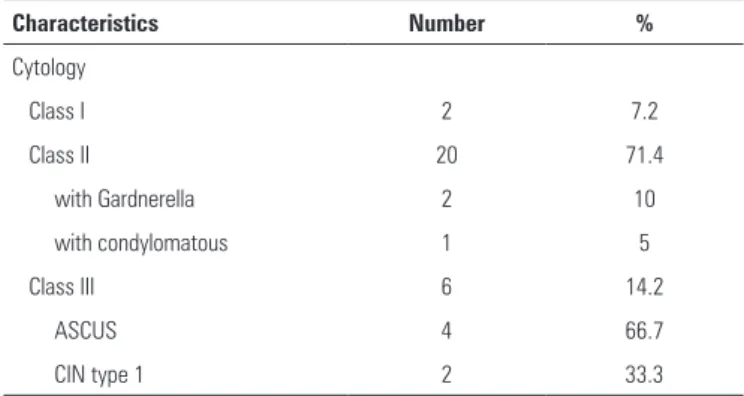
Table 3. Sample cytology characteristics according to Papanicolaou classification
Characteristics Number %
Cytology
Class I 2 7.2
Class II 20 71.4
with Gardnerella 2 10
with condylomatous 1 5
Class III 6 14.2
ASCUS 4 66.7
CIN type 1 2 33.3
ASCUS: atypical squamous cells of undetermined significance; CIN: cervical intraepithelial neoplasia.
7% 14%
79%
No lesion
CIN
ASCUS
CIN: cervical intraepithelial neoplasia; ASCUS: atypical squamous cells of undetermined significance.
Figure 1. Findings grouped by Pap smear cytology
According to the Papanicolaou (Pap) classification, 71.4% of patients had class II Pap and 14.2% had class III Pap (Table 3). The percentages of the classes’ specifications of the Pap smear were based on the absolute number in each class but for the classes, on the sample number. The main findings are shown in figure 1.
As for the reasons for cervical secretion collection, the most common was vaginal discharge (21.4%) and cervical intraepithelial neoplasia (CIN) that was also present in 21.4% of patients. The reason for collection could be more than one, so the total of percentages might be higher than 100%, considering that the same test could be counted twice (Figure 2).
in urban regions, and higher than 10% in asymptomatic
recruits submitted to routine physical tests(17). In
addition, 15 to 20% of heterosexual men seen in STD clinics could be positive for CT. In women, the incidence of CT cervicitis was about 5% among asymptomatic college students and patients seen in a US prenatal program, increasing to 10% in those patients who attended family planning clinics and more than 20% in
those seen in STD clinics(17).
Estimates of the prevalence of CT vary according to the population studied and the diagnostic method used. Teles et al., studying the use of a immunofluorescence test in 407 women from a family planning clinic in
Campinas, found a prevalence of 6.6%(26). Researchers
in the Municipal Division of Jundiai using a different method, found a similar prevalence of 6.9%, in 2009, (49 positive cases within 711 collections) among women
who sought attendance at Prevalence of Chlamydia
trachomatis infection the health service for the yearly cervical/vaginal cytology collection. Both studies presented a lower prevalence than the one found in the present study, probably because our patients were referred to a lower genital tract clinic.
Unlike Oliveira et al.(27) this study did not find
significant associations among CT infection, number of gestations, and parity as well as it did not find a significant association among gestation number, parity, and the presence of low or high genital tract lesions. However, this study might have lacked sufficient power to detect such association if present.
In this study, testing for CT was done in 21.4% of patients because of persistent vaginal discharge and in 3.6% because of vaginal bleeding after intercourse. According to the literature, these symptoms are the most frequent complaints in symptomatic patients with CT. In addition, the CT microorganism is the second more found on the endocervix, and can cause acute and
mucopurulent cervicitis(5,14).
In the present study, we also tested for CT infection in patients who were followed up for CIN, to assess correlations between CT infection and HPV infection. Previous researchers have found reports of CT infection rates ranging from 11% to 16.7% in CIN carriers and can affect 55.6% of asymptomatic squamous cells of undetermined significance (ASCUS) carriers. These indexes are considered high when compared with patients who are negative for HPV and are strongly correlated with such infections and the appearance
of cellular atypia(28,29). In our study, a combination of
CT infection and CIN type I was seen in only in a few number of cases. Simultaneous infection by CT and the presence of ASCUS, CIN II Pap, or CIN III Pap was not observed, perhaps because of the small size of the sample.
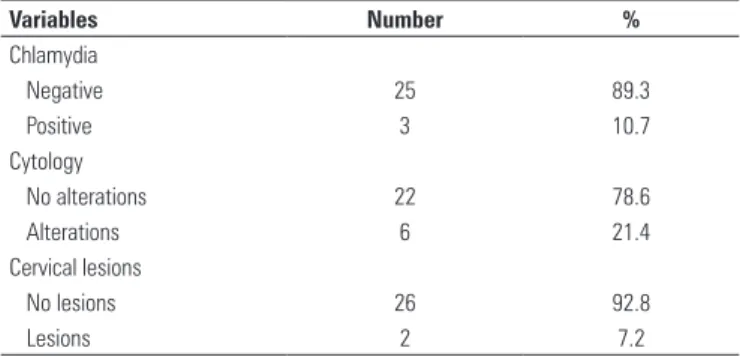
Variables Number %
Chlamydia
Negative 25 89.3
Positive 3 10.7
Cytology
No alterations 22 78.6
Alterations 6 21.4
Cervical lesions
No lesions 26 92.8
Lesions 2 7.2
Table 4. Prevalence of Chlamydia, cytology result and presence of cervical lesions
Figure 2. Main reasons for sample collection
CIN: cervical intraepithelial neoplasia; CT: chemotherapy; ASCUS: undetemined squamous cells
Vaginal bleeding after intercourse Follow-up for CIN type 3 HIV positive Follow-up after CT Follow-up for CIN. Ulcerations on the vulva Condylomata Acute cervicitis/ CIN persistent Previous cytology with ASCUS Irregular periods Acute pelvic pain Vulva burning Follow-up of IUD After treatment of pelvic TB Dyspareunia Ectopic pregnancy Rape Persistent vaginal discharge
0.0 5.0 10.0 15.0 20.0 25.0
3.6 3.6 3.6 3.6
21.4 3.6
14.3 3.6
10.7 3.6
3.6 7.1 3.6
7.1 3.6
21.4
It was also found that cytological alterations were more common in women who had more gestations; and no cytological alterations were found in nulligravida (p = 0.04). Age and parity were not significantly related to the presence of cytological changes (p > 0.05).
DISCUSSION
In countries where CT must be reported, stated prevalence ranges from 2.6% (Hungary) to 13.9% (United States), with a mean prevalence of 5.58%.
Although not being the objective of this study, it is interesting to comment that some American studies have shown that the prevalence of urethral infection by
Considering that CT is an etiologic factor that can induce metaplasia and chronic inflammation in the
cervix(24), in this sample, however, only a minority of
cases showed cytology classes II Pap and III Pap as being positive for CT.
Literature reports risk factors for CT infection as being the use of contraception as well as age (mainly
teenagers and young adults)(3,4), Caucasian women
who had history of STD for more than 12 months, and
chronic pelvic pain(30). In this study, patients with CT
infection showed similar data.
CONCLUSION
The prevalence of CT in the studied group was higher than that found in the city of Jundiai and the global mean referred in the literature. Because of the small sample size and short follow-up we were not able to show an association of CT infection with cellular atypia, and worsening of cervical atypias. Nevertheless, the high prevalence of CT obtained in this study reinforces the need for programs to detect CT infection in patients with symptoms suggestive of this infection or with cervical changes.
REFERENCES
1. Centers for Disease Control and Prevention. Atlanta, Ga, USA: Department of Health and Human Services; 2009. Sexually transmitted disease surveillance, 2010. [Techical Report].
2. Marques CAS, Menezes MLB, Coelho IMG, Marques CRCL, Celestino LC, Melo MC, et al. Infecção genital por Chlamydiatrachomatis em casais atendidos em ambulatório de esterilidade conjugal. DST-J Bras Doenças Sex Transm. 2008;19(1):5-10.
3. Giuliano AR, Denman C, Guernsey de Zapien J, Navarro Henze JL, Ortega L, Djambazov B, et al. Design and results of the USA-Mexico border human papilomavirus (HPV), cervical dysplasia, and Chlamydia trachomatis study. Rev PanamSaludPublica/Pan Am J Public Health. 2001:9(3):172-81.
4. Trabulsi LR, Martinez MB. Chlamydia. In: Trabulsi LR, Alterthum F (Eds.). Microbiologia. São Paulo: Atheneu; 2005. p. 441-3.
5. Miller WC, Ford CA, Morris M, Handcock MS, Schmitz JL, Hobbs MM, et al. Prevalence of chlamydial and gonococcal infections among young adults in the United States. JAMA 2004;291(18):2229-36.
6. Finan RR, Tamim H, Almawi WY. Identification of Chlamydia trachomatis DNA in human papillomavirus (HPV) positive women with normal and abnormal cytology. ArchGynecolObstet. 2002;266(3):168-71.
7. Codes JS, Cohen DA, Melo NA, Santos BA, Codes JG, Silva Júnior JC, et al. Detecção de doenças sexualmente transmissíveis em clínica de planejamento familiar da rede pública no Brasil. RBGO. 2002;24(2):101-6.
8. World Health Organization (WHO). Global prevalence and incidence of selected curable sexually transmitted infections. Overview and estimates. Geneva: WHO; 2001.
9. Seadi CF, Oravec R, Poser B, Cantarelli VV, Rosstti ML. Diagnóstico laboratorial da infecção pela Chlamydiatrachomatis: vantagens e desvantagens das técnicas. J Bras Patol Med Lab. 2002;38(2):125-33.
10.Temmerman M, Kidula N, Tyndall M, Rukaria-Kaumbutho R, Muchiri L, Ndinya-Achola JO. The supermarket for women’s reproductive health: the burden
of genital infections in a family planning clinic in Nairobi, Kenya. Sex Transm Infect.1998;74(3):202-4.
11. Wilkinson D, Ndovela N, Harrison A, Lurie M, Connolly C, Sturm AW. Family planning services in developing countries: an opportunity to treat asymptomatic and unrecognised genital tract infections? Genitourin Med. 1997;73(6): 558-60.
12. Freitas F, Menke CH, Rivone WA, Passos EP. Rotinas em ginecologia. 4a ed. Porto Alegre: Artmed; 2001. p. 110-33.
13. Varella RQ, Passos MRL, Pinheiro VMS, Lopes HR, Santos SB, Guimarães CC, et al. Pesquisa de Chlamydiatrachomatis em mulheres do município de Piraí - Rio de Janeiro. DST- J Bras Doenças Sex Transm. 2000;12(3):27-44. 14. Giffin K, Lowndes CM. Gender, sexuality, and the prevention of sexually
transmissible diseases: a Brazilian study of clinical practice. Soc Sci Med. 1999;48(3):283-92.
15. CDC. Chlamydia trachomatis genital infections - United States, 1995. Morb Mortal Wkly Rep. 1997;46(9):193-8.
16. Black MC. Current methods of laboratory diagnosis of Chlamydia trachomatis infections. Clin Microbiol Rev. 1997;10(1):160-84.
17. Scholes D, Stergachis A, Heidrich FE, Andrilla H, Holmes KK, Stamm WE. Prevention of pelvic inflammatory disease by screnning for cervical Chlamydia infection. N Engl J Med. 1996;334(23):1362-6.
18. Seadi FS, Oravec R, Poser BV, Cantarelli VV, Rossetti ML. Diagnóstico laboratorial da infecção pela Chlamydiatrachomatis: vantagens e desvantagens das técnicas. J Bras Patol Med Lab. 2002;38(2):125-33.
19. Hallsworth PG, Hefford C, Waddell RG, Gordon DL. Comparison of antigen detection, polymerase chain reaction and culture for detection of Chlamydia trachomatis in genital infection. Pathol.1995;27(2):168-71.
20. Sedlecki K, Markovic M, Rajic G. [Risk factors for Clamydia infections of the genital organs in adolescent females]. Srp Arh Celok Lek. 2001;129(7-8):169-74.
21. Weinstock H, Dean D, Bolan G. Chlamydia trachomatis infections. In: Sexually transmitted diseases in the AIDS era: part II. Infect Dis Clin N Am. 1994;8(4):797-819.
22. Gaydos CA, Crotchfelt KA, Howell MR, Kralian S, Hauptman P, Quinn TC. Molecular amplification assays to detect chlamydial infections in urine specimens form high school female students and monitor the persistence of chlamydial DNA after therapy. J Infect Dis. 1998;177(2):417-24.
23. Smith JS, Munoz N, Herrero R, Eluf-Neto J, Ngelangel C, Franceschi SX, et al. Evidence for Chlamydia trachomatis as a human papillomavirus cofactor in the etiology of invasive cervical cancer in Brazil and the Philippines. J Infect Dis. 2002;185(3):324-31.
24. Attapatu AF, Prússia PR, Boyer V, Levett PN. A prospective study of asymptomatic Chlamydia trachomatis in Barbadian women. J Obstet Gynecol. 1999;19(5):506-8.
25. Oliveira ML, Amorim MMR, Souza ASR, Albuquerque LCB, Costa AAR. Infecção por Chlamydia em pacientes com e sem lesões intra-epiteliais cervicais. Rev Assoc Med Bras. 2008;54(6):543-7.
26. Teles E, Hardy E, Oliveira UM, Elias CJ, Faúndes A. Reassessing risk assessment: limits to predicting reproductive tract infection in new contraceptive users. Int Fam Plan Perspect. 1997;23:179-82.
27. Oliveira ML, de Amorim MMR, Souza ASR, Albuquerque LCB, Costa AAR. Infecção por Chlamydia em pacientes com e sem lesões intra-epiteliais cervicais. Rev Assoc Med Bras. 2008;54:506-12.
28. Marques CAS, Menezes MLB. Infecção genital por Chlamydiatrachomatis e esterilidade. DST- J Bras Doenças Sex Transm. 2005;17(1):66-70.
29. Beslagic E, Jasminka G, Mahmutovic S. Detection of Chlamydia trachomatis in cervical smear samples with determined HPV. Med Arh. 2004;58(3):143-4. 30. Stamm WE. Infecção por Chlamydia. In: Fauci AS, editor. Harrison: Medicina