Received on 14 July 2004; revised 20 October 2004.
Address for correspondence: Dr. Dr. Elizabeth N De Gaspari. Instituto Adolfo Lutz.355,11 andar, Zipcode:01246-902, São Paulo, Brazil. Phone (55) (11) 3068-28-98. Fax: (55) (11) 3085-35-05. E-mail : egaspari@ial.sp.gov.br
The Brazilian Journal of Infectious Diseases 2004;8(6):407-418 © 2004 by The Brazilian Journal of Infectious Diseases and Contexto Publishing. All rights reserved.
Production of Monoclonal Antibody to Subtype 9 of
Neisseria meningitidis
and the Distribution of this Subtype in Brazil
Elza F.T. Belo, Ligia M.C.C. Coutinho, Adolfo Lutz Institute, São Paulo/SP, Brazil Aline S. Ferraz and Elizabeth N. De Gaspari
A new monoclonal antibody (5F81A4P1.9), which is specific for subtype 9 antigen of meningococci, was studied. The antibodies were raised against a previously non-typable (NT) serogroup B strain from Brazilian patients and were found to react with the subtype antigen of prototype reference strains for subtype 9 (M982), as well as with those of homologous strains. The subtype 9 epitope was found in 6.8% of serogroup B strains among 602 strains of Neisseria meningitidis case isolates, including representative isolates from Brazilian states. Subtype P1.9 was predominantly related to serogroup B in Brazil among the isolates collected during the N. meningitidis epidemic in 1992. No significant differences were observed in the occurrence of subtype P1.9 among strains isolated from several Brazilian states. Fluorescence-activated cell-sorter analysis showed that 5F81A4 MAb recognized a 46 kDa protein on the surface of a homologous strain of N. meningitidis (B:4:P1.9). These results, in association with a bactericidal activity assay for 5F81A4, and with experimental passive protection in mice, demonstrated the importance of subtype 9 class 1 proteins of N meningitidis in Brazil. Serotyping is essential for the development of vaccination strategies.
Key Words: Neisseria meningitidis, monoclonal antibodies, Brazilian meningococcus strains, serotyping, subtyping.
Neisseria meningitidis is an encapsulated bacteria
that has been the cause of severe diseases, both in developing countries and the western world. Infection with encapsulated bacteria can be circumvented by immunization with type-specific polysaccharide-protein conjugated vaccines generating antibodies to the capsular polysaccharides. The polysaccharide from serogroup B meningococci, a polymer of alfa-2,8-neuraminic acid, is a common epitope that is also found on the surface of human cells. For this reason, many investigators have chosen it to develop outer membrane protein-based vaccines against meningococci [1].
Since 1988, large epidemics of meningococcal disease (MD) due to meningococcus B disease (MenB) have been occurring in the greater São Paulo city (GSP) region, in the state of São Paulo [2].
The outer membrane proteins of N. meningitidis
have attracted attention mainly for two reasons; their use for the classification of meningococcal isolates into serotypes and subtypes, and as potential components of vaccines against this pathogen. Meningococci reveal higher genetic diversity than the most other human pathogenic bacteria. This can be partly explained by horizontal intraspecies recombination and incorporation from closely related
Neisseria species [3]. Traditionally, strains have been
typing nomenclatureis B:4:P1.9, indicating that it is meningococci serogroup B, serotype 4, and subtype P1.9.Based on the antigenic properties of this lipopolysaccharide, renamedlipooligosaccharide (LOS) because of its relatively short sugarchain, another 13 immunotypes, designated by the letter L, canbe distinguished [5]. Additional typing is achievable by using antigenic properties of immunoglobulin A1 (IgA1)proteases and N.
meningitidis pili [6].
Epidemiological studies of MD were not possible prior to the development of methods for serogroup identification of strains [7]. Serotyping has enabled precise studies on the prevalence of serogroup B and C strains as sources of infection. This method has been used to assess strains isolated from meningococcal meningitis outbreaks, and to compare the serotype strains isolated from patients and carriers.
Until recently, hyperimmune sera were used to type bacterial antigens; however, obtaining high-grade antisera was difficult, and typing was time-consuming, as an extensive serum absorption procedure was required, and elucidation of numerous cross-reactions was necessary [8]. The advent of monoclonal antibodies (Mabs) has eliminated many technically problematic issues, and it has greatly improved the accuracy and reproducibility of meningococcal serotyping procedures. Nevertheless, some types and serotypes are still identified by means of polyclonal rabbit sera, due to a lack of corresponding MAbs.
We characterized a monoclonal antibody to the class 1 outer membrane protein of N. meningitidis subtype 9, and we examined the distribution of this subtype epitope among Brazilian meningococcal strains. (Parts of this work were presented at the Tenth International Pathogenic Neisseria Conference, Winchester, England [9]. Whole cell suspensions of 602 case strains of N.
meningitidis from Brazil included 427 strains from
different subtypes, and 175 (NT) strains previously analyzed with P1.9 monoclonal antibody. The group B strains were isolated in 1992 from patients with meningococcal disease, which were screened on dot blots with a panel of serotypes and MAb subtypes, as described elsewhere [10].
Material and Methods
Growth and maintenance of bacterial strains
The N. meningitidis serogroup B strain used for
MAb production was isolated from an MD epidemic that occurred in 1992 in GSP (case strain), and it was initially classified as serotype 4 and subtype NT (N368/ 88). Reference and case strains of N. meningitidis: 120M, 7880, 7889, 44/89, 1002/90, 337/90, 735/ 90, M981, M1027, M992, M978, M982, M136, 1080, B16B6, 2996, S3446, H355, H44/76, 60E, 6557, M990, 190I were used (Table 1). The new monoclonal antibody meningococcal strains were maintained at –70oC in skim milk or were lyophilized
and stored at 4oC. N. meningitidis strainswere grown
on Tryptic Soy Agar (TSA) (Difco Laboratories, Detroit, MI) plates supplemented with 1% horse serum in a 5% CO
2 atmosphere for 16 to 18 h at 37°C in a
candle extinction jar. All of the meningococci were isolated from blood or cerebrospinal fluid specimens from patients with systemic disease, and the bacteria were deposited in the culture collection of the Adolfo Lutz Institute, São Paulo, SP, Brazil. Following biochemical characterization, the strains were serogrouped at the Bacteriology Section of Adolfo Lutz Institute by the slide agglutination technique, using antisera produced against nine major capsular serogroups. These antisera were prepared by the National Reference Center for Meningitis (Adolfo Lutz Institute). The bacteria used in this study were serosubtyped by dot-blotting, using Mabs.
Monoclonal antibody production and characterization
Production of MAb was made using a previously described technique [11]. Briefly, BALB/c mice were immunized with a saline suspension of N. meningitidis
368/88, containing approximately 108 bacteria that had
been heat-inactivated at 56oC for 30 min. Mice were
P3X63-Ag 8.653 mouse myeloma cells at a ratioof 4:1, as previously described. Positive clones were selectedby enzyme-linked immunosorbent assay (ELISA) using plates coatedwith 368/88 outer membrane complex (OMC). Western blot analysiswas performed to confirm the binding of the MAb to class 1 OMP(46 kDa) of the 368/88 strain. Ascites fluids were produced by means injectionof 5 ×10⁶ hybridoma
cells into pristane-primed BALB/c mice. Ascites fluids were pooled, the titers were determined, and they were then storedat -20°C in aliquots. All procedures with animals were in accordance with the principles of the Brazilian Code for the Use of Laboratory Animals. The MAbs class and subclass were determined by ELISA, using peroxidase-conjugated anti-mouse immunoglobulin following the manufacturer’s (mouse hybridoma subtyping kit; Boehringer Mannheim Biochemicals, Indianapolis) instructions.
Dot blot analysis
The cell suspension (1µL of bacteria) was suspended in PBS, pH 7.4, with 0.02% sodium azide. Cells were heat-inactivated at 56oC for 30 min and the absorbance
of the suspension was adjusted to OD 0.1 at 650 nm using a Coleman 6A Junior Spectrophotometer. The
bacteria were dotted onto nitrocellulose membranes, and the membranes were dried for 10 min at room temperature (RT) and used immediately.Membranes were blocked for 30 min with 5% skim milk (Nestle, Brazil). MAbs of various specificities, diluted to 1:10,000 in blocking buffer, were added.Membranes were incubated overnight at RT on a rotator, and then washed three times with PBS. The membranes were placed in a solutioncontaining 1µg/mL of phosphatase-labeled goat anti-mouse immunoglobulinG (IgG) plus IgM (Kirkegaard & Perry Laboratories, Inc., Gaithersburg,MD) diluted in blocking buffer, and allowed to react for2 h at RT on a rotator. The membranes were washed three timeswith PBS, and one time with 50 mM Tris (pH 8.0) and developed adding1 mg of naphthol AS-MX phosphate per mL plus 2 mg of Fast Red TR saltper mL in 50 mM Tris buffer (pH 8.0) (Sigma Chemical Co., St. Louis,Mo).
Preparation of OMC of N. meningitidis
The OMC was prepared from N. meningitidis
Brazilian epidemic strain N368/88. Whole cells were suspended in lithium acetate buffer pH 6.0, containing 20 mM sodium chloride and 10 mM sodium EDTA, and then were shaken with 6-mm glass beads at 300 rpm for 2 h. The supernatant was collected and centrifuged at 40,000 g for 2 h. The pellet was washed three times, and then suspended in distilled water. Each preparation was diluted to a final concentration of 1mg protein per mL and stored at -70oC in aliquots. Protein
concentrations of OMC were determined by the Lowry technique [12].
SDS-PAGE and immunoblotting
SDS-PAGE was carried out in 13% acrylamide gels measuring 16 X 16 X 0.15 cm. OMC at a concentration of 2.5 mg of protein permL and low-molecular-weight protein standards (Amersham & Biociences) were mixed 1:1 with treatment buffer (0.125M Tris-Cl (pH 6.8) , 4% SDS, 20% glycerol, 10% 2-mercaptoethanol)and boiled for 10 min before being loaded onto the gels. Gelswere run at a constant current of 30 mA per gel at 10°C for approximately 4 h. The gels were then stained with Coomassie brilliant blue R-250or blotted onto nitrocellulose filters (0.22-µm pore size) BioRad at30 V overnight at 10°C. Immunoblotting was performed as describedfor dot blotting, except for the primary antibody, which was diluted 1:2000.
Bactericidal activity
The bactericidal assay was carried out using Brazilian epidemicserogroup B N homologous strain 368/88 in 96-wellplates, as described by De Gaspari and Zollinger [13], with some modifications. Briefly, the final reaction mixture (50 µL) contained25 µL of serial twofold dilutions of test serum that hadbeen heat-inactivated at 56oC for 30 min, 12.5 µLbaby rabbit
Table 1. Reference and case strains of Neisseria meningitidis used in Dot ELISA for typing using the monoclonal 5F81A4
suspensionof exponential-phase meningococci (60 cfu per well), grownin agar containing 1% (v/v) horse serum. The reaction mixturewas incubated at 37°C for 30 min, and 130 µL trypticsoy agar (TSA), cooled to approximately 45°C, was addedto each well. The mixture was allowed to solidify and the plateswere incubated for 18 h at 37°C in 5% CO2. Viable bacteria countswere done at time 0 (t0), before incubation with complement,in order to estimate the number of cfu for 100% survival,by plating 12.5 µL bacterial stock suspension onto a 150 x 150mm TSA plate. Control wells included on each microtiter platecontained: (i) bacteria, complement and buffer
(complement-dependentcontrol) or (ii) bacteria, heat-inactivated complement andbuffer (complement-independent control). In addition, a positivecontrol was included in each assay, consisting of serial dilutionsof a known positive serum. Assays were carried out in duplicate [14]. The bactericidaltiter of the serum was defined as the reciprocal of the serumdilution that killed 50% of the bacteria at t0.
Bacteremia
A mouse model of infection [11] was used to determine the protective capacity of 5F81A4MAb .
Strains Serogroup Serotype Subtype
120M A NT NT
7880 A NT P1.3
7889 A 4 NT
44/89 B 4 P1.15
1002/90 C 2b P1.3,6
337/90 B 9 P1.14
735/90 B 8 P1.6
M981 B 4 NT
M1027 A 4 21
M992 B 5 P1.1,7
M990 B 6 P1.6
M978 B 8 P1.1,7
M982 B 9 P1.9
M136 B 11 NT
M1080 B 1 P1.1,7
B16B6 B 2a P1.2
2996 B 2b P1.2
S3446 B 14 P1.6
H355 B 15 P1.15
H44/76 B 15 P1.7,16
60E C 16 P1.1,7
6557 B 17 NT
A B C D E F G H
Figure 2. Level of bacteremia present in BALB/c mice after 0.25 mL 5F81A4 hybridoma ascite fluid inoculation
diluted in PBS. a: PBS as control; b: MAb 1:2,000; C: MAb 1:5,000; d: MAb 1:10,000, e: MAb 1:20,000 dilution. The bacteria B:4:P1.9 (homologous strain) 4x10³ bacteria /mL i.p . The level of bacteremia induced by
the meningococcal strain was calculated from at least two separate experiments as the arithmetic mean CFU per mL of blood ± the standard deviation. Each value represents a mean of the data obtained from at least five animals.
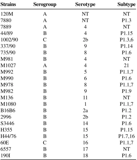
Figure1: Dot-blot assay with whole cells of Neisseria meningitidis of Brazilian isolates serogroup B. Non
typable strains 1a: culture supernatant monoclonal antibody P1.9 diluted 1:500; 1b: ascite monoclonal antibody P1.9 diluted 1:2,000; 2: immunoreactivity of ascite of monoclonal antibody P1.9 with reference strains M 982 (P1.9) and strains described in Table.1; 3: analysis of proteins in N. meningitidis outer membrane components preparations. 3a and 4c: molecular weight standards are indicated in kilodaltons; b: SDS-PAGE profiles after staining with Coomassie blue of OMC preparations of the strains N 368/88; 4a: immunoblot reactivity of N.
meningitidis N 368/88; 4c: N 16/92; 4e: N17/92 case strains with the MAb 5F81A4, P1.9;4b: N368/88; 4d:
N16/92, and 4f: N17/92 N. meningitidis case strains imunoreactivity with the new monoclonal antibody (1A11E10) against class 3 (37 kDa) of N. meningitidis.
1 2 3
Different hours
0 200 400 600 800 1000
B
a
c
te
ri
a
l
le
v
e
ls
/m
Figure 3. Dot-plots of indirect immunofluorescence-activated cell sorter analysis of Neisseria meningitidis
The mice demonstrated uniform and high susceptibility to virulent N. meningitidis strain 368/88 (B:4:P1.9), developing progressive bacteremia. Mouse inoculations were made with meningococci scraped from plates after approximately 20 h culture, suspended in PBS to a concentration of 4 x 10³ bacteria/mL. All inoculations
were completed within 15 min after preparing the suspensions. Based on viable inocula counts before and after injection into the animals, this time lapse did not reduce viability. Female BALB/c mice (C5-sufficient) were obtained from the Adolfo Lutz Institute. Mice were inoculated with heat-inactivated mouse MAb ascites fluid via intravenous (i.v) injection. Five mice were inoculated with each MAb concentration (1:2,000, 1:4,000, 1:8,000 and 1:16,000 dilutions of the IgG2a isotype in 0.25 mL). The bacteria B:4:P1.9 (homologous strain) were administered via intraperitoneal (i.p) injection about 10 minutes later. Bacteremia was monitored by cutting the tip of the mouse’s tail with a sharp scissors and collecting 10 mL of blood with an automatic pipette. Each blood sample was inoculated and spread with a sterile glass road onto TSB-supplemented agar with 1% heat-inactivated normal horse serum containing vancomycin, colistin, and nystatin (Sigma Chemical Co, Biotechnology, Piscataway, NJ) to inhibit the growth of other microorganisms. The bacterial colonies were counted after incubating at 37oC in the presence of 5% CO
2 for
18 h. The rate of bacteremia for each group of animal was calculated as the arithmetic mean log 10 of the number of colony-forming units per milliliter of blood.
Determination of MAb 5F81A4 reactivity with the surface of N. meningitidis by flow cytometry
The ability of 5F81A4 MAb to bind to the surface of pathogenic strains of group B N. meningitidis was determined by means of flow cytometry, through indirect immunofluorescence of N. meningitidis strain N368/88. Whole cells in 0.02% sodium azide were washed and resuspended to an optical density of 1.0 at 650 nm in saline containing 1% (w/v) bovine serum albumin (Sigma). Bacteria were then mixed in 100 mL with MAb (ascitic fluid) diluted in 200 mL S/BSA, and
incubated at 37oC for 30 min. Saline or “nonsense”
MAbs specific for unrelated IgG were employed as negative controls. Antibody-exposed bacteria were washed twice with S/BSA and mixed with 200 mL of appropriately diluted FITC-labeled second antibody. The FITC-labeled probe was affinity-purified goat serum anti-mouse IgG. The bacteria were incubated with FITC-labeled antibodies for 30 min at 4ºC, washed twice with saline and resuspended in 1 mL of saline. The control consisted of bacteria incubated in 1% BSA in saline. All samples were analyzed in triplicate [15]. The positive controls were meningococcal MAbs specific for immunotypes L8 (1C31B8Br4) and L1 (1B81C3 Br5) [20]. Fluorescence emissions of 5000 stained bacteria from each sample were measured in a flow cytometer (FACScan; Becton Dickinson Immunocytochemistry Systems, Mountain View, CA). Forward-scatter threshold and live cell gates were optimized to exclude cell debris and large bacterial aggregates from the analyses. MAb binding was expressed as the percentage of cells with intensely higher fluorescence than those stained with negative control MAb.
Results
Electrophoretic analysis of proteins
Immunochemical characterization of N.
meningitidis OMC was made by 13% SDS-PAGE
(Figure 1:3a).
Monoclonal characterization
B: N. meningitidis serogroup B; C: N. meningitidis serogroup C; Others: W135, poli NG, Y serogroups; P1.2: N. meningitidis
subtype 2; P1.3: N. meningitidis subtype 3; P19: N. meningitidis subtype 9; P1.15: N. meningitidis subtype 15; P1.16: N. meningitidis subtype 16; NT: N. meningitidis not serotyped with monoclonal antibodies used.
(Figure.1:4) to the class 1 protein, when 0.25% of Empigen BB detergent was added to restore the antibody binding activity to this class 1 subtype N368/ 88 protein (Figure.1:4a), N16/92 (4c), N 23/92 (4e). Based on these results, the 5F81A4 MAb was assumed to be subtype P1.9, and it was renamed as MAb 9. Another monoclonal antibody obtained in this fusion (1A11E10) with class 3 (37 kDa) of N. meningitidis
was used as a position marker on immunoblot [17] (Figure.1:4b,d,f), employing case strains N368/88, N16/92 and N23/92.
Bactericidal activity
The functional activity of MAb was measured by means of a bactericidal assay. Bactericidal activity of MAb 5F81A4 (P1.9) tested by a microbactericidal assay was 90%, 80%, 45% and 25%, respectively, with B:4:P1.9 (homologous strain) at 1:50: 1:100: 1:200, and 1:500 ascites dilutions.
Mouse bacteremia
The protective effect of MAb 5F81A4 was assessed in mice. The rate of bacteremia was greatly reduced after 1 h treatment. Passive administration of MAb
5F81A4 to BALB/c mice significantly reduced bacteremia in a concentration-dependent manner (Figure 2).
Flow cytometry analysis
The ability of the 5F81A4 MAb to bind to 46 kDa protein on the surface ofthe pathogenic N. meningitidis
group B strain was also determined by indirect immunofluorescence assay and was analyzed by flow cytometry (Figure 3). The B:4:P1.9 strain of N.
meningitidis was used.
Serotype distribution
The distribution of epitope 9 was estimated by screening 602 Brazilian N. meningitidis strains isolated from meningococcal disease cases during 1992. The new monoclonal antibody specifically recognized N.
meningitidis class 1 of the P1.9 subtype epitope of
reference strain M982 of N. meningitidis (Figure.1:2). Table 2 gives the extent of occurrence of these N.
meningitidis B subtypes in 1992. Subtype P1.9
appears to be related to sorogrup B sorotype 4, contributing 6.8% of the subtype serogroup B strains in Brazil. The use of MAb did not decrease the
Table 2. Distribution of subtypes among Neisseria meningitidis analyzed
Serogroups (subtypes)
B C Others Total
Strains % Strains % Strains % Strains %
P1.2 6 1.5 40 25.5 4 8.7 50 8.3
P1.3 12 3.0 89 56.7 7 15.2 108 18.0
P1.9 27 6.8 0 0 1 2.2 28 4.6
P1.15 194 48.6 3 1.9 10 21.8 207 34.4
P1.16 28 7.0 0 0.0 6 13.0 34 5.6
NT 132 33.1 25 15.9 18 39.1 175 23.1
percentage of nontypable serogroup C strains. However, there was a decrease in nontypable strains to serogroup B, 18.3%. The distribution of the P1.9 epitope in strains isolated from various Brazilian states was determined (Table 3). Through the use of a set of MAbs for the serotypes and subtypes, it was observed that subtype P1.9 is related to serotype B:4 (Table 4 ).
Discussion
The most appropriate screening methods and subtyping analysis are required for an adequate investigation.A panel of well-characterized subtype-specific MAbs should be selected, in order to identify most strains. This epidemiological screening method has proven to be effective in assessingoutbreaks during the last 15 years.
Typing of N. meningitidis includes determination of group, serotype, and subtype by either whole-cell ELISA or with dot blot assays,using MAbs. The subtype specificity of MAb is defined by itsreactivity, by immunoblot analysis, to the approximately 46kDa OMP (PorA) of the immunizingstrain. Subtyping MAbs usually bind toone of two VRs of PorA, and many of these epitopes are known.In recent years, several mutants of subtypeable strains carryingpoint mutations and deletions in VR1 and VR2 that fail to bind to subtype-specific MAbs have been isolated [18]. These identified subtypevariants have been designatedas such by the addition of a lowercase letter to the subtype name.
The PorA, or N. meningitidis class 1 outer membrane porin protein, has been used as the subtyping antigen for the characterizationof meningococci. Monomeric PorA is a transmembrane proteinwith eight
Table 3. Reactivity of P1.9 monoclonal antibody with several Neisseria meningitidis B strains NT from different
Brazilian states (Bahia, Ceará, Espírito Santo, Pernambuco, Paraná, Rio de Janeiro, Santa Catarina, São Paulo)
Table 4. Distribution of P1.9 subtype among Neisseria meningitidis serotypes of serogroup B analyzed
Brazilian states N P1.9 %
Bahia 44 3 0.5
Ceará 3 1 0.2
Espírito Santo 63 4 0.7
Pernambuco 59 6 1.0
Paraná 37 2 0.3
Rio de Janeiro 88 1 0.2
Santa Catarina 36 2 0.3
São Paulo 264 9 1.4
Total 594 28 4.6
Serotypes 2b 4 8 15 NT Total
Strains 2 115 12 3 25 157
MAb P1.9 0 21 1 1 4 27
outer loops, among which the first and the fourth loop contain the hypervariable regions, called variable region 1 (VR1)and VR2, respectively [19]. These two regions definethe dual PorA subtype, designated P1. x,y, where “P1” stands forthe class 1 protein and “x” and “y” stand for numbers denotingthe VR1 and VR2 domains, respectively. The two subtype regionsof PorA are generally determined by an (ELISA) [20] or a blotting assay, with referenceMAbs directed against epitopes in VR1or VR2; consequently, each PorA can bind to two different subtype-specificMAbs. In addition, variations in VR1 and VR2 can be analyzed by sequencing porA genes [19-21].
The value of producing monoclonal antibodies to a nontypable meningococcal strain is probably related to the frequency with which the strains of this antigenic phenotype are isolated from the population. If a nontypable group B strain is representative of a significant percentage of case isolates, it may be desirable to use it in vaccine production or as a target strain in bactericidal assays. Nongroupable N.
meningitidis can constitute one third or more of
meningococcal isolates recovered from the nasopharynx of human carriers [22].
The profile of outer membrane protein bands on SDS polyacrylamide gels can be used to survey nontypable strains and as presumptive data about whether a given strain is representative (presenting the same serotype and subtype) of a significant percentage of all nontypable strains.
MAbs prepared against frequent nontypable strains can be used for a number of purposes: (a) for serotyping, serosubtyping, and immunotyping meningococcal isolates; (b) to identify additional protective antigens; (c) to monitor the stability of antigenic expression of the strain used in bactericidal assays, or for vaccine production; (d) to determine the degree of antigenic variation that occurs in a particular antigen; (e) to select positive clones for the expression of a specific antigen in gene cloning experiments; (f) to identify new strains with epidemic potential, or a change in the antigenic phenotype of an epidemic strain; and (g) to select strains to include in group B vaccines, or for evaluating vaccine potency by means of bacterial assays [23,24].
When fusions are performed, the nature of the immunization antigen, and the technique used for screening antibody-producing clones, will determine which monoclonal antibodies will be the most suitable. With careful planning, a variety of useful antibodies may be obtained from a single fusion [11,13].
Some of the applications of monoclonal antibodies have not been specifically related to nontypable strains, but rather to MAbs in general. Monoclonal antibodies have provided the most accurate and detailed characterization of meningococcal isolates when compared with previously used tools. This capability can lead to new insights into the epidemiology and the pathogenesis of meningococcal disease. For instance, it was previously thought that there was no relationship between nasopharyngeal meningococci and the incidence of disease. By using monoclonal antibodies to identify epidemic strains on the basis of serotype and subtype, a high correlation between carriage of an epidemic strain and the incidence of disease has been shown.
MAb 5F81A4 appeared to bind to an epitope in a conformational-dependent manner. After denaturation in SDS-PAGE, MAb 5F81Br4 bound only to the class 1 antigen of N. meningitidis B (P1.9).
At the appropriate concentrations, the MAb that we have described has effectively defined most of the strains with the P1.9 subtype in screeningprocedures, but it has failed to detect at least one variant strain. It would be impractical to include multiple antibodies for each subtypevariant that has been identified, since epidemiological screening would then require about 15 MAbs. The investigator must be awarethat some subtype variants will not be detected.
designation of subtypes based on their amino acid sequences has been proposed [26], and this information, combined with a knowledge of MAbs structure may ultimately lead to engineering reagents with desired specificities. We used the P1.9 subytype to illustrate the particular effect on the development of comprehensive new reagents for defining meningococcal subtypes, to be used as proteins for vaccine preparation in Brazil [27]. The composition of various group B OMP vaccines, and clinical trials using vaccines against N. meningitidis B were recently reviewed [1].
Acknowledgements
We thank Dr Mirthes Ueda of the Instituto Adolfo Lutz (IAL) for constructive reading of the manuscript, Ana Paula Lemos, Bacteriology Departament, (IAL) for supplying the reference strains, Carmen Aparecida de Freitas, Serology Department (IAL) for technical support in FACS results interpretation, and Drs. WD Zollinger of the Walter Reed Army Institute of Research, Maryland and JT Poolman of the Rijsinstituut voor Volksgezondheit en Milieuhygiene,Bilthoven for supplying the monoclonals used in serotyping. Funding came from FAPESP Grants 99/00638-6 and 99/ 00557-6 .
References
1. Morley S.L., Pollard A.J. Vaccine prevention of meningococcal disease, coming soon? Vaccine
2002;20:666-87.
2. Sacchi C.T., Lemos A.P., Popovic T., et al. Serosubtypes and PorA types of Neisseria meningitidis serogroup B isolated in Brazil during 1997-1998: Overview and implications for vaccine development. J Clin Microbiol
2001;39:2897-903.
3. Caugant D.A. Population genetics and molecular epidemiology of Neisseria meningitidis. APMIS
1998;106:505-25.
4. Frasch C.E., Zollinger W.D., Poolman J.T. Serotype antigens of Neisseria meningitidis and a proposed scheme for designation of serotypes. Rev Infect Dis
1985;7:504-10.
5. Scholten R.J., Kuipers B., Valkenburg H.A., Dankert J., Zollinger W.D., Poolman J.T. Lipo-oligosaccharide immunotyping of Neisseria meningitidis by a whole-cell ELISA with monoclonal antibodies. J Med Microbiol
1994;41:236-43.
6. Tzeng Y.L., Stephens D.S. Epidemiology and pathogenesis of Neisseria meningitidis. Microbes Infect 2000;6: 687-700.
7. Abdillahi H., Poolman J.T. Neisseria meningitidis group B serosubtyping using monoclonal antibodies in whole-cell ELISA. Microbiol Pathol 1988;4:27-32.
8. Poolman J.T., Abdillahi H. Outer membrane protein serosubtyping of Neisseria meningitidis. Eur J Clin Microbiol Infect Dis 1988;7:291-2.
9. De Gaspari E.N., Ribeiro-Filho A.A, Zollinger W.D. Importance of the use of subtype P1.9 monoclonal antibody for the serotyping of Neisseria meningitidis serogroup B strains in Brazil pp. 348. In Evans, JS, Yost, SE, Maiden, MCJ, IM Feavers (eds), Abstracts of the Ninth International Pathogenic Neisseria Conference, The Guildhall Winchester, England, 1994.
10. Wedege E., Caugant D.A., Musacchio A., et al. Redesignation of a purported P1.15 subtype-specific meningococcal monoclonal antibody as a P1.19-specific reagent. Clin Diagn Lab Immunol
1999;6:639-42.
11. De Gaspari E.N. Production and characterization of a new monoclonal antibody against Neisseria meningitidis: study of the cross-reactivity with different bacterial genera. Hybridoma 2000;6:445-53.
12. Lowry O.H., Rosebrough J.N., Farr L., Randall R.J. Protein measurement with the folin phenol reagents. J Biol Chem
1951;193:265-75.
13. De Gaspari E.N., Zollinger W.D. Expression of class 5 antigens by meningococcal strains obtained from patients in Brazil and evaluation of two new monoclonal antibodies. Braz J Infect Dis 2001;3:143-53.
14. Maslanka S.E., Gheesling L.L., Libutti D.E., et al. Standardization and a multilaboratory comparison of
Neisseria meningitidis serogroup A and C serum bactericidal assays. The Multilaboratory Study Group. Clin Diagn Lab Immuno 1977;l4:156-67.
15. De Oliveira L.M.C., De Gaspari E.N. Study of the new cross reactive monoclonal antibody against Neisseria meningitidis using flow cytometry analysis. Analyt celularPathol 1997;14 :191.
17. De Gaspari E.N., Sanchez C.S.R., Zollinger W.D. Monoclonal antibodies to new serotypes and subtypes of Neisseria meningitidis and their use in serotyping . An Acad Bras de Ciênc 1995;96:121-9.
18. Wedege E., Dalseg R., Caugant D.A., et al. Expression of an inaccessible P1.7 subtype epitope on meningococcal class 1 proteins. J Med Microbiol 1993;38:23-8. 19. van der Ley P., Heckels J.E., Virji M., et al. Topology of
outer membrane porins in pathogenic Neisseria spp. Infect Immun 1991;59:2963-71.
20. Zollinger W.D., Moran E.E., Connelly H., et al. Monoclonal antibodies to serotype 2 and serotype 15 outer membrane proteins of Neisseria meningitidis
and their use in serotyping. Infect Immun
1984;46:260-6.
21. Saunders N.B., Shoemaker D.R., Brandt B.L., Zollinger W. D . C o n f i r m a t i o n o f s u s p i c i o u s c a s e s o f meningococcal meningitis by PCR and enzyme-linked immunosorbent assay. J Clin Microbiol
1997;35:3215-9.
22. Feavers I.M., Fox A.J., Gray S., et al. Antigenic diversity of meningococcal outer membrane protein PorA has implications for epidemiological analysis and vaccine design. Clin Diagn Lab Immunol 1996;3:444-50. 23. Saukkonen K., Leinonen M., Kayhty H., Abdillahi H.,
Poolman J.T. Monoclonal antibodies to the rough lipopolysaccharide of Neisseria meningitidis protect infant rats from meningococcal infection. J Infect Dis
1988;158:209-12.
24. Suker J., Feavers I.M., Maiden M.C.J. Monoclonal antibody recognition of members of the meningococcal P1.10 variable region family: implications for serological typing and vaccine design. Microbiology 1996;142: 63-9.
25. Saunders N.B., Brandt B.L., Warren R.L., et al. Immunological and molecular characterization of three variant subtype P1.14 strains of Neisseria meningitidis. Infect Immun 1998;66:3218-22.
26. De Gaspari E.N., Ribeiro-Filho A.A., Zollinger W.D. The use of filter paper monoclonal antibodies in a Dot-blot test for typing Neisseria meningitidis B. Braz J Med Biol Res 1994;27:2889-93.