and HIF-1
a
-Dependent Signaling in Human Prostate
Cancer Cells
Shareen Iqbal1, Shumin Zhang1, Adel Driss2, Zhi-Ren Liu3, Hyeong-Reh Choi Kim4, Yanru Wang1, Chad Ritenour1, Haiyen E. Zhau5, Omer Kucuk6, Leland W. K. Chung5*, Daqing Wu1*
1Department of Urology and Winship Cancer Institute, Emory University School of Medicine, Atlanta, Georgia, United States of America,2Department of Microbiology, Biochemistry and Immunology, Morehouse School of Medicine, Atlanta, Georgia, United States of America,3Department of Biology, Georgia State University, Atlanta, Georgia, United States of America,4Department of Pathology, Barbara Ann Karmanos Cancer Institute, Wayne State University, School of Medicine, Detroit, Michigan, United States of America,5Uro-Oncology Research Program, Department of Medicine, Cedars-Sinai Medical Center, Los Angeles, California, United States of America, 6Department of Hematology and Medical Oncology and Winship Cancer Institute, Emory University School of Medicine, Atlanta, Georgia, United States of America
Abstract
Background:Aberrant platelet derived growth factor (PDGF) signaling has been associated with prostate cancer (PCa) progression. However, its role in the regulation of PCa cell growth and survival has not been well characterized.
Methodology/Principal Findings: Using experimental models that closely mimic clinical pathophysiology of PCa progression, we demonstrated that PDGF is a survival factor in PCa cells through upregulation of myeloid cell leukemia-1 (Mcl-leukemia-1). PDGF treatment induced rapid nuclear translocation of b-catenin, presumably mediated by c-Abl and p68 signaling. Intriguingly, PDGF promoted formation of a nuclear transcriptional complex consisting ofb-catenin and hypoxia-inducible factor (HIF)-1a, and its binding to Mcl-1 promoter. Deletion of a putative hypoxia response element (HRE) within the Mcl-1 promoter attenuated PDGF effects on Mcl-1 expression. Blockade of PDGF receptor (PDGFR) signaling with a pharmacological inhibitor AG-17 abrogated PDGF induction of Mcl-1, and induced apoptosis in metastatic PCa cells.
Conclusions/Significance:Our study elucidated a crucial survival mechanism in PCa cells, indicating that interruption of the PDGF-Mcl-1 survival signal may provide a novel strategy for treating PCa metastasis.
Citation:Iqbal S, Zhang S, Driss A, Liu Z-R, Kim H-RC, et al. (2012) PDGF Upregulates Mcl-1 Through Activation ofb-Catenin and HIF-1a-Dependent Signaling in Human Prostate Cancer Cells. PLoS ONE 7(1): e30764. doi:10.1371/journal.pone.0030764
Editor:Moray Campbell, Roswell Park Cancer Institute, United States of America ReceivedJune 21, 2011;AcceptedDecember 20, 2011;PublishedJanuary 20, 2012
Copyright:ß2012 Iqbal et al. This is an open-access article distributed under the terms of the Creative Commons Attribution License, which permits
unrestricted use, distribution, and reproduction in any medium, provided the original author and source are credited.
Funding:This work was supported by Department of Defense PC060566, American Cancer Society RSG-10-140-01, Emory University Research Committee Award, Kennedy Seed Grant (D.W.), National Cancer Institute grant 1R43CA141870 (A.W.), National Cancer Institute grants P01 CA98912, R01 CA122602, Department of Defense PC060866 (L.W.K.C.), Georgia Cancer Coalition Distinguished Scholar Grant (O.K.). The funders had no role in study design, data collection and analysis, decision to publish, or preparation of the manuscript.
Competing Interests:The authors have declared that no competing interests exist. * E-mail: dwu2@emory.edu (DW); Leland.Chung@cshs.org (LWKC)
Introduction
The platelet-derived growth factors (PDGF) family consists of five dimeric isoforms: PDGF-AA, -AB, -BB, -CC and -DD [1], which exert their cellular effects through two structurally similar tyrosine kinase receptors (PDGFR-a and -b) expressed by many different cell types [2]. Ligand binding to PDGFRs results in the dimerization and autophosphorylation of the receptor kinases, subsequently recruiting certain Src homology 2 (SH2) domain-containing adaptor proteins (e.g., Src, Grb2 and Shc) to specific phosphorylated tyrosine residues. Several signaling cascades, including Ras-mitogen-activated protein kinase (MAPK), phos-pholipase-cand phos-phatidyl-inositol-39-kinase (PI3K)/Akt, have been characterized as the major downstream pathways mediating PDGF functions [3]. Other adaptor molecules (e.g., the Fer and Fes tyrosine kinase family) and transcriptional factors (e.g., b -catenin) are also involved in PDGF signaling in certain cell types [4,5,6,7,8].
a causative role of autocrine PDGF signaling in metastasis [7,8,18,19]. Nonetheless, despite the well-established correlation between deregu-lated paracrine PDGF signaling and tumor progression, the functions and mechanisms of autocrine PDGF signaling in epithelial cancer cells remain elusive [3,17].
Acquisition of apoptosis resistance is characteristic of metastatic tumor cells, which may confer survival advantages during invasion, metastasis and colonization [20]. We recently correlated overexpression of myeloid cell leukemia-1 (Mcl-1), a member of the Bcl-2 family, with the progression of prostate cancer (PCa) towards bone metastasis [21]. In this study, we provide evidence that PDGF-BB is a survival factor in metastatic PCa cells by upregulating Mcl-1 expression through a signaling mechanism mediated by the transcriptional factors b-catenin and hypoxia-inducible factor (HIF)-1a.
Materials and Methods
Cell Culture
Human PCa cell lines ARCaPE, ARCaPM [22], LNCaP
(American Type Culture Collection, ATCC, Manassas, VA), C4-2 [C4-23] and PC3 (ATCC) were routinely maintained in T-medium (Invitrogen, Carlsbad, CA) with 5% fetal bovine serum (FBS). For the treatments with PDGF isoforms, PCa cells seeded in 96-well plates (3,000 cell/well) were serum-starved overnight, replaced with fresh serum-free T-medium, and incubated in the presence of varying concentrations of recombinant human PDGFAA, AB, -BB (R&D Systems, Minneapolis, MN), or phosphate-buffered saline (PBS) for indicated times. Recombinant human interleukin-6 (IL-6) was purchased from R&D Systems. For chemotherapy drug treatment, docetaxel (Sanofi Aventis, Bridgewater, NJ) or dimethyl sulfoxide (DMSO; Sigma-Aldrich, St. Louis, MO) was added to cells and incubated for 72 h. Cell proliferation was measured using the CellTiter 96 AQ proliferation assay according to the manufacturer’s instructions (Promega, Madison, WI). Viable cells were counted in triplicate using a hemacytometer and trypan blue staining.
Plasmids and small interfering RNAs (siRNAs)
The full-length human Mcl-1 promoter region cloned into a firefly luciferase reporter vector pGL3-Basic (Promega, Madison, WI) was kindly provided by Dr. Steven W. Edwards (University of Liverpool, Liverpool, UK) [24]. The hypoxia-responsive element (HRE) (fragment 2900 to 2884)-truncated construct was obtained by digestion of the full-length promoter using KpnI (from position23914 to2855) and then ligated using T4 DNA ligase (New England Biolabs, Ipswich, MA). Both plasmid constructs were confirmed by sequence analysis. The pHIF1-luc reporter was purchased from Panomics (Fremont, CA). TOPFlash and FOPFlash T-cell factor (TCF) reporters were obtained from Upstate (Billerica, MA). pTK-RL plasmid was purchased from Promega. Human Mcl-1 expression vector (pCMV-Mcl-1) was obtained from Origene, Inc. Humanb-catenin expression plasmid was provided by Dr. Zhi-Ren Liu. ON-TARGETplus SMART-pool siRNAs against b-catenin, p68, PDGFR-a and PDGFR-b, and control siRNA were obtained from Dharmacon, Inc (Chicago, IL). HIF-1aand control siRNA were purchased from Santa Cruz Biotechnology, Inc. (Santa Cruz, CA). Transient transfection of DNA constructs and siRNAs was performed using Lipofectamine 2000 or Oligofectamine reagents (Invitrogen), according to the manufacturer’s protocols and our published procedures [21,25].
Western Blot Analysis
Total cell lysates were prepared using radioimmunoprecipita-tion (RIPA) buffer (Santa Cruz Biotechnology, Inc.). Nuclear
proteins were extracted using a Novagen kit (EMD Biosciences, San Diego, CA). Immunoblotting analysis followed standard procedures [25]. ImageJ software (National Institutes of Health) was used to quantitate the relative protein expression as normalized to the loading controls. Information for the antibodies used in this study was described in Supplemental Table S1.
Immunoprecipitation
The Immunoprecipitation Starter Pack (GE Healthcare Bio-Sciences Corp., Piscataway, NJ) was used according to the manufacturer’s instructions. Total nuclear lysates (1 mg) were immunoprecipitated with 5mg rabbit anti-HIF-1a antibody, mouse anti-b-catenin antibody, mouse anti-c-Abl and rabbit anti-p68 antibody (Supplemental Information, Table S1), or normal IgG (R&D Systems). Protein A/G Sepharose 4 Fast Flow beads were added to precipitate proteins, then washed and eluted. The samples were further processed for Western blot analysis.
Immunofluorescence and Confocal Imaging
Immunofluorescence was performed as described previously [21] using mouse anti-b-catenin, rabbit anti-p68 and anti-HIF-1a
antibodies (Supplemental Table S1). Either Alexa 488 or 555 secondary antibodies (Invitrogen) were used at a dilution of 1:500 and were incubated for 1 h at room temperature. Nuclear staining was performed by incubating cells with 0.4mmol/L 49 ,6-diamidino-2-phenylindole (DAPI) to mounting slides. Cells were imaged on a Zeiss LSM 510 META. In all cases, either a 63x or 100x Zeiss Plan-Apo oil objective was used (numerical aperture of 1.3 and 1.4, respectively). All images had contrast expansion performed in Adobe Photoshop.
Quantitative RT-PCR (qRT-PCR) and RT-PCR
Total RNA was prepared with Qiagen RNeasy Kit (Valencia, CA). The first-strand cDNA was synthesized using SuperScript
HIII First-Strand Synthesis System (Invitrogen). Quantitative PCR was performed by the LightCycler 480 system (Roche Applied Science) using a BrilliantH SYBRH Green QPCR Master Mix (Stratagene) according to the manufacturer’s instructions. For end-point RT-PCR, the SuperScriptH III One-Step RT-PCR kit (Invitrogen) was used following the manufacturer’s protocol. The specific primer pairs are described in Supplemental Table S2. Glyceraldehyde-3-phosphate dehydrogenase (GAPDH) mRNA was amplified with a pair of primers described previously [25] and used to normalize RNA inputs.
Chromatin Immunoprecipitation Assay (ChIP)
The sequential ChIP (ChIP-re-ChIP) experiment was per-formed using the Active Motif Re-ChIP-ITH kit (Active Motif, Carlsbad, CA). Briefly, PCa cells were serum-starved overnight and replaced with fresh serum-free medium, incubated with PDGF-BB or PBS for indicated times. Cells were fixed 10 min at room temperature by 1% formaldehyde solution to cross-link DNA-protein interactions. Chromatin was sheared for 8 min using a ChIP-IT Express Enzymatic Shearing kit (Active Motif) [25]. A portion of chromatin was reversed and used as input DNA. For immunoprecipitation, 2mg of anti-b-catenin antibody were added and incubated overnight, with normal IgG as the control (Supplemental Table S1). Eluted chromatin was desalted, and an aliquot was used as control for the first ChIP reaction. The re-ChIP reaction was then performed with 2mg of anti-HIF-1a antibody or with IgG control. PCR primers for the HRE region in human Mcl-1 promoter were described in the Supplemental Table S2. PCR reactions were performed for 40 cycles, with primer
concentration as 10 pmol/20ml, using AmpliTaq Gold 360 Master Mix kit (Invitrogen).
Statistical Analysis
All data represent three or more experiments. Errors are S.E. values of averaged results. For each assay Student’st-testwas used for statistical comparison with the control groups. Values of p#0.05 were taken as a significant difference between means. Statistical analysis was performed using the Sigmaplot software 11.0 (Systat Software, Inc., Chicago, IL).
Results
Mcl-1 is a survival factor in PCa cells
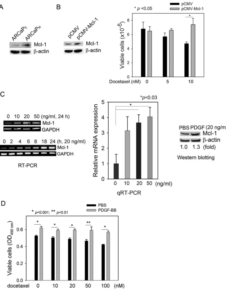
Previously we demonstrated that Mcl-1 overexpression is associated within vivobone metastatic propensity of human PCa cells, and importantly, correlated with clinical PCa bone metastasis [21]. Consistently, using a human PCa ARCaP cellular model that could closely mimic the pathophysiology of bone metastasis in immunocompromised mice [26], we found that Mcl-1 expression was significantly increased in highly bone metastatic ARCaPM
cells when compared to that in the low-invasive counterpart ARCaPEcells (Figure 1A). We hypothesized that upregulation of
Mcl-1 may confer metastatic PCa cells survival advantages, allowing them to escape apoptotic fate during invasion and dissemination and successfully establish distant metastasis [20]. Supporting this notion, ectopic expression of Mcl-1 enhanced PCa cell resistance to docetaxel (Figure 1B), a commonly used chemotherapeutic drug in hormone-refractory and metastatic PCa [27]. These results indicated that upregulation of Mcl-1 may account for, at least in part, resistance to apoptosis in metastatic PCa cells.
PDGF-BB induces Mcl-1 expression and antagonizes apoptosis in PCa cells
Intriguingly, PDGF-BB was found to significantly induce Mcl-1 expression in PCa cells (Figure 1C, Supplemental Figure S1). Treatment with recombinant human PDGF-BB increased Mcl-1 mRNA in a dose- and time-dependent manner, though the optimal conditions for the maximum accumulation of Mcl-1 mRNA varied in different PCa cell lines. Western blot analysis confirmed the inductive effects of PDGF-BB on Mcl-1 expression at protein level. These data identified PDGF-BB as a novel regulator of Mcl-1 expression, which could provide a survival mechanism to protect PCa cells from apoptosis. Indeed, addition of PDGF-BB in PCa cell cultures effectively antagonized the cytotoxicity of docetaxel over a wide range of doses (Figure 1D).
Expression profile of PDGF autocrine signaling components in PCa cells
We examined the expression pattern of PDGFs and their receptors in PCa cells (Figure 2A). RT-PCR analyses showed that the PDGF isoforms were differentially expressed at mRNA level, and among them, increased PDGF-B and PDGF-D were observed in C4-2 and ARCaPM cells when compared to the parental
LNCaP and ARCaPEcells, respectively. Consistent with previous
studies [19], PC3 cells were found to express high levels of PDGF-D, PDGFR-aand -b. Interestingly, PDGFR-amRNAs appeared to be substantially expressed in PCa cells, which was confirmed at protein level by Western blot analysis. In contrary, though PDGFR-bmRNAs were detected by RT-PCR in most PCa cell lines, immunoblotting analysis could only confirm protein expression in ARCaPE and ARCaPM cells (Figure 2A, right
panel). Taken together, these data suggested a functional PDGF autocrine signaling in certain PCa cells.
An autocrine PDGFR signaling mediates PDGF-BB regulation of Mcl-1 in PCa cells
Both PDGFR-a and -b were highly expressed in bone metastatic ARCaPMcells, and rapidly phosphorylated in a
time-dependent manner in response to the stimulation of exogenous PDGF-BB (Figure 2B). Interesting, depletion of either PDGFR-a
or -bby isoform-specific siRNA did not block the inductive effect of PDGF-BB on Mcl-1 expression (Figure 2C, left and central panels), suggesting that activation of either receptors may be sufficient for the upregulation of Mcl-1. Supporting this hypoth-esis, transient transfection with a mixture of siRNAs targeting both PDGFR-a and -b inhibited the basal expression of Mcl-1, and abrogated PDGF-BB induction of Mcl-1 ARCaPM cells
(Figure 2C, right panel). Alternatively, treatment with AG-17 (Tyrphostin), a selective pharmacological inhibitor of PDGFRs [28], reduced Mcl-1 expression at both mRNA and protein levels and markably increased cleavage of poly-ADP ribose polymerase (PARP), an indicator of apoptosis. These effects were attenuated by the presence of PDGF-BB in cultures (Figure 2D). Consistently, AG-17 treatment at low doses (such as 100 nM) effectively induced apoptosis in ARCaPEand ARCaPMcells (Figure 2E), indicating a
pivotal role of PDGFR signaling in the survival of PCa cells.
b-catenin mediates PDGF regulation of Mcl-1 expression in PCa cells
Activation of theb-catenin pathway is a downstream event of PDGF signaling in certain epithelial cancer cells [7,8,29]. Western blot analysis found thatb-catenin and TCF4, a majorb -catenin-interacting transcription factor [30], were differentially expressed in PCa cells (Figure 3A), suggesting a functionalb-catenin-TCF4 signaling in these cells. In fact, an artificial TCF promoter was activated in both the LNCaP-C4-2 and ARCaPE-ARCaPM cell
lineages, and the reporter activities appeared to be associated with increasedin vivometastatic potential in C4-2 and ARCaPM cells
(Figure 3B). It is worth noting that bothb-catenin and TCF4 were substantially presented in the nucleus of ARCaPEand ARCaPM
cells (Figure 3A, low panel), which exhibited markedly higher basal TCF activities than either LNCaP or C4-2 cells (by,100-fold)
(Figure 3B).
Upon PDGF-BB treatment, the nuclear presence ofb-catenin was rapidly increased in ARCaPMcells (Figure 3C, upper panel).
Consistently, TCF reporter activity was also significantly increased following PDGF-BB stimulation, which was attenuated by the pre-treatment with AG-17 (Figure 3C, bottom panel). These data indicated that PDGF-BB activated b-catenin signaling in a PDGFR-dependent manner.
To investigate the role ofb-catenin in the regulation of Mcl-1 expression, ARCaPM cells were transiently transfected with a
construct expressing wild-type b-catenin. RT-PCR and Western blot analyses showed that ectopic epression of b-catenin increseased Mcl-1 at both mRNA and protein levels (Figure 3D). In contrary, b-catenin depletion using a siRNA pool efficiently inhibited both the basal expression of Mcl-1 and its induction by PDGF-BB (Figure 3E). Consistently, whereas PDGF-BB signifi-cantly induced the luciferase activity of a full-length human Mcl-1 promoter in ARCaPMcells transfected with non-targeting control
Figure 1. PDGF-BB upregulates Mcl-1 and protects PCa cells from apoptosis.(A) Mcl-1 protein expression in the lineage-related ARCaPEand ARCaPMcells. (B) Ectopic expression of Mcl-1 in ARCaPMcells and the effects on docetaxel cytotoxicity in ARCaPMcells. pCMV: vector control. (C) Left panel: The dose- and time-dependent effects of PDGF-BB on Mcl-1 mRNA expression in ARCaPMcells; middle panel: qRT-PCR analysis of Mcl-1 mRNA expression in response to PDGF-BB treatment in ARCaPMcells (24 h); right panel: The effects of PDGF-BB treatment (20ng/ml, 72 h) on Mcl-1 protein expression in ARCaPMcells. ImageJ was used to quantitate the relative expression of Mcl-1 protein. (D) The effects of exogenous PDGF-BB on the cytotoxicity of docetaxel in ARCaPMcells, as determined by the MTS assay.
doi:10.1371/journal.pone.0030764.g001
PDGF activates p68-b-catenin signaling in PCa cells
We investigated whether a c-Abl-p68-dependent pathway is involved in the PDGF activation of b-catenin signaling in PCa cells [7]. Western blot analyses found that c-Abl and p68 were differentially expressed in PCa cells (Figure 4A). Upon PDGF-BB treatment, tyrosine phosphorylation of c-Abl and p68 were rapidly activated, as evidenced by immunoprecipitation-immunoblotting assays (Figure 4B, left and middle panels). Importantly, the presence of b-catenin in p68 immunoprecipitates was also increased in a time-dependent manner, suggesting an enhanced physical association between b-catenin and p68 proteins (Figure 4B, middle panel), which was further confirmed by a reciprocal immunoprecipitation experiment (Figure 4B, right panel). In fact, PDGF-BB induced rapid nuclear translocation of p68 within 30 min (Figure 4C), which was associated with increased co-localization of p68 and b-catenin in the nucleus (Figure 4D). These data indicated that PDGF-BB could activate the c-Abl-p68 cascade and subsequentb-catenin signaling in PCa cells.
To examine whether p68 is required for the regulation of Mcl-1 expression, ARCaPM cells were transfected with p68 siRNA or
control siRNA, and analyzed for the expression of Mcl-1 at the mRNA and protein levels. As shown in Figure 4E, depletion of p68 inhibited endogeneous b-catenin and effectively attenuated PDGF-BB induction of Mcl-1 expression. Consistently, Mcl-1 promoter activity was significantly inhibited by the treatment with p68 siRNA in ARCaPMcells, either with or without the presence
of PDGF-BB in the cultures (Figure 4F). These data indicated an indispensible function of p68 in the regulation of Mcl-1 in PCa cells.
PDGF-BB promotes protein interaction between
b-catenin and HIF-1ain PCa cells
Our previous studies demonstrated an important role of HIF-1a
in bone metastatic PCa cells [25]. Interestingly, transfection of a HIF-1a-specific siRNA significantly reduced Mcl-1 protein expression in ARCaPMcells (Figure 5A), suggesting that HIF-1a
may be required for Mcl-1 regulation in PCa cells. To examine Figure 2. Activation of the PDGFR signaling is required for Mcl-1 expression in PCa cells.(A) Expression profile of PDGFR signaling components in PCa cells, as analyzed by RT-PCR and Western blotting. (B) The effects of PDGF-BB (20 ng/ml) on the phosphorylation of PDGFR-aand -bin ARCaPMcells. (C) The effects of depleting PDGFR-aor/and -bon Mcl-1 protein expression in ARCaPMcells. The cells were transfected with either isotype-specific siRNAs targeting PDGFR-a(left panel, 30 nM) or PDGFR-b(central panel, 100 nM), or a mixture of PDGFR-aand -bsiRNAs (right panel) for 48 h, serum-starved overnight, and incubated in the presence or absence of PDGF-BB (20 ng/ml) for 72 h. (D) Upper panel: The time-dependent effects of AG-17 (100 nM) on Mcl-1 mRNA expression in ARCaPMcells; bottom panel: The effects of AG-17 treatment on the expression of Mcl-1 and cleaved PARP in the presence (20 ng/ml) or absence of PDGF-BB (20 ng/ml) in ARCaPMcells. (E) The effects of AG-17 treatment (100 nM, 72 h) on the viability of ARCaPEand ARCaPMcells.
whether PDGF-BB could induce physical interaction between HIF-1a and b-catenin, nuclear proteins were prepared from ARCaPMcells treated with PDGF-BB for varying times. Western
blot analysis found that both HIF-1aand b-catenin were rapidly increased in the nucleus (Figure 5B). A co-immunoprecipitation assay showed that in response to PDGF-BB stimulation, nuclear presence ofb-catenin rapidly increased in the HIF-1a immuno-precipitates (Figure 5C, upper panel). Reciprocal co-immunopre-cipitation with an anti-b-catenin antibody confirmed an increased association of nuclear HIF-1awithb-catenin following PDGF-BB treatment (Figure 5C, bottom panel). The enhanced co-localiza-tion ofb-catenin and HIF-1aproteins was further demonstrated by confocal microscopy, which appeared to acheive the maximum intensity at 30 min upon PDGF-BB stimulation (Figure 5D). These results indicated that in repsonse to PDGF-BB stimulation,
b-catenin physically interacts with HIF-1a in the nucleus, which may lead to the activation of Mcl-1 transcription in PCa cells.
A putative HRE motif is required for PDGF-BB activation of Mcl-1 promoter
HIF-1abinds to the HREcis-elements within the promoters of hypoxia-responsive genes and regulates their expression [31]. We examined whether PDGF-BB-induced nuclear accumulation of HIF-1a was associated with the activation of HRE-dependent transcription. In ARCaPMcells, PDGF-BB treatment significantly
increased luciferase expression driven by an artificial HRE promoter (pHIF-luc) (Figure 6A). Interestingly, a putative HRE motif was identified within human Mcl-1 promoter region, which is located between -900 and -884 nucleotides at the 59-upstream of transcription start site [24]. To investigate the potential role of this cis-element in PDGF regulation of Mcl-1 transcription, we characterized a deletion mutant of the putative HRE motif using human Mcl-1 promoter region as the template (Figure 6B). The resulting reporter construct (p-Mcl-1-Luc: DHRE), or the luciferase reporter driven by the full-length Mcl-1 promoter (p-Figure 3. b-catenin mediates PDGF regulation of Mcl-1 expression in PCa cells. (A) Expression profile of b-catenin-TCF signaling components in PCa cells. (B) TCF reporter activity in the LNCaP-C4-2 and ARCaPE-ARCaPMcells. (C) Upper panel: The effects of PDGF-BB (20 ng/ml) on the nuclear translocation ofb-catenin in ARCaPMcells; Bottom panel: The effects of PDGF-BB (20 ng/ml) on TCF reporter activity in the presence (100 nM) or absence of AG-17. (D) The effects of ectopic expression ofb-catenin (72 h) on Mcl-1 expression at both mRNA and protein levels. (E) The effects ofb-catenin depletion on PDGF-BB regulation of Mcl-1 expression in ARCaPMcells. The cells were transfected withb-catenin siRNA or control siRNA (30 nM) for 48 h, serum-starved overnight, and incubated in the presence or absence of PDGF-BB (20 ng/ml) for 72 h. (F) The effects ofb -catenin depletion on Mcl-1 reporter activity in ARCaPMcells. The cells were transfected withb-catenin or control siRNA (30 nM) for 48 h, and further transfected with a human Mcl-1 reporter for 24 h. Following serum starvation overnight, the cells were incubated in the presence or absence of PDGF-BB (20 ng/ml) for 48 h.
doi:10.1371/journal.pone.0030764.g003
Mcl-1-Luc), was transiently expressed in ARCaPM cells
respec-tively, and treated with PDGF-BB or PBS. IL-6, which has been shown to activate Mcl-1 transcription in PCa and cholangiocarci-noma cells through a signal transducer and activator of transcription 3 (Stat3)-dependent mechanism [32,33], was includ-ed as the positive control. Luciferase activity assay showinclud-ed that PDGF-BB induced the activation of p-Mcl-1-Luc promoter to a greater degree than IL-6 in ARCaPMcells. Significantly, deletion
of the HRE motif not only reduced the basal activity of Mcl-1 promoter, but also abrogated the inductive effects of PDGF-BB on reporter activity. In contrary, p-Mcl-1-Luc:DHRE, containing a Stat3-binding sequence at position between292 and 283 [32], remained activated upon IL-6 treatment (Figure 6C). A similar effect of HRE deletion on the differential response of Mcl-1 promoter to PDGF-BB and IL-6 was also observed in C4-2 cells (Supplemental Figure S2). These data indicated that the putative HRE cis-element is required for PDGF-BB activation of Mcl-1 expression in PCa cells.
PDGF-BB promotes the binding of bothb-catenin and HIF-1ato Mcl-1 promoter region
We investigated whether PDGF-BB promoted specific binding of bothb-catenin and HIF-1ato Mcl-1 promoter by a sequential ChIP assay. Fractionated chromatin from controls and
PDGF-BB-treated ARCaPM cells was firstly immunoprecipitated with a b
-catenin antibody, and the precipitates were subjected to a re-ChIP assay with an HIF-1aantibody. From the isolated DNA, a 151-bp fragment containing the HRE region on the Mcl-1 promoter could be amplified from the re-ChIP precipitates. Upon PDGF-BB stimulation, a considerable increase in the binding of both b -catenin and HIF-1ato the HRE region was observed (Figure 6D). These results demonstrated that PDGF-BB could facilitate the association of Mcl-1 promoter with a transcriptional complex consisting ofb-catenin and HIF-1ain PCa cells.
Discussion
In this study, we uncovered the PDGF-Mcl-1 signaling as a crucial survival mechanism in PCa cells (Figure 7). For the first time, we demonstrated that: 1) PDGF-BB is a novel regulator of Mcl-1 expression; 2) PDGF-BB activation of autocrine PDGFR signaling promotes the interaction betweenb-catenin and HIF-1a, presumably through a c-Abl-p68-dependent mechanism; 3) a putative HRE motif is required for the basal expression and PDGF-BB activation of Mcl-1 promoter; and 4) inhibition of the PDGFR-Mcl-1 signaling using a small-molecule inhibitor AG-17 could activate apoptotic response in metastatic PCa cells. These results support that targeting PDGF-Mcl-1 pathway may provide a novel strategy for treating PCa metastasis.
Figure 4. PDGF-BB activates the c-Abl-p68-b-catenin signaling cascade in PCa cells.(A) Expression of c-Abl and p68 in PCa cells. (B) Left panel: The effects of PDGF-BB (20 ng/ml) on the phosphorylation of c-Abl; middle panel: The effects of PDGF-BB (20 ng/ml) on the phosphorylation of p68 and the expression ofb-catenin in the p68 immunoprecipitates in ARCaPMcells. Phosphorylation of p68 on the tyrosine sites was detected using a pan-phosphorylated-Tyr antibody; right panel: The effects of PDGF-BB (20 ng/ml) on the expression of p68 in theb-catenin immunoprecipitates in ARCaPMcells. (C) The effects of PDGF-BB (20 ng/ml) on the nuclear translocation of p68 in ARCaPMcells. (D) Confocal microscopy analysis of the effects of PDGF-BB on the co-localization ofb-catenin and p68 in the nucleus in a time course experiment in ARCaPMcells. (E) The effects of p68 depletion on PDGF regulation of Mcl-1 in ARCaPMcells. The cells were transfected with p68 or control siRNA (30 nM) for 48 h, serum-starved overnight, and incubated in the presence or absence of PDGF-BB (20 ng/ml) for 24 h (upper panel) or 72 h (bottom panel). Upper panel: RT-PCR analysis of mRNA expression of p68 and Mcl-1; bottom panel: Western blot analysis of protein expression of p68,b-catenin and Mcl-1. (F) The effects of p68 depletion on Mcl-1 reporter activity in ARCaPMcells. The cells were transfected with p68 or control siRNA (30 nM) for 48 h, and further transfected with human Mcl-1 reporter for 24 h. Following serum starvation overnight, the cells were incubated in the presence or absence of PDGF-BB (20 ng/ml) for 48 h.
Activation of PDGFR signaling may be coupled with multiple downstream pathways in the regulation of cell growth, prolifer-ation, migration and survival [3]. In tumor-associated endothelial and fibroblast stromal cells, PDGF has been shown to activate Akt-and MAPK-dependent survival mechanisms [34,35,36,37]. Yet, it remains elusive on the molecular mechanism by which PDGF exerts its functions in epithelial cancer cells. Recent data have linked autocrine PDGF signaling to the activation of b-catenin pathway. For instance, PDGF-AB was found to induce nuclearb -catenin accumulation via a PI3K-dependent mechanism, thereby protecting hepatocellular carcinoma cells from anoikis during metastatic dissemination [8]. In human colon cancer cells, PDGF-BB induces EMT [7] and upregulates cyclin D1 and c-Myc [38] by activatingb-catenin-dependent gene expression. In both cases, PDGF-BB induces the phosphorylation of c-Abl kinase, which subsequently recruits p68, an RNA helicase with ATPase activity, and activates its phosphorylation. Phosphorylated p68 binds b -catenin and promotes its nuclear translocation by displacing Axin from b-catenin and blocking b-catenin degradation, eventually promoting the interaction of b-catenin with TCF/LEF and the assembly of transcription complexes [7]. In this study, we provided molecular evidence demonstrating that in PCa cells that express high basal levels of p68 andb-catenin, PDGF could significantly promote physical interaction and rapid nuclear translocation of p68 andb-catenin. Importantly, p68 depletion in PCa cells led to the inhibition of Mcl-1 expression and induction of apoptosis, as evidenced by the appearance of cleaved PARP (Supplemental Figure S3). These results, for the first time, underscored a critical role of p68 in the regulation of PCa cell survival. Interestingly, a
recent study demonstrated that p68 is actually a novel coactivator of androgen receptor (AR) [39], another transcription factor interacting withb-catenin in certain PCa cells (such as LNCaP and C4-2) [40]. It would be intriguing to further investigate the dynamic interaction between p68, b-catenin and AR, and its biological consequences in these cells. In addition, other pathways may be involved in the PDGF activation ofb-catenin signaling. For example, PDGF-BB treatment was found to induce rapid phosphorylation of both Akt and glycogen synthase kinase 3-b
(GSK-3b) (Supplemental Figure S4), which may also contribute to the elevated intracellular levels and nuclear accumulation of b -catenin [41].
Our data confirmed a highly activeb-catenin/TCF signaling in ARCaP cells and correlated the TCF reporter activity with thein vivo metastatic potential (Figure 3A, 3B), indicating these cells could be used as an excellent model system for investigating b -catenin signaling in PCa progression [42]. Though PDGF-BB activated the full-length human Mcl-1 promoter (Figure 3F) in a similar manner to its effect on the luciferase expression driven by an artificial TCF-binding motif (pTOPFlash), it appeared that human Mcl-1 promoter does not contain any consensus sequences of TCF/lymphoid enhancer-binding factor (LEF). These results suggested that certain transcription factor(s), other than TCF, could be responsible forb-catenin activation of Mcl-1 transcrip-tion. One of such candidates was cAMP-response element-binding protein (CREB), which has been implicated in the regulation of Mcl-1 expression through the PI-3K/Akt signaling pathway [43] and highly expressed in ARCaP cell lineage [25]. Western blotting analyses, however, could not detect a significant increase in Figure 5. PDGF-BB promotes protein interaction betweenb-catenin and HIF-1ain PCa cells.(A) The effects of HIF-1adepletion on Mcl-1 expression in ARCaPMcells. The cells were transfected with HIF-1aor control siRNA (30 nM) for 72 h, and analyzed for Mcl-1 expression by immunoblotting. (B) Western blot analysis of the effects of PDGF-BB (20 ng/ml) on the nuclear translocation ofb-catenin and HIF-1ain ARCaPMcells. (C) Co-immunoprecipitation assays of the effects of PDGF-BB (20 ng/ml) on the interaction betweenb-catenin and HIF-1ain the nucleus in ARCaPM cells. (D) Confocal microscopy of the effects of PDGF-BB (20 ng/ml) on the co-localization ofb-catenin and HIF-1ain the nucleus in ARCaPMcells. doi:10.1371/journal.pone.0030764.g005
nuclear CREB expression upon PDGF treatment (data not shown), suggesting that CREB may not be involved in the b -catenin-dependent activation of Mcl-1 transcription. Intriguingly, the transcription factor HIF-1awas found to be rapidly increased in the nucleus and physically interact with b-catenin following PDGF-BB stimulation, which may mediate Mcl-1 transcription by binding to HRE site(s) within the promoter. These data are consistent with a previous study showing thatb-catenin can switch its binding partner from TCF4 to HIF-1aand enhance HIF-1a -mediated transcription, and this dynamic reassembly ofb-catenin with HIF-1amay allow colorectal cancer cells to rapidly adapt to hypoxic stress and survive [44]. It is important to note that unlike the cited work, our studies were performed in normoxic PCa cell cultures. Since ARCaP cells substantially express HIF-1a even under normoxia [25], PDGF may significantly affect the expression of hypoxia-responsive or HRE-containing genes by promoting the interaction between b-catenin and HIF-1a in a Wnt-independent mechanism. Upregulation of Mcl-1, as a consequence, could provide pivotal protection against apoptotic signals during dissemination and colonization when the majority of cancer cells remain under normoxia.
Earlier studies reported high expression of PDGFRs in both localized and metastatic PCa, which could be detected in 88% of primary tumors and 80% of the metastases [10,45]. However, it remains controversial as to which PDGFR isoforms are expressed
in PCa cells and primarily responsible for autocrine PDGF signaling [12,46,47]. These conflicting results may partially arise from the potential non-specificity of antibodies used in the cited studies, but more importantly, may reflect the intrinsic heteroge-neity of human cancers, especially when at their late-stages. In this study, we were able to detect the expression of both PDGFR isoforms in several established PCa cell lines by RT-PCR and Western blot analyses. Given the fact that both PDGFR-aand -b
have been implicated in the progression of bone metastatic PCa [10,12,13,45,48], our study focused on the function of PDGF-BB since it is the only PDGF isoform that binds all the three receptor dimeric combinations (PDGFR-aa, -bband -ab) with high affinity [2,49]. To determine which PDGFR isoform is required for PDGF regulation of Mcl-1, we transfected PCa cells with specific siRNAs against PDGFR-a or -b. Interestingly, the single depletion of neither PDGFR-a nor PDGFR-b inhibited Mcl-1 expression in ARCaPM cells, suggesting that the PDGF-BB signal could be
transduced via the two independent but complementary receptors to activate Mcl-1 expression in PCa cells expressing both isoforms. Supporting this notion, dual depletion of both receptors simultaneously using a mixture of siRNAs against PDGFR-a
and -b effectively inhibited Mcl-1 expression. Alternatively, treatment with AG-17 or imatinib, two pan-PDGFR inhibitors that could inhibit the tyrosine kinase activity of both PDGFR-a
and -b, also reduced Mcl-1 levels in ARCaPMcells (Supplemental Figure 6. A putative HRE site is required for PDGF-BB activation of Mcl-1 promoter.(A) The effects of PDGF-BB on the HIF-1 reporter activity in ARCaPMcells. The cells were transiently transfected with HIF-1 reporter or pGL3 for 24 h, serum-starved and incubated in the presence or absence of PDGF-BB (20 ng/ml) for 48 h. (B) Schematic diagram of human Mcl-1 promoter and its deletion mutation at the putative HRE site. (C) The effects of deleting the putative HRE site on PDGF regulation of Mcl-1 promoter activity in ARCaPMcells. IL-6 (200 ng/ml) was included as the positive control. (D) ChIP-Re-ChIP assay of the effects of PDGF-BB treatment (20 ng/ml) on the binding ofb-catenin and HIF-1ato human Mcl-1 promoter region in ARCaPMcells.
Figure S5). Furthermore, in PCa cells that predominantly express one PDGFR isoform (for example, PDGFR-ais the major isoform in C4-2 cells; Figure 2A, right panel), it is plausible to expect that inhibition of the isoform alone could affect Mcl-1 expression. Indeed, transfection of PDGFR-asiRNA in C4-2 cells significantly inhibited Mcl-1 (Supplemental Figure S6). These findings support a model that PDGF-BB could activate both PDGFR isoforms in the regulation of Mcl-1 in PCa cells in a context-dependent manner, which may have important implication in the evaluation of PDGFR expression at tissue levels in clinical PCa specimens.
Interaction between PCa and bone microenvironment is crucial to the bone tropism of PCa metastasis, which is identified at autopsy in up to 90% of patients dying from the disease [50]. Tumor-initiated bone resorption promotes the release and activation of multiple growth factors immobilized in bone matrix, including PDGF. These locally expressed and tumor-derived PDGF could activate PDGFR signaling in surrounding stroma (including stromal cells, endothelial cells and pericytes) and promote angiogenesis. As a potent mitogen for osteoblasts, PDGF also significantly contribute to the osteoblastic phenotype of PCa bone metastasis [51]. These effects, taken together, may provide a favorable microenvironment for the survival and outgrowth of bone metastatic PCa. These facts provided rationale for evaluating
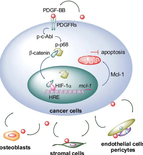
the potential of treating PCa bone metastasis with small-molecule PDGFR inhibitors. In earlier studies, imatinib sensitized bone marrow stromal and endothelial cells to paclitaxel treatment and significantly suppressed PCa bone metastasis in experimental models [52,53]. Disappointingly, however, recent clinical trials with imatinib only achieved limited success due to unexpected severe side effects in patients [48]. These observations highlighted the importance of a better understanding of PDGF signaling in bone metastasis PCa. Our study delineated a novel signaling axis that may allow PCa cells to escape apoptosis during dissemination and colonization by activating PDGF-Mcl-1 pathway in metastatic cancer cells. It is plausible to hypothesize that PDGF-BB may be crucial in mediating the ‘‘vicious cycle’’ between tumor and bone microenvironment, not only promoting angiogenesis in surround-ing stroma but also sustainsurround-ing survival in PCa cells (Figure 7). Supporting this model, PDGF-BB was found to be elevated in PC3-MM2 cells implanted in the mouse bone cortex, and interestingly, activated PDGFR-b was only detected in tumor lesions growing adjacent to bone and the tumor-associated endothelium [53,54]. Given the clinical significance of both PDGF and Mcl-1 in PCa bone metastasis [21,51], specific targeting of PDGF-Mcl-1 survival pathway in PCa cells (autocrine signaling) and co-targeting of microenvironment (paracrine Figure 7. A proposed model for PDGF-BB regulation of Mcl-1 expression in PCa cells.The engagement of PDGF-BB to PDGFR dimers activates the c-Abl-p68 cascade, which subsequently stablizesb-catenin and promotes its nuclear translocation. In the nucleus, interaction between b-catenin and HIF-1aincreases the binding of HIF-1ato the HRE site within Mcl-1 promoter, thereby activating the transcription of Mcl-1 gene. Upregulation of Mcl-1 antagonizes apoptotic signals and confers survival advantages to metastatic PCa cells. Furthermore, tumor-derived and locally expressed PDGF may mediate the interactions between PCa and bone microenvironment. Co-targeting the PDGF signaling in PCa cells (autocrine) and microenvironment (paracrine) could provide a new strategy to disrupt the ‘‘vicious cycle’’ and efficaciously treat metastatic PCa.
doi:10.1371/journal.pone.0030764.g007
signaling) could provide a new strategy to disrupt the vicious cycle and efficaciously treat metastatic PCa.
Supporting Information
Figure S1 The effects of PDGF-BB on the expression of Mcl-1 at mRNA and protein levels in PCa cells.(A–C) RT-PCR and qRT-PCR analyses of the time- and dose-dependent effects of PDGF-BB on Mcl-1 mRNA expression in ARCaPE(A), LNCaP (B) and C4-2
(C) cells. The dose was 20 ng/ml in the time course experiments. (D) Western blot analysis of the effects of PDGF isoforms on the expression of Mcl-1 in several PCa cell lines. Treatment: 20 ng/ml, 72 h. (TIF)
Figure S2 The effects of deleting the putative HRE site on PDGF regulation of Mcl-1 promoter activity in C4-2 cells.IL-6 (200 ng/ml) was included as the positive control. (TIF)
Figure S3 The effects of p68 siRNA on the expression of cleaved PARP, an indicator of apoptosis in PCa cells. ARCaPM cells were transfected with p68 or control siRNA (30
nM) for 48 h, serum-starved overnight, and incubated in the presence or absence of PDGF-BB (20 ng/ml) for 72 h.
(TIF)
Figure S4 The effect of PDGF-BB on the Akt-GSK-3bcascade in PCa cells. PDGF-BB treatment (20 ng/ml) in ARCaPM cells
increased the phosphorylation of Akt and GSK-bat serine residues. (TIF)
Figure S5 Imatinib, a small-molecule inhibitor of PDGFR signaling, inhibits Mcl-1 protein expression in PCa cells.ARCaPMcells were treated with 10mM imatinib for
varying times, western blotting was then performed. (TIF)
Figure S6 Depletion of PDGFR-a abrogates PDGF-BB induction of Mcl-1 in C4-2 cells.The cells were transfected with PDGFR-aor control siRNA (30 nM) for 48 h, serum-starved overnight, and incubated in the presence or absence of PDGF-BB (20 ng/ml) for 72 h.
(TIF)
Table S1 Antibodies used in this study. (PDF)
Table S2 Primers for PCR and RT-PCR. (PDF)
Acknowledgments
We thank Dr. Steven W. Edwards for kindly providing the human Mcl-1 reporter.
Author Contributions
Conceived and designed the experiments: SI ZL HZ OK LC DW HRCK. Performed the experiments: SI SZ AD YW. Analyzed the data: SI SZ AD OK LC DW HRCK. Contributed reagents/materials/analysis tools: AD ZL HRCK. Wrote the paper: SI LC DW. Grant support: CR.
References
1. Heldin CH, Eriksson U, Ostman A (2002) New members of the platelet-derived growth factor family of mitogens. Arch Biochem Biophys 398: 284–290. 2. Pietras K, Sjoblom T, Rubin K, Heldin CH, Ostman A (2003) PDGF receptors
as cancer drug targets. Cancer Cell 3: 439–443.
3. Andrae J, Gallini R, Betsholtz C (2008) Role of platelet-derived growth factors in physiology and medicine. Genes Dev 22: 1276–1312.
4. Kim L, Wong TW (1995) The cytoplasmic tyrosine kinase FER is associated with the catenin-like substrate pp120 and is activated by growth factors. Mol Cell Biol 15: 4553–4561.
5. Yokote K, Margolis B, Heldin CH, Claesson-Welsh L (1996) Grb7 is a downstream signaling component of platelet-derived growth factor alpha- and beta-receptors. J Biol Chem 271: 30942–30949.
6. Darnell JE, Jr. (1997) STATs and gene regulation. Science 277: 1630–1635. 7. Yang L, Lin C, Liu ZR (2006) P68 RNA helicase mediates PDGF-induced
epithelial mesenchymal transition by displacing Axin from beta-catenin. Cell 127: 139–155.
8. Fischer AN, Fuchs E, Mikula M, Huber H, Beug H, et al. (2007) PDGF essentially links TGF-beta signaling to nuclear beta-catenin accumulation in hepatocellular carcinoma progression. Oncogene 26: 3395–3405.
9. Mathew P, Thall PF, Jones D, Perez C, Bucana C, et al. (2004) Platelet-derived growth factor receptor inhibitor imatinib mesylate and docetaxel: a modular phase I trial in androgen-independent prostate cancer. J Clin Oncol 22: 3323–3329.
10. Ko YJ, Small EJ, Kabbinavar F, Chachoua A, Taneja S, et al. (2001) A multi-institutional phase ii study of SU101, a platelet-derived growth factor receptor inhibitor, for patients with hormone-refractory prostate cancer. Clin Cancer Res 7: 800–805.
11. Hofer MD, Fecko A, Shen R, Setlur SR, Pienta KG, et al. (2004) Expression of the platelet-derived growth factor receptor in prostate cancer and treatment implications with tyrosine kinase inhibitors. Neoplasia 6: 503–512.
12. Fudge K, Wang CY, Stearns ME (1994) Immunohistochemistry analysis of platelet-derived growth factor A and B chains and platelet-derived growth factor alpha and beta receptor expression in benign prostatic hyperplasias and Gleason-graded human prostate adenocarcinomas. Mod Pathol 7: 549–554. 13. Singh D, Febbo PG, Ross K, Jackson DG, Manola J, et al. (2002) Gene expression
correlates of clinical prostate cancer behavior. Cancer Cell 1: 203–209. 14. Pietras K, Pahler J, Bergers G, Hanahan D (2008) Functions of paracrine PDGF
signaling in the proangiogenic tumor stroma revealed by pharmacological targeting. PLoS Med 5: e19.
15. Ostman A, Heldin CH (2007) PDGF receptors as targets in tumor treatment. Adv Cancer Res 97: 247–274.
16. Pietras K, Rubin K, Sjoblom T, Buchdunger E, Sjoquist M, et al. (2002) Inhibition of PDGF receptor signaling in tumor stroma enhances antitumor effect of chemotherapy. Cancer Res 62: 5476–5484.
17. Ostman A (2004) PDGF receptors-mediators of autocrine tumor growth and regulators of tumor vasculature and stroma. Cytokine Growth Factor Rev 15: 275–286.
18. Jechlinger M, Sommer A, Moriggl R, Seither P, Kraut N, et al. (2006) Autocrine PDGFR signaling promotes mammary cancer metastasis. J Clin Invest 116: 1561–1570.
19. Kong D, Wang Z, Sarkar SH, Li Y, Banerjee S, et al. (2008) Platelet-derived growth factor-D overexpression contributes to epithelial-mesenchymal transition of PC3 prostate cancer cells. Stem Cells 26: 1425–1435.
20. Mehlen P, Puisieux A (2006) Metastasis: a question of life or death. Nat Rev Cancer 6: 449–458.
21. Zhang S, Zhau HE, Osunkoya AO, Iqbal S, Yang X, et al. (2010) Vascular endothelial growth factor regulates myeloid cell leukemia-1 expression through neuropilin-1-dependent activation of c-MET signaling in human prostate cancer cells. Mol Cancer 9: 9.
22. Zhau HE, Odero-Marah V, Lue HW, Nomura T, Wang R, et al. (2008) Epithelial to mesenchymal transition (EMT) in human prostate cancer: lessons learned from ARCaP model. Clin Exp Metastasis 25: 601–610.
23. Wu TT, Sikes RA, Cui Q, Thalmann GN, Kao C, et al. (1998) Establishing human prostate cancer cell xenografts in bone: induction of osteoblastic reaction by prostate-specific antigen-producing tumors in athymic and SCID/bg mice using LNCaP and lineage-derived metastatic sublines. Int J Cancer 77: 887–894. 24. Akgul C, Turner PC, White MR, Edwards SW (2000) Functional analysis of the
human MCL-1 gene. Cell Mol Life Sci 57: 684–691.
25. Wu D, Zhau HE, Huang WC, Iqbal S, Habib FK, et al. (2007) cAMP-responsive element-binding protein regulates vascular endothelial growth factor expression: implication in human prostate cancer bone metastasis. Oncogene 26: 5070–5077.
26. Xu J, Wang R, Xie ZH, Odero-Marah V, Pathak S, et al. (2006) Prostate cancer metastasis: role of the host microenvironment in promoting epithelial to mesenchymal transition and increased bone and adrenal gland metastasis. Prostate 66: 1664–1673.
27. Pienta KJ, Smith DC (2005) Advances in prostate cancer chemotherapy: a new era begins. CA Cancer J Clin 55: 300–318quiz 323–305.
28. Gazit A, Yaish P, Gilon C, Levitzki A (1989) Tyrphostins I: synthesis and biological activity of protein tyrosine kinase inhibitors. J Med Chem 32: 2344–2352.
29. Singh PK, Wen Y, Swanson BJ, Shanmugam K, Kazlauskas A, et al. (2007) Platelet-derived growth factor receptor beta-mediated phosphorylation of MUC1 enhances invasiveness in pancreatic adenocarcinoma cells. Cancer Res 67: 5201–5210.
30. Clevers H (2004) Wnt breakers in colon cancer. Cancer Cell 5: 5–6. 31. Wenger RH, Stiehl DP, Camenisch G (2005) Integration of oxygen signaling at
32. Isomoto H, Kobayashi S, Werneburg NW, Bronk SF, Guicciardi ME, et al. (2005) Interleukin 6 upregulates myeloid cell leukemia-1 expression through a STAT3 pathway in cholangiocarcinoma cells. Hepatology 42: 1329–1338. 33. Cavarretta IT, Neuwirt H, Untergasser G, Moser PL, Zaki MH, et al. (2007)
The antiapoptotic effect of IL-6 autocrine loop in a cellular model of advanced prostate cancer is mediated by Mcl-1. Oncogene 26: 2822–2832.
34. Langley RR, Fan D, Tsan RZ, Rebhun R, He J, et al. (2004) Activation of the platelet-derived growth factor-receptor enhances survival of murine bone endothelial cells. Cancer Res 64: 3727–3730.
35. Kitadai Y, Sasaki T, Kuwai T, Nakamura T, Bucana CD, et al. (2006) Expression of activated platelet-derived growth factor receptor in stromal cells of human colon carcinomas is associated with metastatic potential. Int J Cancer 119: 2567–2574.
36. Kodama M, Kitadai Y, Sumida T, Ohnishi M, Ohara E, et al. (2010) Expression of platelet-derived growth factor (PDGF)-B and PDGF-receptor beta is associated with lymphatic metastasis in human gastric carcinoma. Cancer Sci 101: 1984–1989.
37. Song N, Huang Y, Shi H, Yuan S, Ding Y, et al. (2009) Overexpression of platelet-derived growth factor-BB increases tumor pericyte content via stromal-derived factor-1alpha/CXCR4 axis. Cancer Res 69: 6057–6064.
38. Yang L, Lin C, Zhao S, Wang H, Liu ZR (2007) Phosphorylation of p68 RNA helicase plays a role in platelet-derived growth factor-induced cell proliferation by up-regulating cyclin D1 and c-Myc expression. J Biol Chem 282: 16811–16819.
39. Clark EL, Coulson A, Dalgliesh C, Rajan P, Nicol SM, et al. (2008) The RNA helicase p68 is a novel androgen receptor coactivator involved in splicing and is overexpressed in prostate cancer. Cancer Res 68: 7938–7946.
40. Truica CI, Byers S, Gelmann EP (2000) Beta-catenin affects androgen receptor transcriptional activity and ligand specificity. Cancer Res 60: 4709–4713. 41. Doble BW, Woodgett JR (2003) GSK-3: tricks of the trade for a multi-tasking
kinase. J Cell Sci 116: 1175–1186.
42. Chesire DR, Ewing CM, Gage WR, Isaacs WB (2002) In vitro evidence for complex modes of nuclear beta-catenin signaling during prostate growth and tumorigenesis. Oncogene 21: 2679–2694.
43. Wang JM, Chao JR, Chen W, Kuo ML, Yen JJ, et al. (1999) The antiapoptotic gene mcl-1 is up-regulated by the phosphatidylinositol 3-kinase/Akt signaling pathway through a transcription factor complex containing CREB. Mol Cell Biol 19: 6195–6206.
44. Kaidi A, Williams AC, Paraskeva C (2007) Interaction between beta-catenin and HIF-1 promotes cellular adaptation to hypoxia. Nat Cell Biol 9: 210–217. 45. Chott A, Sun Z, Morganstern D, Pan J, Li T, et al. (1999) Tyrosine kinases
expressed in vivo by human prostate cancer bone marrow metastases and loss of the type 1 insulin-like growth factor receptor. Am J Pathol 155: 1271–1279. 46. George DJ (2002) Receptor tyrosine kinases as rational targets for prostate
cancer treatment: platelet-derived growth factor receptor and imatinib mesylate. Urology 60: 115–121. discussion 122.
47. Paulsson J, Sjoblom T, Micke P, Ponten F, Landberg G, et al. (2009) Prognostic significance of stromal platelet-derived growth factor beta-receptor expression in human breast cancer. Am J Pathol 175: 334–341.
48. Mathew P, Thall PF, Bucana CD, Oh WK, Morris MJ, et al. (2007) Platelet-derived growth factor receptor inhibition and chemotherapy for castration-resistant prostate cancer with bone metastases. Clin Cancer Res 13: 5816–5824. 49. Williams LT (1989) Signal transduction by the platelet-derived growth factor
receptor. Science 243: 1564–1570.
50. Rana A, Chisholm GD, Khan M, Sekharjit SS, Merrick MV, et al. (1993) Patterns of bone metastasis and their prognostic significance in patients with carcinoma of the prostate. Br J Urol 72: 933–936.
51. Roodman GD (2004) Mechanisms of bone metastasis. N Engl J Med 350: 1655–1664.
52. Kim SJ, Uehara H, Yazici S, Langley RR, He J, et al. (2004) Simultaneous blockade of platelet-derived growth factor-receptor and epidermal growth factor-receptor signaling and systemic administration of paclitaxel as therapy for human prostate cancer metastasis in bone of nude mice. Cancer Res 64: 4201–4208.
53. Uehara H, Kim SJ, Karashima T, Shepherd DL, Fan D, et al. (2003) Effects of blocking platelet-derived growth factor-receptor signaling in a mouse model of experimental prostate cancer bone metastases. J Natl Cancer Inst 95: 458–470. 54. Langley RR, Fidler IJ (2007) Tumor cell-organ microenvironment interactions
in the pathogenesis of cancer metastasis. Endocr Rev 28: 297–321.