Divergent Androgen Receptor and
Beta-Catenin Signaling in Prostate Cancer Cells
Eugine Lee1,3¤, Susan Ha2, Susan K. Logan1,2,3 *
1Department of Biochemistry and Molecular Pharmacology, New York University School of Medicine, New York, NY, United States of America,2Department of Urology New York University School of Medicine, New York, NY, United States of America,3Stem Cell Biology Program, New York University School of Medicine, New York, NY, United States of America
¤ Current address: Human Oncology and Pathogenesis Program, Memorial Sloan Kettering Cancer Center, New York, NY, United States of America
Abstract
Despite decades of effort to develop effective therapy and to identify promising new drugs, prostate cancer is lethal once it progresses to castration-resistant disease. Studies show mis-regulation of multiple pathways in castration-resistant prostate cancer (CRPC), reflect-ing the heterogeneity of the tumors and also hintreflect-ing that targetreflect-ing androgen receptor (AR) pathway alone might not be sufficient to treat CRPC. In this study, we present evidence that the Wnt/β-catenin pathway might be activated in prostate cancer cells after androgen-depri-vation to promote androgen-independent growth, partly through enhanced interaction ofβ -catenin with TCF4. Androgen-independent prostate cancer cells were more prone to acti-vate a Wnt-reporter, and inhibition of the Wnt/β-catenin pathway increased sensitivity of these cells to the second-generation antiandrogen, enzalutamide. Combined treatment of enzalutamide and Wnt/β-catenin inhibitor showed increased growth repression in both androgen-dependent and -independent prostate cancer cells, suggesting therapeutic potential for this approach.
Introduction
Prostate cancer is the most common form of cancer in males [1]. Given the key role of androgen receptor (AR) signaling in disease progression, the current conventional approach to treat pros-tate cancer is androgen deprivation therapy often combined with antiandrogen treatment. Despite the initial tumor regression, aggressive disease progresses to castration-resistant prostate cancer (CRPC), for which treatment is the major challenge in the field. New drugs targeting the AR pathway such as the second-generation antiandrogen, enzalutamide [2] and abiraterone which blocks intratumoral production of androgen [3] have been FDA approved for the treat-ment of CRPC. Despite the promise of these and other therapeutics, they extend life by only 6–8 months [4,5], indicating the need for a new approach to treat advanced disease.
Due to the complex signaling networks in advanced disease, inhibition of one pathway might cause unpredictable responses [6,7]. In this context, it has been suggested that prostate
OPEN ACCESS
Citation:Lee E, Ha S, Logan SK (2015) Divergent Androgen Receptor and Beta-Catenin Signaling in Prostate Cancer Cells. PLoS ONE 10(10): e0141589. doi:10.1371/journal.pone.0141589
Editor:Zoran Culig, Innsbruck Medical University, AUSTRIA
Received:August 4, 2015
Accepted:October 9, 2015
Published:October 28, 2015
Copyright:© 2015 Lee et al. This is an open access article distributed under the terms of theCreative Commons Attribution License, which permits unrestricted use, distribution, and reproduction in any medium, provided the original author and source are credited.
Data Availability Statement:All relevant data are within the paper.
Funding:This study was supported by National Institutes of Health R01CA112226 (SL) and American Cancer Society RSG-11-108-01-CDD (SL). The funders had no role in study design, data collection and analysis, decision to publish, or preparation of the manuscript.
Competing Interests:The authors have declared that no competing interests exist.
tumors may also activate alternative signaling pathways to compensate for the consequences of AR inhibition [8,9]. Interaction between the PI3K and AR pathways has been well studied and in fact reciprocal feedback between the two pathways in PTEN-deleted prostate cancer has been reported, indicating the importance of targeting both pathways in PTEN-deleted disease [10]. More recently, upregulation of glucocorticoid receptor (GR) was reported in enzaluta-mide-resistant tumors [11], and shown to be necessary for the resistant phenotype. Therefore, therapeutic approaches that concomitantly target multiple pathways might be more effective in treating CRPC [6].
Recent genome sequencing data has shown misregulation of the Wnt-pathway in prostate cancer with disease progression. The comparative analysis of two separate whole-exome sequencing sets of data, one from primary tumors [12] and the other from lethal castration-resistant metastatic tumors [13], revealed that the APC (adenomatous polyposis coli) gene was frequently mutated in primary tumors but more significantly mutated in advanced disease [14]. In fact, the later data shows that the Wnt-pathway is one of the most significantly mutated pathways in CRPC [13]. Consistent with this, WNT16B secretion in the tumor microenviron-ment promoted treatmicroenviron-ment-resistance in prostate cancer through activation of the Wnt/β -cate-nin pathway [15]. More recently it was reported that 18% of cases of metastatic castrate resistant prostate cancer exhibited alterations in Wnt pathway signaling [16]. These reports suggest that the Wnt/β-catenin pathway may be one of the compensatory pathways activated in prostate cancer in response to androgen deprivation therapy. Supporting this idea, the expression of an activating mutation ofβ-catenin in mouse prostate enabled continuous pros-tatic growth after castration [17].
Our group previously published proof of concept studies showing that a small molecule inhibitor of the Wnt/β-catenin pathway, iCRT3 (shortened to C3) could decrease AR mRNA expression and transcription of downstream target genes by interfering withβ-catenin/TCF interaction on the AR promoter. We also showed that C3 could interfere with AR andβ -cate-nin protein interaction. The later protein interaction studies were performed in the presence of high levels of androgen to stabilize AR protein levels so that they were not decreased in the presence ofβ-catenin inhibitor [18].
Work described in this manuscript shows that the activity of the Wnt/β-catenin pathway is low in prostate cancer cells likely due to the preference forβ-catenin interaction with AR rather than TCF4 in these cells. We observe that suppression of AR activity by androgen-deprivation, antiandrogen treatment or AR knockdown promoted Wnt/β-catenin-target gene expression and this correlated with increased interaction between TCF4 andβ-catenin. The enhanced activation of the Wnt/β-catenin pathway caused growth of androgen-dependent LNCaP cells in the absence of androgen or in the presence of antiandrogen. Activation of the Wnt/β-catenin pathway was also examined in an androgen-independent subline of LNCaP cells, LNCaP-abl (abl). Abl cells were generated by continuous passage in androgen depleted media and selected for their ability to proliferate in the androgen deprived condition [19]. Abl cells were more prone to Wnt/β -cate-nin activation than LNCaP cells, and inhibition ofβ-catenin activity by a small molecule inhibitor or siRNA increased enzalutamide sensitivity in abl cells. Furthermore, combined treatment of enzalutamide and a Wnt/β-catenin inhibitor exhibited increased growth inhibition in both LNCaP and abl cells, indicating the therapeutic potential of this approach.
Materials and Methods
Cell culture
LNCaP (ATCC, CRL-1740) and LNCaP-abl (abl) [19] (gift by Z. Culig) cells were cultured in RPMI 1640 (Cellgro) supplemented with 10% FBS (Hyclone), and 10% charcoal stripped FBS
(CFBS; serum depleted of steroids including androgens), respectively, and 1% penicillin-strepto-mycin (Cellgro). 22Rv1 (ATCC, CRL-2505) and HEK293 (ATCC, CRL-1573) cells were cultured in DMEM (Cellgro) supplemented with 10% FBS and 1% penicillin-streptomycin. The following compounds were used to treat cells: C3 (ChemDiv, C523-1410), enzalutamide (Selleck Chemi-cals) and GSK-3 inhibitor (CHIR99021; Stemgent). For cell proliferation, qRT-PCR, immuno-blot, immunoprecipitation and ChIP assays, LNCaP cells were hormone deprived in 5% CFBS media for 2–3 days and then treated with androgen with or without the above compounds.
Stable integration of eGFP Wnt-reporter in prostate cancer cells
The lentiviral eGFP Wnt reporter, 7xTcf-eGFP/SV40-mCherry was purchased from Addgene (24304). Packaging cells (HEK293T/17) (ATCC, CRL-11268) were transiently transfected with 6μg of eGFP Wnt reporter construct, 4μg of packaging plasmid (psPAX2) and 2μg of
enve-lope expressing plasmid (pMD2.G) using Lipofectamine 2000 reagent (Invitrogen) according to manufacturer’s instructions. Media conditioned by the transfectants was collected after 24 and 48 h. LNCaP, abl and 22Rv1 cells were infected by incubation in conditioned media over-night and allowed to recover for a day. Infected cells were passaged and selected by mCherry expression using flow cytometry.
Quantitative real-time RT-PCR (qRT-PCR)
Total RNA was isolated using the RNeasy kit (Qiagen), and then reverse transcribed at 55°C for 1 h using Superscript III reverse transcriptase and oligo- (dT) 20 primers (Invitrogen). Real-time PCR was performed using gene-specific primers and 2XSYBR green Taq-ready mix (Sigma). Data were analyzed by the DDCT method using RPL19 as a control gene and normal-ized to control samples, which were arbitrarily set to 1.
Immunoblot, immunoprecipitation and immunostaining
For immunoblot analysis, cells were lysed in Triton buffer and supplemented with 1 mM PMSF, 1 mM Na3VO4, 10 mg/ml of leupeptin and 10 mg/ml of aprotinin. Protein lysates were subjected to SDS-PAGE and immunoblotted with the following antibodies against: AR (441),β-catenin (H-102), TCF4 (H-125, Santa Cruz Biotechnology); activeβ-catenin (clone 8E7, Millipore); tubu-lin (Covance). Protein bands were visualized using ECL Western Blotting detection reagents (GE Healthcare). Image J (NIH) software was used to quantitate protein levels.
In immunoprecipitation experiments cells were lysed as described above. Primary antibod-ies listed above were added to at least 1.5 mg of total protein and incubated overnight at 4°C followed by the addition of Protein A/G agarose beads (Santa Cruz Biotechnology) for 2 h. Immune complexes were extensively washed with Triton buffer and solubilized using Laemmli sample buffer (BioRad). Normal mouse IgG (Santa Cruz Biotechnology) or normal rabbit sera (Sigma) were used as controls.
Cell proliferation assay
For the CyQUANT cell proliferation assay, 3 to 4 X 10³cells were plated in each well of black 96 well plate. Cells were plated in hormone-deprived media containing 5% CFBS and cultured for 2 to 3 days, and then treated with indicated reagents for each experiment. The reagents were added every two days with more media and an equal amount of vehicle added to the con-trol group. At 2–3 day intervals, CyQUANT assay (Invitrogen, C35006) was performed accord-ing to manufacturer’s instructions and analyzed on a SpectraMax M5 plate reader (Molecular Devices) running SoftMax Pro1software.
Chromatin immunoprecipitation (ChIP) assay
ChIP was performed as previously described [20]. Proteins were double cross-linked with DSP (Pierce) for 20 min and 1% formalin for 10 min. Cells were lysed, nuclei collected and resus-pended in sonication buffer, and sonicationed for 12 min (30 sec. on, 30 sec. off) in a Bioruptor sonicator (Diagenode, model XL). Sonicated lysates were pre-cleared for 2 h with Protein A/G agarose beads blocked with salmon sperm DNA (Millipore). Supernatants were then incubated overnight with the following antibodies: a mixture AR (441) and AR (N-20), orβ-catenin (H-102). Control ChIP was performed with normal mouse IgG and normal rabbit IgG sera. Immu-nocomplexes were then washed and cross-linking reversed. DNA was isolated with the Qiagen PCR purification kit and qPCR was performed. Relative enrichment was calculated as a per-centage of 4% input normalized to IgG.
RNA-interference (RNA-i)
Forβ-catenin and AR knockdown, siGENOME SMARTpool siRNAs againstβ-catenin or AR were used (Dharmacon). For APC knockdown, the pool of three Silencer1Select siRNAs against APC (Ambion, s1433, s1434 and s1435) were used. Cells were transfected with HiPer-Fect transfection reagent (Qiagen) according to manufacturer’s instructions. 1 X 10⁵cells were plated in each well of a 24 well plate in hormone-deprived media containing 5% CFBS and the mixture of HiPerFect reagent and siRNAs were added directly on top of the cells. Cells were cultured for 2 days and then treated with indicated reagents for each experiment. For
CyQUANT proliferation assay, 3 to 4 X 10³cells were plated in each well of black 96 well plates in hormone-deprived media containing 5% CFBS and the mixture of HiPerFect reagent and APC siRNAs were added directly on top of the cells. Cells were cultured for 2 days and then treated with indicated reagents for each experiment. CyQUANT assay was performed at 2–3 day intervals as described above.
The cell lines stably depleted ofβ-catenin were generated with lentiviral pGIPZ shRNA againstβ-catenin (Open Biosystems, RHS4430-98912789) or a control shRNA (Open Biosys-tems, RHS4743). After infection, cells were plated at a very low density and selected for 10 days with 1μg/ml puromycin (Sigma). Each resistant cell colony was collected and expanded in the
selection media to screen for beta-catenin protein levels. Two cell lines showing moderate reduction ofβ-catenin were selected to test enzalutamide sensitivity (sh-β-cat-1 and -2) to min-imize the robust growth inhibitory effect ofβ-catenin knockdown (25).
Results
Androgen treatment represses Wnt-reporter activity in
androgen-dependent LNCaP cells
The construct also expresses mCherry under the constitutively active SV40 promoter indicat-ing positively infected cells [21]. LNCaP, LNCaP-abl (abl) and 22Rv1 prostate cancer cells were infected with lentivirus containing the reporter construct and cells stably integrated with the construct were selected by flow cytometry using mCherry expression. Wnt reporter gene activ-ity was tested using GSK-3 inhibitor (GSK3-i; CHIR99021), a potent Wnt-activator [22,23]. eGFP Wnt reporter activity was increased in the presence of GSK3-i, shown by the increased transcription of eGFP in LNCaP-abl cells stably expressing the reporter. The eGFP transcrip-tion was diminished in the presence of siRNA targetingβ-catenin (Fig 1A). Using this reporter, we examined activity of the Wnt/β-catenin pathway in androgen-dependent LNCaP cells, and androgen-independent LNCaP-abl and 22Rv1 cells [24,25]. In spite of abundant levels of active, nuclearβ-catenin (Fig 1B), the three cell lines showed low basal activity of Wnt-reporter (Fig 1C). Treatment with GSK-3 inhibitor (3μM) activated Wnt-reporter activity in
androgen-independent abl and 22Rv1 cells (Fig 1D), suggesting that increased levels ofβ-catenin were required to activate Wnt/β-catenin-responsive transcription. However, treatment of androgen-dependent LNCaP cells with 3μM GSK3-i had very little effect on the Wnt reporter (data not
shown). Additionally, the Wnt-reporter was not activated in the presence of higher concentra-tions of GSK-3 inhibitor (6 or 9μM) (Fig 1D).Fig 1Eshows the relative mRNA levels of eGFP
in each condition, with increased eGFP transcription in GSK-3 inhibitor treated abl cells, but not in LNCaP cells.
Given thatβ-catenin interacts with AR in an androgen-dependent manner [26–28], we rea-soned that androgen-deprivation might enhanceβ-catenin interaction with TCF, thereby increasing Wnt-reporter activity. In support of this idea, treatment of LNCaP cells with GSK-3 inhibitor in regular hormone containing media (condition i) compared to the hormone-deprived media (condition ii) resulted in increased reporter activation (Fig 1F) in the hormone deprived condition. Treatment with di-hydrotestosterone (DHT) diminished this reporter acti-vation (Fig 1F, condition iii), suggesting that androgen treatment represses Wnt reporter activ-ity in LNCaP cells. The relative level of eGFP mRNA in each condition is shown inFig 1G.
Inhibition of AR activity enhances Wnt/
β
-catenin-responsive
transcription
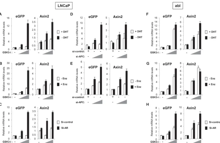
As we observed increased activation of the Wnt-reporter in hormone-deprived conditions, we hypothesized that inhibition of AR activity might enhance Wnt/β-catenin-responsive tran-scription. To test this idea, AR activity was repressed by either hormone-deprivation, antian-drogen treatment (enzalutamide; [2]) or siRNA-mediated AR depletion. Cells were treated with increasing concentrations of GSK-3 inhibitor to induce Wnt/β-catenin-responsive tran-scription and the relative mRNA levels of the Wnt-reporter (eGFP) and endogenous Wnt/β -catenin target gene,Axin2, were analyzed. Compared to control cells, higher induction of
Wnt-reporter andAxin2mRNA levels were observed in LNCaP cells cultured in hormone-deprived
media, in the presence of enzalutamide or treated with AR siRNA (Fig 2A–2C). As an alterna-tive to GSK-3 inhibitor treatment we also repeated the experiment using APC knockdown to activate the Wnt/β-catenin pathway. APC is a component of theβ-catenin destruction complex and APC deletion or loss-of-function mutation has been shown to stabilizeβ-catenin and acti-vate Wnt/β-catenin-responsive transcription [29,30]. Consistent with results in GSK-3 inhibi-tor treated cells (Fig 2A–2C), APC knockdown induced higher activation of the Wnt-reporter gene andAxin2transcription in hormone-deprived or enzalutamide treated cells (Fig 2D and
2E), suggesting that AR repression promotes Wnt/β-catenin-activation in LNCaP cells. How-ever, abl cells exhibited high levels of Wnt reporter andAxin2expression in response to GSK-3
Fig 1. Prostate cancer cells show low Wnt-reporter activity and hormone-deprivation enhances activation of Wnt-reporter in LNCaP cells.A, -catenin knockdown diminished the Wnt-reporter activation in response to GSK-3 inhibitor. abl cells were infected with eGFP Wnt-reporter (7xTcf-eGFP/ SV40-mCherry) and transfected with si-control or si–catenin. Cells were cultured for 2 days and then treated with vehicle, 2μM or 3μM GSK-3 inhibitor for 24
(Fig 2F and 2G) with the exception ofAxin2, which was unaffected by enzalutamide treatment
compared to control cells (Fig 2G). Depletion of AR by siRNA showed an increase in Wnt reporter gene activity andAxin2mRNA in both LNCaP and abl cells (Fig 2C and 2H)
suggest-ing that depletion of AR protein results in release of moreβ-catenin into a cellular pool avail-able to activate the Wnt/β-catenin pathway.
prostate cancer cells were determined by immunofluorescence staining.C, Representative fluorescence images showing the low level of eGFP Wnt reporter activity in LNCaP, abl and 22Rv1 cells: Wnt activity (green), presence of cells (mCherry, red), and nuclear location (DAPI, blue).D and E, Androgen-independent abl and 22Rv1 cells are more prone to activation of Wnt-reporter. Cells were treated with GSK-3 inhibitor for 24 h and subjected to fluorescence imaging (D) or qRT-PCR to measure relative mRNA levels of eGFP (E). InA-E, experiments were performed in the normal growth media of each cell line as described in the Materials and Methods.F and G, Hormone-deprivation enhanced activation of Wnt-reporter in LNCaP cells, which is diminished by androgen treatment. Cells were treated with 9μM GSK-3 inhibitor in complete (i) or hormone-deprived media with/without 10 nM di-hydrotestosterone (DHT) for 24 h (ii and iii). Cells were then subjected to fluorescence imaging (F), or qRT-PCR to measure relative mRNA levels of eGFP (G). The data presented is
representative of three independent experiments and the indicated error is the standard deviation.
doi:10.1371/journal.pone.0141589.g001
Fig 2. Inhibition of AR activity enhances Wnt/β-catenin target gene expression in response to GSK-3 inhibitor or APC knockdown.LNCaP cells were treated with 6μM or 9μM of GSK-3 inhibitor (A-C); abl cells were treated with and 2μM or 3μM of GSK-3 inhibitor (F-H).A and F, Cells were hormone-deprived for 3 days and then treated with vehicle or increasing concentration of GSK-3 inhibitor with/without 10 nM DHT for 24 h.B and G, Cells were treated with vehicle or increasing concentration of GSK-3 inhibitor with/without 10μM enzalutamide (Enz) for 24 h.C and H, Cells were transfected with si-control or si-AR, cultured for 2 days and then treated with vehicle or increasing concentration of GSK-3 inhibitor for 24 h.D and E, LNCaP cells were transfected with si-control or increasing amounts of si-APC and either hormone-deprived for 2 days and then treated with vehicle or 10 nM DHT for 24 h (D), or cultured in complete media for 2 days and then treated with vehicle or 10μM enzalutamide for 24 h (E). The relative mRNA levels of indicated genes are analyzed by qRT-PCR. The data presented is representative of three independent experiments and the indicated error is the standard deviation.
Altogether, these results suggest that in androgen-dependent LNCaP cells, inhibition of AR may allow the tumor cells to activate the Wnt/β-catenin pathway. Under conditions of hor-mone deprivation this may occur upon exposure to Wnt-signals likely present in the tumor microenvironment [15,31,32]. In contrast, androgen-independent abl cells that are derived from LNCaP cells, appear poised for Wnt/β-catenin-activation in both presence and absence of AR activity, indicating that the interaction between AR and Wnt/β-catenin pathways may have been modified during the progression to androgen independence.
Hormone deprivation enhances
β
-catenin interaction with TCF4
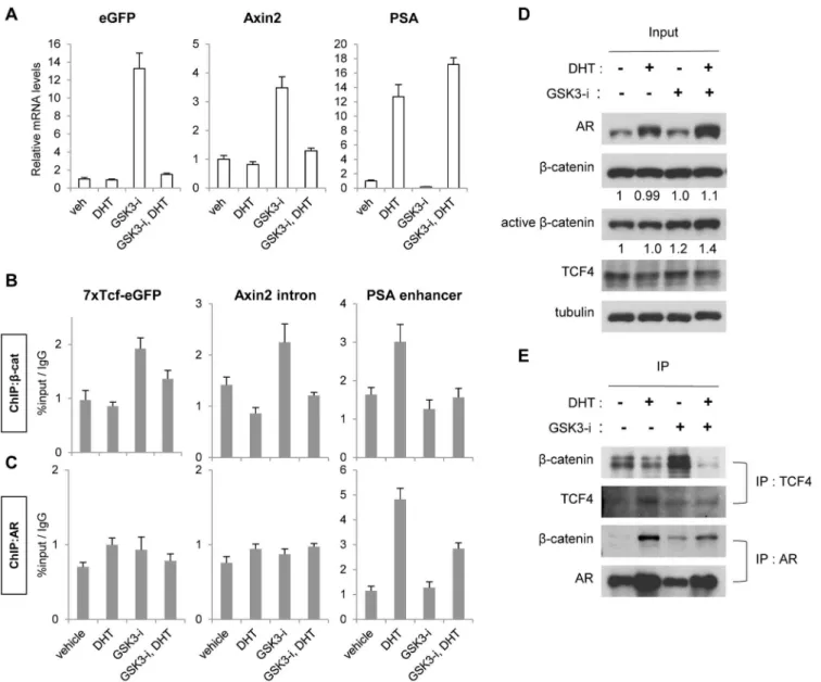
Given thatβ-catenin interacts with AR or TCF to activate AR or Wnt/β-catenin-responsive transcription, respectively, in prostate cancer [33,34], we examined theβ-catenin occupancy on AR and TCF binding sites using chromatin immunoprecipitation (ChIP) assays. LNCaP cells were treated with DHT, GSK-3 inhibitor or a combination of both for 4 h in hormone-deprived media. The relative mRNA levels of Wnt/β-catenin (eGFP Wnt-reporter andAxin2)
and AR-target (PSA) genes in each condition were analyzed (Fig 3A).β-catenin occupancy on
TCF or AR binding sites was also examined (Fig 3B) to determine ifβ-catenin recruitment cor-related with target gene expression. As expected, Wnt-reporter andAxin2mRNA levels were
increased in GSK-3 inhibitor treated cells, but diminished in the presence of DHT and GSK-3 inhibitor (Fig 3A). Consistent with the mRNA levels,β-catenin was recruited to TCF binding sites on the Wnt-reporter andAxin2in response to GSK-3 inhibitor, but not in the cells
co-treated with GSK-3 inhibitor and DHT (Fig 3B). Transcription ofPSAoccurred in response to
DHT, and this was unaffected by co-treatment with DHT and GSK-3 inhibitor (Fig 3A).β -catenin occupancy was observed at thePSAenhancer upon DHT treatment, but was
dimin-ished upon co-treatment with DHT and GSK-3 inhibitor (Fig 3B), suggesting that in this con-textβ-catenin is not essential forPSAtranscription. AR was also analyzed; showing occupancy
onPSAin response to DHT treatment but no binding was detected on TCF sites of Wnt/β -catenin target genes in any treatment (Fig 3C).
To determine if AR andβ-catenin occupancy on target genes correlates withβ-catenin bind-ing to either AR or TCF4 protein, we conducted co-immunoprecipitation assays. LNCaP cells were treated as described for the ChIP assays above, and protein lysates were immunoprecipi-tated withβ-catenin antibody.Fig 3Dshows that AR is stabilized by DHT treatment. While total levels ofβ-catenin were unchanged by 4 hours of GSK-3 inhibitor treatment, GSK-3 inhi-bition slightly increased the protein expression of the active form ofβ-catenin (unphosphory-lated at serine 37 and threonine 41, [35]), independent of the presence of DHT (Fig 3D). Overall, DHT treatment increased AR/β-catenin interaction and decreased TCF4/β-catenin interaction compared to vehicle treatment (Fig 3E), as reported previously [34,36]. Consistent with the ChIP results (Fig 3B), GSK-3 inhibitor enhanced the TCF4/β-catenin interaction but combinatory treatment with DHT diminished this interaction (Fig 3E). In addition, cells treated with DHT showed enhanced AR/β-catenin interaction, which was decreased in the presence of GSK-3 inhibitor (Fig 3E) consistent with decreased AR on the on thePSAenhancer
in the presence of GSK-3 inhibitor and DHT (Fig 3C). It is unknown why AR binding toPSA
and interaction withβ-catenin are decreased when DHT is co-treated with GSK-3 inhibitor, but this reduced level of AR on thePSAenhancer was still sufficient forPSAtranscription (Fig
3A, PSA mRNA levels in DHT vs DHT+GSK3-i).
Inhibition of AR and Wnt/
β
-catenin pathways increases growth
repression of LNCaP cells
Since one of the cellular responses of the Wnt-pathway is to promote proliferation [37,38], we tested if the enhanced activation of Wnt/β-catenin pathway in the absence of androgen or pres-ence of enzalutamide (Fig 2A, 2B, 2D and 2E) also promoted cell proliferation. LNCaP cells cultured in hormone-deprived media were growth arrested as expected [39] but GSK-3 inhibi-tor treatment or APC knockdown induced growth similar to DHT treatment (Fig 4A and 4E).
Fig 3. Hormone deprivation increasedβ-catenin interaction with TCF4 andβ-catenin binding to TCF binding sites.A, Expression of Wnt/-catenin target genes (eGFP reporter andAxin2) and the AR target gene (PSA) was analyzed. LNCaP cells were hormone-deprived for 3 days and then treated with vehicle, 10 nM DHT, 9μM GSK-3 inhibitor or a combination of both for 24 h.B and C, -catenin or AR binding on target genes was analyzed using chromatin-immunoprecipitation. LNCaP cells were hormone-deprived for 3 days and then treated with vehicle, 100 nM DHT, 9μM GSK3-i or a combination of both for 4 h.D and E, -catenin interaction with TCF4 or AR was analyzed using co-immunoprecipitation. LNCaP cells were hormone-deprived for 3 days and then treated with vehicle, 100 nM DHT, 9μM GSK-3 inhibitor or a combination of both for 4 h. The protein lysates were either immunoblotted with indicated antibodies (D) or subjected to co-IP studies (E) Quantification ofβ-catenin and activeβ-catenin protein levels are shown below panels in (D). Relative densitometry is normalized to vehicle alone, set to 1. The data presented is representative of three independent experiments and the indicated error is the standard deviation.
Enzalutamide treatment also repressed growth of LNCaP cells as previously observed [2] how-ever this growth inhibitory effect was relieved upon GSK-3 inhibitor treatment or APC knock-down (Fig 4B and 4F). We also examined transcription of an M phase cell cycle regulatory gene,UBE2C, previously shown to be important for growth of androgen-independent prostate
cancer cells [40]. The GSK-3 inhibitor treatment resulted in upregulation ofUBE2C
transcrip-tion similar to the DHT treatment (Fig 4C), as well as de-repression ofUBE2Cin enzalutamide
co-treated cells (Fig 4D). These results suggest that Wnt/β-catenin activation promotes growth of LNCaP cells in the absence of androgen or in the presence of enzalutamide, accompanied by upregulation ofUBE2C. Treatment of cells with an inhibitor of the Wnt/β-catenin pathway,
iCRT3 (C3) [41], diminished the growth-promoting effect of GSK-3 inhibitor in a dose-responsive manner (Fig 4G and 4H), further indicating that the Wnt/β-catenin pathway directs androgen-independent growth of LNCaP cells.
Since inhibition of AR activity resulted in enhanced Wnt/β-catenin-responsive transcription (Fig 2) and cell growth (Fig 4A, 4B, 4E and 4F), we hypothesized that simultaneous inhibition of both AR and Wnt/β-catenin pathways might be more effective than targeting one pathway alone. To test this idea, LNCaP cells were treated with either vehicle, enzalutamide or C3, or a combination of the two compounds. We used a sub-optimal concentration of the compounds (0.1μM of enzalutamide and 5μM or 10μM of C3) to determine the difference between single
treatment versus co-treatment.Fig 4Ishows that the combined treatment of enzalutamide and
Fig 4. Wnt/β-catenin activation promotes androgen-independent growth of LNCaP cells and inhibition of both AR and Wnt/β-catenin pathways shows increased growth inhibition of LNCaP cells.A-F, GSK-3 inhibitor treatment or APC knockdown promotes growth of LNCaP cells in hormone-deprived or enzalutamide (Enz) treated media.A and B, Cells were hormone-deprived for 3 days and then treated with vehicle, 10 nM DHT or 6μM GSK3-i (A), or 10 nM DHT with/without 10μM Enz or 6μM GSK3-i (B) every two days.C and D, Cells were treated as described in (A) or (B) for 24 h and then the relative mRNA levels ofUBE2Cwere analyzed by qRT-PCR.E and F, Cells were transfected with si-control or si-APC, hormone-deprived for 2 days and then treated with vehicle or 10 nM DHT (E), or 10 nM DHT with/without 10μM Enz (F) every two days.G and H, Treatment of cells with Wnt/β-catenin inhibitor (C3) diminishes the growth-promoting effect of GSK3-i. LNCaP cells were hormone-deprived for 3 days and then treated with vehicle or 6μM GSK3-i (G), or 10 nM DHT plus 10μM Enz (H) with/without increasing concentrations of C3 every two days.I, Co-treatment of Enz and C3 shows increased growth inhibition of LNCaP cells. Cells were hormone-deprived for 3 days and then treated with 10 nM DHT with/without 5μM or 10μM C3, 0.1μM Enz or a combination of both compounds every two days. Cell viability is represented by relative fluorescence units normalized to time zero (prior to treatments). The data presented is representative of three independent experiments and the indicated error is the standard deviation.
C3 has increased effects on growth inhibition of LNCaP cells, indicating the therapeutic poten-tial of targeting both AR and Wnt/β-catenin pathways in prostate cancer.
Inhibition of the Wnt/
β
-catenin pathway sensitizes abl cells to
enzalutamide
Contrary to what we observed in LNCaP cells, treatment of abl cells with GSK-3 inhibitor showed no effect on proliferation in either hormone-deprived or enzalutamide treated culture conditions (Fig 5A and 5B). In fact, consistent with their androgen-independent status, the
Fig 5. Inhibition of both AR and Wnt/β-catenin pathways shows increased growth inhibition of abl cells.A and B, Wnt/β-catenin activation shows no effect on growth of abl cells. Cells were treated with vehicle, 10 nM DHT or 6μM GSK-3 inhibitor (GSK3-i) (A), or vehicle or 10μM enzalutamide (Enz) with/ without 6μM GSK3-i (B) every two days.C, Inhibition of the Wnt/β-catenin pathway throughβ-catenin knockdown sensitized abl cells to Enz. Cells were stably infected with lentiviral vectors encoding control shRNA (sh-control) or -catenin shRNA (sh–cat-1 and 2) and cultured with treatment of vehicle, 0.1μM or
1μM Enz every two days.D, Co-treatment of Enz and Wnt/β-catenin inhibitor (C3) shows increased growth inhibition of abl cells. Cells were treated with vehicle, 5μM or 10μM C3, 10μM Enz or a combination of both compounds every two days. Cell viability is represented by relative fluorescence units normalized to time zero (prior to treatments). The data presented is representative of three independent experiments and the indicated error is the standard deviation.
growth of abl cells was not repressed by either hormone deprivation or enzalutamide treatment (Fig 5A and 5B). Our data suggests that abl cells are more prone to activate the Wnt/β-catenin pathway than LNCaP cells (Figs1D, 1Eand2F–2H). Also, given that previous studies [17,30] and our observations (Fig 4A and 4B) indicate that the Wnt/β-catenin pathway may promote androgen-independent growth of prostate cancer, we hypothesized that inhibition of Wnt/β -catenin pathway might sensitize abl cells to enzalutamide treatment. To test this idea, we exam-ined the effect ofβ-catenin knockdown on enzalutamide treated abl cells. Theβ-catenin knock-down slowed the proliferation of abl cells as previously shown [18] (Fig 5C; vehicle in sh-control vs. sh-b-cat-1 and 2). While enzalutamide treatment did not affect growth of sh-control cells (Fig 5C, sh-control), 0.1μM of enzalutamide showed growth inhibition inβ-catenin
depleted cells (Fig 5C, sh-b-cat-1 and 2). Interestingly, 0.1μM of enzalutamide treatment
showed inhibition comparable to 1μM (Fig 5C, sh-b-cat-1 and 2), suggesting that a lower
con-centration of AR antagonist may be effective under conditions of Wnt/β-catenin pathway inhi-bition. Next, we tested the effect of small molecule inhibition of the Wnt/β-catenin pathway in enzalutamide treated abl cells. The cells were treated with vehicle, enzalutamide, C3, or enzalu-tamide plus C3.Fig 5Dshows a dose-responsive, additive effect of C3 treatment with enzaluta-mide, indicating that targeting the Wnt/β-catenin pathway can be a promising approach for enzalutamide resistant CRPC.
Discussion
Extensive effort has been spent to develop more effective next-generation AR therapies to treat CRPC, including enzalutamide [2] and abiraterone [3]. However, resistance to these treatments inevitably occurs [42], hinting that targeting the AR pathway might not be sufficient, especially given the idea that increased crosstalk between distinct signaling pathways causes activation of AR target genes and regulatory networks in advanced prostate cancer [8,9]. Similarly, despite the clinical success of targeted therapy in many cancer types, overcoming innate or acquired resistance remains a challenge [6,43]. The emerging mechanism underlying acquired treat-ment resistance is that the growth-promoting signals are still active in cancer cells through acti-vation of alternative signaling pathways. For example, kinase inhibitor treated cancers such as melanoma, lung, breast and colorectal cancers exhibit upregulation of other kinases and/or upregulation of ligands [44–46]. Thus, understanding the mechanisms of acquired resistance and development of combination therapy targeting the resistant pathways will be an effective approach to treat advanced cancers [6].
The crosstalk between AR and Wnt/β-catenin pathways in prostate cancer has been well studied [47,48]. In addition, the major downstream effector of the canonical Wnt-pathway,β -catenin, was shown to be an AR co-activator [26,28,36]. More importantly, the AR gene is transcriptionally regulated byβ-catenin through multiple TCF binding sites within the AR pro-moter [49]. Growing evidence has shown the increased mis-regulation of Wnt/β-catenin path-way in CRPC [13,15]. However, the possibility that inhibition of the AR pathway engenders prostate cancer cells that are reliant on Wnt/β-catenin signaling has not been tested
experimentally.
pathway using siRNA againstβ-catenin or a small moleculeβ-catenin inhibitor re-established the sensitivity to enzalutamide in abl cells (Fig 5C and 5D), suggesting that inhibition of the Wnt/β-catenin pathway can be an effective therapeutic approach to treat enzalutamide-resis-tant CRPC.
Previous studies indicate that AR and TCF4 have overlapping interaction domains onβ -catenin [36,50] and the competitive binding of AR and TCF4 toβ-catenin has been shown in both prostate and colon cancer cells [34,36]. The studies showed that androgen enhancedβ -catenin interaction with AR and increased AR transcriptional activity, but resulted in repres-sion of TCF/β-catenin responsive transcription [33,34]. However, these earlier studies utilized the ectopic expression of a constitutively activeβ-catenin mutant, raising the concerns of non-specific effects. Furthermore, the status of the Wnt/β-catenin pathway has not been previously addressed in the absence of androgen (similar to conditions of androgen ablation therapy), to test if inhibition of AR activity enhancesβ-catenin interaction with TCF4.
Here, we activated endogenousβ-catenin through inhibition of theβ-catenin destruction complex via GSK-3 inhibition. We used GSK-3 inhibitor or siRNA against APC to show that the activation of Wnt/β-catenin responsive transcription was enhanced when AR activity was repressed (Fig 2). This was accompanied by increasedβ-catenin interaction with TCF4 (Fig 3E) andβ-catenin recruitment to TCF binding sites on Wnt/β-catenin target genes (Fig 3B). Increased Wnt/β-catenin responsive transcription also promoted the cell growth in the absence of androgen (Fig 4A and 4E) or upon enzalutamide treatment (Fig 4B and 4F), indicating a likely role of the Wnt/β-catenin pathway in CRPC progression. In contrast, the Wnt/β-catenin pathway was readily activated in androgen-independent abl cells in both the presence and absence of androgen or enzalutamide (Fig 2F and 2G), and this activation did not further pro-mote the abl cell growth (Fig 5A and 5B). These findings suggest that the crosstalk between AR and Wnt/β-catenin pathways might have been modified during development of androgen independence, consistent with previous evidence showing enhanced Wnt/β-catenin signaling in CRPC [33,51–53]. Importantly, siRNA mediated or small molecule inhibition of Wnt/β -catenin pathway sensitized abl cells to enzalutamide treatment (Fig 5C and 5D), indicating that enhanced Wnt/β-catenin signaling might be one of the mechanisms that confer enzalutamide resistance.
One promising strategy to overcome acquired resistance is a rationally designed combina-tion therapy against the resistant pathways based on pre-clinical studies [6]. Combinacombina-tion ther-apy has been proven to be effective in multiple cancer types, such as PI3K and MEK inhibition in KRAS or EGFR driven cancers [46] and mTOR and aromatase inhibition in breast cancer [54]. In prostate cancer, combined treatment with AR inhibitors and chemotherapeutic agents has been tested in the clinic, but the efficacy of targeting multiple pathways mis-regulated in advanced prostate cancer has not been well studied [42]. In this context, the Wnt/β-catenin pathway holds great potential to be tested in combination therapy with AR pathway inhibitors based on the well-known crosstalk between the AR and Wnt/β-catenin pathways [47,48]. Sup-porting this idea, our data showed that the simultaneous inhibition of AR and Wnt/β-catenin pathways exhibited increased effects on growth repression of both LNCaP and abl cells (Figs4I and5D). Further studies such as testing the efficacy of this approach in vivo will be required.
Author Contributions
Conceived and designed the experiments: EL SL. Performed the experiments: EL SH. Analyzed the data: EL SL. Wrote the paper: EL SH SL.
References
1. http://apps.nccd.cdc.gov/uscs/.
2. Tran C, Ouk S, Clegg NJ, Chen Y, Watson PA, Arora V, et al. Development of a second-generation anti-androgen for treatment of advanced prostate cancer. Science. 2009; 324(5928):787–90. doi:10.1126/
science.1168175PMID:19359544; PubMed Central PMCID: PMC2981508.
3. Attard G, Reid AH, Olmos D, de Bono JS. Antitumor activity with CYP17 blockade indicates that castra-tion-resistant prostate cancer frequently remains hormone driven. Cancer Res. 2009; 69(12):4937–40.
doi:10.1158/0008-5472.CAN-08-4531PMID:19509232.
4. de Bono JS, Logothetis CJ, Molina A, Fizazi K, North S, Chu L, et al. Abiraterone and increased survival in metastatic prostate cancer. N Engl J Med. 2011; 364(21):1995–2005. doi:10.1056/NEJMoa1014618
PMID:21612468; PubMed Central PMCID: PMC3471149.
5. Scher HI, Fizazi K, Saad F, Taplin ME, Sternberg CN, Miller K, et al. Increased survival with enzaluta-mide in prostate cancer after chemotherapy. N Engl J Med. 2012; 367(13):1187–97. doi:10.1056/
NEJMoa1207506PMID:22894553.
6. Glickman MS, Sawyers CL. Converting cancer therapies into cures: lessons from infectious diseases. Cell. 2012; 148(6):1089–98. doi:10.1016/j.cell.2012.02.015PMID:22424221; PubMed Central
PMCID: PMC3465702.
7. Yap TA, Omlin A, de Bono JS. Development of therapeutic combinations targeting major cancer signal-ing pathways. J Clin Oncol. 2013; 31(12):1592–605. Epub 2013/03/20. doi:10.1200/JCO.2011.37.
6418PMID:23509311.
8. Feldman BJ, Feldman D. The development of androgen-independent prostate cancer. Nat Rev Cancer. 2001; 1(1):34–45. doi:10.1038/35094009PMID:11900250.
9. Saraon P, Jarvi K, Diamandis EP. Molecular alterations during progression of prostate cancer to andro-gen independence. Clin Chem. 2011; 57(10):1366–75. doi:10.1373/clinchem.2011.165977PMID:
21956922.
10. Carver BS, Chapinski C, Wongvipat J, Hieronymus H, Chen Y, Chandarlapaty S, et al. Reciprocal feed-back regulation of PI3K and androgen receptor signaling in PTEN-deficient prostate cancer. Cancer Cell. 2011; 19(5):575–86. doi:10.1016/j.ccr.2011.04.008PMID:21575859; PubMed Central PMCID:
PMC3142785.
11. Arora VK, Schenkein E, Murali R, Subudhi SK, Wongvipat J, Balbas MD, et al. Glucocorticoid receptor confers resistance to antiandrogens by bypassing androgen receptor blockade. Cell. 2013; 155 (6):1309–22. doi:10.1016/j.cell.2013.11.012PMID:24315100; PubMed Central PMCID:
PMC3932525.
12. Barbieri CE, Baca SC, Lawrence MS, Demichelis F, Blattner M, Theurillat JP, et al. Exome sequencing identifies recurrent SPOP, FOXA1 and MED12 mutations in prostate cancer. Nat Genet. 2012; 44 (6):685–9. doi:10.1038/ng.2279PMID:22610119; PubMed Central PMCID: PMC3673022.
13. Grasso CS, Wu YM, Robinson DR, Cao X, Dhanasekaran SM, Khan AP, et al. The mutational land-scape of lethal castration-resistant prostate cancer. Nature. 2012; 487(7406):239–43. doi:10.1038/
nature11125PMID:22722839; PubMed Central PMCID: PMC3396711.
14. Hieronymus H, Sawyers CL. Traversing the genomic landscape of prostate cancer from diagnosis to death. Nat Genet. 2012; 44(6):613–4. doi:10.1038/ng.2301PMID:22641202.
15. Sun Y, Campisi J, Higano C, Beer TM, Porter P, Coleman I, et al. Treatment-induced damage to the tumor microenvironment promotes prostate cancer therapy resistance through WNT16B. Nat Med. 2012; 18(9):1359–68. doi:10.1038/nm.2890PMID:22863786; PubMed Central PMCID:
PMC3677971.
16. Robinson D, Van Allen EM, Wu YM, Schultz N, Lonigro RJ, Mosquera JM, et al. Integrative clinical genomics of advanced prostate cancer. Cell. 2015; 161(5):1215–28. doi:10.1016/j.cell.2015.05.001
PMID:26000489; PubMed Central PMCID: PMC4484602.
17. Yu X, Wang Y, Jiang M, Bierie B, Roy-Burman P, Shen MM, et al. Activation of beta-Catenin in mouse prostate causes HGPIN and continuous prostate growth after castration. Prostate. 2009; 69(3):249–62.
doi:10.1002/pros.20877PMID:18991257.
18. Lee E, Madar A, David G, Garabedian MJ, Dasgupta R, Logan SK. Inhibition of androgen receptor and beta-catenin activity in prostate cancer. Proc Natl Acad Sci U S A. 2013; 110(39):15710–5. doi:10.
19. Culig Z, Hoffmann J, Erdel M, Eder IE, Hobisch A, Hittmair A, et al. Switch from antagonist to agonist of the androgen receptor bicalutamide is associated with prostate tumour progression in a new model sys-tem. Br J Cancer. 1999; 81(2):242–51. doi:10.1038/sj.bjc.6690684PMID:10496349; PubMed Central
PMCID: PMC2362859.
20. Mita P, Savas JN, Djouder N, Yates JR 3rd, Ha S, Ruoff R, et al. Regulation of androgen receptor-medi-ated transcription by RPB5 binding protein URI/RMP. Mol Cell Biol. 2011; 31(17):3639–52. Epub 2011/
07/07. doi:10.1128/MCB.05429-11PMID:21730289; PubMed Central PMCID: PMC3165545. 21. Fuerer C, Nusse R. Lentiviral vectors to probe and manipulate the Wnt signaling pathway. PLoS One.
2010; 5(2):e9370. doi:10.1371/journal.pone.0009370PMID:20186325; PubMed Central PMCID: PMC2826402.
22. Lian X, Hsiao C, Wilson G, Zhu K, Hazeltine LB, Azarin SM, et al. Robust cardiomyocyte differentiation from human pluripotent stem cells via temporal modulation of canonical Wnt signaling. Proc Natl Acad Sci U S A. 2012; 109(27):E1848–57. doi:10.1073/pnas.1200250109PMID:22645348; PubMed
Cen-tral PMCID: PMC3390875.
23. Sineva GS, Pospelov VA. Inhibition of GSK3beta enhances both adhesive and signalling activities of beta-catenin in mouse embryonic stem cells. Biol Cell. 2010; 102(10):549–60. doi:10.1042/
BC20100016PMID:20626347.
24. Hobisch A, Hoffmann J, Lambrinidis L, Eder IE, Bartsch G, Klocker H, et al. Antagonist/agonist balance of the nonsteroidal antiandrogen bicalutamide (Casodex) in a new prostate cancer model. Urol Int. 2000; 65(2):73–9. doi: 64843. PMID:11025427.
25. Sramkoski RM, Pretlow TG 2nd, Giaconia JM, Pretlow TP, Schwartz S, Sy MS, et al. A new human prostate carcinoma cell line, 22Rv1. In Vitro Cell Dev Biol Anim. 1999; 35(7):403–9. doi:10.1007/
s11626-999-0115-4PMID:10462204.
26. Mulholland DJ, Cheng H, Reid K, Rennie PS, Nelson CC. The androgen receptor can promote beta-catenin nuclear translocation independently of adenomatous polyposis coli. J Biol Chem. 2002; 277 (20):17933–43. doi:10.1074/jbc.M200135200PMID:11856748.
27. Pawlowski JE, Ertel JR, Allen MP, Xu M, Butler C, Wilson EM, et al. Liganded androgen receptor inter-action with beta-catenin: nuclear co-localization and modulation of transcriptional activity in neuronal cells. J Biol Chem. 2002; 277(23):20702–10. doi:10.1074/jbc.M200545200PMID:11916967.
28. Truica CI, Byers S, Gelmann EP. Beta-catenin affects androgen receptor transcriptional activity and ligand specificity. Cancer Res. 2000; 60(17):4709–13. PMID:10987273.
29. Morin PJ, Sparks AB, Korinek V, Barker N, Clevers H, Vogelstein B, et al. Activation of beta-catenin-Tcf signaling in colon cancer by mutations in beta-catenin or APC. Science. 1997; 275(5307):1787–90.
PMID:9065402.
30. Bruxvoort KJ, Charbonneau HM, Giambernardi TA, Goolsby JC, Qian CN, Zylstra CR, et al. Inactivation of Apc in the mouse prostate causes prostate carcinoma. Cancer Res. 2007; 67(6):2490–6. doi:10.
1158/0008-5472.CAN-06-3028PMID:17363566.
31. Placencio VR, Sharif-Afshar AR, Li X, Huang H, Uwamariya C, Neilson EG, et al. Stromal transforming growth factor-beta signaling mediates prostatic response to androgen ablation by paracrine Wnt activ-ity. Cancer Res. 2008; 68(12):4709–18. doi:10.1158/0008-5472.CAN-07-6289PMID:18559517;
PubMed Central PMCID: PMC2811537.
32. Zong Y, Huang J, Sankarasharma D, Morikawa T, Fukayama M, Epstein JI, et al. Stromal epigenetic dysregulation is sufficient to initiate mouse prostate cancer via paracrine Wnt signaling. Proc Natl Acad Sci U S A. 2012; 109(50):E3395–404. doi:10.1073/pnas.1217982109PMID:23184966; PubMed
Cen-tral PMCID: PMC3528570.
33. Chesire DR, Isaacs WB. Ligand-dependent inhibition of beta-catenin/TCF signaling by androgen recep-tor. Oncogene. 2002; 21(55):8453–69. doi:10.1038/sj.onc.1206049PMID:12466965.
34. Mulholland DJ, Read JT, Rennie PS, Cox ME, Nelson CC. Functional localization and competition between the androgen receptor and T-cell factor for nuclear beta-catenin: a means for inhibition of the Tcf signaling axis. Oncogene. 2003; 22(36):5602–13. doi:10.1038/sj.onc.1206802PMID:12944908.
35. van Noort M, Meeldijk J, van der Zee R, Destree O, Clevers H. Wnt signaling controls the phosphoryla-tion status of beta-catenin. J Biol Chem. 2002; 277(20):17901–5. doi:10.1074/jbc.M111635200PMID:
11834740.
36. Song LN, Gelmann EP. Interaction of beta-catenin and TIF2/GRIP1 in transcriptional activation by the androgen receptor. J Biol Chem. 2005; 280(45):37853–67. doi:10.1074/jbc.M503850200PMID:
16141201.
38. Clevers H, Nusse R. Wnt/beta-catenin signaling and disease. Cell. 2012; 149(6):1192–205. doi:10.
1016/j.cell.2012.05.012PMID:22682243.
39. Chen CD, Welsbie DS, Tran C, Baek SH, Chen R, Vessella R, et al. Molecular determinants of resis-tance to antiandrogen therapy. Nat Med. 2004; 10(1):33–9. doi:10.1038/nm972PMID:14702632.
40. Wang Q, Li W, Zhang Y, Yuan X, Xu K, Yu J, et al. Androgen receptor regulates a distinct transcription program in androgen-independent prostate cancer. Cell. 2009; 138(2):245–56. doi:10.1016/j.cell.
2009.04.056PMID:19632176; PubMed Central PMCID: PMC2726827.
41. Gonsalves FC, Klein K, Carson BB, Katz S, Ekas LA, Evans S, et al. An RNAi-based chemical genetic screen identifies three small-molecule inhibitors of the Wnt/wingless signaling pathway. Proc Natl Acad Sci U S A. 2011; 108(15):5954–63. doi:10.1073/pnas.1017496108PMID:21393571; PubMed Central
PMCID: PMC3076864.
42. Lorente D, De Bono JS. Molecular alterations and emerging targets in castration resistant prostate can-cer. Eur J Cancan-cer. 2014; 50(4):753–64. Epub 2014/01/15. doi:10.1016/j.ejca.2013.12.004PMID:
24418724.
43. Haber DA, Gray NS, Baselga J. The evolving war on cancer. Cell. 2011; 145(1):19–24. doi:10.1016/j.
cell.2011.03.026PMID:21458664.
44. Chong CR, Janne PA. The quest to overcome resistance to EGFR-targeted therapies in cancer. Nat Med. 2013; 19(11):1389–400. doi:10.1038/nm.3388PMID:24202392.
45. Wilson TR, Fridlyand J, Yan Y, Penuel E, Burton L, Chan E, et al. Widespread potential for growth-fac-tor-driven resistance to anticancer kinase inhibitors. Nature. 2012; 487(7408):505–9. doi:10.1038/
nature11249PMID:22763448; PubMed Central PMCID: PMC3724525.
46. Wong KK, Engelman JA, Cantley LC. Targeting the PI3K signaling pathway in cancer. Curr Opin Genet Dev. 2010; 20(1):87–90. doi:10.1016/j.gde.2009.11.002PMID:20006486; PubMed Central PMCID:
PMC2822054.
47. Terry S, Yang X, Chen MW, Vacherot F, Buttyan R. Multifaceted interaction between the androgen and Wnt signaling pathways and the implication for prostate cancer. J Cell Biochem. 2006; 99(2):402–10.
doi:10.1002/jcb.20983PMID:16741972.
48. Kypta RM, Waxman J. Wnt/beta-catenin signalling in prostate cancer. Nat Rev Urol. 2012. doi:10. 1038/nrurol.2012.116PMID:22710668.
49. Yang X, Chen MW, Terry S, Vacherot F, Bemis DL, Capodice J, et al. Complex regulation of human androgen receptor expression by Wnt signaling in prostate cancer cells. Oncogene. 2006; 25
(24):3436–44. doi:10.1038/sj.onc.1209366PMID:16474850; PubMed Central PMCID: PMC2630384.
50. Yumoto F, Nguyen P, Sablin EP, Baxter JD, Webb P, Fletterick RJ. Structural basis of coactivation of liver receptor homolog-1 by beta-catenin. Proc Natl Acad Sci U S A. 2012; 109(1):143–8. doi:10.1073/
pnas.1117036108PMID:22187462; PubMed Central PMCID: PMC3252924.
51. Chen G, Shukeir N, Potti A, Sircar K, Aprikian A, Goltzman D, et al. Up-regulation of Wnt-1 and beta-catenin production in patients with advanced metastatic prostate carcinoma: potential pathogenetic and prognostic implications. Cancer. 2004; 101(6):1345–56. Epub 2004/08/19. doi:10.1002/cncr.
20518PMID:15316903.
52. de la Taille A, Rubin MA, Chen MW, Vacherot F, de Medina SG, Burchardt M, et al. Beta-catenin-related anomalies in apoptosis-resistant and hormone-refractory prostate cancer cells. Clin Cancer Res. 2003; 9(5):1801–7. Epub 2003/05/10. PMID:12738737.
53. Wang G, Wang J, Sadar MD. Crosstalk between the androgen receptor and beta-catenin in castrate-resistant prostate cancer. Cancer Res. 2008; 68(23):9918–27. doi:10.1158/0008-5472.CAN-08-1718
PMID:19047173; PubMed Central PMCID: PMC2654418.
54. Baselga J, Campone M, Piccart M, Burris HA 3rd, Rugo HS, Sahmoud T, et al. Everolimus in postmeno-pausal hormone-receptor-positive advanced breast cancer. N Engl J Med. 2012; 366(6):520–9. Epub