Molecular Modelling of ABC transporters:
from ATP hydrolysis to substrate transport
Ana Sofia Fernandes de Oliveira
Dissertação para a obtenção de grau de doutor em Bioquímica
pelo Instituto de Tecnologia Química e Biológica
da Universidade Nova de Lisboa
i
Acknowledgements ... 1
List of Publications... 3
Papers presented in this thesis ...3
Papers related to this thesis ...3
Abstract ... 5
Sumário ... 9
List of abbreviations and symbols ... 13
1. Introduction ... 17
1.1. ABC transporters ...17
1.2. Structure and Function of ABC transporters ...21
1.2.1. Transmembrane Domain (TMD) ...25
1.2.2. Nucleotide Binding Domain (NBD) ...26
1.2.2.1. NBDs asymmetry, one of the oldest unanswered question ... 33
1.2.2.2. Stoichiometry of ATP binding/hydrolysis and the role of two binding sites .. 35
1.2.3. Substrate Binding Protein (SBP) ...36
1.3. Molecular Mechanisms in ABC transporters ...41
1.3.1. Mechanisms for NBD dimer functioning ...41
1.3.2. Mechanisms for coupling hydrolysis to allocrite transport ...49
1.3.2.1. Export Systems ... 49
1.3.2.2. SBP-dependent import systems ... 52
ii
1.4.1. Protein dynamics ...55
1.4.1.1. Nucleotide binding domains ... 56
1.4.1.2. Full-length ABC transporters ... 58
1.4.2. Structure prediction ...61
1.5. The ABC transporters studied ...62
1.5.1. ABC Importers: MalFGK2E ...62
1.5.2. ABC Exporters: Sav1866 and MJ0796 ...65
2. Theory and Methods ... 69
2.1. Introduction ...69
2.2. Molecular Mechanics/Dynamics ...72
2.2.1. Empirical Force Field ...72
2.2.2. Bonded interactions ...74
2.2.3. Non-bonded interactions ...75
2.2.4. Solvation and Boundary Conditions ...79
2.2.5. FF parameterization ...80
2.2.6. Computer Simulation methods ...81
2.2.7. Molecular Dynamics simulation ...82
2.2.8. Metropolis Monte Carlo simulations ...84
2.2.9. Membrane Protein simulations ...85
2.3. Continuum Electrostatic Methods ...89
iii
2.3.2. Electrostatic binding free energy ...91
2.3.3. Binding equilibrium of protons ...93
2.3.4. Proton tautomerism ...94
2.3.3. Determination of int pK and Wij ...95
3. Conformational changes in the MJ0796 NBD dimer ... 99
3.1. Abstract... 100
3.2. Introduction ... 101
3.3. Materials and Methods ... 106
3.3.1. Starting Structure ... 106
3.3.2. Protonation state of protonable residues ... 106
3.3.3. General setup of molecular dynamics simulation ... 107
3.3.4. Data analysis ... 111
3.3.5. Catalytic cycles for the ABC NBD dimer ... 112
3.4. Results ... 114
3.4.1. Structural differences between the E171Q mutant and the wild type dimer ... 114
3.4.2. Structural differences during the catalytic cycle ... 114
3.4.3. A possible exit way for Inorganic phosphate ... 122
3.4.4. Localization of the major conformational changes in the full-length transporter: inter-domain interfaces ... 126
iv
3.6. Acknowledgements ... 131
4. Conformational changes in the Sav1866 exporter... 133
4.1. Abstract... 134
4.2. Introduction ... 136
4.3. Materials and Methods ... 141
4.3.1. Starting Structure ... 141
4.3.2. Sav1866 and lipid bilayer assembly ... 141
4.3.3. General setup for the molecular dynamics simulations... 142
4.3.4. Estimation of substrate permeation energy profiles ... 144
4.3.5. Data analysis ... 146
4.4. Results ... 147
4.4.1. Structural differences between 2ATP and 2ADP.IP state ... 148
4.4.2. Opening of the NBDs dimer upon hydrolysis ... 154
4.4.3. An entry point for the allocrite molecules? ... 158
4.5. Concluding Remarks... 162
4.6. Acknowledgements ... 164
5. Conformational changes in the MalFGK
2E importer ... 165
5.1. Abstract... 166
5.2. Introduction ... 167
5.3. Materials and Methods ... 172
v
5.3.2. MalFGK2E insertion into a lipid bilayer ... 172
5.3.3. General setup for the molecular dynamics simulations... 173
5.3.4. Data analysis ... 175
5.4. Results ... 177
5.4.1. Rigid body motion of the MalF Periplasmic region... 179
5.4.2. MalK dimer interface ... 180
5.4.3. Structural differences during the ATP-cycle in the MalFGK2E complex ... 183
5.4.4. Maltose Position during ATP hydrolysis ... 189
5.5. Concluding Remarks... 190
5.6. Acknowledgements ... 192
6. General Discussion ... 193
7. General Conclusion ... 199
8. Supporting Information for chapter 3 ... 201
9. Supporting Information for chapter 4 ... 211
10. Supporting Information for chapter 5 ... 225
1
Acknowledgements
First of all, I want to thank myself for the courage and persistence (although some people still call it stubbornness) in pursuing a scientific career against all difficulties.
A very special thanks and my sincere gratitude goes to Cláudio Soares, who has been my supervisor and friend since the beginning. He provided me with many valuable and helpful suggestions and important advice during all the years.
I would also like to thank António Baptista for the continuous support and motivation along the years and for all the stimulating discussions (not only scientific). I want to thank him for the patience spent listening to my existential scientific doubts and for the time used explaining the most complicated subjects in a way I could truly understand them.
I want to acknowledge all the members (whether past or present) of the Protein Modelling and the Molecular Simulation Laboratory: Vitor Teixeira, Nuno Micaelo, Carlos Cunha, Miguel Machuqueiro, Diana Lousa, João Damas, Sara Campos, Luis Carlos, Pedro Magalhães and Carla Baltazar for the fantastic working environment and for all the fun we have during this last years (especially at lunch time). I have no doubt that their sense of humor helped to ease the hardest moments. A very special thanks to Bruno Victor and Carla Pinheiro for their constant friendship and pertinent advices.
Also, I would like to acknowledge ITQB for the excellent working conditions and FCT for the financial support (SFRH/BD/21433/2005).
2
Also, I would like to thank my children, Cuca and Gonçalinho. How can I say this without becoming too sappy? Both of you lighted my darkest moments and I am very proud of you both.
3
List of Publications
Papers presented in this thesis
1- Oliveira, A.S., A.M. Baptista, and C.M. Soares, Insights into the molecular mechanism of an ABC transporter: conformational changes in the NBD dimer of
MJ0796. J Phys Chem B, 2010. 114(16): p. 5486-96.
2- Oliveira, A.S., A.M. Baptista, and C.M. Soares, Conformational changes induced by ATP-hydrolysis in an ABC transporter: a molecular dynamics study of the
Sav1866 exporter. 2010, submitted to J Phys Chem B.
3- Oliveira, A.S., A.M. Baptista, and C.M. Soares, Inter-domain communication mechanisms in an ABC importer: A molecular dynamics study of the MalFGK2E
complex. 2010, manuscript in preparation.
Papers related to this thesis
4- Damas, J.M., Oliveira, A.S., A.M. Baptista, and C.M. Soares, Structural consequences of ATP hydrolysis on ABC transporter NBDs: molecular dynamics
5
Abstract
Despite the great advances that have been made in the past decades in the ABC transporters field, the molecular mechanisms involved in transport across membranes remains largely an enigma. To date, questions regarding the molecular mechanism of transport, nucleotide hydrolysis and inorganic phosphate exit from the binding sites are still unanswered. In this thesis the dynamic behavior of several ABC transporters during the ATP-hydrolytic cycle is investigated using molecular modeling methods. The content of this thesis is compiled in three main scientific publications [1-3], corresponding to sections 3, 4 and 5, respectively. Although these three works are performed in prokaryotic family ABC transporters, it is likely that eukaryotic ones use similar mechanisms for nucleotide hydrolysis, inter-domain communication and allocrite translocation.
6
In the second study (section 4), we describe a molecular modeling study with the main objective of identifying the major conformational rearrangements induced by nucleotide hydrolysis in a complete model ABC exporter (Sav1866). The Sav1866 exporter is a bacterial ABC protein belonging to the family of the lipid flippase-like ABC transporters, and it is a homologue of the human multidrug resistance proteins. Comparing our MD simulations for the pre- (with ATP-bound in the binding sites) and the post-hydrolysis (ADP and IP-bound) states, we were able to show that segments undergoing significant conformational changes upon hydrolysis are not restricted to the NBDs, but extend deeply to the transmembrane external regions. In the transmembrane domains (TMDs), these segments are localized in the extracellular loops 1 and 3, the coupling helices and in the TMD helix 7, whereas in the NBDs, the conformational changes are located in the helical sub-domain region. Additionally, we report, for the first time, the atomic details associated to the NBD dimer dissociation upon hydrolysis in the context of a complete exporter. The NBD interface opening results from the withdrawal of the ABC signature motif from the nucleotide ribose ring and inorganic phosphate. Moreover, we determined, also for the first time, for the two simulated states, the adiabatic energy profiles along the transmembrane pore for one of known Sav1866 allocrites, doxorubicin. These profiles are similar between the two states and are overall downhill, from the cytoplasmatic to the extracellular side, with local energy barriers relatively small and easy to be overcome. The only difference between the two states is related to an energy barrier located in the intracellular loop (ICL) region, in the ATP-bound state, which, upon hydrolysis, becomes severely reduced facilitating allocrite passage. These adiabatic energy profiles give us new insights into the transport “powering” mechanism and allow us to understand, at a molecular level, the unidirectionality of allocrite translocation.
7
phosphate exit in a model ABC importer, the maltose import system (MalFGK2E)
from Escherichia coli. For that purpose, we simulated the MalFGK2E importer in
three intermediate states of the ATP-hydrolytic cycle: an ATP-bound, an ADP plus inorganic phosphate and an ADP-bound state. From extensive MD simulations performed for the three states, we showed that the major rearrangements during the ATP-cycle are located in the “EAA motif” region (mainly in the “coupling helices”) of the TMDs and in the A-loop and in the helical sub-domain region of the NBDs. Additionally, only in the ADP-bound simulation, we were able to clarify the molecular details associated to the NBD dimer interface dissociation, which in this case, and similarly to what was observed in the Sav1866 exporter, is accompanied by the dissociation of ADP from the ABC signature motif, but not from its corresponding P-loop motif. However, the opening of one of the nucleotide binding sites did not significantly alter the transmembrane domains conformations, at least in our simulated timescale.
9
Sumário
Apesar dos enormes avanços científicos das últimas décadas na área dos transportadores ABC, até hoje, os mecanismos moleculares responsáveis pelo transporte activo através de membranas nesta família permanecem, na generalidade, um enigma. Actualmente, várias questões associadas ao mecanismo molecular de transporte, à hidrólise de nucleotidos e à saída do fosfato inorgânico aguardam uma resposta clara e conclusiva. Ao longo desta tese, o comportamento dinâmico de vários transportadores ABC irá ser estudado, com detalhe atómico, recorrendo a métodos teórico-computacionais. O conteúdo desta tese encontra-se repartido em três publicações científicas [1-3], correspondentes às secções 3, 4 e 5, respectivamente. É importante realçar que apesar dos três trabalhos reportados terem sido realizados unicamente em membros da família ABC de procariotas, é bastante provavél que os mecanismos para a hidrólise de nucleótidos, a comunicação entre domínios e a translocação de substratos nos transportadores ABC eucariotas sejam similares.
10
que continham fosfato inorgânico (IP), foi possível identificar uma provável via de saída para os IPs resultante do movimento dos resíduos 38-43. Após hidrólise, este segmento rearranja-se e altera a sua conformação expondo os IPs ao solvente, aumentando a probabilidade de difusão destes para fora dos centros activos.
11
facilmente ultrapassáveis pelos alócritos. A única diferença observada entre os dois estados estudados localiza-se na região dos loops intracellulares (ICLs). Após hidrólise, a barreira de energia localizada nos ICLs é significativamente reduzida, facilitando assim a passagem dos alócritos. A determinação destes perfis adiabáticos de energia permitiu-nos perceber, com detalhe molecular, a unidirectionalidade do transporte de alócritos, o que ajudou a clarificar o mecânismo de transporte.
No último trabalho (secção 5), apresentamos um estudo de modelação molecular num importador ABC modelo completo (MalFGK2E de Escherichia coli). O objectivo
deste trabalho é identificar as consequências estruturais da hidrólise de ATP e subsequente saída dos IPs. Para isso, simulamos o importador MalFGK2E em três
estados intermédios do ciclo de hidrólise: um primeiro estado com ATP ligado nos centros activos, um segundo com ADP e IP e um terceiro estado com apenas ADP. Com base nas extensas simulações realizadas para os três estados, foi possível mostrar que os rearranjos conformacionais mais relevantes localizam-se no motivo estrutural “EAA” nos TMDs (especialmente nas hélices de acoplamento). Nos NBDs, as maiores alterações localizam-se no loop A e no sub-domínio helicoidal. Adicionalmente, nas simulações do estado ADP, conseguimos observar os detalhes moleculares associados à dissociação da interface do dímero dos NBDs. Neste caso, e de forma semelhante ao que havia sido reportado para o exportador Sav1866, a dissociação da interface dos NBDs é acompanhada pelo afastamento do ADP do motivo assinatura ABC, mas não do motivo estrutural loop P. Todavia, a abertura de um dos centros activos não alterou significativamente a conformação dos domínios transmembranares, pelo menos na escala de tempo simulada.
12
13
List of abbreviations and symbols
Aminoacids
Alanine A Ala Leucine L Leu
Arginine R Arg Lysine K Lys
Asparagine N Asn Methionine M Met
Aspartate D Asp Phenylalanine F Phe
Cysteine C Cys Proline P Pro
Glutamine Q Gln Serine S Ser
Glutamate E Glu Threonine T Thr
Glycine G Gly Tryptophan W Trp
Histidine H His Tyrosine Y Tyr
14 Abbreviations
ADP Adenosine diphosphate ATP Adenosine triphosphate CE Continuum Electrostatic
CFTR Cystic Fibrosis Conductance Regulator DNA Deoxyribonucleic acid
ESP Electrostatic Potential Charges FE Free energy
FF Force field
HlyB Hemolysin Secretion Protein IP Inorganic Phosphate
B
k
Boltzmann Constantb
k
Bond Force Constant
k
Bond Angle Force Constant
k Improper Dihedral Force Constant
k Proper Dihedral Force Constant
15
ns Nanosecond
NBD Nucleotide Binding Domain PB Poisson-Boltzmann
PDB Protein Data Bank ps Picosecond
QM Quantum Mechanics RD Regulatory Domain RESP Restrained ESP RNA Ribonucleic acid
j i
r, Inter-atomic distance between atoms i and
j
rms Root Mean Square SBP Substrate Binding Protein
T Temperature
TMD Transmembrane Domains TMS Transmembrane Segments V Potential Energy
ij
W Electrostatic interaction between site i and
j
Greek Symbols
Energy well depth (in Lennard–Jones Potential); dielectric constant (in electrostatics)0
16
r
Relative Permittivity of the Medium17
1. Introduction
1.1. ABC transporters
All cells and cellular compartments are separated from the external media by membranes. Then, the survival of all organisms depends on the regulation and selective passage of some specific molecules (such as ions, amino-acids and water molecules) across these lipid membranes. These transport processes, allow not only the acquisition of nutrients but also the excretion of waste products from cells. Examples of compounds commonly exchanged across membranes include metabolites (such as glucose and pyruvate), ions (such as sodium, potassium, calcium, and chloride) as well as amino acids and nucleotides [4].
A biological membrane is much more complex than a simple phospholipid bilayer and it can incorporate different lipids (including cholesterol) and membrane proteins. Membrane proteins can be classified into two broad categories: integral (or intrinsic) and peripheral (or extrinsic), based on the nature of their interactions with the membranes [4]. The integral membrane proteins are formed by one or more membrane spanning segments and, in general, contain hydrophobic residues that interact with the fatty acyl groups of the phospholipids anchoring the protein to the membrane [4]. The peripheral membrane proteins do not interact with the hydrophobic core of the phospholipid bilayer and are usually bound to the membrane indirectly, by interactions with integral membrane proteins or directly, through interactions with the lipid polar head groups [4].
18
In a simplified way, in the passive transport, the proteins simply allow the passage of the substrate molecules across membranes at a higher rate than normal diffusion. Since passive transporters do not work against a concentration gradient, no energy input is required. On the other hand, active transport requires the hydrolysis of ATP since it involves the translocation of molecules across a membrane against its concentration gradient.
The membrane transporter proteins are grouped into several families, where all members are related in sequence and have probably a common evolutionary origin and molecular mechanism. In this thesis, we will only focus in one of these families, the ABC family and we will try to answer some of the questions related with ATP-hydrolysis and with its coupling to transport.
ABC (ATP Binding Cassette) family forms one of the largest superfamilies of proteins [5] and all members couple the energy from ATP binding and hydrolysis to a large variety of essential biological phenomena, comprising not only the pumping, against the concentration gradient, of substrates across biological membranes, [5] but also to other non-transport-related processes [6, 7]. This family exists through all phyla ranging from bacteria to man. The term ABC system was assigned because of several highly conserved sequences of residues associated with nucleotide (ATP) binding and hydrolysis [8, 9]. Although the vast majority of the ABC systems are associated with membrane transport, a few members appear to have been sequestered during evolution to serve alternative functions, such as DNA repair [10], translation elongation [11] and chromatin organization [12]. For example, one of the best known atypical ABC member is the UvrA protein, which is a cytoplasmic enzyme involved in DNA repair and is it formed by two typical ATPase domains interrupted by DNA-binding zinc fingers [13].
19
translocates allocrites to the cell exterior. The second category, the importers [7], act as uptake systems retrieving allocrites from the cell exterior, and the third category is apparently not involved in transport but rather in alternative functions [7]. The term ABC transporters is restricted to the first and the second classes and they contain typical integral membrane domains [7].
Despite that each ABC transporter involved in substrate translocation is relatively specific for that given substrate [5], the variety of allocrites is huge, ranging from small organic and inorganic ions, mono- and oligissaccharides, amino-acids, vitamins, peptides and even to proteins, including toxins.
Until this moment, all the identified members of this family present a unidirectional allocrite transport [5] and no ABC transporter is known to be capable of working in both directions. Additionally, importers have only been found in prokaryotes while exporters are widely spread and expressed through all kingdoms of life [14].
Over the last decades, the ABC transporter family has received considerable attention, especially due to its connections to several human genetic diseases [15] and to multidrug resistance [16]. There are ~52 ABC genes identified in the human genome [17] and mutations in several of them lead to human genetic disorders, including a bleeding disorder [18], several eye [19, 20] and liver [21] diseases, and cystic fibrosis [22]. Moreover, in humans some members of this family are known by their function in the extrusion of chemotherapeutic drugs and thus contributing to cancer cells resistance [23]. Similarly, in bacteria [24] and fungi [25], ABC transporters catalyze the export of toxic substances, contributing to drug and antibiotic resistance.
20
21
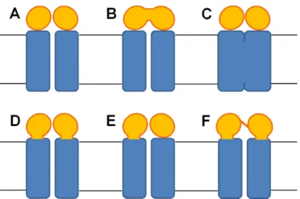
1.2. Structure and Function of ABC transporters
Despite the large diversity of transported allocrites and the difference in the transport direction, both importers and exporters share a common minimum functional unit. A typical ABC transporter is formed by four domains [27] (see Figure 1): two hydrophobic membrane-spanning or integral membrane domains (designated as Trans-Membrane Domains –TMDs) and two hydrophilic domains (peripherally located at the cytoplasmic side), where nucleotide binding and hydrolysis occurs (designated as Nucleotide Binding Domains –NBDs).
Figure 1- Minimum functional unit organization of an ABC transporter. The ABC architecture
comprises two transmembrane domains (in dark blue) and two nucleotide binding domains (in yellow). Some ABC transporters receive their substrate (in red) from the bilayer whereas other ABC members receive it from the aqueous phase or from substrate binding proteins (in blue). In the last case, the substrate binding proteins prime function is the uptake and delivery of the allocrite to the transporter.
The TMD units form the transport pathway through which substrate crosses the membrane [28] and they consist of several membrane-spanning α helices
22
hydrolysis [5], whereas the ABC signature motif is a set of residues specific to the ABC family [2]
In the majority of the bacterial ABC transporters, the four basic domains are frequently coded and expressed as separated single polypeptide chains (Figure 2A) that are assembled to form a complex suitable for allocrite transport (as for example the maltose transporter MalFGK2). However, the four basic domains can be fused
together in almost every possible combination imagined and any domain arrangement can be found in nature (see Figure 2). For example, there are several cases in which the domains are fused together forming larger, multifunctional polypeptide chains (Figure 2B and Figure 2C). This is the case of the ribose transporter (Rbs) from E. coli (where the two ATP binding domains are fused together -Figure 2B) or the siderophore/haem/vitamin B12 transporter (where both
23
Figure 2-Possible domain architecture of ABC transporters. A typical ABC transporter is composed of
two hydrophobic transmembrane domains (TMDs) and two water soluble nucleotide-binding domains (NBDs) bound to the cytosolic face of the TMDs. The TMDs form the translocation pathway and they can form a homo or a heterodimer. The NBDs are the engines of ABC transporters and they couple the ATP energy to the transport process. Schematically indicated are: TMDs (blue rectangles) and NBDs (yellow spheres). The domains are frequently expressed as separate polypeptide chains, but they can be fused in all possibilities represented in this figure. (A) All four basic domains are
expressed as separated single polypeptide chains. An example of such archiquecture is maltose/maltodextrin transporter (MalFGK2) and vitamin B12 transporter (BtuCD) from E. coli. (B) The two NBDs are fused together while the two TMDs are expressed as separated single chains. An example is the ribose transporter (Rbs) from E. coli. (C) The two TMDs are fused together while the
two NBDs are expressed as separated polypeptide chains. An example of this archiquecture is the siderophore/haem/vitamin B12 transporter (Fhu). (D) Each TMD is fused to one NBD (frequently named half-transporter). Some examples of this architecture is the lipid flippase from E. coli (MsbA), the mitochondrial peptide transporter from Saccharomyces cerevisiae (Mdl) and the multidrug lipid flippase from Staphylococcus aureaus (Sav1866). (E) One TMD is fused to one NBD while the other
TMD and NBD are expressed separatly as single polypeptide chains. An example is the YhiGHI transporter. (F) All four domains are expressed as a unique polypeptide chain. Examples of this
24
Despite the fact that the previously described four core domains are sufficient to mediate allocrite translocation, some ABC members possess additional domains with peripheral or regulatory functions. This is the case of CFTR or the maltose transporter. CFTR presents an extra domain with regulatory functions located between the NBDs, which has no equivalence to any other known domain in the ABC family [29, 30]. MalK presents an extension in the C-terminal region of the NBDs, with regulatory and enzymatic functions (which are apparently independent of the transport process itself) [31]. Furthermore, all prokaryotic importers, in order to properly perform their function, rely on high-affinity binding proteins located in the external side of the membrane, and whose function is to retrieve the allocrites from the medium and deliver them to the transporter [32].
Despite the fact that high resolution crystal structures obtained for the isolated [33-49] and the dimeric [46, 47, 50-56] ATPase modules, together with the full-length ABC transporters structures [57-65], provided us with relevant structural information and insights into the transport cycle, there is still much to be done before we have a complete understanding of how ABC transporters work. There are still many open questions about the NBD:TMD communication mechanisms, ATP-hydrolysis or the conformational changes induced during a transport cycle in the TMDs.
25 1.2.1. Transmembrane Domain (TMD)
The TMDs are two highly hydrophobic protein domains that form the channel through which the allocrite is translocated [28]. The TMD dimer can be either formed by two copies of the same monomer or by two distinct monomers, forming by this reason homo- or heterodimers. The TMDs are essentially formed byseveral
α-helices that traverse the membranes. Many ABC members posses a “two-times-six” helices, in a total of 12 transmembrane segments (TMSs) per functional unit [5, 28]. However, this number may vary from 4 to 11 for each individual transporter [5, 28]. The large structural diversity of the TMDs can easily be illustrated looking at the X-ray data available for full-length transporters [57-65] (see Figure 3).
Figure 3- TMDs structural diversity. X-ray structures for A) Sav1866 homodimer [58]. B) MalFGK2E complex [62]. C) ModA-ModBC complex [60]. D) Vitamin B12 (BtuCD) transporter [57]. E) Putative metal chelate transporter H10796 [63]. F) MetNI methionine transporter [66]. G) MsbA [64]. H) P-glycoprotein [65]. The approximate location of the lipid bilayer is indicated by the black lines. Some structures (B, C, F) have an additional extension of the NBD, probably involved in regulation of
26
So, in this figure the nucleotide species is represented by magenta sphere whenever present. In these eight examples, only structures B and H show the transported allocrite bound to the TMDs.
TMDs present low sequence identity between family members. If we consider that all ABC transporters share a common evolutionary origin [32], the low sequence identity may imply that each TMDs is designed to satisfy different structural constraints, depending on the transported allocrite. In this case, the TMDs low sequence identity among the family most probably reflects the large diversity of transported molecules. Another factor that can help explain the large differences (whether in sequence and in structure) in the TMD region is the physiological lipid environment where each transporter is inserted.
Despite the sequence variability, there is one sequence of residues highly conserved in the ABC importers [67]. This sequence is named “EAA” motif or L-loop [67, 68], and it is usually located in one cytoplasmic loop and appropriately positioned to interact with the ATP-binding domains [69], being most probably involved in inter-domain communication [58]
.
1.2.2. Nucleotide Binding Domain (NBD)
27
E . c.MalK 1
T . 1.MalK 1
Glcv 1
MJ0796 1
E . c.MsbA 351 V. C.MsbA 351
cTAPI 489
HlyB
HisP
MJ 1267 BtuO
E . c.MalK 50
T .1.MalK 50
Glcv 52
MJ 0796 52
E . C.MsbA 390
V. C .~lsbA 390
TAPl 552
HlyB 50
HisP 53
MJ1267 54
BtuO 47
E . c.MalK 99
T .1. ~!alK 105
GlcV 106
14J0796 107
E . c . MsbA 441
V . C.~lsbA 441
TAP1 603
HlyB 101
HisI' 117
14J 1267 106
Bt uD 95
E.C . MalK 146 T .1.~ l alK 154
GlcV 155
MJ 0796 160
E . c . MsbA 495 V. C. MsbA 495
TAp1 657
HlyB 154
HisI' 168
/1J1267 168
BtuD 141
E . c.MalK 205 T .1. MalK 211
GlcV 212
HJ0796 216
E. C.MsbA 550 V.C.MsbA 550
TAp1 714
HlyB 209
Hi.P 224
HJ1267 224
BtuD 204
sl s2 s3 ... Wal.ker A hI
~:.:. .:::.
---MASvQLQNVTKANGE:VV----vSKD1NLD1HE:GEFVVFV PSGCGKS LLRM1 ---MAGVRLVOVWKVFGEVT----AVREMSLEVKOGEFM1LL PSGCGKT TLRM1 ---MVR11VKNVSKVFKKGK--VVALDNVN1N1ENGERFG1L PSGAGKT FMR11 ---X1KLKNVTKTYKXGEEIIYALKNVNLNIKEGEFVSIX PSGSGKS XLNII --- yPGR-- DVPALRN1NLKIPAGKTVALV RSGSGKS 1ASL1 --- --- --- yQGK--EKPALSHVSFSIPQGKTVALV RSGSGKS 1ANLF PPSGLLTPLHLEGLVQFQOVSFAY PNRP- DVLVLQGLTFTLRPGEVTALV PNGSGKS VAALL ---OITFRN1RFRYKPO--SPV1LDN1NLSIKQGEVIGIV RSGSGKS LTKLI ---MMSENKLHVIOLHKRYGGHE----VLKGVSL ARAGOVISII SSGSGKS FLRCI ---MROTMEI LRTENIVKYFGEFK----ALDGVSISVNKGOVTLII PNGSGKS LINVI
---MSI~ l QLQDVAESTR---LGPLSGEVRAGEILHLV PNGAGKS LLARM
s 4 s5 h2 s6 Q loop
AGLETITSGOLFIGEKRMNDT---PPAERGVGMVFQSYALYPHLSVAENMSF AGLEEPSRGQIYIGDKLVAD?EKG---IFVPPKDRDIAMVFQSYALYPIlI4TVYDNIAF AGLOVPSTGELYFDDRLVASNGKL- - ---- - -- IVPPEOR-KIGMVFQnIAL Y PNLTAfENIAf GCLOKPTEGEVYIDNIKTNDLDOO--- ELTKIRRDKIGFVFQQfNLIPLLTALENVEL TRFYDIDEGEILMDGHDLREYT- ---- ---LASLRNQVALVSQNVHLFNDTVANNIAYA TRFYOVOSGSICLDGHDVRDYK--- ---LTNLRRHfALVSQNVHLFNDTIANNIAYA QNLYQPTGGQLLLDGKPLPQYE---HRYLHRQVAAVGQEPQVFGRSLQENIAYG QRFYIPENGQVLIDGHDLALAO---PNWLRRQVGVVLQONVLLNRSIIONISLA NFLEKPSEGAIIVNGQNINLVROKDGQLKVADKNQLRLLRTRLTMVFQHfNLWSHMTVLENVME TGFLKAOEGRVYFENKOITNKEP---AE:LYHYGIVRTFQTPQPLKEMTVLENLL1 AGMTS-GKGSI FAGQPLEAWS---ATKLALHRAYLSQQQTPPFATPVWHYLTL
h4 LSGGQ h5
GLKLAGAKK- --- ---EVINQRVNQVAEVLQLAH--LLDRKPKA SGGQRQR PLKLRKVPR---- - - - ---QEIOQRVREVAEL LGLTE- - LLNRKPRE SGGQRQR PLTNMKMSK--- - - - EE1RKRVEEVAKI LD1HH- - VLNHFPRE SGGQQQR PL1FKYRGAX---SGEERRKRALECLKXAELEER-FANHKPNQ SGGQQQR RTEQYSREQ1---EEAARMAYAMDF1NKMDNGLDTV1GENGVL SGGQRQR AEGEYTREQI---EQAARQAHAHEFIENMpQGLDTVIGENGTS SGGQRQR LTQKPTHEEI---TAAAVKSGAHSFISGLpQGyDTEVDEAGSQ SGGQRQA NPG-MSVEKV---IYAAKLAGAHDFISELREGyNTIVGEQGAG SGGQRQR Ap1QVLGLS --- KHOARERALKYLAKVGIOER- AQGKYpVH SGGQQQR GE1CPGESPLNSLFYKKWIPKEEEMVEKAFK1LEFLKLSH--LYDRKAGE SGGQMKL H--QHDKTRT--- ELLNDVAGALALDDK--LGRSTNQ
Walker B
57 56 B motif
AIGRT ALGRA ALARA A1ARA A1ARA AIARA ALARA AIARA SIARA E1GRA LAlIV
LVl\E- P--- pLSNLDAALRVQMRIEISRLHKRLGRTMIYVT DQV£AMTLADKI
IVRK- P--- PLSNLDAKLRVRMRAELKKLQRQLGVTTI YVT DQVEAMTMGORI
LVKD-P--- pFSNLDARHRDSARALVKEVQSRLGVTLLVVSHDPAOIFA1ADRV
LANN-P--- PTGALDSKTGEKI X LLKKLNEEOGKTVVVVTHq1NVARFGE-R1
LLRDSP--- L1 LO ATSALDTESERAI AALDEL K--NRTS L V1AH~S-TIEKADE1
LLRDAP--- L1 LD ATSALDTESERA1QAALDEL K--NKTV L V1~RLS-TIEQADE1
LIRK-P--- LILO ATSALDANSQLQV£QLLY£SPERYSRSVLLIT HLS - LVEQADHI
LVNN-P- - ---- LIFD ATSALDYES£HVIMRNMHKICK - -GRTVIIIA RLS-TVKNADRI
LAME-P-- ---- LLFD PTSi\LDPELVGEVLRIMQQLAEE-GKTMVVVT £MGFARHVSSHV
LMTN-P--- - -- p1AGVAPGLAHDIFNHVLELKAK-GITFLIIE ~DIVLNYIDHL
VLQ1TPQANPA P/1NSLDVAQQSALDKI LSALC Q-GLAIVMSS~LNHT L RHAHRA
s
VVLDAGRVAQVGKPLELYHYPAORFVAGFIG--- 235 (to 371)
AVMNRGVLQQVGSPDEVYOK?ANTFVAGFIG--- 241 (to 372)
GVLVKGKLVQVGKPEDLYON?VSIQVASLIG--- 242 ( to 353)
IY LKDGEVER-EKLRGFDDR--- 235 (e nd)
VVVEDGVIVERGTHNDLLEH- RGVYAQLHKMQFGQ--- 562 (end)
LVVOEGEIIERGRHADLLAQ- DGAYAQLHRIQFGE--- 562 (end)
LFLEGGAIREGGTHQQU1EK - KGCYWAMVQAPADAPE:--- 748 (end)
IVMEKGKIVEQGKHKELLSEpESLySyLYQLQSD--- 241 (end)
IFLH GKIEEEGOPEQVFGN?QSPRLQQFLKGSLKKLEH--- 262 (end)
YVMFNGQIIAEGRGEEEIKNVLSDPKVVEIY1GE--- 257 (end)
28
Figure 4- Sequence alignment of several ABC ATPases whose structures have been determined. The sequences are ordered based on their homology to the maltose transporter, MalK. Some of the conserved sequence motifs are highlighted. Secondary structure elements for MalK are shown above the sequence. Figure adapted from [72].
The NBDs include several highly conserved short motifs (see Figure 4 and Table 1) associated with nucleotide binding and hydrolysis, such as the Walker [8, 73, 74] and the ABC signature motifs [72]. Mutations in these regions often severely reduce or eliminate ATPase activity and allocrite transport [72, 75]. However, it is important to refer that the sequence identity between the NBDs is far more extensive than the nucleotide binding motifs and spans over the entire catalytic domain.
The X-ray structures of several NBDs, either isolated or in the dimeric form, from different ABC members (including both importers, such as HisP, GlcV, MJ1267, MalK, as well as exporters, like TAP, HlyB, MJ0796), have been reported [33-56]. The structures, like the sequences, are all very similar to each other, and no clear distinction can be made between importer and exporter ABC NBDs. Due to the sequence and structural similarities of these domains, it is reasonable to expect similar mechanisms for the coupling of ATP hydrolysis to an appropriate biological event.
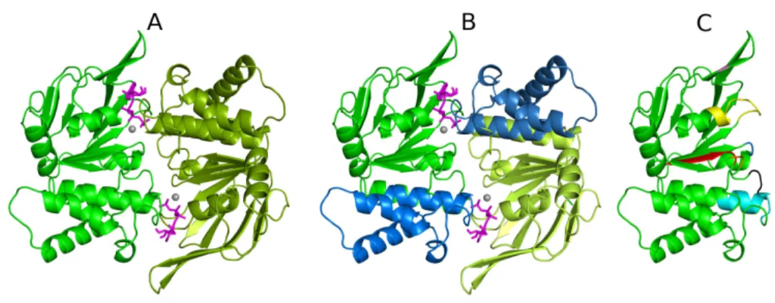
29
Figure 5- Structure of an ABC transporter NBD dimer. A-The NBDs are colored in green (NBD1 in dark green and NBD2 in lighter green) and represent the closed dimer conformation, with ATP, (colored in magenta) bound at the interface. The magnesium ions are represented as gray spheres located in the nucleotide binding sites. B- Each NBD monomer has a bilobal design and is composed by two distinct sub-domains (the RecA-like and the helical sub-domain). The RecA sub-domain is colored in green while the helical sub-domain in blue. C- Schematic representation of NBD1 with the ATP-binding family motifs highlighted in different colors. The residues and motifs coordinating the nucleotide are colored as follows: A-loop (pink), P-loop (yellow), Q-loop (orange), ABC Signature Motif (cyan), D-loop (black), Walker B (red) and the H-motif (blue).
30
Walker-B motif is located in β-strand 7 and its terminal acidic residues coordinate the magnesium ion through a H2O molecule [35, 36, 38]. Additionally, the A-loop
[77] is located just above the adenine ring of the nucleotide and contains a highly conserved aromatic residue, which is involved in – stacking with the nucleotides.
Table 1-Conserved sequence motifs in the NBD and its functions. Table adapted from [72].
Motif Consensus sequence
Function Representative
Structuresa Walker A
or
P-loop GxxGxGKS/T
b ATP binding MJ1267, E.c.MalK, HisP, MJ076,
Rad50 Q-loop
or
lid Q
a) Interdomain interaction
b) H-bond with Mg c) Binding to the attacking water
a) BtuCD
b) MJ0796(E171Q), GlcV(ADP)
c) MJ0796 (E171Q) LSGGQ
or
ABC-motif
LSGGQxQRb ATP binding MJ0796 (E171Q),
E.c.MalK
Walker B ϕϕϕϕD
b contact mediated by a water with Mg
molecule
GlcV, MJ1267 (ADP), MJ0796 (ADP)
E (next to Walker B)
a) Binds to attacking water
b) Mg water bridged contact
a) MJ0796 (ADP) b) GlcV
H motif or switch region
H Binds to the γ-phosphate MJ0796 (E171Q),
E.c.MalK A-loop A Binds to the Adenine ring
of the nucleotide MJ0796 (E171Q)
a References for some exemplifying structures: HisP [33], MJ0796 [36], MJ1267 [35], Rad50 [78], TAP1 [34], GlcV [38], MalK [52], BtuCD [57], MJ0796 (E171Q) [51].
31
Lastly, another very important residue located in the RecA sub-domain, is the acidic residue (a glutamate in many cases) immediately following the Walker-B motif, which is thought to act as a general base in hydrolysis (“general base” model) [79, 80]. This residue is known to interact with the magnesium ion [51, 80] and in several of the X-ray structures available (e.g. [51, 80]), it makes a hydrogen bond with the alleged hydrolytic water in the binding site. Furthermore, the mutation of this acidic residue by its corresponding amide completely abolishes, in some cases, the ATPase activity [46, 80, 81] and triggers the formation of a trapped ATP-bound dimer (as for example in [51, 80]). These observations led to the hypothesis that this residue is capable of polarizing the attacking water molecule for hydrolysis [79, 80] and that the corresponding amide residue is not able to act as a base for hydrolysis.
32
Additionally to the RecA-like sub-domain, each NBD monomer is also constituted by a smaller sub-domain (called helical sub-domain), which is formed by three or four helices and by the ABC-signature motif (which is a sequence unique for the ABC family and distinguishes ABC proteins from other nucleotide-binding proteins). The ABC- signature motif (see Table 1) binds the ATP γ-phosphate and this, together with the P-loop and the Walker-B motif, forms the nucleotide binding site [72].
The Rec-A and the helical sub-domains are joined together by two flexible loops, one of which contain a highly conserved glutamine residue (called Q-loop) and the other a conserved histidine residue (named H-loop). The Q-loop (also named “the lid” [50] or the “γ-phosphate switch” [36]) is located in β-strand 6 and contains a glutamine residue that binds to the magnesiun ion and to the attacking water [38, 51] (see Table 1). Based in the high-B factor values reported in several of the NBD X-ray structures, the Q-loop region appears to be highly flexible when ATP is not present. Furthermore, over the last years, several authors have suggested that the Q-loop glutamine residue plays a central role in mediating the allostery between the nucleotide binding site and the helical sub-domain [51, 86, 87]. The potential relation between this glutamine residue and the helical sub-domain will be extensively addressed later in section 3 and 4. The H-motif or the “switch” [50] (see Table 1), is located in β-strand 8, and contains a highly conserved histidine residue
that forms hydrogen bonds with the γ-phosphate of ATP [51, 52].
33
observation of positive cooperativity in hydrolysis reported for some members (such as MalK [88], YvcC [89] and HlyB [90]) can be easily understood.
Over the last years, several high-resolution structures for the NBD monomers and dimers have been determined [33-43, 47, 50-54, 91, 92], allowing us to gain relevant insights into the working mechanisms of these nanomachines. In order to obtain crystals for the pre-hydrolysis dimer (with ATP-bound in the binding sites), some residues in the active site were mutated [46, 51, 54] or EDTA was used to avoid the ATP hydrolysis [52]. Based in the ATP-bound crystal structures available, a complex network of interactions between the protein and the nucleotides is now unraveled. First of all, the nucleotide adenine ring is normally stabilized by π-π
stacking interactions with the A-loop conserved aromatic residue [77, 79]. Moreover, the P-loop conserved lysine residue forms hydrogen bonds with the oxygen atoms of the α- and γ- phosphates, thereby holding them in a defined orientation, whereas the magnesium ion is coordinated by the oxygen atoms of the
β- and γ-phosphates and by some P-loop residues [34, 38]. Additionally, the highly conserved H-loop histidine forms a hydrogen bond with the γ-phosphate, which is known to be essential for hydrolysis [52 , 54]. The side chain of the serine and the backbone amide groups of the ABC signature glycine residues are known to coordinate the γ-phosphate in ATP.
1.2.2.1. NBDs asymmetry, one of the oldest unanswered question
34
dimer, e.g. [1, 47, 87, 93-98] or in full length transporters, e.g. [99-101]) pointed out to the fact that a functional asymmetry can be also observed in NBDs homodimers, such as HlyB [47], histidine [99] and maltose transporter [100, 101]. For example, in the histidine transporter [99], it has been reported that thiol-specific reagents react more readily with a cysteine residue from one monomer than with the equivalent cysteine in the other. In the maltose transporter, it has been reported that the cross-linking pattern between cysteines located in the helical sub-domains and in the TMDs is different for the two monomers [100, 101]. Nevertheless, in some cases, like in the maltose transporter, the NBD dimer asymmetric behavior reported can easily be explained by the effect exerted by the TMDs in the NBDs, since the former can have different amino acid residue sequences. Moreover, in the case of importers, the existence of a substrate binding protein (SBP) may introduce an extra source of asymmetry, since the SBPs are themselves asymmetric [102] and by this reason imposing functional asymmetries in the NBD monomers.
Despite all the information available until this moment, the question whether the two NBD monomers and the two binding sites are functional equivalents, does not have a straightforward answer and it may depend on the transporter being homo- or heterodimeric (both for the TMDs and for the NBDs) or if it requires a SBP. However, there are some experimental evidences (such as [47, 100, 101, 103-105]), which suggest that functional asymmetries in the binding sites can arise during the catalytic cycle. These experimental observations lead to the formulation of the hypothesis that the observed NBD asymmetry is an intrinsic and essential characteristic of an ABC pump and the structural basis for the ATP hydrolysis mechanism.
35
1.2.2.2. Stoichiometry of ATP binding/hydrolysis and the role of two binding sites
36
Over the last years, many efforts have been made in order to measure the stoichiometric ratio of substrate transport and ATP hydrolysis, both in vivo and in vitro, for several ABC members, hoping to finally determine how many ATP molecules are hydrolyzed per transport cycle. Experimental technical details make these measurements very difficult to achieve, due, especially, to the high levels of uncoupled ATP hydrolysis and the possible leakage of substrate from membrane vesicles. Moreover, when the stoichiometric ratio can be effectively measured, most of the times, the values reported are so disparate that is impossible to conclude anything, like for example in the maltose transporter (where rates ranging from 1 [109, 110] to 17 [111] ATPs per transported sugar have been reported).
After this brief description of the ABC catalytic domains chemistry and despite the available information (many times contradictory), the reader clearly understand that we are still very far away from a complete understanding of the ABC molecular mechanisms. Nevertheless, the fact that the ABC modules are highly conserved (both in sequence and in structure) between all ABC members raises the hypothesis that the basic mechanisms for nucleotide hydrolysis and their coupling to allocrite transport, are probably conserved throughout the ABC transporter family.
1.2.3. Substrate Binding Protein (SBP)
37
In Gram-negative bacteria, SBPs are located in the periplasmatic space between the inner and outer membranes whereas in Gram-positive organisms and in Archea (which lack the periplasm), SBPs are often lipoproteins bound to the external face of the membrane or directly fused to the membrane transporter itself [115].
The range of allocrites retrieved by the SBPs is extremely diverse [7] and includes mono- and oligosaccharides, organic and inorganic ions, amino acids and short peptides, iron-siderophores, metals, polyamine cations opines, and vitamins. Moreover, most SBPs are specific for a single allocrite or for a family of structurally related substrates [7]. However, there exceptions to this rule, when a single SBP is very versatile and is able to handle structurally unrelated substrates [7].
SBPs are usually monomeric and both structural and kinetic data indicate that there is only one substrate-binding site per SBP [116]. Moreover, the X-ray structures of several full length transporters (MalFGK2E [62], ModABC [60] and BtuFCD [61]) in
complex to their SBPs revealed that only one SBP directly interacts with the transporter membrane domains at each time. However, in order to increase the transporter versatility, there are cases where multiple SBPs, with different binding specificity, are able to interact with the same transporter (like in the lysine/histidine/arginine transporter [117]). Until this moment, most of the SBP knowledge arises from the study of negative bacteria, as only a few Gram-positive and Archaea substrate-binding lipoproteins have been characterized, either biochemically or structurally [118-120].
38
is composed of an alpha-beta fold consisting of several β-sheets surrounded by α -helices and connected by loops. Over the years, high-resolution structures of many periplasmic SBPs in multiple conformations (open-unliganded, closed-unliganded, open-liganded, and closed-liganded) conformations have been determined [91, 102, 121-124, 125 ] which allows us to understand the conformational changes induced upon substrate binding (see Figure 6). Based on all X-ray structures, three distinct folding patterns have been identified for the SBPs, which allow the division of SBPs in three main groups [121, 127]. The classes I and II [72], represented by the simple sugar (arabinose and ribose) binding protein and the maltose/maltodextrin binding protein [128], respectively, differ primarily in the fold of the two domains. In class I, the β-sheet topology for the N- and the C-lobes is
β2β1β3β4β5, whereas class II has a different topology (β2β1β3βnβ5 with n representing
39
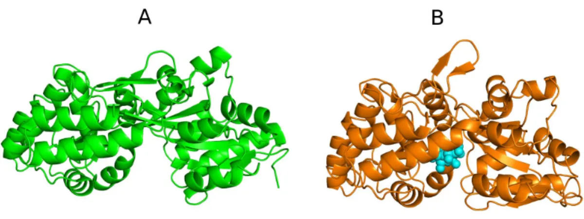
Figure 6- Maltose binding protein (MBP) in two distinct substrate-bound states. A- open-free state (PDB entry 1OMP [129]). B- closed maltose-bound conformation (PDB entry 1ANF [130]). The
substrate, when present, is represented by cyan spheres and it is located in the cleft between the N- and the C-lobe.
Later, a third class of SBPs (class III) was defined and its distinguishing feature is a single α-helix segment linking the two α/β lobes. The class III SBPs, represented by the iron/siderophore/vitamin B12 SBP [91, 131], present a more rigid hinge structure
and substrate binding is not accompanied by a large domain movement, like the one documented for classes I and II [91, 132].
40
Figure 7-Representation of the complex formed by the putative molybdate importer (ModB2C2) and its SBP (ModA) [60]. The ModB subunits are colored in orange and yellow whereas the ATPase domains (ModC) are highlighted in green and red. The ModA protein is colored in magenta. The horizontal black lines depict the probable location of the lipid membrane, based on the hydrophobicity of the protein residues. The reader should note that the SBP substrate-binding cleft is more or less located close to the interface between the TMDs.
41
1.3. Molecular Mechanisms in ABC transporters
1.3.1. Mechanisms for NBD dimer functioning
Despite the large amount of experimental data available and all the detailed insights into the structure of the ABC modules, the precise mechanism for NBD dimer functioning in ABC transporters is still controversial. However, based on the NBDs highly conserved sequences and fold, a common and unique mechanism for ATP hydrolysis is likely to exist, independently of the nature of the transported allocrite. Until this moment, three models have been proposed for the ABC ATPase domains functioning [135]: the “processive-clamp”, the “alternating two sites” and the “constant-contact” model. The major differences between these three models are related to the number of ATP molecules required to open the dimer interface and the extension of the dimer interface opening. In this chapter, the current models for NBD dimer formation coupled to ATP hydrolysis will be briefly described.
42
A
2ATP5
g+fj
~ -r fj~
2ADP~10
31
B
R Fl
RFl RFl
lj
V
ljv s-lj!:)
fg
7r
lP4r
lPRFl
8RFl
~
R Fl
lj!j
T
lJ.V
lj!:)
IP
R Fl----+ R Fl----+ ' •
'oclj
Q
lj
V
ABC . P1
K Fl R Fl
P - ABClj v 'T lj
v}
'"'.'
C
R ~
-L
Rrl ----+
m
lj)) ljv lj"L)
~ ~
~
~
43
Figure 8-Mechanistic models of dimer formation coupled to ATP hydrolysis in the NBDs of an ABC transporter. ATP is represented as a red hexagon, whereas ADP and IP are depicted by yellow and blue spheres. Each NBD monomer is formed by two domains (RecA-like and helical sub-domains). The letters P and ABC refer to the P-loop and to the ABC signature motif. A- “Processive-clamp” model [51, 52, 136-138]. B- “Alternating two sites” model [6, 139]. C- “Constant-contact” model
[87, 97, 140].
44
mechanisms are not common to all NBDs, and alternative mechanisms can appear for other ABC members (for a more intensive discussion in this subject we suggest reading Section 3.4.3).
It is noteworthy that the majority of the experimental data supporting the existence of symmetrical sandwich dimer states (such as the two ATP- ot two ADP-bound states) comes from structural and biochemical studies performed in the absence of the catalytic magnesium ion or in ATPase inactive mutants, or using non-hydrolysable ATP analogues [135, 141]. Additionally, there are several X-ray structures in which the NBD monomers are separated more than 20 Å (e.g. [52, 65, 66, 142]). These high distances values observed for several nucleotide-free structures, together with the fact that ADP and ATP differ by only one charge unit, lead some researchers that claimed to be unlikely that electrostatic forces could bring the monomers together at all, or at least in a biological relevant timescale [135]. Due to all these questions, the earlier version of the “processive-clamp” model [138] was recently updated to a new version [143], where the authors propose that structural differences between the open and closed dimer conformation are probably less extended than the complete physical separation of the dimer.
45
145]. Additionally, it is implicit in this model that the transporter retains memory of which site last hydrolyzed ATP, so that the next hydrolysis would occur in the opposite site, resulting in an alternating ATP hydrolysis [6, 139]. In this model (see Figure 8B), in the ATP-bound closed dimer, only one of the nucleotides is hydrolyzed before the respective monomer opens up. ADP is then exchanged and the binding of a new ATP molecule can then occur, thus inducing the formation of a closed dimer again. After the first hydrolysis and nucleotide exchange in one of the monomers, the ATP in the second monomer will then be hydrolyzed. However, this model, although derived from biochemical studies, is incompatible with the X-ray structures available, since all structures unambiguously show symmetric bound nucleotides states (with two ATP or ADP-bound) and, until this moment, no hybrid nucleotide bound state (e.g one ATP and one ADP-bound) was reported.
46
resting state, the NBDs are dissociated and separated by, in some cases, distances up to 20 Å (e.g. [52, 65, 66]).
Despite all three previous models being corroborated by experimental data, it is important to refer that, until now, no single experiment was able to convincingly rule out any one of them.
One essential task for ABC transporters is the harnessing of the energy released from nucleotide binding/hydrolysis and its conversion into mechanical work (most probably through conformational changes). Consequently, one of the major questions still awaiting for an answer is which are the conformational rearrangements induced by nucleotide binding and hydrolysis in the NBDs, and how are these structural changes transferred to the TMDs, ultimately allowing allocrite transport. Upon comparison of the different nucleotide-bound NBDs monomeric structures (e.g. [35-37]), the major conformational change observed between different nucleotide-bound states is the rigid-body rotation of the helical sub-domain relative to the RecA domain [7, 72], a movement which has been suggested to facilitate the nucleotide exchange upon hydrolysis [35].
47
Figure 9- Structures of the MalK dimer in the nucleotide-free [52, 53], ATP- [52, 53] and ADP-bound
states [52, 53]. In the absence of nucleotides (ground state), the two MalK monomers are separated from each other, held as a dimer primarily through contacts between the C-terminal RDs. In the presence of ATP (pre-hydrolysis state), the NBDs are closed, allowing ATP hydrolysis to occur. In the ADP-bound state (post-hydrolysis state), the NBDs are again separated in a conformation similar to the resting state. In this figure the NBDs are colored in green while the RDs are colored in orange. Corresponding domains in the second MalK monomer follow the same color scheme but are rendered in lighter colors. ATP and ADP are represented as red and magenta sticks, respectively, and magnesium as a gray sphere.
pre-48
49
1.3.2. Mechanisms for coupling hydrolysis to allocrite transport
Several experimental studies have early showed that the NBDs bind and hydrolyze ATP and that nucleotide hydrolysis is coupled to transport [74, 110, 152, 153]. Nevertheless, how ATP hydrolysis is coupled to allocrite translocation is a question still waiting to be answered. Additionally, the conformational changes (both in the NBDs and the TMDs) responsible for vectorial transport in ABC transporters are also mostly unknown.
There are several models proposed for coupling ATP hydrolysis, whether to exporters (Figure 10 and Figure 11) or to SBP-dependent importers (Figure 12). These models differ in a number of aspects, including the number of hydrolyzed ATP molecules per transport cycle, the number of allocrite binding sites and the point in the transport cycle when the allocrite is translocated. In the following sub-chapters, these proposed transport models will be briefly described.
1.3.2.1. Export Systems
50
Figure 10- Three of the four possible models for exporter pumps. In this figure, ATP and ADP are
represented as red hexagons and yellow spheres, respectively (A) In this model [139], the magenta
sphere represents the single allocrite binding site shared between the two TMDs (B) In this model
[154], the magenta sphere represents the allocrite binding site inward-facing conformation whereas the magenta ellipse represents the outward-facing conformation. (C) In this model, two binding sites coexist [155], an inward-facing site (magenta sphere) and an outward-facing site (magenta ellipse).
The model A (Figure 10A), called the “alternating catalytic sites” model, was proposed by Senior and co-workers [139] based on observations made on the mammalian P-glycoprotein. In this model, only one interchangeable drug-binding site is shared between the two halves of the pump, and the conversion from an inside-facing, higher affinity, to an outside-facing, lower-affinity conformation, is associated with one ATP hydrolysis [139].
51
The model C (Figure 10C) was proposed by Sauna & Ambudkar [155] based also in the mammalian P-glycoprotein. In this model, both high- and low-affinity sites coexist, and during the transport cycle, the loss of high-affinity binding promotes the transfer of the allocrite from the high-affinity to the low-affinity site [155]. While in the first two models, drug transport occurs each time ATP is hydrolyzed, the third model suggests that ATP hydrolysis at the second site is required to reset the system before transport can occur again [155].
A fourth model for transport (see Figure 11), the “ATP-switch” model, was proposed by Higgins and colleagues [138, 156], based on their work with the mammalian P-glycoprotein. In this model, the authors suggest that nucleotide binding to the transporter, rather than ATP hydrolysis, drives the major conformational changes that reorient the drug-binding site from the inside, high-affinity to the outside, low-affinity conformation [156].
52
as blues and pink shapes. The yellow and blue stars, located in the TMDs region, represent the high affinity and the low affinity allocrite-binding site, respectively. Figure adapted from [138].
In the “ATP-switch” model [138, 156], transport is a multistep process involving communication via conformational changes, in both directions, between the NBDs and TMDs. In this model, the driver for transport is the switch between two principal NBD conformations: the “closed dimer” (with the two monomers in straight contact and two ATP molecules bound at the interface) and the “open dimer” (where the monomers are separated). The conversion from the “open” to the “closed” NBD dimer conformation induces the conformational changes in the TMDs necessary for transport. The conversion from the “closed” to the “open dimer” conformation happens after ATP hydrolysis and resets the transporter ready for the next transport cycle. The precise conformational changes that trigger the ‘closed dimer’ formation are yet unclear, but experimental studies in intact transporters suggest that the conformational changes at the NBDs are relatively small [157, 158].
1.3.2.2. SBP-dependent import systems
53
Figure 12- SBP-dependent translocation mechanism for an ABC importer. This model is composed by four distinct steps. For a detailed explanation, see the following text
54
Nowadays, the essential role played by the SBP in the transport cycle is clearly accepted, and it was shown that the interaction of the allocrite-bounded SBP with the TMDs truly initiates this cycle [159]. It is also known that allocrite analogues failing to induce SBP closure do not stimulate ATPase activity and are not transported, a fact that suggests that the SBP closed conformation is necessary to initiate a cycle of transport and hydrolysis [160].
55
1.4. Computer simulations of ABC transporters
Computational theoretical techniques, such as homology modeling and molecular mechanics/dynamics simulations, are nowadays considered important and powerful tools that can contribute to increase our understanding of protein structure, function and dynamics. In particular for membrane proteins, which are difficult to crystallize due to technical limitations, computational methods may be useful tools for complementing the experimental structural information and providing properties that are hard to be measured experimentally. In this chapter, we will briefly describe the evolution of the computer simulations field in the ABC transporter family and we will show how computational techniques may help obtain important insights into the ABC mechanisms.
1.4.1. Protein dynamics
When a NMR or X-ray structure (preferentially high-resolution structure) is available, the intrinsic dynamic behavior of that protein can be studied using molecular mechanics/dynamics simulations and normal model analysis. Classic atomistic MD simulations of proteins are limited, until this moment, to several tens of nanoseconds. Although this timescale is too short to study the major conformational changes occurring during a transport cycle (≥ 100ms for a transport
event in P-glycoprotein [161]), this technique can be used to study individual states or initial transitions in the transport cycle, and can facilitate the interpretation of the experimental data available.
56 1.4.1.1. Nucleotide binding domains
Over the last decade, the dynamic behavior of the NBDs (both in the isolated and in the dimeric form) has been extensively studied [1, 56, 87, 94, 96, 97, 171-173]. In 2002, the first molecular dynamics simulation study of an ABC transporter domain was published. In this work [173], the authors report a 390 ps simulation for the monomeric NBD of a histidine transporter (HisP), aimed at identifying the regions that may be involved in conformational changes in the NBDs. Since then, multiple, much longer (5-20 ns), simulations of the monomeric HisP have been reported [171]. Using 3 different nucleotide-bound HisP conformations (nucleotide free-, ATP- and MgATP-bound), Campbell and co-workers [171] tried to understand how the NBD responds to the presence of ATP. From the ATP-bound simulation, the authors observed that the presence of ATP was associated to the movement of the helical sub-domain region towards the core RecA-like sub-domain. Curiously, a similar movement was not observable in the MgATP-bound simulation, presumably due to the presence of the Mg ion.
57
ADP/ATP-bound form for the dimer). The dimer simulations revealed a large asymmetrical transition of one helical sub-domain, resulting in an asymmetric conformation of the dimer.
Molecular modeling studies have also been able to give us important insights into the dynamics of nucleotide driven NBD closure [94]. In 2006, Oloo and co-workers [94] used molecular dynamics simulations to study the effect of the presence of ATP in the binding sites of the maltose importer NBDs (MalK). In their simulations, the introduction of MgATP into the binding sites transformed a semi-open state into a closed state, whereas the open dimer evolved to a semi-open state. In contrast, in the absence of MgATP, the NBDs of both the open and semi-open states drifted further apart.
Recently, molecular dynamics techniques were used in order to understand the mechanisms of dimer opening upon ATP hydrolysis in the NBD dimer [96, 97]. In 2008, Wen and Tajkhorshid [96] applied molecular dynamics simulations to study the detailed mechanism of dimer opening in the MalK dimer. In this work, 50 ns MD simulations of four possible ATP and ADP plus Inorganic Phosphate combinations were performed. After 30 ns (in the fastest cases), it was possible to observe opening of one or both binding sites (for all systems with the exception of the one with two ATPs) and to identify the atomic details associated to this process. Later in 2009, Jones and George [97], also used molecular dynamics simulations to study the opening of the isolated NBD MJ0796 dimer. After extending the dimer simulations performed previously [87], the authors were able to observe the rotation of the helical sub-domain relative to the catalytic domain, which allowed the opening of the ADP-bound binding site.
![Figure 3- TMDs structural diversity. X-ray structures for A) Sav1866 homodimer [58]. B) MalFGK 2 E complex [62]](https://thumb-eu.123doks.com/thumbv2/123dok_br/15769781.641150/33.774.192.580.474.746/figure-tmds-structural-diversity-structures-homodimer-malfgk-complex.webp)
![Figure 9- Structures of the MalK dimer in the nucleotide-free [52, 53], ATP- [52, 53] and ADP-bound states [52, 53]](https://thumb-eu.123doks.com/thumbv2/123dok_br/15769781.641150/55.774.99.665.150.526/figure-structures-malk-dimer-nucleotide-free-bound-states.webp)
![Figure 10- Three of the four possible models for exporter pumps. In this figure, ATP and ADP are represented as red hexagons and yellow spheres, respectively (A) In this model [139], the magenta sphere represents the single allocrite binding sit](https://thumb-eu.123doks.com/thumbv2/123dok_br/15769781.641150/58.774.192.590.142.395/figure-possible-exporter-represented-hexagons-respectively-represents-allocrite.webp)
![Figure 11- The ‘ATP switch’ model for the transport cycle of an ABC transporter [138, 156]](https://thumb-eu.123doks.com/thumbv2/123dok_br/15769781.641150/59.774.157.619.580.906/figure-atp-switch-model-transport-cycle-abc-transporter.webp)

![Figure 14- X-ray structures available for the Sav1866 homodimer [58, 59]. Two Sav1866 crystal structure are represented in this image: (A) the AMP-PNP bound [59] and (B) the ADP-bound state [58]](https://thumb-eu.123doks.com/thumbv2/123dok_br/15769781.641150/74.774.141.639.141.455/figure-structures-available-homodimer-crystal-structure-represented-image.webp)

