S
TENOSTOMUMLEUCOPSD
UGÈS, 1828 (P
LATYHELMINTHES, C
ATENULIDA):
A PUTATIVESPECIESCOMPLEXWITHPHENOTYPICPLASTICITYM
ARCOST. R
OSA1C
AMILA. M. P
EREIRA1G
EOVANI. T. R
AGAGNIN2E
LGION. L.S. L
ORETO1,3,4ABSTRACT
Species of Stenostomum are small flatworms that live in freshwater and normally reproduce asexually by paratomy. They are basal in the phylogeny of Platyhelminthes. For more than a century, species of this genus, especially S. leucops, have been used in regeneration and other biological studies. However, some basic aspects of their biology are poorly known. Here, we characterized a strain of S. leucops that has been maintained in the laboratory for five years and a recent strain of S. grande. The time required for complete formation of zooids of S. leu-cops by asexual reproduction is approximately 42.5 hours at 28°C. The number of cells in the zooids, soon after paratomy, is approximately 2,500. The number of zooids formed in the chain is a plastic characteristic and is dependent on the conditions for cultivation. In some cul-tivation conditions of S. leucops, only worms with two zooids are formed. However, in other conditions, worms with up to five zooids are observed. Phylogenetic analyses of a fragment of the Cytochrome C Oxidase I (COI) sequence showed S. leucops and S. grande species consti-tute a species complex, the lineages of which having high intraspecific divergences.
Key-Words: Stenostomum grande; Paratomy; Zooids; Number of cells; Microturbellaria.
INTRODUCTION
The order Catenulida Meixner, 1924, is repre-sented by small flatworms that generally range from 0.5 to 2 mm long. They have unpaired, dorsomedi-ally located protonephridium, anterodorsal testes and
male genital pore, and aciliary nonmobile sperm. Normally they are white in reflected light, and trans-lucent in transmitted light. The intestine varies in col-or depending on their food content (Larsson, 2008). Approximately 100 species have been described worldwide, and the majority lives in freshwater. The
http://dx.doi.org/10.1590/0031-1049.2015.55.27
1. Universidade Federal de Santa Maria (UFSM), Programa de Pós-Graduação em Biodiversidade Animal. Avenida Roraima, 1.000, Camobi, CEP 97105-900, Santa Maria, RS, Brasil.
2. Universidade Federal de Santa Maria (UFSM), Curso de Ciências Biológicas. Avenida Roraima, 1.000, Camobi, CEP 97105-900, Santa Maria, RS, Brasil.
3. Universidade Federal de Santa Maria (UFSM), Departamento de Bioquímica e Biologia Molecular. Avenida Roraima, 1.000, Camobi, CEP 97105-900, Santa Maria, RS, Brasil.
sexually mature stage is rarely found because
Caten-ulida normally reproduce by paratomy, an asexual
form of reproduction in which structures typical of the anterior region develop followed by a fission per-pendicular to the antero-posterior axis, which forms zooids (a movie of this process can be seen in http:// w3.ufsm.br/labdros/permanente/paratomy.mp4). In some species, chains with up to nine zooids can occur (Hyman, 1951). One of the most studied Catenulida is Stenostomum leucops (Dugès, 1828), a cosmopoli-tan species exhibiting characteristics that make it an excellent experimental organism. For example, it can be easily maintained and reproduces quickly in cul-ture, regenerates extensively and has internal organs that can be readily observed (Nuttycombe & Waters,
1938). For more than a century, Stenostomum,
pri-marily S. leucops, have been used for several experi-mental biology studies, such as studies of fission and regeneration (Ritter & Congdon, 1900; Child, 1990, 1902; Hartmann, 1922; Ruhl, 1927; Van Cleave, 1929), stem cells (Palmberg, 1990), ultrastructure of sensory organs (Reuter et al., 1993; Palmberg & Re-uter, 1992; Ruppert & Schreiner 1980), ultrastructure of the digestive tract (Antoniazzi & Silveira, 1996), senescence (Martínez & Levinton, 1992), neuropep-tides (Grahn et al., 1995; Wikgren & Reuter, 1985) and ecology (Nandini & Sarma, 2013; Nandini et al., 2011). However, some basic aspects of the biology of these worms, such as the time required for paratomy or the number of cells constituting the body, are not well known.
The validity of S. leucops as a species has been questioned by Nuttycombe & Waters (1938) and Marcus (1945). However, further studies by Noreña et al. (2005) have validated this species. More recent-ly, Larsson et al. (2008) used DNA sequences of 18 S rDNA and COI to show that S. leucops constitutes a monophyletic group. Yamazaki et al. (2012) used the same molecular markers and also included species col-lected in Japan; in their analysis, S. grande was placed in a cluster with representatives of S. leucops. Further-more, the genetic divergences observed among the se-quences of S. leucops collected in different places cor-respond to that expected for species, suggesting that it is a species complex. In order to solve the problem of whether S. leucops is one species or a species com-plex, it is necessary to morphologically and biologi-cally characterize populations worldwide, and DNA barcoding can be useful for this task.
The basis for DNA barcoding is that short nu-cleotide sequences can be used to distinguish species, because the genetic variation between species is nor-mally higher than that observed within species. For
animals, the sequence used the most is a 650-base fragment of the 5’ end of the mitochondrial gene
Cytochrome C Oxidase I (COI, cox1) (Hebert et al.,
2003). Although several shortcomings have been as-sociated with DNA barcoding, the methodology is now well established and has been shown to be useful in various fields of biological research, including the identification of cryptic species or species complexes (Collins & Cruickshank, 2013; Albu et al., 2010).
The aim of this study was to characterize some morphological and biological characteristics, such as the time required for paratomy and the number of cells in each zooid of a strain maintained in the laboratory for 5 years. A morphological plasticity was observed in the number of zooids formed, which depended on the growing conditions. We also per-formed a DNA barcoding and phylogenetic analysis. We found that S. leucops collected in Sweden, London and Brazil are paraphyletic with regard to S. grande, from Japan and Brazil, supporting the hypothesis that this taxon corresponds to a species complex.
MATERIALS AND METHODS
Animal sampling and cultures
The worms were collected in a pond at the Fed-eral University of Santa Maria, Santa Maria, Brazil (53°17’W; 29°28’S). Stenostomum leucops was
collect-ed in March of 2009 and S. grande Child, 1902, in
the medium; ii) Condition 2 – the culture flasks were maintained at room temperature under indirect solar illumination. In these conditions, cyanobacteria be-longing to the Chroococcales order become abundant in the medium and promote differential growth in the worms.
Vouchers of the used strains were deposited in UFSM Department of Biology collection under num-bers SL01-sm01 and SL01-sm01.
Estimation of the time required for asexual reproduction of S. leucops
To assess the time required for asexual reproduc-tion by paratomy, a total of 98 worms under Condi-tion 1 were analyzed. They were put in a drop of water in Kline concavity slides with one worm per concav-ity. The slides were maintained in a wet chamber, and the water was replenished every day. The worms were observed through a stereo microscope every 12 hours. After fission, each “new” worm was transferred to a new well, and the time for the process was registered.
Estimating the number of cells of S. leucops
Worms soon after paratomy or showing the con-striction between zooids, which characterize that the fission will occur shortly, were put on a slide with a drop of distilled water and 2.5 µl of ethidium bromide (0.5 mg/ml). For each preparation, one worm was put on the slide, covered with a coverslip and squashed, and they were observed through an Olympus BX41 fluorescence microscope. Pictures were taken using a 518 nm absorption filter and a 605 nm emission filter. Additionally, estimates were performed for worms with two, three and four zooids. The nuclei were counted directly from a computer screen, which marked the nuclei that had already been counted.
A second procedure was performed using an ace-tic orcein (2%) stain. The worms were photographed, and the number of cells was estimated as previously described.
DNA barcoding
Genomic DNA was isolated from approximate-ly 100 worms of each strain following the method de-scribed by Oliveira et al. (2009). As previously high-lighted, the strains were made from a single worm. Thus, as only asexual reproduction has been observed,
the worms in each culture are clones, therefore, homo-geneous mitochondrial sequences are expected. The primers and PCR conditions used to amplify Cyto-chrome C Oxidase I (COI) were described by Telford et al. (2000). A fragment of 497 bp was sequenced using a MegaBace 500 automatic sequencer. The dideoxy chain-termination reaction was performed using the DYEnamicET kit (GE Healthcare). The sequences obtained were deposited in GenBank (ac-cession numbers: S. leucops KJ476143 and S. grande KM056359).
For the phylogenetic analyses, other
Stenosto-mum COI sequences were obtained from GenBank
(accession numbers: AB665116 to AB665124, FJ384873 to FJ38910, AJ405975, and AJ405976). These nucleotide sequences were aligned using Clust-al W (Thompson et al., 1994) according to the de-fault program parameters. The phylogenetic analyses were implemented in Mega 5.0 software (Tamura et al., 2011) by neighbor-joining and maximum like-lihood with 1,000 bootstrap replications. The genetic distances among different taxa were estimated by P distance also using Mega 5.0 software.
RESULTS
Life cycle and biological characterization of S. leucops
The morphological characteristics of S. leucops here studied correspond to those described by Noreña et al. (2005) and Damborenea et al. (2011). The same occurs for S. grande.
five zooids and an average size of 1.03 ± 0.05 mm. Af-ter paratomy, the size of the worms with one zooid, which were formed from chains of multiple zooids, was 0.45 ± 0.03. An absence of perturbation in the culture flasks is a necessary condition for the growth of more than two zooids. When the culture is agitat-ed, such as when collecting worms using a pipette, the development of multiple zooids is interrupted, and as a consequence the two zooid pattern of paratomy re-turns. For this reason, we were not able to determine the time necessary for paratomy with multiple zo-oids, once the transferring to a Kline slide promotes a change of the reproduction pattern of multiple zooids to two zooids.
The cell number estimate for worms with one zooid growing in Condition 1, soon after paratomy, is approximately 2,500. The number of cells in two zooid worms preceding the fission is approximately 5,000. As seen in Table 1, for worms growing in medium supplemented with cyanobacteria (Condi-tion 2), the number of cells in animals with one, two, three and four zooids increases by 2,000 cells for each zooid formed. This suggests that the number of cells necessary to form one zooid is approximately 2,000.
DNA Barcoding
Using the available Stenostomum COI sequences, the phylogenetic analysis conducted through neigh-bor-joining showed that both S. leucops and S. grande, constitute a complex of species sincespecimens of S. leucops, from Sweden, grouped with a specimen of S. grande, from Japan, while specimens of S. leucops, from Brazil and London, grouped in a clade includ-ing a representative of S. grande, from Brazil (Fig. 3). A similar tree was obtained by maximum likelihood (data not shown). These analyses indicate that the two nominal species S. leucops and S. grande are poly-phyletic. The estimates of evolutionary divergence
us-ing P distance were very small within the sequences of S. leucops from Sweden (zero to 0.004). Howev-er, larger distances were observed between these se-quences and those collected in Sweden and the south of Brazil (approximately 0.14) as well as between the samples from Brazil and London (0.127) (Table 2). The distance found between the S. grande from Bra-zil and Japan is remarkable (0.154), but the distance observed between S. grande from Brazil and S. leucops from London is lower (0.110). Similar distances were observed between S. grande from Japan and the
S. leu-cops samples from Sweden. The P distances observed
among the twelve species here studied ranged from 0.076 to 0.213.
DISCUSSION
The easy cultivation, fast asexual reproduction, and regeneration are characteristics that make Ste-nostomum, particularly S. leucops, a potential model organism for studies of regeneration, stem cells, ag-ing and other biological topics. However, there are very few records in the literature that describe the time necessary for the worms to proceed the natural fission. Hyman (1951) described the time necessary for Stenostomum to undergo paratomy as around two days. Reuter & Kuusisto (1992) noted that the time required for a new zooid development in both S. leu-cops and Microstomum lineare Müller OF, 1773 var-ied from 20 to 45 h owing to environmental factors. These studies do not describe the conditions under which the time required for paratomy was deter-mined. Our results showed that the worms required 42.5 hours to carry out the fission at 28°C.
No records were found in the literature for the
number of cells that make up Stenostomum. For
Ca-tenulida, Simanow et al. (2012) reported that Macros-tomum lignano consists of approximately 25,000 cells. This worm is approximately 1.5 mm long, which is about three times larger than the worms used in our study. The number of cells found in S. leucops soon after paratomy is approximately 2.500 and, if an ad-justment to the length of the body is applied between
24 h %
50
40
30
20
10
0
48 h 72 h 96 h
FIGURE 1: The frequency of fission in Stenostomum leucops under Conditions 1. The percentage of zooids formed is shown on the Y-axis. The X-axis shows the time (in hours) required for fission to occur.
TABLE 1: Estimates of cell number in worms with 1, 2, 3 and 4 zooids growing in cultivation Procedure 1 and Procedure 2 (average ± standard deviation).
Number of Zooids Procedure 1 Procedure 2
1 2.400 ± 280 2.158 ± 112
2 4.850 ± 325 3.964 ± 365
3 — 5.166 ± 91
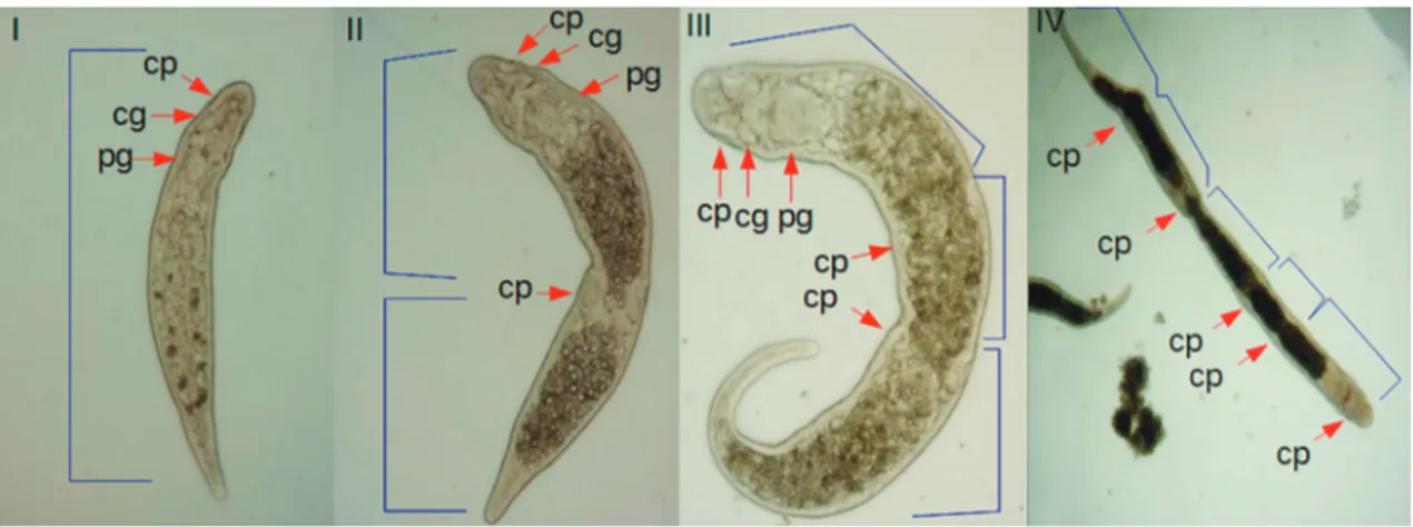
FIGURE 2: General view of Stenostomum leucops showing one zooid (I), two zooids (II), three zooids (III) and five zooids (IV). cp = cili-ated pits; cg = cerebral ganglia; pg = pharyngeal glands.
S. leucops909 Sweden
S. leucops899 Sweden
S. leucopsSM Brazil
S. bryophilum874 Sweden
S. grabbskogense880 Sweden
S. grabbskogense907 Sweden
S. leucops898 Sweden
S. grande116 Japan
S. grandeSM Brazil
S. tuberculosum122 Japan
S. saliens123 Japan
S. averaloi910 Sweden
S. simplex117 Japan
S. simplex120 Japan
S. sphagnetorum904 Sweden
S. sphagnetorum873 Sweden
S. island901 Sweden
S. saliens124 Japan
S. longpich906 Sweden
S. leucops908 Sweden
S. leucops976 London 71
71 92
93
59 61
97 97
97
99 62
28 20
100
100
100
100
0.02
number, of GenBank accession number, informed in Materials and Methods.
1 2 3 4 5 6 7 8 9 10 11 12 13 14 15 16 17 18 19 20
1 S. sphagnetorum 904 Sweden
2 S. sphagnetorum 873 Sweden 0,002
3 S. simplex 120 Japan 0,076 0,078
4 S. island 901 Sweden 0,114 0,116 0,122 5 S. simplex 117 Japan 0,139 0,141 0,148 0,133
6 S. saliens 124 Japan 0,190 0,190 0,207 0,188 0,194 7 S. saliens 123 Japan 0,186 0,186 0,198 0,188 0,181 0,030
8 S. tuberculosum 122 Japan 0,175 0,177 0,188 0,173 0,188 0,105 0,108 9 S. longpich 906 Sweden 0,179 0,179 0,177 0,186 0,194 0,148 0,143 0,137
10 S. leucops SM Brazil 0,181 0,184 0,181 0,184 0,198 0,173 0,165 0,181 0,190
11 S. leucops 976 London 0,156 0,158 0,152 0,167 0,177 0,169 0,162 0,171 0,152 0,127 12 S. leucops 909 Sweden 0,184 0,186 0,173 0,181 0,177 0,196 0,192 0,196 0,175 0,143 0,124
13 S. leucops 908 Sweden 0,186 0,188 0,175 0,184 0,179 0,198 0,194 0,198 0,177 0,146 0,127 0,002 14 S. leucops 899 Sweden 0,184 0,186 0,173 0,181 0,177 0,194 0,192 0,196 0,175 0,143 0,124 0,002 0,004
15 S. leucops 898 Sweden 0,184 0,186 0,173 0,181 0,177 0,194 0,192 0,196 0,175 0,143 0,124 0,002 0,004 0,000
16 S. grande 116 Japan 0,181 0,184 0,184 0,177 0,184 0,186 0,184 0,171 0,173 0,150 0,133 0,118 0,120 0,116 0,116 17 S. grande SM Brazil 0,179 0,181 0,169 0,188 0,175 0,179 0,171 0,192 0,175 0,135 0,110 0,146 0,148 0,143 0,143 0,154
18 S. bryophilum 874 Sweden 0,184 0,181 0,177 0,181 0,213 0,194 0,196 0,190 0,181 0,177 0,171 0,186 0,188 0,186 0,186 0,190 0,194 19 S. grabbskogense 880 Sweden 0,184 0,181 0,177 0,181 0,213 0,194 0,196 0,190 0,181 0,177 0,171 0,186 0,188 0,186 0,186 0,190 0,194 0,000
20 S. grabbskogense 907 Sweden 0,184 0,181 0,177 0,181 0,213 0,194 0,196 0,190 0,181 0,177 0,171 0,186 0,188 0,186 0,186 0,190 0,1994 0,000 0,000
21 S. averaloi 910 Sweden 0,179 0,161 0,196 0,207 0,179 0,192 0,198 0,181 0,186 0,203 0,177 0,188 0,190 0,186 0,186 0,181 0,184 0,190 0,190 0,190
Rosa, M.T
.
et
al
.: S
tenoSt
omum
leuc
opS
:
a sp
ecies c
S. leucops and M. lignano, it can be estimated that S. leucops has 30% of the cells found in M. lignano. Nevertheless, Caenorhabditis elegans, a nematode simi-lar in size to S. leucops, has fewer cells, 959 in the adult hermaphrodite and 1031 in the adult male (Sulston & Horvitz, 1977). A small number of cells was a use-ful characteristic for transforming C. elegans into one of the most prestigious animal models, as it allows precise descriptions of development; for example, it was the first organism to have its connectome (neural wiring diagram) completed (White et al., 1986). The
small number of cells observed in Stenostomum may
also be important to the usefulness of these worms as models for biological studies.
Although the number of zooids is not a diag-nostic characteristic for Stenostomum species identi-fication, this characteristic is always cited in the de-scriptions of species of this genus (Van Cleave, 1929; Nuttycombe & Waters 1938; Noreña et al., 2005;
Damborenea et al., 2011; Gamo & Leal-Zanchet,
2004). Van Der Land (1965), Gamo & Leal-Zanchet (2004) and Noreña et al. (2005) describe S. leucops as having only two zooids. On the other hand, Van Claeve (1929) and Palmberg (1990) report that the worms they studied showed multiple zooids (up to five), but do not described the conditions un-der which these animals were cultivated. Our data show that zooid number is a plastic phenotype and is highly dependent on environmental conditions. We have maintained our cultures for four years and have only observed two zooids. During this time, we were led to think that it was a characteristic of the species, but along the fifth year, the alteration of culture conditions resulted in worms with multiple zooids. This phenotypic plasticity could explain the differences in the descriptions from various authors. Our results suggest that the reproductive pattern with multiple zooids occurs only when the worms are in Condition 2 of maintenance. Even in this condition, disturbance in the cultures, as pipetting the worms, promote the changes of reproductive patterns from multiple for two zooids.
The number of cells in each zooid in worms with multiple zooids is smaller than that observed in animals with only two zooids, approximately 2,000 for the former and 2,500 for the latter on average. Furthermore, the size of worms with two zooids, originated from the fission process of two zooids, is similar to that observed in worms with four or five zooids produced, approximately 1 mm long. Never-theless, the number of cells in these animals is very different. The two-zooid worms have approximately 5,000 cells compared to 7,000 cells in animals with
four zooids. It is likely that some of the cells present in the four or five zooid worms are smaller. This could indicate a faster reproductive process in animals pro-ducing multiple zooids. However, as we are not able to measure the time required in paratomy in multiple zooids process, this hypothesis needs additional assays to be clarified.
Stenostomum leucops is distributed worldwide, with registers for North America, Europe, Africa (Larsson, 2008 and references therein) and Japan
(Yamazaki et al., 2012). In South America, it has
been recorded from Surinam (Van der Land, 1965),
Argentina (Noreña et al., 1995), Peru
(Dambore-nea et al., 2011) and the South Brazilian State Rio Grande do Sul (Gamo & Leal-Zanchet 2004, Brac-cini & Leal-Zanchet, 2013 and in this study). As mentioned previously, the validity of this species has been questioned by Nuttycombe & Waters (1938) and Marcus (1945) who consider the descriptions of this species ambiguous and broad to make its recogni-tion difficult. Molecular analyses performed by Lars-son et al. (2008) for Swedish Catenulida showed that the nominal species S. leucops is strongly supported as monophyletic group. However, the authors noted that variation in the branch lengths may be evidence for ongoing cladogenesis of some S. leucops popula-tions, which makes it a candidate for a species com-plex. Yamazaki et al. (2012) performed a phylogenetic analysis for Japanese Stenostomum and included se-quences of S. leucops from Europe. They found that a sequence of S. grande from Japan was included in the cluster of S. leucops from Europe. Our phylogenetic analysis of the available Stenostomum COI sequences showed that S. leucops and S. grande constitute a spe-cies complex. This is supported by by the high genetic divergence observed among sequences from same spe-cies. In S. leucops, the P distance among samples from Sweden, London and Brazil have the same range as observed among the species here sampled, strongly suggesting it is a species complex, as previously not-ed by Larsson et al. (2008). Although the status of S. grande as a valid species has never been questioned, the results of our phylogenetic analyses and the large genetic distance observed between the COI sequences of S. grande from Japan and Brazil suggest that it may also be a candidate for a species complex.
Since it was proposed, DNA Barcoding has al-lowed the increase of cryptic species discovery, even for species that are not distinguishable morphological-ly. For some examples see Gill et al. (2014), Crawford et al. (2012), Clare et al. (2011), Hebert et al. (2004).
complex and thus we did not propose any taxonomic change for S. leucops and S. grande.
Stenostomum leucops, while putatively a species complex, has many characteristics that make it an ex-cellent organism for these comparative studies, main-ly with planarians. However, as these worms have a simple anatomy and few diagnostic characters to al-low the species identification, DNA barcoding can be a good supplementary tool for the characterization of these species for those who are thinking in using these animals as experimental models.
RESUMO
Stenostomum são pequenos vermes que vivem em água
doce e normalmente se reproduzem assexualmente por pa-ratomia. Eles estão na base da filogenia dos platelmintes. Por mais de um século, espécies desse gênero, especialmente S. leucops, vêm sendo empregadas em estudos biológicos, principalmente sobre regeneração. Entretanto, alguns aspectos básicos da biologia destes vermes são ainda po-bremente conhecidos. Neste estudo, caracterizamos uma linhagem que vem sendo mantida no laboratório por cin-co anos. O tempo necessário para reprodução assexuada e completa formação de zoóides, a 28°C, é de aproxima-damente 42,5 horas. O número de células nos zoóides, logo após a paratomia, é de aproximadamente 2.500. O número de zoóides presentes nos vermes é uma característi-ca variável e depende das condições de cultivo. Em alguns procedimentos de cultivo de S. leucops, apenas cadeias com dois zoóides são formadas. No entanto, em outras condições de cultivo, cadeias de até cinco zoóides podem ser observadas. Análise filogenética empregando sequência do gene de Citocromo C Oxidase (COI) mostrou que S. leu-cops e S. grande constituem um complexo de espécies cujas linhagens mostram altas divergências intraespecíficas.
Palavras-Chave: Stenostomum grande; Paratomia;
Zooides; Número de células; Microturbellaria.
ACKNOWLEDGEMENTS
We thank Dr. Sandro Santos for introducing us to the Catenulida’s world, Dr. Lizandra Robe for critical comments on the manuscript, and the anony-mous reviewer for valuable suggestions. This study was supported by research grants and fellowships from CNPq-Conselho Nacional de Desenvolvimento Científico e Tecnológico, Pq 303244/2010-0, PIBIC/ UFSM and Fapergs (Probic/UFSM) and PRONEX FAPERGS (10/0028-7).
REFERENCES
Albu, M.; Nikbakht, H.; Hajibabaei, M. & Hickey, D.A. 2010. The DNA Barcode Linker. Molecular Ecology Resources, 11: 84-88.
Antoniazzi, M.M. & Silveira, M. 1996. Studies of Stenostomum grande Child, 1902 (Platyhelminthes, Catenulida): fine structure of the digestive tract and the endocytotic activity of the gastrodermis. Acta Zoologica, Stockholm, 77: 25-32. Braccini, J.A.L. & Leal-Zanchet, A.M. 2013. Turbellarian
assemblages in freshwater lagoons in southern Brazil. Invertebrate Biology, 132: 305-314.
Child, C.M. 1900. Fission and regulation in Stenostomum leucops. Biological Bulletin, 2(6): 329-331, 1901. (Meeting of the American Morphological Society. Abstracts of Papers, 27-28 december, Baltimore).
Child, C.M. 1902. Studies on regulation. Archiv für Entwicklungsmechanik der Organismen, 15: 187-234.
Clare, E.L.; Lim, B.K.; Fenton, M.B. & Hebert, P.D.N. 2011. Neotropical Bats: estimating species diversity with DNA Barcodes. PLoS ONE, 6: e22648.
Collins, R.A. & Cruikshank, R.H. 2013. The seven deadly sins of DNA barcoding. Molecular Ecology Resources, 13: 969-975. Crawford, A.J.; Cruz, C.; Griffth, E.; Ross, H.; Ibáñez, R.;
Lips, K.R.; Driskell, A.C.; Bermingham, E. & Crump, P. 2012. DNA barcoding applied to ex situ tropical amphibian conservation programme reveals cryptic diversity in captive populations. Molecular Ecology Resources, 13: 1005-1018. Damborenea, C.; Brusa, F.; Almagro, I. & Noreña, C. 2011.
A phylogenetic analysis of Stenostomum and its neotropical congeners, with a description of a new species from the Peruvian Amazon Basin. Invertebrate Systematics, 25: 155-169. Dugès, A. 1828. Recherches sur l’organisation et les moeurs des
planariees. Annales des sciences naturelles, 15: 139-182.
Gamo, J. & Leal-Zanchet, A.M. 2004. Freshwater microturbellarians (Platyhelminthes) from Rio Grande do Sul, Brazil. Revista Brasileira de Zoologia, 21: 897-903.
Gill, B.A.; Harrington, R.A.; Kondratieff, B.C.; Zamudio, K.R.; Poff, N.L. & Funk, W.C. 2014. Morphological taxonomy, DNA barcoding, and species diversity in southern Rocky Mountain headwater streams. Freshwater Science, 33: 288-301.
Grahn, M.; Maule, A.G.; Elo, I.; Shaw, C.; Reuter, M. & Halton, D.W. 1995. Antigenicity to neuropeptide-f (NPF) in Stenostomum leucops and Microstomum lineare. Hydrobiologia, 305: 307-308.
Hartmann, M. 1922. Uber den dauernden Ersatz der ungeschlechtlichen Fortpflanzung durch fortgesetzte Regenerationen. Biologisches Zentralblatt, 42: 364-381.
Hebert, P.D.N.; Cywinska, A.; Ball, S.L. & de Waard, J.R. 2003. Biological identifications through DNA barcodes. Proceedingsof the Royal Society of London. B, Biological Science, 270: 313-321.
Hebert, P.D.N.; Penton, E.H.; Burns, J.M.; Janzen, D.H. & Hallwachs, W. 2004. Ten species in one: DNA barcoding reveals cryptic species in the neotropical skipper butterfly Astraptes fulgerator. Proceedings of the National Academy of Sciences, United States of America, 101: 14812-14817. Hyman, L.H. 1951. The invertebrates: Platyhelminthes and
Rhynchocoela The acoelomate Bilateria. New York, McGraw-Hill, v. 2.
Larsson, K. 2008. Taxonomy and phylogeny of Catenulida (Platyhelminthes) with emphasis on the Swedish Fauna. Acta Universitatis Upsaliensis, 2008: 1-48. (Digital Comprehensive Summaries of Uppsala Dissertations from the Faculty of Science and Technology 395).
Larsson, K.; Ahmadzadeh, A. & Jondelius, U. 2008. DNA taxonomy of Swedish Catenulida (Platyhelminthes) and a phylogenetic framework for catenulid classification. Organisms, Diversity & Evolution, 8: 399-412.
Marcus, E. 1945. Sobre Microturbelários do Brasil. Comunicaciones Zoologicas del Museo de Historia Natural de Montevideo, 1: 1-74. Martínez, D.E. & Levinton, J.S. 1992. Asexual metazoans
undergo senescence. Proceedings of the National Academy of Sciences, United States of America, 86: 9920-9923.
Nandini, S. & Sarma, S.S.S. 2013. Demographic characteristic of cladocerans subject to predation by the flatworm Stenostomum leucops. Hydrobiologia, 715: 159-168.
Nandini, S.; Sarma, S.S.S. & Dumont, H.J. 2011. Predatory and toxic effects of the turbellarian (Stenostomum cf. leucops) on the population dynamics of Euchlanis dilatata, Plationus patulus (Rotifera) and Moina macrocopa (Cladocera). Hydrobiologia, 662: 171-177.
Noreña, C.; Damborenea, C. & Brusa, F. 2005. A taxonomic revision of South American species of the genus Stenostomum O. Schmidt (Platyhelminthes: Catenulida) based on morphological characters. Zoological Journal of the Linnean Society, 144: 37-58.
Nuttycombe, J. & Waters, A. 1938. The American species of the genus Stenostomum. Proceedings of the American Philosophical Society, 79: 213-301.
Oliveira, L.F.V.; Wallau, G.L. & Loreto, E.L.S. 2009. Isolation of high quality DNA: a protocol combining rennet and glass milk. Electronic Journal of Biotechnology, 12: 1-6.
Palmberg, I. 1990. Stem cells in microturbellarians an autoradiographic and immunocytochemical study. Protoplasma, 158: 109-120.
Palmberg, I. & Reuter, M. 1992. Sensory receptors in the head
of Stenostomum leucops. 1. Presumptive photoreceptors. Acta
Biologica Hungarica, 43: 259-267.
Reuter, M. & Kuusisto, A. 1992. Growth factors in asexually reproducing Catenulida and Macrostomida (Plathelminthes): A confocal, immunocytochemical and experimental study. Zoomorphology, 112: 155-166.
Reuter, M.; Joffe, B. & Palmberg, I. 1993. Sensory receptors in the head of Stenostomum leucops. II localization of catecholaminergic histofluorescence-ultrastructure of surface receptors. Acta Biologica Hungarica, 44: 125-131.
Ritter, W.E. & Congdon, E.M. 1900. On the inhibition by artificial section of the normal fission plane in Stenostoma. Proceedings California Academy of Sciences, 2: 365.
Ruhl, L. 1927. Regeneration in rhabdocoeles. Zoologischer Anzeiger, 72: 160-175.
Ruppert, E.E. & Schreiner, S.P. 1980. Ultrastructure and potential significance of cerebral light-refracting bodies of Stenostomum virginianum (Turbellaria, Catenulida). Zoomorphology, 96: 21-31.
Simanow, D.; Mellaart-Straver, I.; Sormacheva, I. & Berizikov, E. 2012. The flatworm Macrostomum lignano is a powerful model organism for ion channel and stem cell research. Stem Cells International, (2012): 1-10, ID 167265, doi: 10.1155/2012/167265.
Sulston, J.E. & Horvitz, H.R. 1977. Post-embryonic cell lineages of the nematode, Caenorhabditis elegans. Developmental Biology, 56: 110-56.
Tamura, K.; Peterson, D.; Peterson, N.; Stecher, G.; Nei, M. & Kumar, S. 2011. MEGA5: molecular evolutionary genetics analysis using maximum likelihood, evolutionary distance, and maximum parsimony methods. Molecular Biology and Evolution, 28(10): 2731-2739.
Telford, M.J.; Herniou, E.A.; Russell, R.B. & Littlewood, T.J. 2000. Changes in mitochondrial genetic codes as phylogenetic characters: two examples from the flatworms. Proceedings of the National Academy of Sciences, United States of America, 97: 11359-11364.
Thompson, J.D.; Higgins, D.G. & Gibson, T.J. 1994. CLUSTAL W: improving the sensitivity of progressive multiple sequence alignment through sequence weighting, position-specific gap penalties and weight matrix choice. Nucleic Acids Research, 22(22): 4673-4680.
Van Cleave, C.D. 1929. An experimental study of fission and reconstitution in Stenostomum. Physiological Zoology, Chicago, 2: 18-58.
Van Der Land, J. 1965. Notes on microturbellaria from freshwater habitats in the Netherlands. Zooologische Mededelingen, 40: 235-251.
White, J.G.; Southgate, E.; Thomson, J.N. & Brenner, S. 1986. The structure of the nervous system of the nematode Caenorhabditis elegans. Philosophical Transations of the Royal Society of London, B Biological Sciences, 314: 1-340.
Wikgren, M.C. & Reuter, M. 1985. Neuropeptides in a microturbellarian-whole mount immunocytochemistry. Peptides, 6: 471-5.
Yamazaki, M.; Asakawa, S.; Murase, J. & Kimura, M. 2012. Phylogenetic diversity of microturbellarians in Japanese rice paddy fields, with special attention to the genus Stenostomum. Soil Science and Plant Nutrition, 58: 11-23.