Factors influencing electroporation-mediated gene transfer to
Stylosanthes
guianensis
(Aubl.) Sw. protoplasts
V.M. Quecini, C.A. de Oliveira, A.C. Alves, M.L.C. Vieira
Laboratório de Biologia Celular e Molecular de Plantas, Departamento de Genética - ESALQ/USP,
Piracicaba, SP, Brazil.
Abstract
In order to develop a high-efficiency and reproducible transformation protocol for Stylosanthes guianensis we assessed the biological and physical parameters affecting plant electroporation protoplasts. Energy input, as combinations of electric field strengths discharged by different capacitors, electroporation buffer and DNA form were evaluated. Transformation efficiency was assayed in vivo as transient reporter gene expression, using the GFP-coding genemgfp5 driven by a CaMV 35S constitutive promoter. Energy input and electric field strength had a critical influence on transgene expression with higher transformation levels being achieved with 250 V.cm-1 discharged by 900 and 1000µF capacitors. Linear plasmid DNA, the absence of chloride and the presence of calcium ions also increased transient gene expression, albeit not significantly.
Key words:direct gene transfer, plant transformation, forage legume,Stylosanthes guianensis.
Received: February 4, 2000; accepted: March 14, 2002.
Introduction
The genus Stylosanthes (Fabaceae) comprises ap-proximately 50 predominantly herbaceous species and sub-species native to tropical and subtropical regions of Asia, Africa and the Americas, principally South America. In the last two decades considerable progress has been made to-wards the identification of promising species for use as pas-ture in Brazil and Colombia (van der Stappenet al., 1998; Vieiraet al., 1997; Liuet al. 1996).
Due to their vigorous growth habit, deep rooting ability and resistance to poor and infertile soilsStylosanthes spe-cies, associated with a wide range of grasses, are currently being used as protein banks, green manure and, principally, as forage crops (Lovato and Martins, 1997; Partridge, 1996). Although its characteristics favor the use ofStylosanthesas animal feed, the quantity and, especially, the quality of its proteins serve as constraints on its use as the sole feed source for extensive and intensive cattle feeding.
Stylosanthesis considered one of the least recalcitrant legume genus in respect to regeneration (Cônsoli et al., 1996; Dornelaset al., 1992; Dornelaset al., 1991; Meijer and Szabados, 1990; Vieiraet al., 1990) andin vitrocell and tissue culture ofStylosanthes is well established, making molecular breeding strategies feasible (Portykus, 1990). The
main drawback to long-termin vitro Stylosanthesculture is the induction of genetic and epigenetic changes (Cônsoliet al., 1996; Valariniet al., 1997).
To our knowledge, genetic transformation of Stylo-santheshas only been mediated by the use ofAgrobacterium tumefaciensandAgrobacterium rhizogenes(Manners, 1987; Manners, 1988; Manners and Way, 1989) and transgenic plants have only recently been obtained (Sarriaet al., 1994). These methods, depending on complex plant-pathogen inter-actions, result in low transformation efficiency due to le-gume-bacteria incompatibility (Ankenbauer and Nester, 1993; Cônsoliet al., 1995; Mauroet al., 1995; Yang, 1993) or selective marker negative effect on shoot regeneration (Hoffmann, 1998; Colby and Meredith, 1990).
Direct genetic transformation relies on physical and chemical forces to introduce foreign nucleic acid into a host genome, these methods having the advantage that they can be universally employed because no biological interaction is involved. Electroporation and microparticle bombard-ment are the principal systems for direct gene delivery to le-gumes (Aragãoet al., 1999; Quorinet al., 1997; Potrykus, 1991).
Biological membranes are composed of phospholi-pids, amphipatic molecules that have a hydrophilic head group attached to a hydrophobic tail, and are able to be po-larized when submitted to electric fields. Electric pulses raise the transmembrane potential, promoting transient pore formation due to the increased dipole moment of the Send correspondence to M.L.C. Vieira. Departamento de Genética,
hydrophilic lipid heads (Kinosita and Tsong, 1977; Neumannet al., 1982), allowing charged macromolecules to migrate through the pores and eventually reach the nu-cleus where they can promote genetic transformation.
Protoplast electroporation allows the introduction of foreign DNA into a great variety of cells, and these cells can be regenerated into transformed plants depending on the effectiveness of the shoot regeneration protocol. Under these conditions, transformation efficiency is higher than that obtained using high efficiency systemse.g. biolistics (Quecini, 1999).
In this article we describe a highly effective protocol for the transformation ofStylosanthes guianensis (Aubl.) Sw. (cv. Mineirão) protoplasts by electroporation. Electric field strength, energy input, DNA form and electroporation buffer were assessed in order to optimize transformation rates. Reporter gene expression and transgene PCR ampli-fication was observed in regenerated plants.
Material and Methods
Plant material and tissue culture methods
Seeds of Stylosanthes guianensis (Aubl.) Sw. (cv. Mineirão) were provided by Embrapa/Cerrados (Brasília, Brasil). After surface sterilization (30 s in 70% ethanol
followed by 15 min in 2% calcium hypochloride and abun-dant rinsing in autoclaved water) seeds were germinatedin vitroin half-strength Murashige and Skoog (MS) medium (Murashige and Skoog, 1962) without growth regulators but containing 1.5% (w/v) sucrose and solidified with 0.18% (w/v) Phytagel (Sigma). Fully expanded cotyledons excised from 7-8 day old seedlings were used for protoplast isolation as described by Vieiraet al. (1990).
Freshly isolated protoplasts were re-suspended in electroporation buffer containing 10µg of plasmid DNA per 800µL (the volume of the electroporation cuvette) to give 2x10⁶protoplasts mL-1and the mixture gently agitated on ice for 30 min, after which the mixture was transferred to 0.4cm electrode gap cuvettes (BioRad). The electropo-ration buffers investigated were designate buffer I (Fromm
et al., 1987), buffer II (Tada et al., 1990) and buffer III (Walbot, 1993).
After electroporation, the protoplasts were carefully removed from the cuvettes and cultivated as described by Vieiraet al. (1990) in liquid media (Kao, 1977 modified by Gilmouret al., 1989) at a density of 1x10⁶protoplasts mL-1. Cultures were maintained under a 16 h light regime (75 ± 3µE m-2.s-1) at 25 ± 2.4°C. Shoots (2-4cm long) were base excised from the callus and induced to root in MS me-dium containing 1% (w/v) sucrose, a regenerated plantlet being shown in Figure 1E. Individual shoots were
histochemically assayed (Jeffersonet al., 1987) forβ -glu-curonidase (GUS) expression. A specific polymerase chain reaction (PCR) transgene amplification method was also used.
Plasmid characteristics
The transient expression assay vector pCambia 1304 (CAMBIA GPO Box 3200, Canberra ACT 2601, Austra-lia) carries a selective marker gene (hpt II) conferring hygromycin resistance and a fusion between the reporter genes coding for GUS (uidA) and a green fluorescent pro-tein (GFP) (mgfp5), both driven by a 35S promoter from cauliflower mosaic virus (CaMV) as shown in Figure 2.
Plasmid DNA was independently transformed intoE. coliJM 109 cells (Doweret al., 1988) or DH5a (Hanahan, 1983) and purified by the cesium chloride method (Sam-brooket al., 1989). Supercoiled plasmid DNA was directly employed in transformation experiments or linearized by digestion with Eco RI (GIBCO BRL). Concentrations of plasmid DNA were spectrophotometrically determined at 260 nm and confirmed by Tris-borate EDTA (TBE)-gel electrophoresis in 0.8% agarose (Sambrooket al., 1989).
Electroporation conditions
Exponential decay pulses were applied by a commer-cial device (Bio Rad Gene Pulser II with Capacitance Ex-tender) to 0.4cm electrode gap cuvettes (800 µL) containing 1.6x10⁶protoplasts and 10µg of plasmid DNA.
Electric discharges from 400, 800, 900 and 1000µF capacitors at electric field strengths of 125, 200, 250 and 300 V.cm-1were used, providing the following energy in-puts to the system: 500, 1280, 2000, 2880 mJ for the 400µF capacitor; 1000, 2560, 4000 and 5760 mJ for the 800µF ca-pacitor; 1125, 2880, 4500 and 6480 mJ for the 900µF ca-pacitor; 1250, 3200, 5000 and 7200 mJ for the 1000µF capacitor. Sample resistance was 20Ω(buffers I and III)
and 50Ω(buffer II) as measured by the Gene Pulser. The length of the pulse (time) was established by the internal settings of the apparatus, and can be expressed as the pulse decay constant (τ= RC).
Analysis of GFP expression
Electroporated protoplasts were re-suspended in cul-ture medium (Kao, 1977 modified by Gilmouret al., 1989) to give 1.0x10⁶protoplasts mL-1and kept in the dark for 24h at 4°C. Non-electroporated protoplasts in 800µL of electroporation buffer with 10µg of plasmid DNA were ice-incubated for 45 min. As a measure of transformation efficiency, the number of transgene-expressing and non-expressing protoplasts were scored using fluorescence mi-croscopy atλ= 395 nm.
Polymerase chain reaction (PCR) analysis
Genomic DNA was isolated from plantlets derived from electroporated or non-electroporated protoplasts as described by Edwardset al. (1991).
Each 25µL of reaction mixture contained 20 ng of genomic DNA, 10 mM TRIS-HCl (pH 8.4), 50 mM KCl, 2.0 mM MgCl2, 160µM of each dNTP, 1.0 U ofTaq
poly-merase (GIBCO BRL) and 200 nM of eachuidAspecific primer (gus 1= CCT GTA GAA ACC CCA CAA CG and
gus 2= TGC AGC GCT ACC TAA GGC CG) (Figure 2), which provide an amplification product of 795 bp.
The mixture was overlaid with sterile mineral oil. De-naturation was performed at 95 °C for 2 min, followed by 25 amplification cycles in a Thermus (Perkin Elmer) ther-mal cycler (1 min at 94°C, 1 min at 45 °C and 1.5 min at 72 °C) with a final extension of 7 min at 72 °C. DNA ampli-fication products migrated on 1.4% (w/v) agarose TBE gels, were stained with ethidium bromide (10µg mL-1) and detected with ultraviolet light.
Results and Discussion
Optimizing the field strength for the delivery of foreign DNA toS. guianensisprotoplasts
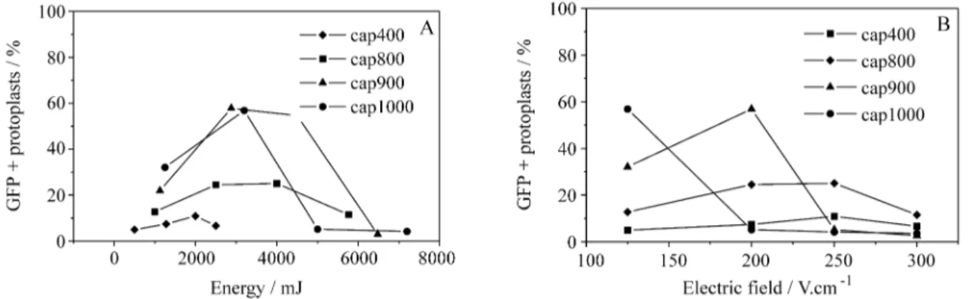
We used the transient expression of a gene coding for a green fluorescent protein (GFP) (Chalfieet al., 1994) for thein vivoevaluation of transformation efficiency. The fre-quency of transformants increased up to 3000 mJ of energy input (Figure 3A). While capacitors with a higher charge accumulation capacity (900 and 1000 µF) gave higher transformation frequencies ( 50%) when used in the range of 2000 to 4000 mJ, capacitors of lower charge accumula-tion (400 and 800µF) led to the death of approximately 70% of the protoplasts (Figure 3A). Maximum transient gene expression has been reported under electric field strengths causing more than 50% reduction in protoplast vi-ability (Frommet al., 1985; Hauptmannet al., 1987; Oard
et al., 1989).
Electric field (EF) strength is related to the electrode gap (d) and the discharged voltage (V) according to the equation EF = V.d-1, while the induced energy discharge (ε) is dependent on the capacitor and the applied voltage (V) according toε= CV2. Figure 3B shows that EF values of 250 V.cm-1discharged by 900 and 1000µF capacitors re-sulted in high transformation rates in electroporated proto-plasts. Less intense electric fields were unable to promote reporter gene expression in more than 20% of electro-porated protoplasts, while stronger electric fields induced irreparable plasma membrane damage, causing extensive cellular death and virtually no transformation.
Transient membrane pores are caused by an increase in the dipole moment of hydrophilic phospholipid heads, which move in the same direction as the applied electric field and provoke highly localized dielectric breakages in membrane structure (Kinosita and Tsong, 1977; Neumann
et al., 1982; Neumann et al., 1996). Irreversible pores which lead to cellular death are caused by longer pulses and stronger electric fields (Langridgeet al., 1987; Shillito et al., 1985), and we found that this occurred with energy
inputs of more than 5000 mJ (Figure 3A) or electric fields stronger than 300 V.cm-1(Figure 3B). Even so, protoplast electroporation has been reported as a mean of increasing cell division (Rechet al., 1987), plant regeneration (Chand
et al., 1988; Ochattet al., 1988) and DNA synthesis (Rech
et al., 1988). However, in our experiments, plant regenera-tion was slower from electroporated than from non-elec-troporated protoplasts (Table I).
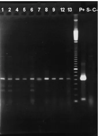
We observed specific transgene amplification in shoots regenerated from electroporated protoplasts (Figure 4).
Effect of the electroporation buffer on DNA introduction
Figure 5 shows that transient expression frequency of the GFP was significantly (p < 0.01) higher with buffer II and lowest with buffer I, while an intermediary frequency was obtained with buffer III. Non-viable and damaged protoplasts were rarely seen after electroporation in buffer II, which gave almost 100 % expression of GFP (Figure 6A). However, we found that the frequency of stable
Table I- Comparison of plant regeneration efficiency from electroporated and non-electroporated S. guianensis protoplasts. See text for electroporation conditions.
Non-electroporated Electroporated
Protoplasts per cotyledon pair (x 105)
4.57 ± 0.73 5.67 ± 0.85
Protoplast viability1(%) 76.00 ± 2.46 21.00 ± 7.09 Division frequency (%) 4.50 ± 1.66 0.70 ± 3.33
Plating efficiency2(%) 1.90 ± 0.140.02 ± 6.67 Regeneration frequency3(%) 85.00 ± 3.41 72.00 ± 7.29
*Mean values of 4independent experiments ± standard errors. 1 = 24h after electroporation.
2 = N. of colonies after 45 days culture divided by the N. of plated proto-plasts.
3 = N. of calli with shoots after 90 days of culture divided by the N. of plated calli.
transformation was lower than that expected from transient gene expression in electroporated protoplasts (Hainet al., 1985; Shillitoet al., 1985), probably due to some retention of transgene expression ability in damaged protoplasts for periods shorter than 72 h. The chloride ions in buffer I re-sulted in the production of toxic chlorine gas when electric pulses were applied, leading to a reduction in cellular via-bility (Figure 6B). In contrast to the data reported by Sena-ratna et al. (1991), we found that non-electroporated protoplasts incubated with plasmid DNA under the same
conditions for longer periods of time never showed tran-sient expression of the reporter gene (Figure 6C).
In exponential wave electric circuits, the voltage de-cays exponentially in the electroporation buffer between the electrodes and the induced energy is dependent on the initially applied voltage and the charge accumulation ca-pacity of the capacitor (Neumannet al., 1982, 1996). In this kind of circuit, the time constant (τ) is a function of the electrical resistance (R) and the capacitance (C) of the cir-cuit according to the equationτ= RC, so that for a given ca-pacitor (C = constant) electroporation buffers with reduced ionic force impose less resistance to the movement of charge through the circuit and pulse decay time is longer. Kinosita and Tsong (1977) have demonstrated that longer duration pulses promote greater membrane permeability due to pore enlargement, and this probably contributed to the higher transformation rates which we observed with the low ionic force buffers II and III (Figure 5).
Influence of DNA form on reporter gene expression
Figure 7 shows that linear DNA was more efficiently introduced into electroporated protoplasts. Higher Figure 4- Transgene PCR amplification in genomic DNA ofS. guianensis
regenerants derived from electroporated protoplasts. Regenerated plants are indicated as numbers on the lanes,p+= pCambia 1340,S- = non-elec-troporatedS. guianensis,C= without template DNA. Molecular weight maker = 100 bp DNA ladder (GIBCO BRL).
Figure 5- GFP expression in electroporatedS. guianensisprotoplasts in
buffer I(Frommet al., 1987),buffer II(Tadaet al., 1990) andbuffer III
(Wlalbot, 1993). Electroporation conditions were: 500µF, 100 V.cm-1, 20 Ωfor buffer I and III, 50Ωfor buffer II, 10µg of supercoiled DNA. Bars indicate standard errors and small letters indicate significant differences at p < 0.05.
transformation efficiency using linear plasmid DNA has been reported for bacteria (Rittich and Spanova, 1996), yeasts and filamentous fungi (Kwon-Chunget al., 1998), plant protoplasts (Linet al., 1997) and intact tissue (Akella and Lurquin, 1993; Dillenet al., 1995; Linet al., 1997; Sabriet al., 1996; Saunderset al., 1995). Due to the inner twist of the molecule and to the absence of distortions im-posed on the double helix structure (Drewet al., 1988), lin-ear plasmid DNA is highly mobile under electric fields (Courey and Wang, 1983). Macromolecular movement across pores is facilitated when the molecules are linear be-cause the absence of tertiary and quaternary structures re-duces their volume (Tanaka, 1988) and allows a more uniform superficial polarization to be induced by the elec-tric field (Neumannet al., 1982). The transient nature of membrane pores and of structural alterations to the cell wall induced by such electric fields imposes conformational re-strictions on the type of macromolecules that can be intro-duced (Neumannet al., 1982).
Nandadeva et al. (1999) reported that the physical structure of DNA is critical for efficient transformation, with denatured molecules being much more favorable for transformation, although high transformation efficiency has been obtained with supercoiled plasmid DNA (Bateset al., 1990; Frommet al., 1985; Oardet al., 1989).
In summary, we found that the critical factors regard-ing transgene expression in electroporated Stylosanthes guianensisprotoplasts were energy input and electric field strength. A field strength of 250 V.cm-1discharged by 900 and 1000µF capacitors gave the highest transient transfor-mation frequencies. Linear plasmid DNA, absence of chlo-ride and the presence of calcium ions also increased tran-sient gene expression, although not significantly. Our data suggest that putative transgenic plants ofS. guianensiscan be obtained at high frequencies from electroporated proto-plasts, on condition that an effective plant regeneration pro-tocol is used.
Acknowledgments
We thank Dr. C. Karia (Embrapa/Cerrados) for kindly providing theStylosanthes guianensis seeds. This research was supported by the Brazilian agency Fundação de Amparo à Pesquisa do Estado de São Paulo (FAPESP) Grant No.98/11270-7. VMQ was the recipient of a Doctoral fellowship (Grant No. 141294/97-3) from the Brazilian agency Conselho Nacional de Desenvolvimento Científico e Tecnológico (CNPq).
References
Akella V, and Lurquin PF (1993) Expression in cowpea seedlings of chimeric transgenes after electroporation into seed-deri-ved embryos. Plant Cell Rep. 12:110-117.
Ankenbauer RG and Nester EW (1993) TheAgrobacteriumTi plasmid and crown gall tumorigenesis: A model for signal transduction in host-pathogen interactions. In: Nester ER (ed) Signal transduction: prokaryotic and simple eukaryotic systems. Academic Press Inc., New York, pp. 67-104. Aragão FJL, Barros LMG, de Souza MV, Grossi de Sá MF,
Almeida ERP, Gander ES and Rech EL (1999) Expression of a methionine-rich storage albumin from the Brazil nut (Bertholletia excelsaH.B.K., Lecythidaceae) in transgenic bean plants (Phaseolus vulgarisL., Fabaceae). Genet. Mol. Biol. 22:445-449.
Bates GW, Carle SA and Piastuch WC (1990) Linear DNA intro-duced into carrot protoplasts by electroporation undergoes ligation and recircularization. Plant Mol. Biol. 14:899-908. Chalfie M, Tu Y, Euskirchen G, Ward WW and Prasher DC
(1994) Green fluorescent protein as a marker for gene ex-pression. Science 263:802-805.
Chand PK, Ochatt SJ, Rech EL, Power JB and Davey MR (1988) Electroporation stimulates plant regeneration from proto-plasts of the woody medicinal speciesSolanum dulcamara
L. J. Exp. Bot. 39:1267-1274.
Colby SM and Meredith CP (1990) Kanamycin sensitivity of cul-tured tissues ofVitissp. Plant Cell Rep. 9:237-240. Cônsoli L, Gaziola SA and Vieira MLC (1995) Plant
transforma-tion mediated byAgrobacterium rhizogenes: Optimization of the infection process. Braz. J. Genet. 18:115-119. Cônsoli L, Vieira MLC, de Souza Jr. CL and Garcia AAF (1996)
Tissue culture effects on quantitative traits inStylosanthes guianensis(Leguminosae). Braz. J. Genet. 19:469-474. Courey AJ and Wang JC (1983) Cruciform formation in a
nega-tively supercoiled DNA may be kinetically forbidden under physiological conditions. Cell 33:817-829.
Dillen W, Engler G, van Montagu M. and Angenon G (1995) Electroporation-mediated DNA delivery to seedling tissues ofPhaseolus vulgaris L. (common bean). Plant Cell Rep. 15:119-124.
Dornelas MC, Vieira MLC and Appezzato-da-Glória B (1992) Histological analysis of organogenesis and somatic embryo-genesis induced in immature tissues ofStylosanthes scabra. Ann. Bot. 70:477-482.
Dornelas MC, Vieira MLC and Souza Jr. CL de (1991) Fonte e idade de explante, radiação luminosa e combinações de NAA e BAP na indução de calos emStylosanthes scabra
Vog. Pesq. Agropec. Bras. 26:1901-1909.
Dower WJ, Miller JF and Ragsdale CW (1988) High efficiency transformation ofE. coli by high voltage electroporation. Nucleic Acid Res. 16:6127-6145.
Drew HR, McCall MJ and Calladine CR (1988) Recent studies of DNA in the crystal. Ann. Rev. Cell. Biol 4:1-20.
Edwards K, Johnstone C and Thompson C (1991) A simple and rapid method for the preparation of plant genomic DNA for PCR analysis. Nucleic Acid Res. 19:1349.
Fromm M, Callis J, Taylor LP and Walbot V (1987) Electroporation of DNA and RNA into plant protoplasts. Methods Enzymol. 153:351-365.
Fromm ME, Taylor LP and Walbot V (1985) Expression of genes transferred into monocot and dicot plant cells by electroporation. Proc. Natl. Acad. Sci. USA 82:5824-5828. Gilmour DM, Davey MR and Cocking EC (1989) Production of
somatic hybrid tissues following chemical and electrical fu-sion of protoplast from albino cell suspenfu-sions ofMedicago sativaandM. borealis. Plant Cell Rep. 8:29-32.
Hain R, Stabel P, Czernilofsky AP, Steibib HH, Herrera-Estrella L, and Schell J (1985) Uptake, integration, expression and genetic transmission of a selectable chimaeric gene by plant protoplasts. Mol. Gen. Genet. 199:161-168.
Hanahan D (1983) Studies on the transformation ofEscherichia coliwith plasmids. J. Mol. Biol. 166:557-580.
Hauptmann RM, Ozias-Akin P, Vasil V, Tabaeizaden Z, Rogers SG, Horsch RB, Vasil IK and Fralwy RT (1987) Transient expression of electroporated DNA in monocotyledonous and dicotyledonous species. Plant Cell Rep. 6:265-270. Hoffmann LV (1998) Regeneração de plantas e expressão
transi-ente de GUS emStylosanthes guianensis (Aubl.) Sw. via
Agrobacterium. M.Sc. Thesis, Escola Superior de Agricul-tura “Luiz de Queiroz”, Universidade de São Paulo, Pira-cicaba, São Paulo.
Jefferson RA, Kavanagh TA and Bevan MW (1987) GUS fusion : the β-glucuronidase as sensitive and versatile gene fusion marker in higher plants. EMBO J. 6:3301-3307.
Kao N (1977) Chromosomal behavior of somatic hybrids of soy-bean xNicotiana glauca. Mol. Gen. Genet. 150:225-230. Kinosita K and Tsong TY (1977) Voltage-induced pore formation
and hemolysis of human erythrocytes. Biochem. Biophys. Acta 471:227-242.
Kwon-Chung KJ, Goldman WE, Klein B, Szaniszla PJ, Polonelli LO and Walsh TJ (1998) Fate of transforming DNA in pathogenic fungi. Med. Mycol. 36:38-44.
Langridge WHR, Li BJ and Szalay AA (1987) Uptake of DNA and RNA into cells mediated by electroporation. Methods Enzymol. 153:336-350.
Lin CH, Xiao L and Hou BH (1997) Optimization of electropo-ration conditions for expression of GUS activity in electro-porated protoplasts and intact cells. Plant Physiol. Biochem. 35:959-968.
Liu CJ, Musial JM and Smith FW (1996) Evidence for a low level of genomic specificity of sequence-tagged sites in
Stylosanthes. Theor. Appl. Genet. 93:864-868.
Lovato MB and Martins PS (1997) Genetic variability in salt tol-erance during germination ofStylosanthes humilisH.B.K. and association between salt tolerance and isozymes. Braz. J. Genet. 20:435-441.
Manner JM (1987) Transformation of Stylosanthes spp. using
Agrobacterium tumefaciens. Plant Cell Rep. 6:204-207.
Manners JM and Way H (1989) Efficient transformation with re-generation of tropical pasture legumeStylosanthes humilis
using Agrobacterium rhizogenes and a Ti plasmid binary vector system. Plant Cell Rep. 8:341-345.
Manners JM (1988) Transgenic plants of the tropical pasture le-gumeStylosanthes humilis. Plant Sci. 55:61-68.
Mauro AO, Nóbrega JC, Baraldi GL and Collins GB (1995) Sus-ceptibility of some Brazilian soybean genotypes to three strains of Agrobacterium tumefaciens. Braz. J. Genet. 18:417-420.
McCabe DE, Swain WF, Matinell JB and Christou P (1988) Sta-ble transformation of soybean (Glicine max) by particle ac-celeration. Bio/Tech. 6:923-926.
Meijer EGM and Szabados L (1990) Cell and tissue culture of
Stylosanthesspp. In:Biotechnology in agriculture and for-estry. Legumes and oilseed crops. (Bajaj, Y.P.S., ed.). Springer Verlag, Heidelberg, pp.312-322.
Murashige T. and Skoog F (1962) A revised medium for rapid growth and bioassays with tobacco tissue cultures. Physiol. Plant. 15:473-497.
Nandadeva YL, Lupi CG, Meyer CS, Devi PS, Potrykus I and Bilang R (1999) Microprojectile-mediated transient and in-tegrative transformation of rice embryogenic suspension cells: effects of osmotic cell conditioning and of the physical configuration of plasmid DNA. Plant Cell Rep. 18:500-504. Neumann E, Kakorin S and Tsoneva I (1996) Calcium-mediated
DNA adsorption to yeast cells and kinetics of cell transfor-mation by electroporation. Biophys. J. 71:868-877. Neumann E, Schäefer-Ridder M, Wang M and Hofschneider PH
(1982) Gene transfer into mouse lyoma cells by electro-poration in high electric fields. EMBO J. 1:841-845. Oard JH, Paige DF, Simmonds JA and Gradziel TM (1989)
Tran-sient gene expression in maize, rice and wheat cells using an airgun apparatus. Plant Physiol. 92:334-339.
Ochatt SJ, Chand PK, Rech EL, Davey MR and Power JB (1988) Electroporation-mediated improvement of plant regenera-tion from colt cherry (Prunus aviumxpseudocerasus) pro-toplasts. Plant Sci. 54:165-169.
Partridge I (1996) Protecting stylos from anthracnose. Rural Res. 171:29-32.
Potrykus I (1990) Gene transfer to plants: assessment and per-spectives. Physiol. Plant. 79:125-134.
Potrykus I (1991) Gene transfer to plants: assessment of published approaches and results. Ann. Rev. Plant Physiol. Plant Mol. Biol. 42:205-225.
Quecini VM (1999) Transferência direta de genes para plantas de
Stylosanthes guianensis (Aubl.) Sw. Ph.D. Thesis, Escola Superior de Agricultura “Luiz de Queiroz”, Universidade de São Paulo, Piracicaba, São Paulo.
Quoirin M, Aragão F, Rech E and de Oliveira DE (1997) Tran-sient expression of a reporter gene introduced by bioballistic bombardment into Racosperma mangium (Leguminosae family) tissues. Braz. J. Genet. 20:507-510.
Rech EL, Ochatt SJ, Chand PK, Davey MR, Mulligan BJ and Power JB (1988) Electroporation increases DNA synthesis in cultured plant protoplasts. Bio/Tech. 6:1091-1093. Rech EL, Ochatt SJ, Chand PK, Power JB and Davey MR (1987)
Electro-enhancement of division of plant protoplast-derived cells. Protoplasma 141:169-176.
quan-titatively describing the relationship between the number of electrotransformants and DNA concentration. Bioelectroch. Bioener. 40:233-238.
Sabri N, Pelissier B and Teissie J (1996) Transient and stable electrotransformation of intact black Mexican sweet maize cells are obtained after preplasmolysis. Plant Cell Rep. 15:924-928.
Sambrook J, Fritsch EF and Maniatis T (1989) Molecular cloning: a laboratory manual. Cold Spring Harbor Laboratory Press, New York.
Sarria R, Calderón A, Thro AM, Torres E, Mayer JE and Roca WM (1994) Agrobacterium-mediated transformation of
Stylosanthes guianensisand production of transgenic plants. Plant Sci. 96:119-127.
Saunders JA, Lin CH and Hou BH (1995) Rapid optimization of electroporation conditions for plant cells, protoplasts and pollen. Mol. Biotechnol. 3:181-190.
Senaratna T, Mckersie BD, Kasha KJ and Procunier JD (1991) Di-rect DNA uptake during the imbibition of cells. Plant Sci. 79:223-228.
Shillito RD, Saul MW, Paszkowski J, Müller M and Potrykus I (1985) High efficiency direct gene transfer to plants. Bio/Tech. 3:1099-1103.
Tada Y, Sakamoto M and Fujimura T (1990) Efficient gene intro-duction into rice by electroporation and analysis of transgen-ic plants: use of electroporation buffer lacking chloride ions. Theor. Appl. Genet. 80:475-480.
Tanaka T (1988) Gels. Sci. Amer. 244:124-138.
Valarini MJ, Otsuk IP and Vieira MLC (1997) Changes in N2
fixa-tion in Stylosanthes scabra derived from tissue culture. Braz. J. Genet. 20:713-716.
van der Stappen J, van Campenhout S, and Lopez SG (1998) Se-quencing of the internal transcribed region ITS1 as a molec-ular tool detecting variation in the Stylosanthes species complex. Theor. Appl. Genet. 96:869-877.
Vieira MLC, Fungaro MHP, Jubier MF and Lejeune B (1997) De-termination of taxonomic relationship among Brazilian taxa ofStylosanthesSW., Leguminosae, using RAPD markers. Pesq. Agrop. Bras. 3:305-310.
Vieira MLC, Jones B, Cocking EC and Davey MR (1990) Plant regeneration from protoplasts isolated from seedling cotyle-dons of Stylosanthes guianensis, S. macrocephalaand S. scabra. Plant Cell Rep. 9:289-292.
Walbot V. (1993) Gene pulser electroprotocols, survey number 181, Bio-Rad Laboratories, Hercules-CA.