Mutation in the
xpsD
gene of
Xanthomonas axonopodis
pv.
citri
affects cellulose degradation and virulence
Juliana Cristina Baptista
1,2, Marcos Antonio Machado
1, Rafael Augusto Homem
1,2,
Pablo Sebastián Torres
3, Adrian Alberto Vojnov
3and Alexandre Morais do Amaral
1,41
Centro APTA Citros “Sylvio Moreira”, Cordeirópolis, SP, Brazil.
2Universidade Estadual de Campinas, Campinas, SP, Brazil.
3
Fundación Pablo Cassará, Instituto de Ciencia y Tecnología Dr. Cesar Milstein,
Ciudad de Buenos Aires, Argentina.
4EMBRAPA Recursos Genéticos e Biotecnologia, Brasília, DF, Brazil.
Abstract
The Gram-negative bacteriumXanthomonas axonopodis pv. citri, the causal agent of citrus canker, is a major threat to the citrus industry worldwide. Although this is a leaf spot pathogen, it bears genes highly related to degradation of plant cell walls, which are typically found in plant pathogens that cause symptoms of tissue maceration. Little is known onXac capacity to cause disease and hydrolyze cellulose. We investigated the contribution of various open reading frames on degradation of a cellulose compound by means of a global mutational assay to selectively screen for a defect in carboxymethyl cellulase (CMCase) secretion inX. axonopodis pv. citri. Screening on CMC agar re-vealed one mutant clone defective in extracellular glycanase activity, out of nearly 3,000 clones. The insertion was lo-cated in thexpsD gene, a component of the type II secretion system (T2SS) showing an influence in the ability of Xac to colonize tissues and hydrolyze cellulose. In summary, these data show for the first time, thatX. axonopodis pv. citri is capable of hydrolyzing cellulose in a T2SS-dependent process. Furthermore, it was demonstrated that the ability to degrade cellulose contributes to the infection process as a whole.
Key words:citrus canker, transposome, type II secretion system.
Received: November 24, 2008; Accepted: July 6, 2009.
Introduction
Citrus canker is a highly destructive disease of citrus crops, which occurs worldwide and affects most of the commercial varieties of citrus. The disease is caused by the bacterium Xanthomonas axonopodis pv. citri. In areas where citrus canker has been established, its control is very difficult. Control is extremely difficult in areas where the disease is already firmly established, and is based on eradi-cation programs involving the heavy use of copper com-pounds. Bacterial plant pathogens, such as xanthomonads deliver various virulence proteins that play a role during in-fection. Knowledge on the complete repertoires of specific types of effectors would be beneficial towards understand-ing the mechanisms by which bacteria interact with plants, thereby causing diseases (Collmeret al., 2002, Guttmanet al., 2002, Rodenet al., 2004, Changet al., 2005, Nomura and He, 2005).
The complete genome sequencing ofX. axonopodis
pv.citrirevealed several open reading frames that were
an-notated as related to pathogenicity and virulence (da Silva
et al., 2002). Unexpectedly, given that the symptoms caused byX. axonopodispv.citri, a leaf-spot pathogen, are not typically associated to massive degradation of the plant-cell wall, such sequences include a number of genes that share similarity with those that code for plant cell wall degradation in other organisms. Plant cell walls are prised mainly of cellulose and hemicellulose as major com-ponents, and also lignin, pectin, among others. Apparently there is no correlation between citrus canker symptoms and citrus tissue degradation caused by cell-wall cleavage. In-terestingly,X. axonopodis pv.citriwas found to express cell-wall degrading enzymes when grown in a medium that supposedly mimics the environment of plant intercellular spaces (Astua-Mongeet al., 2005).
InErwiniaspecies, plant-cell wall cleavage is a key step for the pathogenesis. In fact, virulence depends basi-cally on the production and secretion of host-cell wall de-grading enzymes (Matsumotoet al., 2003, Corbettet al., 2005). To date, most of the proteins that have been charac-terized in bacterial plant pathogens, and also involved in the degradation of several components of plant-cell walls, are www.sbg.org.br
Send correspondence to Alexandre Morais do Amaral. EMBRAPA Recursos Genéticos e Biotecnologia, Caixa Postal 02372, 70770-917 Brasília, DF, Brazil. E-mail: aamaral@cenargen.embrapa.br.
secreted by the type II secretion system (T2SS) (Jhaet al., 2005). A functional T2SS is known to be required for viru-lence and the secretion of cell-wall degrading enzymes, in at least two xanthomonads that are vascular pathogens in their plant hosts, Xanthomonas oryzae pv. oryzae and
Xanthomonas campestrispv.campestris(Chenet al., 2005; Leeet al., 2005; Jhaet al., 2007).
AlthoughX. axonopodispv.citriharbors the T2SS, little is known regarding the effects of this system and the proteins that are delivered during its action. This study re-ports on the role ofxpsD, a T2SS component, in the ability ofX. axonopodispv.citrito cause the disease and degrade cellulose.
Materials and Methods
Bacterial strains, plasmids and growth conditions
The bacterial strains and plasmids used in the present study are listed in Table 1.Xanthomonas axonopodispv.
citri306 strain, already described (da Silvaet al., 2002), was routinely cultured on nutrient broth (NB) medium [containing per liter, 15 g of peptone, 3 g of yeast extract, 6 g of NaCl and 1 g D(+) glucose] at 28 °C, while Esche-richia colicultures were grown on Luria-Bertani medium at 37 °C. Kanamycin (15mg/mL) was used for selection in
E. coliandX. axonopodispv.citri. The plasmid for site-directed mutagenesis was prepared from theE. colihost strain by the alkaline lysis method.
DNA manipulation
Insertion points were confirmed by DNA sequencing with the forward and reverse primers of EZ::TN (Epicentre Technologies, Madison, WI) (Table 2). For genomic DNA isolation, strains were cultured overnight in 50 mL of NB. Cultures were then adjusted to optical density of 0.6 at 600 nm in sterile NB, and DNA was isolated using the CTAB (hexadecyltrimethylammonium bromide) method (Ausubel et al., 1998). DNA was stored in Tris-EDTA buffer (10 mM Tris, 1 mM EDTA, pH 8.0) at -20 °C. For
Southern blotting and hybridization, 2mg of genomic DNA fromX. axonopodispv.citriwere digested withEcoRI and
EcoRV, separated by electrophoresis in an 0.8% agarose gel, and transferred onto Hybond-N nylon membrane (Amersham Pharmacia Biotech, Little Chalfont, Buckinghamshire, England) in 5 M NaOH, with subse-quent cross-linking by exposure to UV irradiation. Hybrid-ization was carried out at 65 °C. Southern blotting analysis was performed with digoxigenin (DIG)-labeled PCR prod-ucts corresponding to a 399-bp fragment ofaph gene as probe, by using the PCR DIG Probe Synthesis Kit and DIG DNA Labeling and Detection Kit (Roche Diagnosis, India-napolis, IN). The analysis was used to confirm single EZ:TN insertions and homologous recombination.
Construction of mutant strains
For random insertion mutagenesis, the EZ::TN < KAN-2 > transposome complex, a mixture of the transpo-son EZ::TN < KAN-2 > and EZ::TN transposase (Epicentre Technologies, Madison, WI), was introduced by electropo-ration directly into X. axonopodis pv. citri 306 electro-competent cells. These cells were prepared as described by Amaral et al. (2005). A Gene Pulser II Electroporator (Bio-Rad, Hercules, CA) was used to transform 50mL of
Table 1- Bacterial strains and plasmids.
Strain or plasmid Characteristics Source or reference
Escherichia coli
DH5a F-f80dlacZDM15D(lacZYA-argF)U169 endA1 deoR recA1 hsdR17(r
K-mK+)phoA supE44l-thi-1
gyrA96 relA1
Gibco
Xanthomonas axonopodispv.citri
306 Wild type, Apr R.P. Leite
30E9 Kanr, XpsD- This study
DxpsD Kanr, site-directed XpsD- This study
Plasmid
pCR2.1-TOPO vector pUC18 derivative, kanr,bla(Apr),lacZ Invitrogen
TOPO-xpsD PCR-amplifiedxpsD(725-1307) in pCR2.1-TOPO; Kanr This study
Table 2- Oligonucleotides used.
Primer Nucleotide sequence (5’®3’)a
KAN RP-1 GCAATGTAACATCAGAGATTTTGAG
KAN2 FP-1 ACCTACAACAAAGCTCTCATCAACC
KanA CATGCAAGCTTCAGGGTTGA
AD1 NTCGA(G/C)T(A/T)T(G/C)G(A/T)GTT
AD2 NGTCGA(G/C)(A/T)GANA(A/T)GAA
AD3 (A/T)GTGNAG(A/T)ANCANAGA
AD4 AG(A/T)GNAG(A/T)ANCA(A/T)AGG
XpsD_EcoRI_F GAATTCCAAGGCCGAAAAAGTCTCTG
XpsD_EcoRI_R GAATTCCACCAGCAGGGTATTGGTCT
cell suspension with 1mL of the transposome complex, un-der the following conditions: 50W, 50mF, and 2.5 kV in a 0.2-cm cuvette. After electroporation, cells were suspended in 1 mL of SOC broth (containing, per liter, 20 g of trypto-ne, 5 g of yeast extract, 0.5544 g of NaCl, 0.1864 g of KCl, 1.2038 g of MgSO4, 0.9522 g of MgCl2and 3.2 g of glu-cose) and allowed to recover for 2-3 h at 28 °C with shak-ing. To confirm the phenotypes, the site-directedDxpsD
mutant was produced by using the primers XpsD_EcoRI_F and XpsD_EcoRI_R to PCR amplify an internal fragment of thexpsD region fromX. axonopodispv.citri306 geno-mic DNA. The 583-kb PCR product was cloned into the pCR2.1-TOPO vector (Invitrogen, Carlsbad, CA), thereby generating TOPO-xpsD, and then electroporated into E. coli, whereupon the plasmid DNA was extracted and elec-troporated intoX. axonopodispv.citri.
Plate assay
Carboxymethyl cellulose (CMC) (0.5%) was incor-porated into agar plates with culture medium (0.1% NaNO3, 0.1% K2HPO4, 0.1% KCl, 0.05% MgSO4, 0.05% yeast extract, 0.1% glucose and 1.7% agar). Colonies were grown at 28 °C for 48 h and then washed off with water. The plates were flooded with 1% Congo Red for 30 min and washed with 1 M NaCl. CMC degradation was detected by yellow halos under and around the colonies, and CMCase activity analyzed by using the (H2- C2)/ C2ratio (H - diame-ter of the halo, C - diamediame-ter of the colony). For each strain, the hypothesis of equivalent CMC degradation in the two groups (wild-type and mutant strains) was tested by means of the Student two-samplettest.
DNA sequencing
The general PCR procedure has been described by Sambrooket al.(1989). For mapping the location of trans-poson insertion, TAIL-PCR (thermal asymmetric inter-laced PCR) was performed to amplify unknown DNA sequences contiguous to knownkangene sequences, ac-cording to the method of Liu and Whittier (1995). Three successive high- and low-stringency PCR amplifications were performed with nested sequence-specific primers and shorter arbitrary degenerate primers with genomic tem-plates from the mutant strains generated by random inser-tion. Sequences were compared and aligned with sequences from the GenBank database, by using the BLAST program of the National Center for Biotechnology Information website.
Sodium dodecyl sulfate-polyacrylamide gel electrophoresis (SDS-PAGE)
For comparative analysis of proteins secreted by both
X. axonopodis pv. citri306 andDxpsDmutant strains, the bacteria were grown for 24 h at 28 °C in XMV2 medium (Schulte and Bonas, 1992) to an optical density of 0.6 and the culture-supernatant proteins were concentrated by
pre-cipitation with 10% trichloroacetic acid, as previously described (Economouet al., 1990), except that after precip-itation, the trichloroacetic acid was extracted by washing the precipitate with acetone. Proteins from an equivalent of 10 mL of culture supernatant were separated by SDS-PAGE (Bradleyet al., 1988) with 12% acrylamide and vi-sualized by staining with Silver Stain Kit (Bio-Rad, Hercu-les, CA).
Plant material and plant inoculation
Pathogenicity and virulence assays were performed by using sweet orange cv. Baia (Citrus sinensisL. Osbeck) as host of all mutant strains. All plants were grown in growth chambers at 28 °C with a 12-h photoperiod. Ino-culum concentrations were adjusted to an optical density of 0.6 at 600 nm. For pathogenicity and virulence tests, plants were inoculated by syringe infiltration with needle and, to mimic the natural infection process (bacteria entering the plant through the stomata), leaves were sprayed with the bacterial suspension on the abaxial surface. Symptoms were scored by any visual modification on lesion. Bacterial growth in citrus leaves was measured by harvesting leaf discs for eachX. axonopodispv.citri mutant strain. The leaves were ground with a mortar and pestle in 1 mL of 0.01 M phosphate buffer, pH 7.2. The solution was serially diluted and spread onto NB plates, with proper antibiotics. The mean number of colonies in plates of the proper dilu-tions was calculated.
Results
Construction of a knockout mutant library and isolation of cellulose degradation deficientX. axonopodispv.citrimutants
To confirm the presence of functional genes for CMC degradation in X. axonopodis pv. citri, a collection of nearly 3,000 knock-out mutants was produced by random insertion of a cassette for resistance to kanamycin.
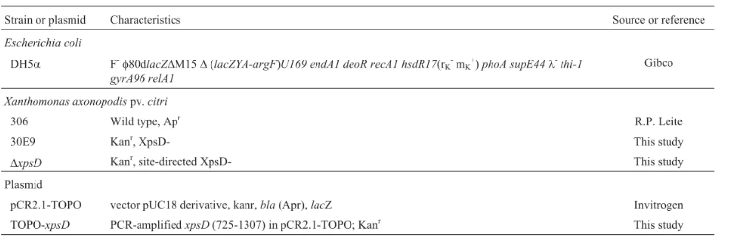
To evaluate the capacity ofX. axonopodispv.citriin hydrolyzing cellulose-containing material, we performed a global screening to search strains for lack-of-function (Fig-ure 1 A and B). Individual transformants from the random mutant libraries were replicated onto squared CMC plates for phenotypic screening.
From the knock-out mutants produced by random in-sertion, the transformant named 30E9 exhibited a smaller, superficial clear zone on the CMC plate, suggesting that it is merely partially functional (Figure 1C and D). The ability of the mutant 30E9 to promote CMC degradation from agar plates was very weak and limited, only occurring below the colony. This inconsiderable CMC degradation remained stable for several days after inoculation. Areas with CMC degradation were significantly lower on spots with the 30E9 mutant strain than on those with the wild-type (ttest with paired two samples for mean, p < 0.001;n= 24 spots analyzed per each strain) (Table 3). To check whether the CMC degradation in the mutant strain could be induced by the plant tissue, infected foliar discs were placed on a CMC plate (Figure 1D). Despite the fact that the wild-type strain showed CMCase activity below the edge of the discs, the mutant strain had little effect.
To identify the site of transposon integration, we de-termined the DNA sequence adjacent to thekanRcassette for this strain. The 30E9 mutant had inactivation of the gene
xpsD (GenBank accession n. AAM38377.1), with the transposon located at the position 4,178,942 in the genome
(GCACGATG < KAN-2 > TCCAGAA). Since the transposon EZ::TN < KAN-2 > shows no origin of replica-tion it cannot be rescued by digesting genomic DNA of the mutant strain, therefore the TAIL-PCR technique was used to map the site of cassette insertion. Southern blot analysis verified that a single insertion was responsible for the phe-notype (Figure 2A).
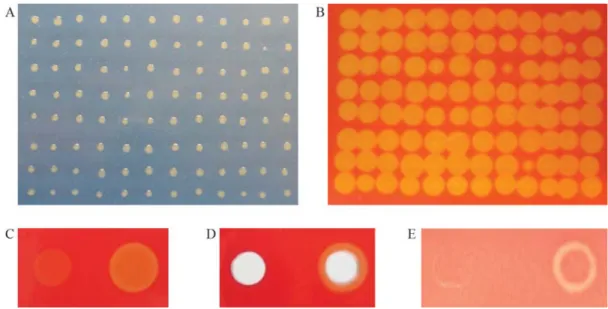
Interruption ofX. axonopodispv.citri xpsD gene results in the modification of bacterial secretion, disease symptoms and multiplication on citrus leaves
The screening of the transposon library led to the identification of a clone with an impaired ability to hydro-lyze cellulose. Also, the lack-of-function for the gene in-vestigated in this study had effect on bacterial secretion (Figure 2B), disease symptoms (Figure 3 A-E), and growth
in planta(Figure 4). Nevertheless, no apparent differences in symptom severity were observed on comparing mutant and wild-type strains when the inoculation method (spray) reproduced natural-infection phenomena (Figure 3F and G).
Table 3- Hydrolysis of CMC by wild-type and mutant strains.
X. axonopodispv.citristrain CMCase activity index*, # Standard deviation
Wild type 5.07a&±0.1487 0.7285
XpsD-defective mutant (30E9) 0.86b±0.0804 0.3941
*
(H2- C2)/ C2(H - diameter of the halo, C - diameter of the colony).
#
Average±standard error of the means (n = 24).
&
Statistically significant differences (p < 0.001), as determined by the Studentttest, are indicated by letters “a” and “b” for comparison to the strains.
Figure 1- Selection ofX. axonopodispv.citrimutant colonies defective in CMC degradation. In panel A, Petri dish with 96 colonies grown on CMC dium, 2 days after inoculation; B, Halo of CMC degradation in Petri dish after colonies were washed off and Congo red staining was applied. C. Agar
me-dium topped with CMC was seeded with lawn of mutant strain 30E9 (left) and wild-type (right) (40mL per spot), grown for 24 h, washed off and then
stained with Congo red. In panel D, the same procedure as that in panel C, however wells were made in the medium before seeding the cells. In panel E, fo-liar discs of citrus plants were infiltrated, placed onto the medium and then removed after 24 h, whereupon plates were stained. All strains were grown at
As shown by protein profiles (Figure 2B), CMCases certainly are not the only enzymes whose secretion is af-fected by mutation inxpsD ofX. axonopodispv.citri. In fact, the xpsD gene characterized in X. campestris pv.
campestriswas involved in the secretion of a number of ex-tra-cellular enzymes (Huet al., 1992).
To confirm that during infection on leaves, the transposon was not excised from the bacterium due to
se-lection, the strains were re-isolated from the infected tis-sues and plated using an appropriate antibiotic. Unlike the wild-type strain, the mutant strain was capable of growing on plates with kanamycin, and the 399-bp fragment ofaph
(kanamycin-resistance gene) was amplified by PCR.
A clear difference in symptomatology was found ac-cording to the method of inoculation used in the study. Al-though no differences were detected in leaves inoculated by spray (Figure3A and B), we found clear altered symptoms in the inoculated zone of the mutant strain compared to the wild-type strain in infiltrated leaves (Figure 3C to G). The differences were especially notable at the first two days
af-Figure 2- A.Southern blotby using a fragment ofnptII (399 pb) as a probe (1) to confirm single insertion. Total genomic DNA of the strain 30E9 (xpsD) fromXac306 digested with restriction enzymes (2:EcoRI, 3:
EcoRV, and 4:NcOI) and wild strain (5). B. SDS-PAGE of the secreted
proteins (supernatant) fromXanthomonas axonopodispv. citri in the
XVM2 medium. Lane 1, molecular mass marker; lane 2, wild-type strain; lane 3, xpsD defective strain.
Figure 3- Foliar symptoms of inoculation withX. axonopodispv.citriin citrus. In panels A (mutant strain) and B (wild-type strain), symptoms of spray inoculation with Xac (O.D.600= 0.5) on the abaxial surface of sweet orange leaves. The photographs were taken 30 days post-inoculation. In panels C, D,
E, F and G, the strains were inoculated (O.D.600= 0.5) into the side of the mid-vein (mutant strain, left; wild-type strain, right), and respective photographs
taken 2, 4, 6, 8 and 10 days post-infiltration, in each case.
Figure 4- Growth curve ofXanthomonas axonopodispv.citri(Xac) strains on leaves of susceptible sweet orange cultivar ‘Baia’ 0, 1, 3, 6 and 10 days after inoculation by syringe infiltration with needle. Xac WT:
wild-type strain; Xac 30E9 (xpsD-):xpsD mutant strain. Data represent
ter inoculation (Figure 3C), by a delay of appearance of symptoms in the mutant strain, and at ten days (Figure 3G), when the wild-type strain showed necrosis in the inoculated zone, in opposition to the mutant strain.
To ascertain the influence of thexpsD genes in citrus canker development, the growth ofX. axonopodispv.citri
was compared and differences between the two strains ob-served (Figure 4). A smaller population of the mutant strain was observed within 3 days after inoculation and remained until the stationary phase.
Discussion
In this study, we demonstrate that the T2SS exerts an influence on the ability ofX. axonopodispv.citrito colo-nize host tissues and is able to mediate the hydrolysis of cel-lulose.X. axonopodispv.citrienters citrus tissues through stomata and wounds, and has been traditionally described as causing lesions where the center becomes raised and spongy, or corky (Graham et al., 1992; Graham et al., 2004), aspects not typically related to symptoms of plant-cell degradation. However, the results presented above showed thatX. axonopodispv.citricells have the ability to hydrolyze cellulose.
The lack of a halo of CMC degradation around colo-nies ofX. axonopodispv.citriwas expected to result from the inactivation of either a gene for a major cellulose-degrading enzyme (Walkeret al., 1994), a gene-regulator (Vincent-Sealyet al., 1999), or a component of the secre-tion system itself (Rayet al., 2000). The fact that the scant CMC degradation on agar plates only occurred below the colony, and furthermore that no surrounding degradation halo was detected through staining, all implies that the mechanism is cell bound (Zorreguietaet al., 2000).
Studies indicated that when thexpsD gene was inter-rupted, extracellular enzymes polygalacturonate lyase, al-pha-amylase and endoglucanase accumulated in the peri-plasm ofXanthomonas campestrispv.campestris(Hu et al., 1992). Likewise, our study has demonstrated by protein profile thatX. axonopodispv.citriwhich lacks a functional
xpsD gene was clearly affected in its secretion.
XpsD is the outer membrane single protein of the xps cluster-encoded T2SS in Gram-negative bacteria, which re-quires a multicomponent assembly apparatus for the secre-tion of extracellular enzymes. Recent articles on several plant pathogens report a key role of the xps cluster during pathogenesis (Jhaet al., 2005, 2007). This finding has shed light on the possibility ofX. axonopodispv.citribeing ca-pable of somehow modifying cellulose components in its environment, and is consistent with the effects of the T2SS found in other bacteria (Rayet al., 2000; Zhou and Ingram, 2000; Zorreguietaet al., 2000; Jhaet al., 2005, 2007). In fact, X. axonopodis pv. citri, like X. campestris pv.
vesicatoriaandX. campestrispv.campestris, harbors two gene clusters coding for different T2SSs (the xcs and xps type II secretion systems), whereasX. oryzaepv.oryzae
en-codes only the xps cluster. So far, there are no reports on the role played by the xcs-encoded T2SS in xanthomonads.
We had no means of complementingX. axonopodis
pv.citrito evaluate whether wild-typexpsDgenes are capa-ble of inducing virulence and cellulose degradation, since amplification of the full gene (2,291 bp in length) from the genome or cosmid has so far been unsuccessful. Thus, complementation tests were not accomplished.
Although it remains unknown whether the degrada-tion of cellulose can be unequivocally imputed to a major gene or is the result of the joint-action of a number of genes, it seems clear that such activity is mediated by the T2SS. Likewise, the same machinery is in some way responsible for the secretion of proteins that play important roles in vir-ulence.
InErwinia chrysanthemi, two endoglucanases (CelZ and CelY) are produced, however CelZ represents approxi-mately 95% of the total carboxymethyl cellulase activity (Zhou and Ingram, 2000). The particularX. axonopodispv.
citrienzymes involved in cellulose hydrolysis have not, as yet, been identified. Even though many more insertion strains must be tested for this screen to reach saturation, these results suggest that the proteins related to CMC deg-radation seem to be exclusively secreted by the xps -en-coded T2SS present in X. axonopodis pv. citri. Further-more, even though incapable strains represent only a small fraction of the total number of sequences annotated as puta-tively involved in the process, this mutant strain could pos-sibly make a significant difference for studies involving plant cellulose and the action of plant pathogens.
The multiplication ofX. axonopodispv.citriwas sig-nificantly suppressed when inoculated into sweet orange leaves. The observation of reduced disease severity follow-ing inoculation with the 30E9 was different from that ob-served whenxpsD-defectiveX. campestrispv.campestris
was inoculated into cabbage (Huet al., 1992). In fact, apart from these two pathogens infecting distinct plant hosts, a major difference between these xanthomonads is their mode of action. WhereasX. axonopodispv.citriis a leaf-spot pathogen,X. campestrispv.campestrisis a vascular bacterium. Moreover, in the spray process emulating natu-ral infection, there were no visible modifications in lesions. Collectively, these data may indicate specific roles played by the T2SS according to the pathosystem.
Acknowledgments
J. C. B. was supported by a fellowship from FAPESP (04/02815-2). This work was sponsored in part by grants from FAPESP (05/00719-9, 2008/00070-0) and the PRODETAB program. The collaborative work between re-search groups from Brazil and Argentina was supported by the CNPq-CONICET cooperative program (490766/2006-4).
References
Amaral AM do, Toledo CP, Baptista JC and Machado MA (2005) Transformation of Xanthomonas axonopodis pv. citri by eletroporation. Fitopatol Brasil 30:292-294.
Astua-Monge G, Freitas-Astua J, Bacocina G, Roncoletta J, Car-valho AS and Machado MA (2005) Expression profiling of
virulence and pathogenicity genes of Xanthomonas
axonopodispv. citri. J Bacteriol 187:1201-1205.
Ausubel FM, Brent R, Kingston RE, Moore DD, Seidman JG, Smith JA and Struhl K (1998) Current Protocols in Molecu-lar Biology. John Wiley & Sons, Inc., New York.
Bradley DJ, Wood EA, Larkins AP, Galfrè G, Butcher GW and Brewin NJ (1988) Isolation of monoclonal antibodies react-ing with peribacteroid membranes and other components of pea root nodules containing Rhizobium leguminosarum. Planta 173:149-160.
Chang JH, Urbach JM, Law TF, Arnold LW, Hu A, Gombar S, Grant SR, Ausubel FM and Dangl JL (2005) A high-throughput, near-saturating screen for type III effector genes from Pseudomonas syringae. Proc Natl Acad Sci USA 102:3527-3528.
Chen Y, Shiue S-J, Huang C-W, Chang J-L, Chien Y-L, Hu N-T and Chan N-L (2005) Structure and function of the XpsE N-terminal domain, an essential component of the
Xanthomonas campestris type II secretion system. J Biol Chem 280:42356-42363.
Collmer A, Lindeberg M, Petnicki-Ocwieja T, Schnieder DJ and Alfano JR (2002) Genomic mining type III secretion system effectors inPseudomonas syringaeyields new picks for all TTSS prospectors. Trends Microbiol 10:462-469.
Corbett M, Virtue S, Bell K, Birch P, Burr T, Hyman L, Lilley K, Poock S, Toth I and Salmond G (2005) Identification of a new quorum-sensing-controlled virulence factor inErwinia carotovorasubsp.atrosepticasecreted via the type II target-ing pathway. Mol Plant-Microb Interact 18:334-342. da Silva AC, Ferro JA, Reinach FC, Farah CS, Furlan LR,
Quag-gio RB, Monteiro-Vitorello CB, Van Sluys MA, Almeida NF, Alves LMC,et al.(2002) Comparison of the genomes of two Xanthomonas pathogens with differing host specifi-cities. Nature 417:459-463.
Economou A, Hamilton WDO, Johnston AWB and Downie JA
(1990) The Rhizobium nodulation gene nodO encodes a
Ca2+-binding protein that is exported without N-terminal cleavage and is homologous to haemolysin and related pro-teins. EMBO J 9:349-354.
Graham JH, Gottwald TR, Riley TD and Achor D (1992) Penetra-tion through leaf stomata and growth of strains of Xantho-monas campestrisin citrus cultivars varying in susceptibil-ity to bacterial diseases. Phytopathology 82:1319-1325.
Graham JH, Gottwald TR, Cubero J and Achor DS (2004)
Xanthomonas axonopodis pv. citri: Factors affecting suc-cessful eradication of citrus canker. Mol Plant Pathol 5:1-15. Guttman DS, Vinatzer BA, Sarkar SF, Ranall MV, Kettler G and Greenberg JT (2002) A functional screen for the type III (Hrp) secretome of the plant pathogenPseudomonas syrin-gae. Science 295:1722-1726.
Hu NT, Hung MN, Chiou SJ, Tang F, Chiang DC, Huang HY and Wu CY (1992) Cloning and characterization of a gene re-quired for the secretion of extracellular enzymes across the outer membrane byXanthomonas campestrispv. campes-tris. J Bacteriol 174:2679-2687.
Jha G, Rajeshwari R and Sonti RV (2005) Bacterial type two se-cretion system secreted proteins: Double-edged swords for plant pathogens. Mol Plant-Microb Interact 18:891-898. Jha G, Rajeshwari R and Sonti RV (2007) Functional interplay
be-tween two Xanthomonas oryzaepv.oryzaesecretion sys-tems in modulating virulence on rice. Mol Plant-Microb In-teract 20:31-40.
Kim Y-S, Jung H-C and Pan J-G (2000) Bacterial cell surface dis-play of an enzyme library for selective screening of im-proved cellulase variants. Appl Environ Microbiol 66:788-793.
Lee M-S, Chen L-Y, Leu W-M, Shiau R-J and Hu N-T (2005) As-sociations of the major pseudopilin XpsG with XpsN (GspC) and secretin XpsD ofXanthomonas campestrispv. campestris type II secretion apparatus revealed by cross-linking analysis. J Biol Chem 280:4585-4591.
Liu YG and Whittier RF (1995) Thermal asymmetric interlaced PCR: Automatable amplification and sequencing of insert end fragments from P1 and YAC clones for chromosome walking. Genomics 25:674-681.
Matsumoto H, Jitareerat P, Baba Y and Tsuyumu S (2003) Com-parative study of regulatory mechanisms for pectinase
pro-duction by Erwinia carotovora subsp. carotovora and
Erwinia chrysanthemi. Mol Plant-Microb Interact 16:226-237.
Nomura K and He SY (2005) Powerful screens for bacterial viru-lence proteins. Proc Natl Acad Sci USA 102:3527-3528. Ray SK, Rajeshwari R and Sonti RV (2000) Mutants of
Xanthomonas oryzaepv. oryzae deficient in general secre-tory pathway are virulence deficient and unable to secrete xylanase. Mol Plant-Microb Interact 4:394-401.
Roden JA, Belt B, Ross JB, Tachibana T, Vargas J and Mudgett MB (2004) A genetic screen to isolate type III effectors translocated into pepper cells during Xanthomonas infec-tion. Proc Natl Acad Sci USA 101:16624-16629.
Sambrook JE, Fritsch EF and Maniatis TA (1989) Molecular Cloning: A Laboratory Manual, 2nd edition. Cold Spring Harbor Laboratory, Cold Spring Harbor.
Schulte R and Bonas U (1992) Expression of Xanthomonas
campestris pv. vesicatoria hrp cluster, which determines pathogenicity and hypersensitivity on pepper and tomato, is plant inducible. J Bacteriol 174:815-823.
Vincent-Sealy LV, Thomas JD, Commander P and Salmond GPC (1999)Erwinia carotovoraDsbA mutants: Evidence for a periplasmic-stress signal transduction system affecting tran-scription of genes encoding secreted proteins. Microbiology 145:1945-1958.
aroto-vorais an important soft-rot virulence factor. Mol Plant-Mi-crobe Interact 7:425-431.
Zhou S and Ingram LO (2000) Synergistic hydrolysis of carboxy-methyl cellulose and acid-swollen cellulose by two endoglu-canases (CelZ and CelY) from Erwinia chrysanthemi. J Bacteriol 182:5676-5682.
Zorreguieta A, Finnie C and Downie JA (2000) Extracellular glycanases ofRhizobium leguminosarumare activated on the cell surface by an exopolysaccharide-related component. J Bacteriol 182:1304-1312.
Associate Editor: Luís Carlos de Souza Ferreira