Comparative morphology among representatives of main taxa
of Scaphopoda and basal protobranch Bivalvia (Mollusca)
Luiz Ricardo L. Simone
ABStRACt
This study deals with detailed morphology and anatomy of 4 species of Scaphopoda and 5 species of protobranch Bivalvia. Both classes are traditionally grouped in the taxon Diasoma, which has been questioned by different methodologies, such as molecular and developmental. This study is developed under a phylogenetic methodology with the main concern in performing it in an intelligible and testable methodology. The analyzed Scaphopoda species came from the Brazilian coast and belong to the family Dentaliidae [(1) Coccodentalium carduus; (2) Paradentalium disparile] and Gadiliidae; [(3) Polyschides noronhensis, n. sp. from Fernando de Noronha Archipelago; (4) Gadila braziliensis]. These species represent the main branches of the class Scaphopoda. From protobranch bivalves, representatives of the families Solemyidae [(5) Solemya occidentalis, from Florida; S. notialis, n. sp. from S.E. Brazil], Nuculanidae [(6) Propeleda carpentieri from Florida], and Nuculidae [(7) Ennucula puelcha, from south Brazil] are included. These species represent the main branches of the basal Bivalvia. The descriptions on the anatomy of S. occidentalis and of
P. carpentieri are published elsewhere. The remaining are included here, for which a complete taxonomical treatment is performed. Beyond these species, representatives of other taxa are operationally included as part of the ingroup (indices are then shared with them), as a procedure to test the morphological monophyly of Diasoma. These taxa are: two lamellibranch bivalves [(8) Barbatia – Arcidae; (9) Serratina – Tellinidae; both published elsewhere;, and
Propilidium (10) Patellogastropoda, and (11) Nautilus, basal Cephalopoda, based on basal taxa. The effective outgroups are (12) Neopilina (Monoplacophora) and (13) Hanleya
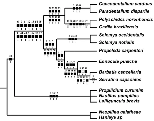
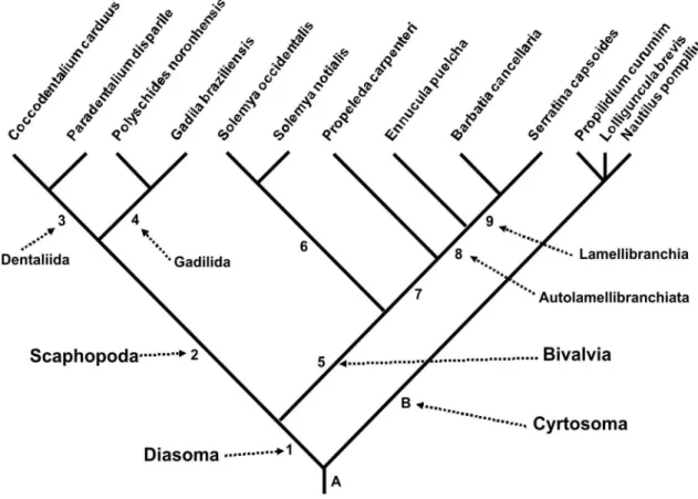
(Polyplacophora). The phylogenetic analysis based on morphology revealed that the taxon Diasoma is supported by 14 synapomorphies, and is separated from Cyrtosoma (Gastropoda + Cephalopoda). Although they are not the main goal of this paper, the taxa Scaphopoda and Bivalvia are supported by 8 and by 7 synapomorphies respectively. The taxon Protobranchia resulted paraphyletic. Both scaphopod orders resulted monophyletic. The obtained cladogram is: ((((Coccodentalium carduus – Paradentalium disparile) (Polyschides noronhensis – Gadila brasiliensis)) ((Solemya occidentalis – S. notialis) (Propeleda carpenteri (Ennucula puelcha (Barbatia cancellaria – Serratina capsoides))))) (Propilidium curumim – Nautilus pompilius – Lolliguncula brevis)).
Keywords: Scaphopoda; Bivalvia; Diasoma; Phylogeny; Morphology.
IntRoduCtIon
As explained below, this paper is the result of a larger project related to a group of Bivalvia, of which some of the presently analyzed species are outgroups. As the information obtained with the comparative analyzes is interesting, and helps to answer some im‑ portant questions on the Mollusca higher inter‑re‑ lationship, it was organized as a separate paper. The main intention is to furnish a comparative and phy‑ logenetic scenario with a theoretical background only based on morpho‑anatomy. This is certainly interest‑ ing for further analysis, since analyses for the past, present or future have been produced using other equally important methodologies. The relationship of the Classes Scaphopoda and Bivalvia has been fluid, and even their monophyly has been questioned in some nom‑morphological approaches. Nevertheless, as also explained below, both classes were grouped in the presently controversial taxon called Diasoma.
Diasoma, also called Loboconcha, was coined by Runnegar & Pojeta (1974), reuniting the classes pos‑ sibly derived from Rostroconchia, Scaphopoda and Bivalvia (Pojeta et al., 1972; Runnegar, 1996) (Lo‑ boconcha was named by Salvini‑Plawén, 1980). This molluscan branch is regarded to have diverged at the early Cambrian, bearing representatives with shells opening on both sides, and the digestive tube in a somewhat straight organization, i.e., mouth and anus located on opposite sides. This somatic conformation is adapted to an infaunal mode of life, and to deposit feeding. Diasoma was, then, subsequently accepted and used in the current technical literature (e.g., Po‑ jeta & Runnegar, 1976, 1985; Salvini‑Plawén, 1980 as Loboconcha; Haszprunar, 1988; Salvini‑Plawén & Steiner, 1996; Wagner, 1997), which sometimes even complemented the concept of the taxon. One example is the taxon Ancrypoda (= Diasoma), meaning anchor foot (Hennig, 1979; Lauterbach, 1984).
In the early 1990s, the concept of Diasoma was relatively well established in every phylogenetic vision of the Mollusca. However, another affinity for the Scaphopoda gradually appeared, approximating the taxon to the Gastropoda‑Cephalopoda branch (e.g.,
Peel, 1991; Haszprunar, 2000; Wanninger & Hasz‑ prunar, 2001), collectively called Cyrtosoma Run‑ negar & Pojeta, 1974.
Two main approaches were responsible for this change of concepts: 1) microscopic and developmen‑ tal studies (Wanninger & Haszprunar, 2002); 2) mo‑ lecular studies (Steiner & Dreyer, 2003; Passamaneck
et al., 2004). Both, which are explained in more detail below, gradually transferred Scaphopoda to Cyrtoso‑
ma. The new point of view has persuaded more gener‑ ic literature on Malacology (e.g., Coan et al. 2000:13) and on Zoology (Brusca & Brusca, 2003).
Of course most of this controversial relationship of the Scaphopoda, whether it be related to the Bi‑ valvia or Cyrtosoma classes, was stressed in early lit‑ erature (e.g., Lacaze‑Duthiers, 1857‑1858; Grobben, 1886; Plate, 1892; Simroth, 1894). However, with the introduction of phylogenetic methodologies, the dis‑ cussion becomes more intelligible. This methodology has been applied mainly in the last three decades; con‑ versely, and despite this, the scaphopod affinities are still inconclusive. A summary of this conceptual his‑ tory is provided in the recent literature (e.g., Steiner & Dreyer, 2003), details of which are not reported here. As the data of early literature was already explored by those papers, the present paper is, then, mainly con‑ cerned with argumentation of papers produced in the last three decades.
Some recent issues have directly or indirectly influenced analysis of the Scaphopoda relationship. For example, the appearance of a distinct pair of ce‑ phalic retractors during the development of a scaph‑ opod species, which is also found in gastropods and cephalopods, was used as additional argumentation to consider Scaphopoda within Cyrtosoma (Wanninger & Haszprunar, 2002). multiple cephalic appendages, scaphopod captacula, have been compared to cepha‑ lopod arms (Steiner & Dreyer, 2003). Molecular ap‑ proaches, on the other hand, sometimes demonstrate scaphopod’s affinity to Cephalopoda (Steiner & Dreyer, 2003 – 18S rDNA; Passamaneck et al., 2004: LSU + SSU rRNA), and sometimes to Bivalvia (Drey‑ er & Steiner, 2004 – mtDNA). In favor of the cyrto‑ some link, the scaphopod affinity to Bivalvia has even been considered as a mere consequence of homopla‑ sies, resulting from the similar infaunal mode of life (Steiner & Dreyer, 2003). Some combined molecular approaches have even demonstrated that scaphopods are related to a set grouping cephalopods and non‑ conchiferan aplacophorans, at the base of Mollusca (Giribet et al., 2006).
Although the number of taxa studied here is equivalent to that found in most papers, above related, referent to molecular and ultrastructural aspects, the main objective is not to test the other methodologies, nor their importance for comparative approaches. The main goal, instead, is to furnish another point of view and further argumentation to the still inconclu‑ sive relationships of bivalves and tusk‑shells. Another argument is that morphology is an autonomous sci‑ ence, and even if its importance in phylogeny and tax‑ onomy has been proven to be weak, morpho‑anatomy of the animals still must be studied. The final result, if only morphology is applied in the phylogeny at higher levels, must be exposed and debated.
The present study has, subsequently, the ob‑ jective of testing the relationship of a set of Scaph‑ opoda and Bivalvia, themselves and with remaining main branches of Mollusca, for the first time based on holistic morpho‑anatomy. For this task, a set of species were selected and examined in the same sort of details. From Scaphopoda, two species of each main branch were chosen – Dentaliida and Gadilida (Scarabino, 1995; Steiner, 1999). From Bivalvia, one or two samples of the main branches of basal taxa,
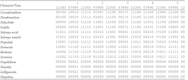
i.e., the Protobranchia, were selected, as well as two representatives of the higher taxa, i.e., Lamellibran‑ chia (a filibranch and an eulamellibranch). From protobranchs, Nuculoidea (1 species), Nuculanoidea (1 species) and Solemyoidea (2 species) were chosen. Lamellibranchs include an Arcidae and a Tellinidae. Representatives of other molluscan classes were also selected as outgroups, however, some of them, as ex‑ plained below, were operationally analyzed as part of the ingroup. This providence is a method for testing monophylies (Simone, 2006a). Further investigation on the phylogenies of Bivalvia and Scaphopoda was not performed, as they already exist in literature, such as in Bivalvia: Morton (1996), Giribet & Wheeler, (2005); and Scaphopoda: Steiner (1992a); of which data and concepts are hereby also applied.
Beyond the self‑searched data, information from the literature was used to increase the dataset of char‑ acters. For Scaphopoda, the following papers, which dealt with anatomical aspects, are included: Deshayes (1825); Lacaze‑Duthiers (1856‑1857); Yonge (1937); Cotton & Godfrey (1940); Gainey (1972); Steiner (1991, 1992a, b, 1993, 1994, 1996, 1998, 1999); Palmer & Steiner (1998); Lamprell & Healy (1998); Reynolds (2002). For Bivalvia, the following general papers are considered: Yonge (1939); Allen (1978); Starobogatov (1992); Morton, 1996; Morton et al.
(1998); Reid (1998); Villarroel & Stuardo (1998); Coan et al. (2000); Harper et al. (2000); and several
other more specific papers discussed in following sec‑ tions. These papers also bring comprehensive history of the classification and evolution of both classes that are not reproduced here. Some points, however, are certainly broached in the discussion.
It is important to emphasize that some studies on the phylogeny of scaphopods and bivalves have been produced in the last decades, mostly or totally based on molecular biology. Although they are con‑ sidered herein, all kinds of phylogenetic arrangements are found, which sometimes complicate comparisons (Giribet & Wheeler, 2002: 274‑275). In those stud‑ ies, morphology is not applied or is a secondary part of the dataset. This demonstrates that the morphology has not been properly evaluated, an impression that the present study has the objective of altering. Some interesting examples are Campbell (2000); Steiner & Hammer (2000); Steiner & Dreyer (2003); Dreyer & Steiner (2004).
As has been done for more general papers with phylogenetic approaches (Simone, 1999‑2000, 2001, 2002, 2004a, b, 2005, 2006a, b, 2007), this paper initiates with the systematic part, containing taxo‑ nomical treatment and morphological descriptions. Afterwards, a phylogenetic treatment is given, with presentation and discussion of the characters. This paper ends with a discussion of the obtained clado‑ gram in the light of present knowledge, with further implications mainly in taxonomy and phylogeny.
MAteRIAL And MethodS
The part of this paper related to comparative biology is performed under a phylogenetic (cladis‑ tic) methodology, which is the most practical and testable method. However, there is no intention to consider the analysis of this paper as “the phylogeny of Bivalvia” nor “of Scaphopoda”. Nevertheless, it is expected that the putative phylogenetic relationships among the species will remain even with the addition of more species, and that some taxonomical infer‑ ences can already be made. Analysis was performed with the aid of the program Tree‑Gardener (Ramos, 1997) (under a few modifications for Windows XP and Vista), which basically is an interface for the pro‑ gram Hennig86 (Farris, 1988). The algorithm used was “ie”. As outgroups, two species are used to root the cladogram, Monoplacophora Neopilina galatheae
Lemche, 1957 (Lemche & Wingstrand, 1959) and Polyplacophora Hanleya sp. Another five species are outgroups, but they are operationally analyzed as part of the ingroup. The species are Cephalopoda Nautilus pompilius Linné, 1758 (Griffin, 1897) and Loligun-cula brevis (Blainville, 1823) (Simone, 1997); Patel‑ logastropoda Propilidium curumim Leal & Simone, 1998, Lamellibranchia Barbatia cancellaria (Lamarck, 1819) (Simone & Chichvarkhin, 2004 – Arcidae), and Serratina capsoides (Lamarck, 1818) (Simone & Wilkinson, 2008 – Tellinidae). This measure is for testing the monophyly of Diasoma; conversely, the indices are shared with these taxa. In the discussion of the character, a short descriptive sentence is given for each one, followed by plesiomorphic and apomorphic states and conditions in the most parsimonious hy‑ pothesis. The consistency index (CI) and the retention index (RI) are given last, expressed as %. Several other characters were selected but excluded from this analy‑ sis, because their states were overlapping or purely au‑ tapomorphic. However, some autapomorphic states were maintained; this measure is based on the interest of the state or to test its importance. Study on two of the protobranchs considered herein are published elsewhere: Nuculanidae Propeleda carpenteri (Dall, 1881) and a second Solemyidae Solemya occidentalis
Deshayes, 1857. Partial anatomical descriptions and figures of these species are already published (Mik‑ kelsen & Bieler, 2008: 31‑33, 38‑40).
Some characters were introduced in order to organize outgroups, i.e., the representatives of other classes. However, the search for such characters was very superficial, sufficient only to this objective.
In the Discussion, some terms are used to des‑ ignate a collective set of taxa, with no taxonomical purpose. Some of them are “protobranchs” in apposi‑ tion to “lamellibranchs”; the former refers to the para‑
phyletic bivalve taxon Protobranchia, the last to the remaining Bivalvia. The term “lamellibranch” sets the filibranchs and the eulamellibranchs (and possibly the septibranchs), i.e., the filter‑feeding taxa.
Abbreviations of institutions: BMnh, The Natural History Museum, London, UK; IBuFRJ, Instituto de Biologia da Universidade Federal do Rio de Janei‑ ro, Ilha do Fundão, Brazil; MCZ, Museum of Com‑ parative Zoology, Harvard University, USA; MZuSP, Museu de Zoologia da Universidade de São Paulo, Brazil; uSnM, National Museum of Natural History, Smithsonian Institution, Washington D.C., USA.
SySteMAtICS Family dentaliidae Genus Coccodentalium Sacco, 1896 (type: Dentalium radula Schröter, 1784, od)
Coccodentalium carduus (dall, 1889) (Figs. 1‑9, 47‑64)
Dentalium carduus Dall, 1889: 423 (pl. 27, fig. 3); Pilsbry & Sharp, 1897: 30 (pl. 7, fig. 6); Hen‑ derson, 1920: 33 (pl. 3, figs. 4, 5, 7); Steiner & Kabat, 2004: 573‑574.
Dentalium (Fissidentalium) floridense Henderson, 1920: 64‑65 (pl. 10, figs. 1, 2, 6, 7) [off Sand Key, Florida (Eolis sta. 301, 173 m depth]; Penna‑Neme, 1974: 113; Scarabino, 1975: 184 (pl. 59, fig. 902); Steiner & Kabat, 2004: 591.
Dentalium (Coccodentalium) carduus: Emerson, 1952: 2.
Dentalium (Dentalium) carduum: Turner, 1955: 311.
Dentalium (Coccodentalium) carduum: Abbott, 1974: 384 (fig. 4497).
Dentalium (Fissidentalium) amphialum: Penna‑Neme, 1974: 113 (non Watson, 1879).
Fissidentalium floridense: Scarabino, 1985: 199 (pl. 72, fig. 1021); Emerson in Turgeon, 1988: 50; 1998: 54, 200; Sumida & Pires‑Van‑ in, 1997: 781.
Fissidentalium carduum: Scarabino, 1994: 306 (pl. 106, fig. 1509); Steiner & Kabat, 2001: 444.
Coccodentalium carduum: Steiner, 1998: 81.
Fissidentalium carduus: Steiner & Kabat, 2004: 574.
Coccodentalium carduus: Caetano et al., 2006: 18 (figs. 33‑38); Caetano, 2007: 94‑97 (figs. 73‑83).
Types: lectotype MCZ 7692 (designed by Hen‑ derson, 1920: 30); paralectotypes: USNM 95321, 1 shell; USNM 95322, 1 shell; MCZ 7691, 1 shell.
D. floridense: Holotype USNM 314457 (examined).
Type locality: Lesser Antilles, 13°50’N 61°03’W, off St. Lucia, 211 m depth (Blake sta. 220).
description
Shell (Figs. 1-5): Size about 80 mm, walls thick, color white, weakly curved, section circular (Fig. 4). Color yellowish. Relatively abrupt widening (tax of increase about 0.145 mm/mm of length). Sculpture six lon‑ gitudinal, equidistant primary ribs close to posterior aperture; gradually secondary ribs appearing between primary ribs towards anterior, about 30 similar‑sized ribs close to anterior aperture (Figs. 2‑3); each lon‑ gitudinal rib spaced from neighboring ribs by space equivalent to is width, this space filled by transverse, uniform threads, located close to each other (space about half their width), each thread about three times smaller than longitudinal ribs, transversely aligned, producing uniform reticulate effect. Posterior aper‑ ture with narrow slit in ventral surface (Fig. 5), from 3 to 8% of shell length.
Main muscle system (Figs. 47-50): Pair of longitudinal muscles originated in ventral side of base of apical flap of posterior aperture (Fig. 49), occupying about half of this base. Both longitudinal muscles running straight towards anterior, gradually crossing from ventral to lateral, coming away from one another (Figs. 47, 48). Posterior half of each muscle incompletely divided lon‑ gitudinally in two portions of equivalent sizes. Thin‑ ness of both longitudinal muscles equivalent to that of shell wall, gradually becoming slightly thicker anteri‑ orly. Both longitudinal muscles bifurcating between middle and anterior thirds of animal’s body; external branches splaying like a fan in anterior pallial wall up to anterior 1/6, where both muscles touch each other along medial line, thickness about half of that of shell wall, both muscles ending in anterior mantle edge; in‑ ternal branches splaying in foot base as longitudinal layer of foot retractors, thickness equivalent to that of shell wall. Thin internal layer of circular muscles sur‑ rounding posterior half of foot, with thickness about half of external layer of longitudinal muscles. Longi‑ tudinal pedal muscles becoming thicker in anterior pedal half, forming two distinguishable thick layers, a dorsal layer covered by integument, another ventral layer running laterally, forming lateral walls of vis‑ ceral mass, in direction of bifurcation of longitudinal muscles (Fig. 53). Posterior mantle flap with a very thin layer of longitudinal muscles inserted in a ring of entire flap base, just posterior to longitudinal muscles origin in ventral half.
adjacent shell width. Proximal 3/4 somewhat cylin‑ drical, abruptly expanding in distal tip as an umbrella, about 1/3 wider. This umbrella‑like edge surrounding a central elevation, tall, with about half of foot width and 1/6 of its length; tip rounded. Foot edges pro‑ ducing a concavity turned anteriorly, uniform, except for a ventral notch becoming a longitudinal furrow, running along ventral side of central projection. Basal portion of foot divided by a pair of muscular bundles, one ventral and another dorsal to visceral mass, both gradually converging to longitudinal muscles.
Mantle (Figs. 47-49): Very thin, translucent, with anterior aperture about 4 times wider than poste‑ rior aperture. No pigment. Anterior edge very thick (Figs. 47, 48), a pair of folds; outer fold relatively narrow, thickness equivalent to that of shell wall; in‑ ner fold 5‑6 times wider and taller than inner fold, positioned inward. Posterior mantle flap tall, about 1/7 of shell length; aperture with ventral notch very deep (Figs. 47, 49), almost reaching flap base; poste‑ rior region of aperture wider; edges simple and thin. Posterior aperture preceded by wide notch, occupy‑ ing almost entire ventral length; edges thin, simple; length of notch about 1/7 of total shell length.
Pallial cavity: Compressed by pedal and visceral structures, with approximately 1/3 of internal shell space. Mantle with about 7‑8 transverse folds pres‑ ent in basal level of foot, uniform, very narrow (width equivalent to mantle thickness), spaced by equivalent width (of folds); surrounding entire man‑ tle; length about 1/20 of shell length (Fig. 47: pf ). Glandular area located just posterior to folds, area about 3 times longer than folds, composed by irregu‑ lar, yellow acini, very low, plane, close to each other (Fig. 47: pl).
Visceral mass (Figs. 47, 48, 52, 53, 55, 56): Elongated and conical as internal mould of shell. Anterior region (about 1/4 of shell length) located inside posterior half of foot; posterior region of foot bulged ventrally, containing almost exclusively digestive tubes (foregut and intestinal loops) (Figs. 53, 55). Middle region with about 1/8 of shell length containing reno‑peri‑ cardial structures, with about same width of posterior region of foot. Posterior region almost as long as an‑ terior region, containing stomach in anterior half and gonad in posterior half (Figs. 47, 52). All structures described below. Region preceding kidney and anus, continuous with foot, ventrally bulged; this form maintained by cruciform‑like musculature of local wall of integument (Fig. 50: cm).
Circulatory and excretory systems (Figs. 47, 50, 51):
Pericardium a simple hollow chamber located in center of reno‑pericardial ventral surface (pc). Dor‑ sal surface touching stomach centro‑anterior region; ventral surface bulging inside pallial cavity; anterior and a portion of dorsal surface touching kidney. No detectable heart. Kidney solid, pale beige, triangular, occupying about 3/4 of reno‑pericardial area. No de‑ tectable inner folds or chamber. Nephropore a pair of slits as lateral ends of kidney; apparently lacking sphincter, protected by pair of low flaps, one anterior and another posterior. Anterior third of kidney sur‑ rounding rectum.
wide, originating on outer edge of anterior surface of odontophore cartilages, running posteriorly covering cartilages posterior edges, after running medially to‑ wards anterior, inserting in radular sac along its in‑ ternal portion inside odontophore, origins almost as wide as cartilages, medial region with muscular fibers coming from each muscle imbricating, about half thick of m3; m6, single muscle of approximator of cartilages (Figs. 59‑62), uniting medial‑ventral edges of both odontophore cartilages, mainly in internal edges, with about half of cartilages length, posterior region with about 1/4 of each cartilage width, gradu‑ ating becoming wider, anterior region with about double of posterior region, about half thick as m3; m7, pair of small muscles located inside radular sac lateral walls (Fig. 60); each with about 1/4 of radular sac wall width, originating in insertion of m4 pair, running towards posterior, gradually disappearing along radular sac lateral walls. Odontophore carti‑ lages (oc) somewhat squared (with rounded vertices) (Figs. 61, 62), weakly curved and concave internally; thickness about 1/5 of that of odontophore; anterior edge about 85% shorter than posterior edge. Sub‑ radular cartilage expanding about 20% beyond radula in exposed (in use) region of odontophoral chamber connected to esophagus, performing a circular protec‑ tive layer covering entire odontophore exposition in‑ side this chamber (Figs. 6, 7). Radula relatively short, about 50% longer than odontophore length (Figs. 57, 58); color uniform pale brown; about half of radu‑ la located inside radular sac, possessing about same length of odontophore, positioned approximately in its center, and surrounded by pair of m4 muscles (Figs. 59, 60: rs); distal half or radula expanded, cov‑ ering exposed portion of odontophore; about 20 rows of radular teeth along its length (Figs. 6, 7). Radular teeth (Figs. 6, 8, 9): rachidian (central) tooth almost rectangular, weakly curved; with about 1/4 of total radula width and about three times wider than long; no cusps or projections; distal edge shallowly concave and encased in preceding tooth, distal edge slightly thicker and shallowly convex. Pair of lateral tooth sig‑ moid, marginal half thinner and located at a position more distal than at medial half; width about 70% that of rachidian; medial half flattened, with rounded, rel‑ atively thick proximal edges, located approximately in same level of rachidian; lateral half with strong, sub‑ terminal concavity in proximal edge with about 1/3 of tooth’s width, located approximately at level of follow‑ ing distal rachidian; proximal‑marginal vertex marked by small cusp turned proximally. Pair of marginal teeth similarly shaped to rachidian, except in being flatter and obliquely positioned (approximate angle of
55° in relation to longitudinal axis of radula); medial edge thicker than lateral edge; medial edge articulat‑ ing with lateral edge of lateral tooth. Esophagus run‑ ning directly towards posterior (Figs. 53, 55, 56: es), flattened dorso‑ventrally; length slightly longer than 1/3 of that of shell, width about 1/3 of local visceral width in anterior half, and gradually about half of that in posterior half. Esophageal insertion simple, on right side of stomach middle‑dorsal region. Stom‑ ach (Figs. 47, 52, 53, 55: st) constituted by central chamber, with about 1/7 of total shell length, some‑ what flattened dorso‑ventrally, smooth, simple inner surface; and marginal digestive diverticles in lateral edges; about 20 pairs of similar‑sized diverticles, each pair of diverticles performing lateral, flat expansions like wings, being slightly longer posteriorly (Figs. 50, 53, 55: dg); diverticles staying somewhat aligned in a virtual longitudinal line, directed externally, lo‑ cated immersed in lateral walls of visceral mass and part exposed in middle region of pallial cavity, each conjunct of diverticles with about 1/5 of shell length and about same width of stomach; surrounding ani‑ mal walls towards ventral, performing, both conjunct of diverticles and ventral concavity. Each digestive diverticle with rounded tip; walls thin, translucent; performing an undulating cylinder with about 1/4 of local shell width; inner region hollow and continu‑ ous with stomach inner surface. Intestine originating in dorsal gastric wall just at left of esophageal inser‑ tion (Figs. 55, 56); both situated side by side and of equivalent size (about 1/4 of gastric main chamber). Intestine running direct towards anterior at a distance equivalent to 1/6 of shell length, up to visceral region just posterior to odontophore; in this region intes‑ tine performing a complex set of loops as shown in Fig. 55. Intestine with thin, translucent walls, with uniform width along its length, average width ap‑ proximately 1/10 of local shell width. Rectum marked by gradual diminishment of width, crossing through kidney (Figs. 55, 56). Anus a small papilla, projected ventrally, edges somewhat expanded and thicker than preceding intestinal walls; located approximately at middle level of pallial cavity, on median line (Figs. 47, 50, 55: an).
gonad continuously connected to gonoduct, very thin walled (Figs. 56: gd), transparent. Gonoduct running towards anterior, between stomach and mantle, grad‑ ually running obliquely, from median line to right, crossing esophageal insertion; insertion on dorsal‑ right surface of kidney, just by side of local portion of esophagus.
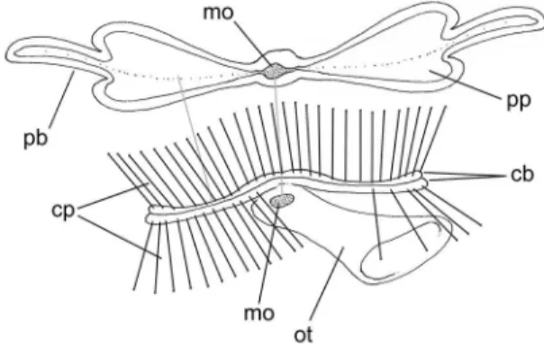
Central nervous system (Figs. 53, 63, 64): Pair of ce‑ rebral ganglia located just dorsal to mouth (between bases of captacula and oral tube); each cerebral gan‑ glion spherical, with approximate diameter equivalent to 1/15 that of anterior shell aperture; both ganglia located very close to one another and to median line; cerebral commissure very short, narrow, located in middle region of ganglia median surface; two pairs of large anterior nerves. Pair of pedal ganglia of equiva‑ lent size than cerebral ganglia, located on foot base at same level of cerebral ganglia, on opposite side of mouth; staying attached to ventral surface of pedal musculature; form of each ganglion roughly spheri‑ cal, except for low expansions correspondent to each main nerve, being two anterior pairs and another pair postero‑lateral. Pedal commissure short, very nar‑ row, located in anterior region of medial surface of both ganglia. Pair of statocysts located just posterior to pedal ganglia, closer to median line; volume of each statocyst about 1/5 of each pedal ganglion; in‑ ternally several statoconia. Visceral ganglia not seen. Pair of buccal ganglia very small (each ganglion about 1/100 of odontophore), located in ventral wall con‑ necting odontophore to esophagus; distance between both buccal ganglia equivalent to 1/4 of odontophore width; a pair of anterior nerves and two pairs of pos‑ terior nerves.
Measurements (respectively antero-posterior length, dorso-ventral maximal lateral inflation in mm; finally tax of increase in mm/mm of length): MZUSP 32977: #1: 80.2 by 11.4, 0.142; #2: 74.5 by 11.0, 0.147; MZUSP 47189: 52.3 by 8.1, 0.154.
Distribution: From North Carolina, USA, to Rio Grande do Sul, Brazil.
Habitat: Sandy bottoms, from 40 to 1980 m depth; living specimens from 180 to 270 m depth (Steiner & Kabat, 2004; Caetano et al., 2006). Content of buccal tube several foranmifer testa and rarely small mollusks.
Material examined: UNITES STATES. Florida. Dry Tortugas, Sand Key, 174 m depth, USNM 314457 (holotype of Dentalium floridense; Eolis sta. 301,
1915). BRAZIL. Rio de Janeiro. Off Cabo Frio, 350‑400 m depth, MZUSP 32977, 21 specimens (o.t.; C. Cunha col iv/2001). São Paulo. Off Ubatuba, 320 m depth, MZUSP 47189, 17 specimens (R.V.W. Besnard sta. 5365; 07/xii/1988).
Genus Paradentalium Cotton & Godfrey, 1933 (type: Dentalium intercalatum Gould, 1859, od)
Paradentalium disparile (d’orbigny, 1853) (Figs. 10‑15, 65‑74)
Dentalium disparile d’Orbigny, 1853: 202 (pl. 25, figs. 14‑17); Scarabino, 1973: 201 (fig. 8); Rios, 1970: 144; Matthews & Rios, 1974: 47; Steiner & Kabat, 2004: 584, 712.
Dentalium (Antalis) disparile: Pilsbry & Sharp, 1897: 56 (pl. 14, figs. 16‑21); Henderson, 1920: 47 (pl. 6, figs. 4‑8); Maury, 1922: 35; Haas, 1953: 203; Morretes, 1949: 53; Turner, 1955: 311; Penna, 1972: 230; 1974: 111; Ab‑ bott, 1974: 385 (fig. 4505); Almeida & Olivei‑ ra, 2000: 48, 54 (fig. 2).
Dentalium oerstedii: Jaeckel, 1927: 130 (non Mörch, 1860).
Dentalium (Dentale) disparile: Rios, 1970: 144.
Dentalium (Heteroschimoides) callinthrix: Penna, 1972: 231; Almeida & Oliveira, 2000: 49 (fig. 3) (non Dall, 1889).
Antalis disparile: Scarabino, 1985: 198 (pl. 72, fig. 1012); 1994: 306 (pl. 106, fig. 1500); Díaz & Puyana, 1994: 256 (pl. 71, fig. 1040); Gar‑ cía‑Valencia & Díaz, 2000: 79; Steiner & Ka‑ bat, 2001: 440; 2004: 584.
Dentalium dispareli: Almeida & Oliveira, 2000: 48 (fig. 2) (err.).
Paradentalium disparile: Caetano et al., 2006: 11 (figs. 11‑15); 2007: 797 (figs. 4‑6); Caetano, 2007: 38‑43 (figs. 19‑25).
Types: BMNH 1854.10.4.465, 3 syntypes.
Type locality: Martinique. distinctive description
FIGuReS 1‑15: Shell, radular and anatomical aspects of Dentaliidae: 1‑9) Coccodentalium caduum;1) Shell, whole right view, MZUSP 32977, length = 74.0 mm; 2) Detail of anterior aperture, profile, right view; 3) Detail of sculpture of middle region. 4) Anterior aperture, anterior view, animal still inside; 5) detail of posterior aperture, dorsal view, scale = 2 mm; 6) Radula in situ, anterior view, scale = 1 mm; 7) Radula removed from odontophore, ventral view, scale = 1 mm; 8‑9) Radulae of 2 specimens in SEM, scale = 200 µm;
10‑15) Paradentalium disparile;10) Shell, whole left view, MZUSP 25589#1, length = 35.1 mm. 11) Same, detail of posterior aperture;
spaces smooth and somewhat irregular. Posterior pipe normally present. Posterior slit as in preceding species.
Main muscle system (Figs. 65, 66): Very similar in fea‑ tures to those of preceding species, except in being weakly thinner and narrower, and by pallial muscles in anterior region thinner.
Foot (Figs. 13, 68, 69): Character as described for pre‑ ceding species, except in being somewhat narrower.
Mantle (Figs. 65-68): General features similar to those of preceding species. Main differences following. Ante‑ rior edge thick, feebly thicker than shell wall, length of thickness equivalent to 5% of total shell length; unclear separation in pair of edge folds. Pallial gland (Fig. 67: pl) a narrow transverse band located at short distance from, and weakly narrower than mantle anterior edge. Poste‑ rior mantle flap with narrower slit (Fig. 66).
Pallial cavity (Figs. 67, 68): Characters as described for preceding species. About 7‑8 transverse folds also present in basal level of foot (Fig. 67: pf ). Glandular area located just posterior to folds absent, presenting another located closer to mantle edge (Fig. 67: pl).
Visceral mass (Figs. 67, 68): Characters similar to those of preceding species, except in being somewhat narrower and with stomach proportionally shorter. Cruciform‑like musculature of integument in pos‑ tero‑ventral region of foot absent.
Circulatory and excretory systems (Fig. 67): Pericardium and kidney with similar characters of previous species.
Digestive system (Fig. 68): Characters similar to F. carduus,
except for the following features: Quantity of captacula apparently smaller (Fig. 13) but with similar remaining attributes; Oral tube edge straight; Odontophore mus‑ cles (Figs. 69‑72): mj, pair of thin protractor muscles, slightly thicker; m3, shorter, about half of odontophore width (Fig. 71); m7, pair narrower and placed along me‑ dian line (Fig. 69). Odontophore cartilages (oc) some‑ what rectangular (with rounded vertices), about twice long as tall. Radula (Figs. 14, 15) with about 15 rows of radular teeth along its length. Radular teeth: rachidian (central) tooth with slightly more rounded cutting edge. Esophagus with glandular inner surface. Pair of lateral tooth with more irregular distal‑medial tip, barely form‑ ing hooks. Stomach with about 1/9 of total shell length; marginal digestive diverticles in lateral edges; about 15 pairs of similar‑sized diverticles. Intestine performing a complex set of loops as shown in Fig. 68.
Genital system: Characters somewhat similar to those of preceding species, except for more elongated and narrower gonad fashion (Fig. 67).
Central nervous system (Figs. 68): Main characters similar to those of Coccodentalium carduus, with the following remarkable features: Pair of cerebral ganglia more elongated laterally, possessing a single anterior nerve each (Fig. 74); Pair of pedal ganglia located in ventral surface of dorsal wall of foot base (Fig. 73); Each statocyst with volume approximately 1/3 of each pedal ganglion; Pair of buccal ganglia located more laterally in ventral wall connecting odontophore to esophagus (Figs. 70: bg); distance between both buc‑ cal ganglia equivalent to 3/4 of odontophore width.
Measurements (respectively antero-posterior length, dor-so-ventral maximal lateral inflation in mm; finally tax of increase in mm/mm of length): MZUSP 25589: #1: 34.1 by 3.0, 0.088; #2: 30.6 by 2.8, 0.092.
Distribution: Florida, USA to Santa Catarina, Brazil.
Habitat: Sandy bottoms, from intertidal to 103 m depth, living species from 5 to 50 m depth. Content of buccal tube several foranmifer testa.
Material examined: BRAZIL. Rio de Janeiro. Angra dos Reis (iii/1969), MZUSP 25586, 19 specimens (sta. 345), MZUSP 25589, 2 specimens (sta. 334), MZUSP 25590, 1 specimen (sta. 341); Ilha Grande (R.V. Emilia), MZUSP 18135, 1 specimen (sta. 38, 12/xii/1965), 13 m depth, MZUSP 18137, 2 speci‑ mens (sta. 46, 10/xii/1965), MZUSP 25573, 1 speci‑ men (sta. 30, 13/xii/1965). São Paulo. Ubatuba (Seção Bentos col.), Boqueirão, MZUSP 25645, 62 speci‑ mens (9/v/1962), Praia Sul, 16 m depth, MZUSP 25639, 2 specimens (23/ii/1962), Enseada do Fla‑ mengo, MZUSP 25627, 4 specimens (17/i/1962).
Family Gadilidae
Genus Polyschides Pilsbry & Sharp, 1898 (type: Siphodentalium tetraschistum
Watson, 1879, od)
Polyschides noronhensis new species (Figs. 16‑22, 75‑86)
FIGuReS 16‑29: Shell and radular aspects of Gadilidae: 16‑22) Polyschides noronhensis;16) Holotype shell, left view, length = 8.6 mm;
17) same, paratype MZUSP 32011, length = 7.8 mm; 18) Paratype MZUSP 32011#1, left view, detail of anterior aperture in profile with some captacula, scale = 0.5 mm; 19‑20) Paratype MZUSP 46736, detail of posterior aperture in profile, right and left views respectively, scale = 0.25 mm; 21‑22) Radulae of 2 specimens in SEM, scale = 50 µm; 23‑29) Gadila braziliensis MZUSP 46736; 23) #3, right view, length = 7.5 mm; 24) #2, dorsal view, length = 7.9 mm; 25) #4, young specimen, length = 3.4 mm; 26) Radula in SEM, scale = 50 µm;
distinctive description
Shell (Figs. 16-20): Size about 8 mm, walls thin, color white, translucent; weakly curved; middle and anteri‑ or thirds almost straight, posterior third more curved (Figs. 16, 17). Softly widening (tax of increase about 0.125 mm/mm of length), general form almost cy‑ lindrical. Maximum width about half of shell length, located between middle and anterior thirds. Outer surface smooth, glossy. Anterior aperture oblique (ventral region slightly more posterior) (Figs. 16‑18); weakly flattened dorso‑ventrally; about 75% narrower than broader region of shell. Posterior aperture about half of anterior aperture size, rounded, weakly turned dorsally, normally pale‑brown pigmented; four equi‑ distant projections (Figs. 19, 20); dorsal and ventral projections about 50% wider than lateral projections; each projection with about 3% of total shell length.
Main muscle system (Figs. 75, 76): Pair of longitudi‑ nal muscles originating on ventral and lateral sides of base of apical flap, occupying about 3/4 of this base. Both longitudinal muscles running towards anterior, restricted to ventral side, touching one another along median line. Middle region of both muscles narrow‑ er than both ends (Fig. 76: lm). Both longitudinal muscles trifurcating in middle level of animal’s body; external branches thin, splaying along anterior pallial wall; middle branches splaying in foot base as longi‑ tudinal layer of foot retractors (Fig. 79: mv); internal branches as pedal retractor muscles (Fig. 79: mf ). Each pedal retractor muscle running anteriorly thought haemocoel, flanking some intestinal loops (Fig. 80), connecting with its pair at middle level of haemocoel; after this, running as single bundle up to retracted pedal distal tip.
Foot (Figs. 78-80): Mostly hollow, looking like a re‑ tractile gastropod proboscis, with pair of above de‑ scribed pedal retractor muscles inserted in distal tip. Foot capable of enfolding along itself at about half its length (Figs. 79, 80). Foot with about 1/4 of animal’s volume and approximately 1/3 of total shell length in retractile condition; width about half of adjacent shell width. Foot tip with central foramen corresponding to its enfolding portion. Transition between foot and insertion of pedal retractor muscles unclear; solid por‑ tion of foot about 1/6 its length; from this up to re‑ gion of splaying longitudinal muscles of haemocoel (Fig. 79: mv) simple, thin walls.
Mantle (Figs. 75-78): Anterior edge thick, a pair of folds; outer fold relatively narrow, thickness equiva‑
lent to that of shell wall; inner fold similarly thick‑ ened and 5‑6 times longer than inner fold, positioned inwards (Figs. 75, 76, 78: mb). Posterior mantle flap tall, about 1/8 of shell length (Fig. 77). Low trans‑ verse septum present in base of posterior mantle edge (Fig. 78: ms).
Pallial cavity (Fig. 78): About 8‑9 transverse folds present at posterior level of foot‑visceral mass (Fig. 78: vm), uniform, very narrow (width equiva‑ lent adjacent to mantle thickness), spaced by equiva‑ lent width (of folds); only present in ventral half. Pallial gland with oblique folds (Fig. 78: pl) white, lo‑ cated approximately in middle region of mantle cav‑ ity, occupying about 1/6 of shell length; ventral folds slightly longer than dorsal folds and more anteriorly located; anterior edge of folds rounded and slightly taller, posterior end unclear. Transverse fold located close to mantle edge, with approximately same height of outer fold of mantle edge (Fig. 78: pf ).
Visceral mass (Figs. 78-81): Middle region with about 1/10 of shell length containing renal structures. Pos‑ terior region of almost half of shell length containing practically only gonad (Figs. 78: go).
Circulatory and excretory systems (Fig. 78): Pericardium totally absent. Kidney solid, antero‑posteriorly short, but with about same shell width.
gradually narrowing; inner surface simple, smooth. No differentiated stomach, neither clear separation between esophagus and intestine. Intestine very nar‑ row (about 1/8 odontophore thickness); perform‑ ing 3‑4 simple loops (Figs. 80, 81) of similar width, partially compressed between pedal retractor muscles (Fig. 80). Anus a relative wide papilla (Fig. 78: an).
Genital system (Figs. 78, 81): Gonad somewhat trian‑ gular, with almost half of shell length; anterior region as wide as kidney, gradually narrowing towards poste‑ rior, ending at base of posterior mantle border. Gonad constituted for relatively long digital acini forming transverse folds; each acinus with almost half of go‑ nad width and bifid distal end; gonadal folds highly protruding inside posterior half of pallial cavity, sur‑ rounding almost completely adjacent animal’s diam‑ eter. Gonoduct running towards anterior and right, crossing rectum (Fig. 81).
Central nervous system (Figs. 79-81, 86): Located sur‑ rounding esophageal connection to mouth, edging base of oral tube as a nerve ring. Pair of cerebral gan‑ glia located just dorsal to mouth; each cerebral gan‑ glion spherical, with approximate diameter equiva‑ lent to 1/10 of that of anterior shell aperture. Pair of pedal ganglia of equivalent size to cerebral ganglia, located in opposite side of cerebral ganglia, ventrally to mouth; form of each ganglion roughly spherical. Pedal commissure with about half each ganglion length (Fig. 86). Cerebro‑pedal connectives of simi‑ lar length, lying on esophageal wall. Pair of statocysts located just posterior to pedal ganglia; volume of each statocyst about 1/3 of each pedal ganglion. Pair of buccal ganglia absent.
Measurements (respectively antero-posterior length, dor-so-ventral maximal lateral inflation in mm; finally tax of increase in mm/mm of length): MZUSP 32011: #1: 8.0 by 1.1, 0.137; #2: 7.4 by 0.9, 0.122.
Distribution: Endemic to Fernando de Noronha Ar‑ chipelago, Brazil.
Habitat: Sandy bottoms, about 6 m depth. Content of buccal tube several foranmifer testa.
Material examined: Types.
Discussion: Polyschides noronhensis possesses the shell character arrangement typical of the genus, as the maximum diameter located in the anterior third, and wide posterior aperture bearing four lobes. The
new species differs from the other co‑generic spe‑ cies from the region (Caetano et al., 2006; Caetano, 2007) mainly by the shape of the shell. P. noronhensis
is wider than P. tetraschistus (Watson, 1879), and than
P. xavante Caetano & Absalão, 2005, from which it also differs by a straighter shape. the other two spe‑ cies have a more arched shell, P. noronhensis still dif‑ fers from P. portoricensis (Henderson, 1920) and from
P. tetrodon (Pilsbry & Sharp, 1897) in being narrower, particularly close to the anterior aperture, and in hav‑ ing a more uniform width along its length; P. noron-hensis has the wider portion of the shell closer to the anterior aperture than the other two species, as it is in the anterior third in P. portoricensis and almost in the middle in P. tetrodon.
Genus Gadila Gray, 1847
(type: Dentalium gadus Montagu, 1803, od)
Gadila braziliensis (henderson, 1920) (Figs. 23‑29, 87‑94)
Cadulus (Platyschides) braziliensis Henderson, 1920: 124 (pl. 19, fig. 16); Turner, 1955: 316; Scarabino, 1970: 41 (pl. 1, fig. 1); 1973, 198‑199 (fig. 6); 1975: 182 (pl. 58, fig. 887); 1985: 201 (pl. 73, fig. 1032); 1994: 309 (pl. 107, fig. 1520); Penna‑Neme, 1974: 115; Steiner & Kabat, 2004: 570, 715.
Cadulus (Platyschides) brasiliensis: Rios, 1966: 7; 1970: 143; 1975: 182 (pl. 58, fig. 887) (err).
Platyschides braziliensis: Scarabino, 1980: 113 (pl. 1, fig. 9).
Gadila braziliensis: Steiner & Kabat, 2001: 445; 2004: 570; Caetano, 2007: 150‑153 (figs. 141‑142).
Cadulus braziliensis: Absalão et al., 2005: 175‑178 (fig. 2); 2006: 67.
Type: holotype and paratype USNM 96113.
Type locality: BRAZIL, off Rio de Janeiro, 23°08’S 41°34’W, 108 m depth (USBF).
distinctive description
half of anterior aperture, circular. Other details in above given references.
Main muscle system (Fig. 88): Components similar to those described for Polyschides noronhensis.
Foot (Figs. 87, 88): Mostly similar in features to that of preceding species. Tip clearly rounded, stubby.
Mantle (Figs. 87, 88): General features as described for Polyschides noronhensis.
Pallial cavity (Fig. 87): Characters similar to those described for Polyschides noronhensis, except in lack‑ ing transverse folds in posterior level of foot‑visceral mass, pallial gland, and transverse fold located close to mantle edge.
Visceral mass (Figs. 88): Similar organization of pre‑ ceding species.
Digestive system (Figs. 88-92): Mostly similar to Poly-schides noronhensis characters, distinctive features fol‑ lowing: Odontophore muscles (Figs. 28, 89‑92): mj, inconspicuous; m4, pair slightly narrower (Figs. 90, 91); m6, narrower 1/8 of each odontophore cartilages thickness (Figs. 28, 91, 92). Odontophore cartilages (oc) with ventral edge with wide furrow in middle level (Fig. 92). Radular teeth (Figs. 26, 27): pair of lateral teeth with three sub‑terminal cusps in median third, being that more lateral approximately twice larger than remaining (Fig. 26). Intestine slightly broader; performing 3‑4 simple loops (Fig. 88: in). Anus a relative wide papilla (Fig. 87: an).
Genital system (Figs. 87): Features similar to those of preceding species.
Central nervous system (Figs. 88, 94, 95): Features similar to those described for Polyschides noronhensis.
Nerve connected to each cerebral ganglion wider in vicinity of these ganglia, forming almost a ganglion.
Measurements (respectively antero-posterior length, dor-so-ventral maximal lateral inflation in mm; finally tax of increase in mm/mm of length): MZUSP 46736: #1: 7.8 by 1.3, 0.166; #2: 7.4 by 1.1, 0.149.
Distribution: Rio de Janeiro, Brazil, to La Plata river, Argentina.
Habitat: Sandy bottoms, from 23 to 220 m depth. Content of buccal tube several foranmifer testa.
Material examined: Types. BRAZIL (R.V.W. Besnard). Rio de Janeiro. Angra dos Reis; 23°13’S 44°24’W, 36 m depth, MZUSP 18761, 2 specimens (R.V.W. Besnard sta. 340, 19/iii/1969). São Paulo. Ubatuba; 23°47’S 44°58’W, 47 m depth, MZUSP 46736, 11 specimens (P.I. sta. 4854, 17/xii/1985).
Class Bivalvia Family nuculidae Genus Ennucula Iredale, 1931 (type: Nucula obliqua Lamarck, 1819, od)
Ennucula puelcha (d’orbigny, 1842) (Figs. 30‑38, 95‑105)
Nucula puelcha d’Orbigny, 1842: 162; 1846: 624 (pl. 84, figs. 24‑26); Schenck, 1939: 30; Carcelles, 1944: 268; Ihering, 1907: 371; Castellanos, 1967: 189 (pl. 14, fig. 5); Figueiras, 1976: 73; Roux et al., 1995: 295, 301‑303; Bremec & Roux, 1997: 157; Paiva, 2001: 428; Soares‑Gomes & Pires‑Vanin, 2003: 721; Acha et al., 2004: 93; Absalão et al., 2006: 67; Gib‑ erto et al., 2006: 5; Vinuesa & Varisco, 2007: 29.
Nucula puelchana d’Orbigny, 1842 (pl. 84, fig. 24‑26); Borchert, 1901: 32 (pl. 3) [error].
Nucula uruguayensis E.A. Smith, 1880: 320‑321 [loc: 36°47’S, 55°17’W, 51.2 m depth, off Rio de la Plata mouth].
Nucula savatieri Mabille & Rochebrune in Rochebrune & Mabille, 1889: 112 (pl. 8, fig. 2) [loc: Canal du Beagle; Baie Orange, Tierra del Fuego].
Ennucula puelcha: Dell, 1964: 141; Camacho, 1966: 53 (pl. 8, fig. 6); Scarabino, 2003: 229; Rosenberg, 2005; Clavijo et al., 2005: 391.
Nucula (Ennucula) puelcha: Rios, 1970: 146 (pl. 50); 1975: 188 (pl. 60, fig. 918); Figueiras & Broggi, 1973: 203.
Nucula (Leionucula) puelcha: Abbott, 1974: 411; Del Rio, 1991: 27 (pl. 27, fig. 1); Rios, 1985: 203 (pl. 74, fig. 1040); 1994: 225 (pl. 78, fig. 1111).
Nucula (Nucula) semiornata: Del Rio, 1992: 12 (pl. 1, fig. 11).
Leionucula puelcha: Del Rio, 1998: 14, 48 (pl. 4, figs. 10, 11; pl. 16, fig. 5; pl. 24, fig. 10).
Types: lectotype BMNH 1854.12.4.774/1 (single valve; designation Aguirre, 1994). Paralectotypes BMNH 1854.12.4.774/2‑3 (possibly of another spe‑ cies, see below).
Redescription
Shell (Figs. 30-38): Size about 15 mm, color pale to dark brown. Height about 80% of length; width about 60% of length. Periostracum glossy, smooth, relatively thick. Sculpture lacking except for growth
lines (Figs. 32, 38). Umbos tall, rounded, located close to each other (Fig. 31) approximate angle 110°, located about 20% of total length from posterior mar‑ gin, extending about 10% of total height dorsal from hinge (Figs. 30, 31). Resilium internal, conic (wider region posterior), located approximately between pos‑
FIGuReS 30‑38: Shell and anatomical aspects of Ennucula puelcha (MZUSP 19101; length = 13.0 mm): 30) Right valve, inner view;
terior and middle thirds of hinge (Figs. 33, 34). Liga‑ ment length about 10% of shell length and height about 5% of shell height. Inner surface glossy, silver‑ nacred, including hinge. Hinge with approximately 18 teeth anterior and 8 posterior to ligament; teeth increasing height from umbonal region towards both (anterior and posterior) ends (Figs. 35, 36); anterior set of teeth becoming dorso‑ventrally wider towards anterior (Figs. 30, 31, 33‑35); posterior set of teeth of similar width. Scar of anterior adductor muscle el‑ liptical, about twice tall than long, located in middle level of anterior edge, about 15% of shell length away from anterior margin; occupying about 4% of inner surface of valve (Figs. 30, 31. 36). Scar of posterior adductor muscle with similar characters than anterior adductor scar, located in opposed side and with about 75% anterior scar size. Pallial line simple, located edg‑ ing ventral edge a distance equivalent to 17% of shell height.
Main muscle system (Figs. 37, 95, 99): Anterior ad‑ ductor muscle elliptical in section; about twice high than wide; occupying approximately 4% of valve; lo‑ cated between middle and ventral thirds of animal’s height, and about 15% of shell length posterior to anterior edge; anterior region about half of poste‑ rior region (Fig. 35). Posterior adductor muscle ap‑ proximately 75% anterior adductor muscle size, and positioned slightly in opposite region; remaining characters, including horizontal level, similar; clearly divided into two equally sized portions (quick and slow components) along dorso‑ventral axis (Figs. 95, 98‑100: pa). Pair of auxiliary protractor muscle of foot (Fig. 99: ap) very narrow and long; each one originating in dorso‑posterior region of anterior ad‑ ductor muscle in area approximately 1/150 of that of this adductor; running posteriorly and ventrally between integument and anterior foot musculature, splaying superficially along anterior foot base. Pair of foot protractor muscle (Fig. 99: fp), relatively thick and long; each one originating dorsally to anterior adductor muscle in area equivalent to 1/30 of this ad‑ ductor; running towards posterior and slightly ventral a distance equivalent to half shell length, gradually broadening; inserting along lateral walls of visceral sac and middle pedal base. Pair of anterior pedal retractor muscle, very broad and thick (Fig. 99: fa); each one originating just dorsal and slightly posterior to origin of anterior protractor muscle in area equivalent to half of anterior adductor muscle; running towards ventral and slightly posterior, close to median plane, broad‑ ening weakly along their length; inserting along ante‑ rior foot base, fulfilling almost entire anterior volume
of visceral sac. Pair of middle pedal retractor muscle, broad and thick (Figs. 95, 98, 99: rm); each one originating in umbonal cavity, between posterior and middle thirds of distance between umbo and anterior shell margin, close to dorsal medial line, in area equiv‑ alent to 1/5 that of anterior adductor muscle; running towards ventral and slightly posterior, weakly curved (concavity anterior), widening gradually, positioning closer to medial plane; insertion in middle‑posterior region of foot base, fulfilling most of middle‑ventral volume of visceral sac. Pair of auxiliary middle pedal retractor muscle, narrow and long (Fig. 99: fr); each one originating just ventral to origin of middle pedal retractor muscle; running almost vertically towards ventral (slightly posteriorly), somewhat away from middle retractor muscle; inserting in middle region of foot base, flanked externally by middle pedal retractor muscles. Pair of posterior pedal retractor muscles, very broad and thick (Figs. 95, 98, 99: fm); each one origi‑ nating at some distance dorsal and slightly anterior to posterior adductor muscle, in area slightly larger than half that of anterior adductor muscle, antero‑posteri‑ orly long (about three times longer than wide); run‑ ning close to median plane towards ventral, almost vertically (slightly anterior); inserting along posterior pedal base.
Foot (Figs. 45, 95, 101): Laterally flattened, about 1/3 of shell volume. Distal region with expanded edges, umbrella‑like, extending about twice foot width be‑ yond lateral edges. Posterior vertical posterior flap (Fig. 101: ff ) rounded, extending about 1/6 of entire foot length towards posterior, occupying ventral half of posterior foot surface, along medial plane.
Pallial cavity (Figs. 37, 95, 101): Occupying about half of inner shell volume. Palps with about 1/3 of valves size; main (broader) region oval, slightly lon‑ ger than half shell length and height (Figs. 101: pp); inner surface entirely covered by narrow transverse folds; each fold very narrow and close to each other, ventral end rounded, dorsal end connected with its pair of other hemipalp; inner palp folds diminishing in both ends, posterior folds situated slightly more separated from each other and in oblique, curved way (Figs. 101). Palp inner folds end before palp ventral edges, producing smooth, uniform margin. Proboscis of palps about as long as main portion of palps, and about 1/6 its width; located as posterior continuation of furrow between both hemipalps; inner surface as wide groove, smooth; proboscis tapering gradually, tip slightly rounded; edges undulating. Two pairs of small projections located only in inner hemipalps, by side and internally from proboscis; similar in characters to proboscis but about 1/10 its length and half its width; more ventral projection weakly smaller than dorsal projection (Fig. 101: pj). Palps inner folds reach‑ ing region close to mouth. Pair of small palp mus‑ cles located in postero‑dorsal corner between both hemipalps (Figs. 95, 99: mu), running immersed in adjacent integument up to posterior region carving base of both posterior pedal retractor muscles. Gills bipectinate and proportionally small (about 1/15 of shell volume), about 6 times longer than tall; locat‑ ed obliquely from pericardial area to region ventral to posterior adductor muscle; anterior end rounded, gradually narrowing up to pointed posterior end. Gills posterior end supported by pair of suspensory stalks (Figs. 98, 100: gs), as thick membranes connected to ventral surface of posterior adductor muscle, close to median line. Suspensory membrane becoming short‑ er and wider towards anterior, supporting entire gills (Figs. 97, 98); in anterior region bearing some muscu‑ lar fibers, and thin hypobranchial gland in both sides (Fig. 97: hg). Gills periphery connected to mantle and to visceral sac by cilia. Gill filaments symmetrical in both sides (Fig. 97), edges somewhat thicker, rounded ventrally and bluntly angled dorsally; afferent and ef‑ ferent gill vessels (Fig. 97: af, cv) narrow, located in opposed sides of central rod, efferent vessel slightly broader than afferent vessel. Supra‑branchial chamber about 1/6 of infra‑branchial chamber.
Visceral mass (Figs. 35, 37, 98, 99, 102): With about half shell volume, placed as dorsal continuation of foot; strongly compressed by pedal musculature (Fig. 99). Stomach as central structure, positioned vertically from umbonal cavity up to region close to
ventral foot surface. Digestive diverticula pale green, located surrounding dorsal half of stomach, occupy‑ ing about 1/4 of inner visceral volume. Gonad fulfill‑ ing remaining regions, mainly umbonal cavities, color cream to white. Digestive tubes mainly positioned at right from stomach, looping through digestive diver‑ ticula and gonad. Pericardium occupying about 1/5 of visceral volume, located posteriorly to umbos, flank‑ ing posterior pedal retractor muscles (Fig. 98); about twice wider than long. Transverse muscles well devel‑ oped in region surrounding stomach (Fig. 102: tm), crossing through gonad, connecting both sides of pedal base integument; generally four anterior and five posterior to stomach.
Circulatory and excretory systems (Fig. 98): Located compressed between pair of posterior pedal retractor muscles and middle pedal retractor muscles. Pair of auricles elongated, connected to efferent gill vessel in ventral region of gills anterior end; crossing per‑ pendicularly towards medial a distance equivalent to 1/4 shell width. Ventricle relatively small, located in middle region of pericardium, on median line, sur‑ rounding intestinal portion crossing pericardium. Kidneys solid, small (about 1/8 of pericardial vol‑ ume), pale brown; located in both sides of pericar‑ dium, covering anterior region of gill.
between posterior series of transverse muscles and middle pedal retractor muscles (Figs. 99, 102); after this, performing complex series of loops (Fig. 99) at right from stomach; finally crossing from anterior to posterior in dorsal region of visceral mass along me‑ dial line, crossing through origins of middle and pos‑ terior retractor pedal muscles (Fig. 99) and pericar‑ dium. Entire intestine narrow (about 3/4 esophageal width), uniform width along its length. Rectum cross‑ ing along median line attached to dorsal and posterior surface of posterior adductor muscle. Anus simple, sessile (Figs. 98, 100).
Genital system: Gonad above described. No gonoducts detected.
Central nervous system (Fig. 99): Pair of cerebral gan‑ glia located close to origins of foot protractor muscle (Fig. 99; ce); each one rounded, size equivalent to half esophageal transverse section. Cerebral commissure with about half shell maximum width. Pair of pedal ganglia (Figs. 99, 102: pg) 6‑7 times larger than cere‑ bral ganglia, about three times longer than wide; lo‑ cated close to median plane, flanking posterior surface of base of anterior pedal retractor muscle; each pedal ganglion with single main connective in both ends. Pair of visceral ganglia of similar size than cerebral ganglia (Figs. 98, 99: vg), located in space between pair of posterior pedal retractor muscle and ventro‑ posterior region of posterior adductor muscle. Cere‑ bro‑visceral connectives crossing gonad close to lateral regions of integument of visceral sac.
Measurements (respectively length, height and maximum inflation in mm): MZUSP 19101 #1: 13.0 by 10.0 by 7.6.
Distribution: From south Bahia, Brazil, to north Ar‑ gentina (Pacific records contested, see below).
Habitat: Muddy bottoms, from infratidal to ~ 100 m depth.
Material examined: BRAZIL. Bahia. Alcobaça; Parcel de Paredes, 2‑3 m depth, MZUSP 46333, 7 valves (Souza & Gonçalves col., 2005). Espírito Santo. Guarapari; MZUSP 77268, 6 specimens (Coltro col., 2006). Rio de Janeiro. off Campos, 22°34’S 40°29’W, 213 m depth, MZUSP 18793, 1 specimen (R.V.W. Besnard sta. 9, 11/ii/1969, laminarias); Angra dos Reis; MZUSP 56232, 1 shell (IOUSP sta. 327, iii/1969); Ilha Grande Bay (R.V. Emilia); MZUSP 18287, 1 specimen (sta. 5B, 1968) MZUSP 18284,
45 shells (sta. 132, 12/v/1966), 25.5 m depth, MZUSP 18289, 2 shells (sta. 7B, v/1965), 17.5 m depth, MZUSP 18276, 1 shell (sta. 31, 13/xii/1965), MZUSP 18281, 1 shell (sta. 65, 18/v/1966), MZUSP 18277, 4 specimens, (sta. 40, 12/xii/1965), MZUSP 18288, 1 specimen (sta. 6B), MZUSP 18278, 1 specimen (sta. 43, 11/xii/1965), MZUSP 18283, 1 specimen (sta. 118, 2/vii/1966), MZUSP 18286, 1 specimen (sta. 137, 4/vii/1966), MZUSP 18282, 1 specimen (sta. 104, 1/vii/1966), MZUSP 18280, 2 specimens (sta. 99, 1/vii/1966), MZUSP 18285, 6 specimens (sta. 134, 12/v/1966), 50‑60 m depth, MZUSP 38458, 1 specimen (o.t., Magenta leg. vii/2003), (R.V.W. Besnard) 22 m depth, MZUSP 23658, 1 specimen (sta. 331, 21/iii/1969), MZUSP 23660, 1 specimen (sta. 341, 14/iii/1969), 30 m depth, MZUSP 23659 m, 2 specimens (sta. 339, 19/iii/1969). São Paulo. Ubatuba (Projeto Integra‑ do; R.V. Veliger II); 23°37’24”S 45°03’48”W, 35 m depth, MZUSP 86366, 25 valves (sta. 2, 26/x/1985), 23°50’S 45°10’W, MZUSP 83141, 1 shell (sta. 22, 16/iv/1986); off Queimada Grande Island, 40‑50 m depth (o.t., Coltro leg.), MZUSP 65891, 2 specimens (vii/2002), MZUSP 65892, 2 specimens (vii/2000), 50‑60 m depth (Magenta leg., viii/2002), MZUSP 35717, 20 specimens, MZUSP 35753, 12 specimens. Paraná. off Paranaguá, MZUSP 35371, 8 specimens (o.t., Magenta leg, xi/1999) Santa Catarina. Bom‑ binhas; Zimbros Bay, 5‑8 m depth, MZUSP 32079, 1 specimen, MZUSP 32080, 1 specimen (in starfish stomach, o.t., Tarasconi leg. vii/1993); off Gaivotas, 29°33’S 48°57’W, 91 m depth, MZUSP 18787, 13 specimens (R.V.W. Besnard sta. 1706, 6/iv/1972). Rio Grande do Sul (R.V.W. Besnard). Off Traman‑ dai, 30°12’S 50°11’W, 90 m depth, MZUSP 18788, 37 specimens (sta. 1723, 10/iv/1972); off Mostardas, 30°50’S 50°06’W, 79 m depth, MZUSP 18790, 10 specimens (sta. 1860, 6/viii/1972); Off Rio Grande, 32°48’S 50°27’W, 197 m depth, MZUSP 18789, 2 specimens (sta. 1758, 22/iv/1972). URUGUAY. Off Maldonado, 35°00’S 54°50’W, 23 m depth, MZUSP 19101, 62 specimens (GEDIP‑RS sta. 1866; R.V.W. Besnard col., 11/viii/1972). Rocha (R.V.W. Bes‑ nard). Off Punta del Diablo, 34°05’S 53°30’W, 20 m depth, MZUSP 18792, 2 specimens (sta. 1877, 14/ viii/1972); off La Paloma, 35°51’S 53°06’W, 206 m depth, MZUSP 18791, 1 specimen (sta. 1870, 12/viii/1972).
discussion
America [Bernard, 1983: 10; Villarroel & Stuardo, 1998: 134‑136 (figs. 29‑32, 72, 110‑112); Osorio & Reid, 2004: 78‑79 (fig. 2J)]. However, the shells of the Pacific samples have more pointed umbones. This shell difference, allied to a relatively wide geographic distance and the glacial separation between that re‑ gion and the south Atlantic coast of South America, are indicative of the reports from Chile and Peru that the species actually belongs to another, possible new species. This shell difference has also been pointed out in the literature (Osorio & Reid, 2004: 79). Howev‑ er, remarkably, the papers on the Pacific samples are those that changed the species from Nucula (Lamarck, 1799) to the genus Ennucula.
The type specimens of Ennucula puelcha were examined at BMNH (Figs. 104‑110). The lectotype (Figs. 104, 105) matches with the specimens exam‑ ined herein. On the other hand, the paralectotypes (Figs. 106‑110) do not; in the meantime, they are somewhat similar to Nucula semiornata d’Orbigny, 1846, of which they can possibly be the types. The paralectotypes (Figs. 106‑110) have taller umbones, the apical angle is narrower, the outer concentric sculpture is more evident (Figs. 106, 107, 110), the resilium is more projected and the hinge is narrower and possesses slightly more teeth (Figs. 108, 109). These characters fit the description of N. semiornata
and differentiates it from that of E. puelcha. With the above mentioned possibility that the Pacific and Atlantic specimens belong to separate species, some species supposedly synonymous to E. puelcha were not included in the present synonymic list. These species are: Nucula agujana Dall, 1908 (described from Aguja, Peru; 1895 m depth). N. pigafettae Dall, 1908 (described from Magellan Strait; 494 m depth); and possibly N. felipponei Marshall, 1928 (Bernard, 1983); as well as some Pacific citations of E. puelcha,
such as Villarroel & Stuardo (1998) and Osorio & Reid (2004). Accounts on the anatomy of Pacific samples identified as E. puelcha are provided by Vil‑ larroel & Stuardo (1998, figs. 29‑31, 72, 110‑112). The stomach of the Atlantic specimens has a shorter style sac, about 20% in contrast to 60% of the Pacific species, and less developed dorsal sorting area and dorsal hood. Additionally, the gills are proportionally smaller, and the papillae in mantle edge are restricted to the posterior region in Atlantic species, while papil‑ lae occur in most mantle edges in Pacific samples.
Another Ennucula has accounts written on its anatomy, E. tenuis (Montagu, 1818) (Kuznetsov et al.
1983). in such E. puelcha differs in having propor‑ tionally larger palps and respective proboscises, as well as larger adductor muscles.
Family Solemyidae Genus Solemya Lamarck, 1818 (type: Solemya mediterranea Lamarck,
1818, Sd Children, 1823)
Solemya notialis new species (Figs. 39‑46, 111‑117)
Solemya patagonica: Rios, 1975: 186 (pl. 60, fig. 912‑left) (part) (non E.A. Smith, 1885).
Solemya occidentalis: Rios, 1985: 207 (pl. 75, fig. 1058); 1994: 224 (pl. 78, fig. 1105) (non Deshayes, 1857).
Types: Holotype MZUSP 88440. Paratypes: BRA‑ ZIL. Rio de Janeiro. Cabo Frio, MZUSP 35257, 1 specimen (Paulo Gonçalves col.; v/2002); Off Mar‑ icá, 23°08’S 42°47’W, IBUFRJ 2172. 1 specimen (NOAS sta. CF‑VII‑6165); off Saquarema, 22°59’S 42°19’W, IBUFRJ 1335, 1 specimen (Geocosta Rio II sta. B3, 20/iii/1986); Angra dos Reis, 31 m depth, MZUSP 20396, 2 specimens (R.V. Emilia sta. 53; 29/vi/1966), 27 m depth, MZUSP 20397, 1 speci‑ men (R.V. Emilia sta. 54; 29/vi/1966). São Paulo. Off Ubatuba, 33.5 m depth, MZUSP 20395, 1 specimen (R.V. Emilia sta. 12; 17/xii/1965).
Type locality: BRAZIL. São Paulo. Off Ubatuba, 23°25’S 44°43’W, 33.5 m depth (R.V. Emilia sta. 12, 17/xii/1965).
description
mately equal‑sized; each one equivalent to 15% of cal‑ careous portion of each valve (Figs. 44, 46); anterior adductor scar located close to dorsal edge, just pos‑ terior to anterior quarter of valve’s length; posterior adductor scar located at short distance from posterior end (Figs. 46, 111). Pallial line simple, thick, located between middle and ventral thirds of shell height.
Main muscle system (Figs. 111, 113, 116): Anterior adductor muscle elliptical (with longer axis parallel to adjacent shell border) in section, attached in area equivalent to 15% of each valve area. Posterior adduc‑ tor muscle similar in characters to anterior muscle, section slightly more rounded (for position of adduc‑ tor muscles, see shell description). Pedal protractor
muscle of foot (Fig. 116: fp) broad and thin; origi‑ nating surrounding ventral edge of anterior adductor muscle on both sides; running immersed in ventro‑ anterior wall of visceral mass, disappearing along foot dorsal base. Pair of anterior pedal retractor muscles (Figs. 111, 116: fa) narrow and long; originating just dorsal and posterior to anterior adductor muscle, in area equivalent to 1/15 of adductor muscle, antero‑ posteriorly elongated; running ventral and posteriorly with uniform width along its length; inserting splay‑ ing along wall between visceral mass and antero‑dor‑ sal foot base. Pair of median‑anterior foot retractor muscles (Fig. 116: fb) very narrow and long, filiform; originating at small points approximately in middle of distance between umbones and anterior adductor