CASE REPORT
J of Evidence Based Med & Hlthcare, pISSN- 2349-2562, eISSN- 2349-2570/ Vol. 2/Issue 10/Mar 09, 2015 Page 1573
VARIATIONS IN THE ORIGIN OF SARTORIUS MUSCLE
D. A. V. S. Sesi1
HOW TO CITE THIS ARTICLE:
D. A. V. S. Sesi. ”Variations in the origin of Sartorius Muscle”. Journal of Evidence based Medicine and Healthcare; Volume 2, Issue 10, March 09, 2015; Page: 1573-1576.
ABSTRACT: Sartorius is the longest muscle in the body. The location and the length, vascular supply, along with innervations makes the muscle unique in its plastic surgical procedures. Variations in the origin and insertion of this muscle impact surgical applications and phylogenetic interests. Here a rare variety of variation in the origin of Sartorius muscle apart from its normal origin below anterior superior iliac spine a separate head originated from the lateral one fifth of the inguinal ligament is described.
KEYWORDS: Sartorius muscle, origin from inguinal ligament.
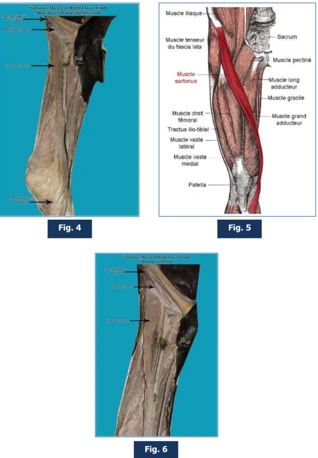
CASE REPORT: Normally this muscle originates from the inferior aspect of anterior superior iliac spine from a single head.
Though variations in the origin and insertions of this muscle have been described earlier this variation of origin as a separate head from the lateral one fifth of the inguinal ligament has not been described.
Here an anatomical variation of origin of Sartorius muscle in which the muscle originated from lateral one fifth of inguinal ligament in addition to its normal origin from inferior aspect of anterior superior iliac spine, in the department of anatomy in our institute, is described.
As the electromyography activity of the muscle increases during lateral rotation of thigh at the end of the swing phase immediately preceding heel strike (CARVALHO et al 1972): thus it has substantial involvement in climbing.
DISCUSSION: Normal anatomy: Origin - inferior to the anterior superior iliac spine. Insertion - anteromedial surface of the upper tibia in the pes anserinus. Arteial supply -femoral artery. Nerve supply- femoral nerve (sometimes from the intermediate cutaneous nerve of thigh). Actions - flexion, abduction, and lateral rotation of the hip, and flexion of the knee.
An anatomical significance of the sartorius muscle is that it forms one of the boundaries of the femoral triangle along with the inguinal ligament and the adductor longus muscle. The femoral triangle contains the femoral artery, vein and nerve.
SEPARATE OR ACCESSORY HEADS OF ORIGIN OCCASIONALLY ARISE FROM: The notch below the anterior superior spine, the iliopectineal line, the pubic bone close to the symphysis, the inguinal ligament. Macalister reports the variations in the sartorius muscle as follows:[1,2] 1.
Absent (meckel); 2. Doubled (by otto, by rosenmüller, by gantzer and by sömmerring);[3,4] 3.
Doubled, the lower of the two representatives being inserted into the femur (meckel);[3,4] 4. Or
into the tendon of the normal one (huber); 5. Splitting of the lower border into two parts (horner)[5]; 6. It has been found much broader than usual in a negro by horne[6]r; 7. Crossing the
thigh more transversley than usual (quain);[7] 8. Ending in the fascia lata; 9. With two heads, one
from the anterior superior iliac spine, and another, perfectly separate, below it, from the incisura semilunaris;[3,8] 10. With a central tendinous intersection (kelch and by hyrtl); 11. With its tendon
inserted into the medial side of the capsule of the knee, and not extending any farther;[9,10] 12.
With some of its medial fibers arising from the splitting of the iliac portion of the fascia lata, which forms its sheath, as it forms the outer wall of the triangle of scarparare presentations biceps (two-headed) sartoriussartorius bicaudatus both specimens available at dept anatomy university of iowa.[2,5,11]
CASE REPORT
J of Evidence Based Med & Hlthcare, pISSN- 2349-2562, eISSN- 2349-2570/ Vol. 2/Issue 10/Mar 09, 2015 Page 1574 PHYLOGENETICS: Phylogenetically the muscle is linked to the ilio tibialis cranialis in aves which did not appear in crocodilia though the muscle has been described in amphibians with a sciatic nerve innervation with opposing function. In primates the muscle represents the orientation in dorso lateral orientation in quadripeds. During evolution the muscle lost its spinal attachment and moved anteriorly with femoral anti version and pelvic rotation along with pelvic cephalad rotation as a part of evolution of erect posture.
SURGICAL IMPLICATIONS: In cerebral palsy and in poliomyelitis while releasing the origin of Sartorius muscle (by SOUTTER’S release) for flexion contracture of hip, the medial origin should also be released.
In plastic surgical procedures the flap (neuron vascular muscle flap) has to be mobilized along with the second variant head to completely achieve transfer.
Fig. 1
CASE REPORT
J of Evidence Based Med & Hlthcare, pISSN- 2349-2562, eISSN- 2349-2570/ Vol. 2/Issue 10/Mar 09, 2015 Page 1575 CONCLUSION: Sartorius muscle is the narrow strap muscle- longest muscle in the body. The knowledge of variation in the origin helps in complete release of the muscle in the contractures developed in cerebral palsy and in poliomyelitis to achieve correction of deformities. This knowledge also helps in planning the muscle flaps during plastic surgical procedures. As the activity of the muscle increases at the end of swing phase immediately preceding heel strike and substantially involved in and contributes to the climbing, this variation may influence the gait cycle.
REFERENCES:
1. Henle, J. (1871) Handbuch der Muskellehre des Menschen, in Handbuch der systematischen Anatomie. Verlag von Friedrich Vieweg un Sohn, Braunschweig.
2. Macalister, A. Observations on muscular anomalies in the human anatomy. Third series with a catalog of muscular variations hitherto published. Trans. Roy. Irish Acad. Sci. (1871) 25:1-130.
Fig. 4 Fig. 5
CASE REPORT
J of Evidence Based Med & Hlthcare, pISSN- 2349-2562, eISSN- 2349-2570/ Vol. 2/Issue 10/Mar 09, 2015 Page 1576 3. Bergeron, H. Muscle couturier double. Bulletins et Mem. de la Société anatomique de Paris
XLI, (1866) (1): 2.
4. Bhatnagar, B.N.S. and D. Narayan. Bicipital sartorius. J. Anat. Soc. India (1959) 8: 32-33. 5. Mori, M. Statistics on the musculature of the Japanese. Okajimas Fol. Anat. Jap. (1964) 40:
195-300.
6. Schaefer, E.A, Symington, J. and T.H. Bryce., Eds. (1923) Quain's Essentials of Human Anatomy.11th ed. Longmans, Green and Co., London.
7. Nakano, C. (1913) rare variations of the muscles observed in the dissecting room (M. sartorius and muscles of the arm). Juzenkai Zasshi (1913) 18: 10-13.
8. Brock, G.S. A two headed sartorius. J. Anat. Physiol. (1879) 13: 578.
9. Miyasaki, M., Hirohashi, A., Yoshikawa, H. and Y. Nishio. One case of the variation of the sartorius. Kurume Igakkai Zasshi. In Japanese, (1958) 21: 671-674.
10.Morita, M. One case of the variation of the M. sartorius. Kaibogaku Zasshi, In Japanese. (1947) 21: 3.
11.MARQUES, M. A.; VASCONCELLOS, H. A. & AZEVEDO, N. L. The union betwen gracilis and sartorius muscles in Leontipithecus Morphofunctional analisys. Int. J. Morphol., 24(2): 215-220, 2006.
AUTHORS:
1. D. A. V. S. Sesi
PARTICULARS OF CONTRIBUTORS:
1. Associate Professor, Department of Anatomy, Rangaraya Medical College, Kakinada, Andhra Pradesh.
NAME ADDRESS EMAIL ID OF THE CORRESPONDING AUTHOR:
Dr. D. A. V. S. Sesi,
# 25-3-5, Kommi Reddy Street, Kakinada-533001, Andhra Pradesh. E-mail: venkatorthokka@gmail.com

