INTRODUCTION
More than 30 years have passed since Vaubel’s 1975 description of an arterialized venous flap (AVF) to re-construct the dorsum of the hand and the highly cited Nakayama’s 1981 article on the creation of an exper-imental AVF model in the abdomen of the rat.1,2 Ini-tially, there was great enthusiasm with AVFs, since they allowed the transference from composite blocks of
tis-sues based exclusively on the venous system. This in turn allowed the creation of thin, pliable, and versatile flaps, that could be tailored rapidly and with minimal morbidity in the donor zone.3 However, reports of high necrosis rates and a poorly understood physiology have been deterring many surgeons of using AVFs in clinical practice.3–8
Although numerous experimental models of AVFs have been proposed, no model has gained widespread accep-tance, which hinders comparison of observations on the physiology and interventions on these flaps. Additionally, lack of a standardized model of AVF can be an obstacle for the novice in microsurgery while preparing to execute these flaps in a training environment.9 Therefore, the main aim of this work was to evaluate the flap survival area of AVFs produced with different vascular constructs in the abdomen of the rat. Secondary endpoints were
determina-Disclosure: Supported by a grant from “The Programme for Advanced Medical Education” (D.C.) sponsored by “Fundação Calouste Gulbenkian, Fundação Champalimaud, Ministério da Saúde and Fundação para a Ciência e Tecnologia, Portugal.” The Article Processing Charge was paid for by the authors.
Optimization of an Arterialized Venous
Fasciocutaneous Flap in the Abdomen of the Rat
Diogo Casal, MD*†‡Eduarda Mota-Silva, MSc§ Diogo Pais, MD, PhD† Inês Iria, MSc¶ Paula A. Videira, PhD¶ David Tanganho, MD*† Sara Alves, MSc‖
Luís Mascarenhas-Lemos, MD‖
José Martins Ferreira, BSc‖
Mário Ferraz-Oliveira, MD‖
Valentina Vassilenko, PhD§ João Goyri O’Neill, MD, PhD†
From the *Plastic and Reconstructive Surgery Department and Burn Unit, Centro Hospitalar de Lisboa Central, Lisbon, Portugal; †Anatomy Department, Nova Medical School, Lisbon, Portugal; ‡Glycoimmunology, CEDOC, NOVA Medical School, Lisbon, Portugal; §LIBPhys, Physics Department, Faculdade de Ciências e Tecnologias, Universidade NOVA de Lisboa, Caparica, Portugal; ¶Department of Life Sciences, Faculdade de Ciências e Tecnologias, Universidade NOVA de Lisboa, Caparica, Portugal; and ‖Pathology Department, Centro Hospitalar de Lisboa Central, Lisbon, Portugal.
Received for publication April 6, 2017; accepted June 14, 2017.
Copyright © 2017 The Authors. Published by Wolters Kluwer Health, Inc. on behalf of The American Society of Plastic Surgeons. This is an open-access article distributed under the terms of the Creative Commons Attribution-Non Commercial-No Derivatives License 4.0 (CCBY-NC-ND), where it is permissible to download and share the work provided it is properly cited. The work cannot be changed in any way or used commercially without permission from the journal. DOI: 10.1097/GOX.0000000000001436
Background: Although numerous experimental models of arterialized venous flaps (AVFs) have been proposed, no single model has gained widespread acceptance. The main aim of this work was to evaluate the survival area of AVFs produced with different vascular constructs in the abdomen of the rat.
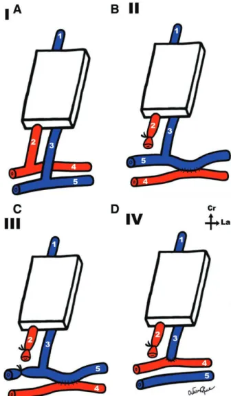
Methods: Fifty-three male rats were divided into 4 groups. In group I (n = 12), a 5-cm-long and 3-cm-wide conventional epigastric flap was raised on the left side of the abdomen. This flap was pedicled on the superficial caudal epigastric ves-sels caudally and on the lateral thoracic vein cranially. In groups II, III, and IV, a similar flap was raised, but the superficial epigastric artery was ligated. In these groups, AVFs were created using the following arterial venous anastomosis at the caudal end of the flap: group II (n = 13) a 1-mm-long side-to-side anastomosis was performed between the femoral artery and vein laterally to the ending of the su-perficial caudal epigastric vein. In group III (n = 14), in addition to the procedure described for group II, the femoral vein was ligated medially. Finally, in group IV (n = 14), the superficial caudal epigastric vein was cut from the femoral vein with a 1-mm-long ellipse of adjacent tissue, and an end-to-side arterial venous anastomo-sis was established between it and the femoral artery.
Results: Seven days postoperatively, the percentage of flap survival was 98.89 ± 1.69, 68.84 ± 7.36, 63.84 ± 10.38, 76.86 ± 13.67 in groups I–IV, respectively.
Conclusion: An optimized AVF can be produced using the vascular architecture described for group IV. (Plast Reconstr Surg Glob Open 2017;5:e1436; doi: 10.1097/ GOX.0000000000001436; Published online 17 August 2017.)
PRS Global Open
•
2017
tion of the time required to produce the flap, animal mor-tality, and surgical complications, as well as thermographic, histological, and microvascular characterization of the dif-ferent constructs. The ultimate goal of all these evaluations was to define an optimized model of AVF that could be eas-ily replicated for research and teaching purposes.
METHODS
Fifty-three male rats weighing 250–350 g were used. Only male rats were used to prevent potential confound-ing effects of cyclical hormonal changes in female rats.10 All the animals were housed under standard environmen-tal conditions and given nothing by mouth 6 hours before surgical procedures. No antibiotic prophylaxis was given.
Rats were anesthetized with a mixture of ketamine (5 mg/kg) and diazepam (0.25 mg/kg) given intraperito-neally. The depth of anesthesia was evaluated by toe pinch and by observance of respiration rate throughout the en-tire procedure. Supplementary doses of the anesthetic mixture were provided as needed.11
After shaving the abdomen and placing the animals on the operation table, the skin was disinfected with an anti-septic solution (Cutasept, Hartmann, Heidenheim, Ger-many). Hypothermia was avoided by placing the rat over a heating pad for the duration of the surgery.
Under a surgical operating microscope, a 5-cm-long and 3-cm-wide fasciocutaneous flap was raised on the left side of the rat’s abdomen immediately deep to the panniculus car-nosus layer (Fig. 1). This flap was initially pedicled on the superficial caudal vessels caudally and on the lateral thorac-ic vein cranially. All other vessels were carefully ligated.9,11
Rats were then randomly assigned to the following groups (Fig. 2):
In group I (n = 12), a conventional perfusion flap (CPF) was raised as described above.
In groups II, III, and IV, the superficial epigastric ar-tery was ligated with an 8/0 nylon suture. In these groups, AVFs were created using the following arterial venous anastomosis (AVA) at the caudal end of the flap:
In group II (n = 13), a 1-mm-long side-to-side anasto-mosis was performed between the femoral artery and vein laterally to the ending of the superficial caudal epigastric vein (SCEV) in the femoral vein. The AVA was performed after making a 1-mm-long ostium in adjacent flanks of the femoral artery and vein. A monofilament nylon 11/0–in-terrupted suture was used for the vascular anastomosis.12
In group III (n = 14), in addition to the procedure de-scribed for group II, the femoral vein was ligated immedi-ately medial to the ending of the SCEV, to increase blood flow through the AVA.
Finally, in group IV (n = 14), the SCEV was cut from the femoral vein with a 1-mm-long ellipse of adjacent fem-oral vein tissue. The ostium in the femfem-oral vein was closed with a monofilament nylon 11/0 continuous suture. The same suture line was used to perform a side-to-end AVA between the SCEV and the ventral flank of the femoral artery through a 1-mm-long ostium previously created in the latter vessel. Interrupted stitches were used for this anastomosis.
Surgical wounds were closed with 5/0 nylon stitches. No anticoagulants were administered pre-, intra-, or post-operatively.
After surgery, rats were kept in solitary rat cages and offered rat chow and water ad libitum.
Seven days after the surgery, rats were anesthetized as described above and AVA patency was noted. Only animals with patent AVAs were included in the study.
All surgical procedures were performed under aseptic conditions by the same microsurgeon (D.C.), to minimize intersurgeon variability. The operative time was registered in all animals by a blinded observer.
One hour postoperatively, 2 rats in each group were submitted to infrared thermography with a FLIR E6 cam-era (FLIR Systems, Wilsonville, Or.) placed 25 cm above the abdomen. Rats were placed on their backs for 10 min-utes before this evaluation. Thermographic measurements were made at a constant room temperature (22 ± 0.05ºC) and humidity (50%).13
Rats were assessed daily by the same researcher, to re-duce interobserver bias and variability.8 The following pa-rameters were evaluated: animal wellbeing, flap viability, flap ischemia, and presence of complications. Objective measurement of flap survival was performed on the third and seventh days postoperatively based on digital photo-graphs, which were later analyzed by a blinded observer using the free Image J software (National Institutes of Health, Bethesda, Md.).11 AVF survival was expressed as a percentage of the total flap surface area.14
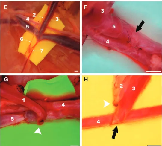
Half of the rats in each group were prepared for conventional histological examination, whereas the other half was submitted to processing to obtain vas-cular corrosion casts for scanning electron microscopy (SEM) evaluation. For histological processing, rats were submitted to axial sections in the caudal, middle, and cranial aspect of the AVF that were later stained using hematoxylin–eosin and Masson’s trichrome. These rats were euthanized by exsanguination after dividing the neck vessels under sedation.
The rats destined to SEM analysis were submitted to in-travascular injection of a resin cast (Mercox, Ladd Research, Williston, Vt.) and latter processed.15 SEM images were ob-tained using a JEOL JSM-7001F with an acceleration voltage of 2–30 kV. Vascular cast interpretation of microvascular find-ings was made according to Aharinejad and Lametschwandt-ner.15 These rats were euthanized by exsanguination after left and right ventricular catheterization.
All in vivo studies involving rats were carried out in strict accordance with the recommendations in the Guide for Proper Conduct of Animal Experiments and Related Activities in Academic Research and Technology.16
The protocol was approved by the Institutional Animal Care and Use Committee and Ethical Committee at the authors’ institution (CEFCM/08/2012).
Statistical Analysis
Kolmogorov-Smirnov test was used to assess whether variables were distributed normally. Analysis of variance and t test were used to compare averages in normally distributed data. Kruskal-Wallis and Mann-Whitney tests were used to compare means in nonnormally distributed data. Proportions were analyzed with the chi-square test or Fisher’s exact test. Kaplan Meier survival analysis was performed to identify differences in mortality between groups.
A 2-tail value of P < 0.05 was considered to be statisti-cally significant.
RESULTS
Contrarily to CPFs (group I), all AVFs presented ve-nous congestion, marked edema, epidermolysis, and ar-eas of necrosis (see figure, Supplemental Digital Content 1, which shows representative photographs of CPFs and AVFs from the end of surgery to the seventh postoperative day, http://links.lww.com/PRSGO/A494).
Most of the necrotic areas were clearly defined on the third day after surgery (Fig. 3; see figures, Supplemen-tal DigiSupplemen-tal Content 1, http://links.lww.com/PRSGO/A494 Fig. 1. Epigastric flap surgical anatomy. a, Schematic drawing of the blood supply to the rat’s epigastric
PRS Global Open
•
2017
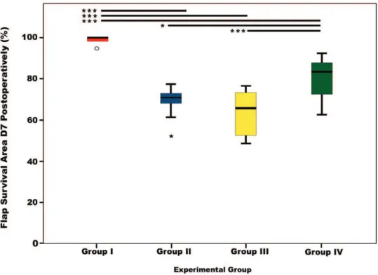
and Supplemental Digital Content 2, which shows box plot graphics illustrating flap survival in the different experi-mental groups 3 days postoperatively. Horizontal lines over boxplots indicate statistically significant differences, http:// links.lww.com/PRSGO/A495). On this day, the percentage of flap survival was 99.16 ± 1.46, 71.48 ± 7.80, 68.01 ± 12.39, and 83.21 ± 11.36 for groups I, II, III, and IV, respectively. Seven days postoperatively, the percentage of flap survival in these groups was 98.89 ± 1.69, 68.84 ± 7.36, 63.84 ± 10.38, 76.86 ± 13.67. Necrotic areas were more extensive in the caudal third of the flap (Supplemental Digital Content 1, http://links.lww.com/PRSGO/A494). Flap survival was high-er in the CPF group than in any of the AVF groups (P < 0.01). Among AVFs, group IV presented a higher flap sur-vival than groups II and III (P < 0.05). There were no sta-tistically significant differences between groups II and III.
Average operating time was increasing longer in groups I, II, III, and IV (Table 1; P < 0.0001). On average, it took twice the time to produce a group IV AVF com-pared with a CPF (group I).
There were no statistically significant differences in rat mortality rates among the different groups (Table 1).
The most common complication was hematoma, which occurred in 8.3%, 23.1%, 21.4%, and 14.3% of cas-es of groups I, II, III, and IV, rcas-espectively. Five rats (35.7%) in group III developed venous congestion and a swollen left hind limb. Four of these rats died within the first 48 hours after surgery. At the end of the experiment, surgical inspection of the AVA revealed an aneurysm in 1 of the rats in group III and thrombosis in 1 of the rats in group IV. No infections were noted. Overall, complications were more common in group III, although this difference was not statistically significant.
Thermographic evaluation revealed that all flaps, in-cluding CPFs, presented a lower temperature than the contralateral nonoperated region (Fig. 4). However, the temperature difference was higher in the AVFs, being of at least of 2°C in these flaps. In all AVFs, temperature was lower in the caudal third of the flap. No significant differ-ence was found among the different AVF groups.
Histological and SEM evaluation of vascular corrosion casts revealed great morphological homogeneity among the different AVFs (see figure, Supplemental Digital Con-tent 3, which displays a comparison of the histological features of AVFs compared with the CPFs controls, http:// links.lww.com/PRSGO/A496; see figure, Supplemental Digital Content 4, which displays a comparison of the mi-crovasculature of the conventional flap and the AVF using scanning electron microscope images of vascular corro-sion casts, http://links.lww.com/PRSGO/A497). In fact, the authors were not able to identify distinctive morphologi-cal patterns for any of the AVFs groups, based on quali-tative and/or quantiquali-tative features. Nevertheless, from a histological standpoint, comparatively to CPFs, AVFs presented greater flap edema, epidermolysis, loss of skin appendages, venous congestion and rupture, subcutane-ous hematoma, and necrosis. In AVFs, there were regions of necrosis scattered throughout all integumentary lay-ers. These histological features were more prominent in sections taken from the caudal third of AVFs. Moreover,
all AVFs presented signs of SCEV arterialization and sig-nificant increment of the lumen diameter of the lateral thoracic vein (Supplemental Digital Content 3, http:// links.lww.com/PRSGO/A496).
The study of the microvasculature through SEM vas-cular corrosion casts revealed higher vasvas-cular density in AVFs. In addition, these flaps also presented loss of venu-lar valves and/or venous valve incompetency particuvenu-larly in the caudal half of the flap. Signs of sprouting angiogen-esis were present in both CPFs and AVFs, although more markedly in the latter group. In AVFs, capillary vessels sprouted more commonly from venules, whereas in CPFs, new capillary vessels sprouted mostly from neighboring capillaries. In AVFs, it was also frequent to find evidence of intussusceptive angiogenesis (Supplemental Digital Con-tent 4, http://links.lww.com/PRSGO/A497).
DISCUSSION
Average flap survival area of the best AVF model in this work (group IV) was 76.86 ± 13.67%. This value is slightly inferior to that reported on a recent systematic review and meta-analysis on the clinical application of AVFs. In fact, it was estimated that the survival area of un-conventional perfusion flaps used in the clinical context varies between 87.30% and 91.30% (P < 0.001).3 However, as the cited authors mention, this estimation may be af-fected by several biasses associated with any meta-analysis, namely publication bias, which tend to overestimate posi-tive outcomes.3
AVF survival in the present work was worse than that described in the early description of Nakayama et al.2 These authors described a mean area of survival of 98% in nondelayed AVFs. However, they left a superior skin pedicle that no doubt contributed arterial axial and ran-dom perfusion to the flap.2 Nevertheless, the results here-in presented concernhere-ing AVF survival are similar to those reported by other authors in the rat.17–20
The impact of different anastomotic layouts in flap sur-vival had already been tested in CPFs.21,22 In these flaps, it was shown that side-to-end and end-to-end arterial anasto-moses of similarly sized arteries guaranteed comparable survivals.21 However, as far as the authors could deter-mine, this is the first time that the impact of microvascular
anastomotic type on the survival of AVFs is studied. In the present work, the vascular layout used in group IV (side-to-end AVA) proved to be superior regarding AVF survival than that used in groups II and III (side-to-side AVAs). Many authors have used side-to-side AVAs of the femoral rats to obtain AVFs of the rat’s abdomen. In this way, they avoid the technical challenging AVAs of very small vessels such as the SCEV and the homonymous artery, which are considerably prone to thrombosis.12 Moreover, Nakayama et al.2 had already demonstrated in a pilot study that direct end-to-end AVA of the femoral artery and SCEV was associ-ated with significant hemodynamic disturbance and death of 13 of 15 rats.
The data herein presented suggest that the incorpo-ration of a 1-mm-ellipse of the femoral vein adjacent to the draining point of the SCEV (group IV) allows a safe AVA. This pattern probably ensures a more direct blood flow into the SCEV and thus greater venous valves’ incom-petency, in comparison with AVAs in groups II and III. Furthermore, it is reasonable to expect that after remov-ing the vascular clamps, the AVA in group IV is pulled by the distension of walls of the femoral artery, resulting in radial distraction of the SCEV. This, in turn, will further lead to venous valve incompetency in the later vessel, thus facilitating the entry of blood through the afferent vein of the group IV AVF.
PRS Global Open
•
2017
Interestingly, mathematical modeling using Laplace’s law has suggested that the fine structure of venules un-der 100 µm in diameter renun-ders the valves in these vessels readily incompetent in the presence of venous arterializa-tion of ischemic lower limbs in humans.23 Our SEM data give empirical support to this assertion in the rat model, as in all AVFs groups there were signs of loss valve compe-tency particularly in venules located in the afferent half of the flap (Supplemental Digital Content 4, http://links.lww. com/PRSGO/A497). Similar findings have been reported by other authors.12
Multiple examples of intussusceptive angiogenesis in the vascular molds of AVFs were observed in the present study. These may be justified by the fact that increases in blood pressure inside small vessels have been shown to be associated with transluminal tissue pillar formation and subsequent vascular splitting and neovessel forma-tion.24 This mechanism has been largely neglected in the literature and may have a pivotal role in AVF hemodynam-ic adaptation.12
To the best of the authors’ knowledge, thermogra-phy imaging had never been employed before to study Fig. 3. Box plots graphics illustrating flap survival in the different experimental groups 7 days
postop-eratively. Horizontal lines over boxplots indicate statistically significant differences. *P < 0.05; **P < 0.01; ***P < 0.001.
Table 1. Comparison of the Different Vascular Constructs for Producing Arterialized Venous Fasciocutaneous Epigastric Flaps in the Rat
Assessed Parameters
Group I, CPF (n = 12)
Group II, AVF Produced by
Side-to-Side Anastomosis (n = 13)
Group III AVF Produced by Side-to-Side Anastomosis
and Femoral Vein Ligation (n = 14)
Group IV, AVF Produced by Terminal-Lateral Anastomosis (n = 14)
Statistically Significant Differences
Average operating time
(min) 43.0 ± 5.4 72.2 ± 10.0 75.1 ± 9.4 89.9 ± 10.1
I < II < III < IV (P < 0.0001)
Rat mortality (%) 16.7 23.1 28.6 21.4 None
Surgical complications (%) Arteriovenous
anastomo-sis thromboanastomo-sis
0 0 0 7.1 None
Arteriovenous anastomo-sis aneurysm
0 0 7.1 0 None
Hematoma 8.3 23.1 21.4 14.3 None
Hind limb ischemia 0 0 35.7 0 Larger in group III (P = 0.002) Complications other
than necrosis
8.3 23.1 42.8 21.4 None
Technical difficulty Easy Moderate Moderate Challenging N/A
perfusion of AVFs. Notwithstanding, skin temperature has been used as surrogate marker of perfusion in hind limb vein arterialization in rats. The rationale for this is that skin temperature is proportional to integumentary per-fusion.25 Our study lends support to the use of infrared thermography imaging for AVF perfusion evaluation, be-cause it confirmed an inferior temperature in these flaps comparatively to CPFs and to the contralateral side of the abdomen. Additionally, in all AVFs, temperature was lower in the caudal third of the flap, where necrosis was more commonly found.
Recently, it has been shown that this region of the abdomen of the rat can be used to produce axial flaps simulating arterial ischemia or venous congestion, read-ily observed macroscopically by a pale and dark violet color, respectively.14 Remarkably, in the present study, all AVFs groups presented a dark bluish color suggestive of venous congestion (Supplemental Digital Content 1, http://links.lww.com/PRSGO/A494). Another common finding in these 2 studies was that necrotic areas were clearly defined in both CPFs and AVFs on the third post-operative day.14 This information may be of great inter-est for future research works using similar flaps in this region of the rat.
Rats were chosen in the present study because they have been the most widely used animal model in the realm of experimental flap surgery.26 This is certainly due to the fact they are readily available in most
coun-tries, they are easy to keep, and they are among the cheapest animals to obtain and to maintain.26 Nonethe-less, it should be noted that AVFs performed in humans are usually based on vessels of a larger caliber.3 Hence, extrapolation of data obtained with the AVFs used in this article to humans must be done with this limita-tion in mind. In fact, it would be interesting to study the various vascular constructions described in these ar-ticles in other animal species, where larger flaps could be produced.
Furthermore, it is well known that in loose-skinned an-imals, the well-developed panniculus carnosus in the deep aspect of the integument leads to significant contraction of wounds and flaps.27 To tackle this problem, multiple strategies have been devised, namely choosing anatomi-cal sites where the integument is firmly adherent to the deep structures (e.g., rabbit ear) or using various devices or splints to fixate the integumentary layer and the sur-gical flaps.27 Despite these limitations, in plastic surgery experimental research, it is customary to consider flap’s survival and necrosis as a percentage of flap’s total area, as the authors did in the present article.9,11,14,26 However, this difference in flap and wound behavior between rodents and humans should be taken into consideration when generalizing the data presented in this article to the clini-cal scenario.
PRS Global Open
•
2017
sion flap that can be easily replicated for research and teaching purposes. However, further studies are warrant-ed to confirm or dismiss their usefulness in these contexts.
CONCLUSIONS
An optimized AVF can be reliably produced in the ven-trolateral aspect of the abdomen of the rat by performing an end-to-side AVA between the femoral artery and the SCEV including the adjacent portion of the femoral vein, to produce a 1-mm-wide afferent vein. This model pre-sented an average flap survival area of 76.86% ± 13.67%.
Diogo Casal, MD
Anatomy Department NOVA Medical School Campo dos Mártires da Pátria, 130 1169-056, Lisbon Portugal E-mail: [email protected]
ACKNOWLEDGMENTS
The authors are very grateful to Mr. Carlos Lopes and Mr. Octávio Chaveiro for their help in producing and observing the scanning electron microscope specimens. The authors also thank Mr. Nuno Folque for producing all the drawings contained in this article.
REFERENCES
1. Vaubel, W. Indikationen und Technik des arterialisierten Lappens zur Deckung großer Defekte im Handbereich. Hefte Unfallheilkd. 1975;126:381.
2. Nakayama Y, Soeda S, Kasai Y. Flaps nourished by arterial inflow through the venous system: an experimental investigation. Plast Reconstr Surg. 1981;67:328–334.
3. Casal D, Cunha T, Pais D, et al. Systematic review and meta-anal-ysis of unconventional perfusion flaps in clinical practice. Plast Reconstr Surg. 2016;138:459–479.
4. Goldschlager R, Rozen WM, Ting JW, et al. The nomenclature of venous flow-through flaps: updated classification and review of the literature. Microsurgery. 2012;32:497–501.
5. Yan H, Brooks D, Ladner R, et al. Arterialized venous flaps: a review of the literature. Microsurgery. 2010;30:472–478.
6. Yan H, Zhang F, Akdemir O, et al. Clinical applications of venous flaps in the reconstruction of hands and fingers. Arch Orthop Trauma Surg. 2011;131:65–74.
7. Yan H, Fan C, Zhang F, et al. Reconstruction of large dorsal digital defects with arterialized venous flaps: our experience and com-prehensive review of literature. Ann Plast Surg. 2013;70:666–671. 8. Weng W, Zhang F, Zhao B, et al. The complicated role of venous
drainage on the survival of arterialized venous flaps. Oncotarget. 2017;8:16414–16420.
9. Hirase Y. Skin and muscle flaps in the rat. In: Tamai S, Usui M, Yoshizu T, eds. Experimental and Clinical Reconstructive Microsurgery. Vol. 1, 1st ed. Japan: Springer-Verlag; 2004:111–114.
10. Thatte M, Healy C, McGrouther D. Laser Doppler and microvas-cular pulsed Doppler studies of the physiology of venous flaps.
Eur J Plast Surg. 1993;16:134–138.
11. Casal D, Pais D, Iria I, et al. A model of free tissue transfer: the rat epigastric free flap. J Vis Exp. 2017;1:e55281.
12. Wungcharoen B, Pradidarcheep W, Santidhananon Y, et al. Pre-arterialisation of the arterialised venous flap: an experimental study in the rat. Br J Plast Surg. 2001;54:621–630.
13. Sheena Y, Jennison T, Hardwicke JT, et al. Detection of perfo-rators using thermal imaging. Plast Reconstr Surg. 2013;132: 1603–1610.
14. Matsumoto NM, Aoki M, Nakao J, et al. Experimental rat skin flap model that distinguishes between venous congestion and arterial ischemia: the reverse U-shaped bipedicled superficial inferior epigastric artery and venous system flap. Plast Reconstr Surg. 2017;139:79e–84e.
15. Aharinejad SH, Lametschwandtner A. Identification and in-terpretation of cast vessel structures. In: Aharinejad SH, Lametschwandtner A, eds. Microvascular Corrosion Casting in Scanning Electron Microscopy: Tecnhiques and Applications. 1st ed. New York, N.Y.: Springer-Verlag; 1992:103–115.
16. National Research Council (U.S.). Committee for the update of the guide for the care and use of laboratory animals., Institute for Laboratory Animal Research (U.S.), National Academies Press (U.S.). Guide for the Care and Use of Laboratory Animals. 8th ed. Washington, D.C.: National Academies Press; 2011:xxv:220.
17. Başer NT, Silistreli OK, Sişman N, et al. Effects of surgical or chemical delaying procedures on the survival of proximal pred-icled venous island flaps: an experimental study in rats. Scand J Plast Reconstr Surg Hand Surg. 2005;39:197–203.
18. Chow SP, Chen DZ, Gu YD. A comparison of arterial and venous flaps. J Hand Surg Br. 1992;17:359–364.
19. Miles DA, Crosby NL, Clapson JB. The role of the venous system in the abdominal flap of the rat. Plast Reconstr Surg. 1997;99:2030– 2033.
20. Mutaf M, Tasaki Y, Fujii T. Expansion of venous flaps: an experi-mental study in rats. Br J Plast Surg. 1998;51:393–401.
21. Miyamoto S, Takushima A, Okazaki M, et al. Relationship between microvascular arterial anastomotic type and area of free flap sur-vival: comparison of end-to-end, end-to-side, and retrograde arte-rial anastomosis. Plast Reconstr Surg. 2008;121:1901–1908. 22. Parsa FD, Spira M. Evaluation of anastomotic techniques in
the experimental transfer of free skin flaps. Plast Reconstr Surg. 1979;63:696–699.
23. Koyama T, Sugihara-Seki M, Sasajima T, et al. Venular valves and retrograde perfusion. In: Swartz HM, Harrison DK, Bruley DF, eds. Oxygen Transport to Tissue XXXVI. 1st ed. New York, N.Y.: Springer; 2014;1:317–323.
24. Makanya AN, Hlushchuk R, Djonov VG. Intussusceptive angio-genesis and its role in vascular morphoangio-genesis, patterning, and remodeling. Angiogenesis. 2009;12:113–123.
25. Sasajima T, Kikuchi S, Ishikawa N, et al. Skin temperature in lower hind limb subjected to distal vein arterialization in rats. In: Swartz HM, Harrison DK, Bruley DF eds. Oxygen Transport to Tissue XXXVI. 1st ed. New York, N.Y.: Springer; 2014;1:361– 368.
26. Dunn RM, Mancoll J. Flap models in the rat: a review and reap-praisal. Plast Reconstr Surg. 1992;90:319–328.