University
of Algarve
Effect of Cisplatin in Mytilus galloprovincialis and of
cyclophosphamide, tamoxifen and cisplatin on
Ca
2+ATPase.
Matilde de Brito Morais, aluna nº45242
Project realized in the framework of completing the degree in Marine Biology
Work supervised by: Prof. Doctora Maria João da Anunciação Franco Bebianno and Prof. Doctor Manuel Aureliano Pereira Martins Alves
Abstract
In the last decade anti-cancer drugs, such as cisplatin, tamoxifen and cyclophosphamide, have seen their use greatly increase. Cisplatin is one of the most common anti-cancer drugs used in the EU which ubiquitous occurrence in surface waters, like rivers and estuaries, is related to the poor removal capacities of waste-water treatment plants (WWTPs). These drugs are genotoxic, cytotoxic, mutagenic and teratogenic. Therefore, this study includes a multibiomarker response analysis on mussel Mytilus galloprovincialis during two weeks of exposure to 100ng/l of cisplatin assessing antioxidant enzyme activity - catalase (CAT), glutathion-S-transferase (GST); lipid peroxidation (LPO) coupled with an enzyme assay to determine the inhibition effect of cisplatin, tamoxifen and cyclophosphamide (IC50) in the Ca2+-ATPase. Results
show that cisplatin does not seem to have an effect in the activity of CAT, GST and LPO and that its IC50 is 13.7 mM. The effect of the other two anti-cancer drugs were
tamoxifen 0.271 mM and cyclophosphamide 1.134 μM. which makes Cisplatin much less dangerous than the other two anti-cancer drugs.
Resumo
Na última década, compostos anticancerígenos, como a cisplatina, tamoxifen e ciclofosfamida, tem tido um grande aumento de uso. A cisplatina é um dos anticancerígenos mais usados na UE cuja presença nas águas superficiais tal como rios e estuários resulta da falta de capacidade de remoção por parte das estações de tratamento de água (ETARs). Estes medicamentos tem capacidades genotóxica, citotóxicas, mutagénicas e teratogénica. O objetivo deste estudo é testar o efeito de vários biomarcadores em Mytilus galloprovincialis durante uma exposição de duas semanas a 100ng/l de cisplatina e avaliar a atividade das enzimas antioxidantes- catálase (CAT), glutationa-S-transferase (GST); peroxidação lipídica (LPO) bem como determinar a concentração de inibição 50% (IC50) da cisplatina, tamoxifen e ciclofosfamida em Ca2+
-ATPase. Os resultados mostram que a cisplatina não aparenta ter efeitos na atividade da CAT, GST e LPO e a sua IC50 é de 13.7 mM. Comparado os valores de IC50 com os de
tamoxifen (0.271 mM) e ciclofosfamida (1.134 μM) indica que a cisplatina representa um risco muito inferior para Ca2+-ATPase do que os dois outros anticancerígenos.
1. Introduction
With the demographical increase of the world population the use and necessity of pharmaceutical drugs has greatly increased. And with this, the input of these drugs in the environment also has increased. Which has become one of the principal areas of interest in ecotoxicolocal investigation with the scope to understand the effect that the drugs have in the biodiversity and in the organisms.
1.1. Anticancer drugs
In the last decade the use of pharmaceuticals as increased and it’s detection in the environment has well. (Kosjek & Heath, 2011; Xie, 2012) Part of the pharmaceuticals consumed are excreted un-metabolized or are substances that can be reactivated in the sewage systems (Rowney, Johnson, & Williams, 2009). One of the import pharmaceuticals classes due to their genotoxicity, cytotoxicity, mutagenicity and teratogenicity that need to be much studied are the anticancer drugs (Kosjek & Heath, 2011).
Anticancer drugs are classified as antineoplastic and immunomodulation agents (class L) and divided in alkylating agents (L01A), antimetabolites (L01B), natural products (L01C), cytotoxic antibiotics (L10D) and other antineoplasics (L01X) ( Kosjek and Heath 2011; Xie 2012; Booker et al. 2014).
These drugs achieve the antitumor effect by causing direct DNA damage, inhibition of DNA synthesis, cell proliferation and induction of cell apoptosis ( Rowney et al., 2009; Kosjek & Heath, 2011; Xie, 2012; Booker et al., 2014; Turner & Mascorda, 2014; Vyas, Turner, & Sewell, 2014). Cytostatics act in a non-selective way in developing cells and may become carcinogen, until now it was not possible to say with certainty whether they have or not an environmental impact (Kosjek & Heath, 2011; Xie, 2012; Vyas et al., 2014). Many of these substances are polar, soluble and non-volatile and have as principal source of liberation hospital and municipal effluents, due to the administration of the drugs to inpatients and outpatients (Kosjek & Heath, 2011; Booker et al., 2014). The wastewater-treatment plants do not have adequate equipment to remove totally these drugs which leads to their ongoing
Figure 1:
Cyclophosphamide molecular structure
release in surface waters, rivers e estuaries ( Rowney et al., 2009; Booker et al., 2014).
To handle cytotoxic substances precaution measures are necessary. The drugs must be preserved in a dedicated and isolated local, the experiments must be done in a class II contaminant safety cabinet. And personal protection must be appropriate (Turner & Mascorda, 2014).
Cyclophosphamide is classified as a classical alkylating agent. It is structurally similar to mustard gas, having a bis(2-chlorethyl)
amine group capable of forming aziridinium. This group alkylates N7 of guanine bases and creates interstrand crosslinks in the DNA. This damage to the DNA blocks the transcription and replication of DNA, therefore being highly cytotoxic (Rowney et al., 2009; Xie, 2012).
Cyclophosphamide is a polar compound with a small Kow (octanol-water partition coefficient), high
solubility in water (solubility: 0.1-0.5 g/L), it is a substance with low biodegrability, the EC50 > 1000mg/L in various essays. The environmental
concentration of cyclophosphamide reaches 10 μg/L in hospital effluents and 0.2ng/L in surface waters ( Xie, 2012; Booker et al., 2014).
Tamoxifen is classified as an endocrine disrupting drug that interfere with the endocrine system and disarray the reproduction system. It is a drug that contains 4-hydroxytamoxifen and
N-desmethyl4-hydroxytamoxifen. Both
compounds are inhibitors of estrogen receptors which leads to the inhibition of the transcription of estrogen responsive genes (Xie, 2012).
Figure 2: Tamoxifen molecular structure
Tamoxifen is a lipophilic substance, with a high log Kow and low water
solubility (solubility:17 mg/L), the EC50 > 5 μg/L in various essays. The
environmental concentration of tamoxifen ranges from 0.2 to 8.2 ng/L in hospital effluents and <5.8ng/L in surface waters. Endocrine disruption caused by tamoxifen were only observed for concentration levels much higher than relevant environmental concentrations (Xie, 2012).
1.1.1. Cisplatin
Cisplatin is classified as a non-classical alkylating agent because it does not have the alkyl group like the mustard gas does. However it has a similar effect on DNA as the classic alkylating agent.
In aqueous solutions that have few electrolytes the chloride ligands are replaced by water molecules:
cis-PtCl2(NH3)2 + H2O ↔ cis-Pt(OH2)(𝑁𝐻3)2++ 𝐶𝑙−
cis-Pt(OH2)(𝑁𝐻3)2++ 𝐶𝑙− ↔ cis-Pt(OH2)2(𝑁𝐻3)22++ 𝐶𝑙−
This reaction results in a aqueous species that is more reactive, known as monoaquacisplatin and diaquacisplatin, which binds to guanine base of the DNA and interfere with DNA replication and transcription (Bonnet et al. 2003; Kosjek and Heath 2011; Xie 2012; Turner and Mascorda 2014; Vyas, Turner, and Sewell 2014)
Cisplatin is a compound with a high polarity and a low Kow (Kow =-2.2), high
solubility (solubility: 3*103 mg/L). The
biodegradation of cisplatin is close to zero. DNA damage was noticed only with concentrations higher than 1mg/L (Kosjek and Heath 2011; Xie 2012; Turner and
Figure 3: Cisplatin molecular structure
Mascorda 2014; Vyas, Turner, and Sewell 2014). Bonnet et al. (2003) determined the cisplatin EC50 of 37.31 ± 2.24mg/L for the ciliated
protozoan tetraphymena pyriformis.
1.1.1.1. Production, use and application
Cancerostatic platinum compounds (CPCs) are one of the most used classes of antineoplastic in cancer therapy. Cisplatin is one of the three CPCs authorized in Europe.
The production and use of CPCs in the last decade increased with the increase of cancer cases due to their efficiency in treatments. Usually the in and outpatients are administered a dose of 75-100 mg.m-2 body surface of cisplatin and 28±4% of the medication is excreted in the first 24h to hospital or urban effluents (Hann, Stefánka, Lenz, & Stingeder, 2005; Lenz et al., 2005).
1.1.2. Environmental impact and toxicological effects
Part of the cisplatin consumed by patients is eliminated from the human body unmetabolised or as conjugated substances that can be reactivated in urban effluents (Rowney et al., 2009).
The concentration of cisplatin in hospital effluents range from 38 to 176 ng/L. The constant release of cisplatin and other CPC’s to hospital effluents is problematic due to their capacity to interfere with DNA replication and transcription and so prevent cell development and even kill them (Xie, 2012).
IARC (1897) classified the cisplatin in group “2A- possibly cancerogenic to humans, an agent for which there is sufficient evidence of carcinogenicity in experimental animals” (in this case the assay were done in mouses).
1.1.3. Aquatic organisms
In the present any experiment exists with cisplatin in aquatic organisms.
1.2. Biomarkers
Biomarkers are quantitative measurements of changes at biochemical, molecular, cellular and physiological levels in organisms that may be related to effect and exposure to xenobiotic and environmental chemicals (Van der Oost, Porte-Visa, Van den Brink 2005; Gonzalez-Rey and Bebianno 2012; Vidal-Liñán and Bellas 2013).
In a situation of stress induced by pollutants it a sequence of biological reaction are triggered, which are used as a biomarker. Once a certain pollutant dose or exposure time is exceeded the pollutant-responsive biomarker signal deviate from the range of a normal situation (Van der Oost et al., 2005).
The most important feature of biomarker research is the early identification of potential toxic effects relatively to time of exposure time and concentration (Van der Oost et al., 2005). Biomarker can be divided in three categories: ( Van der Oost, Beyer, & Vermeulen, 2003)
I. Exposure biomarkers: that permit to measure and interaction with a target molecule or a xenobiotic substance;
II. Effect biomarker: which measure biochemical, physiological or other alterations within tissues that can be associated to a possible disease or health damage;
III. Susceptibility biomarker: indicates the capacity of an organism to respond and adapt to the exposure to certain xenobiotic. This includes genetic factors and changes in receptors that change the susceptibility to that xenobiotic.
1.2.1. Phase II biotransformation enzymes
Glutathion-S-transferase (GST) is classified as a phase II biotransformation enzyme which catalyse the process of conjugation of xenobiotics compounds or its metabolites with an endogenous ligand (Van der Oost et al., 2003) and so facilitate the excretion of chemicals by adding a more polar group like glutathione to the molecule (Van der Oost et al., 2005). A critical role of the GST is the defence against oxidative damage and peroxidative products of DNA (Van der Oost et al., 2003).
1.2.2. Oxidative stress biomarkers
Oxidative stress may be caused in organisms due to many environmental contaminants. When reactive oxygen species (ROS), or oxyradicals are present from exposure to cytotoxic compounds there occur oxygen toxicity (Van der Oost et al., 2003; Vlahogianni & Valavanidis, 2007) . The products of oxygen (O2) reduction are superoxide anion radical, hydrogen
peroxide and hydroxyl radical, a potent oxidant capable of reacting with cellular molecules, leading to enzyme inhibition and lipid peroxidation (LPO) (Van der Oost et al., 2003). There are defence systems that inhibit oxyradicals formation being antioxidant enzymes such as catalase (CAT) one of them (Van der Oost et al., 2005).
1.2.3. Biochemical indices of oxidative damage
Many biochemical and physiological effects have been linked to increase of oxyradical production. One of the principal biochemical perturbations used as biomarker of oxidative damage is lipid peroxidation (LPO), or the oxidation of polyunsaturated fatty acids. Many studies demonstrated the increase of LPO in tissues of fish exposed to various chemicals ( Van der Oost et al., 2003; Vlahogianni & Valavanidis, 2007).
1.3. Model system
Bivalves, like mussels, have been used as sentinel organisms because they are sessile, filter feeders and wildly distributed. These organisms also have the capacity to accumulate contaminants from the water during their life ( Letendre et al. 2008; Gonzalez-Rey and Bebianno 2012; Marigómez et al. 2013; Jarque et al. 2014).
Most of coastal monitoring programs use mussels from the intertidal zone, which are of easy access. It is necessary to take into account that the physiology of intertidal organisms is affected by the periods of submersion and emersion (Vidal-Liñán & Bellas, 2013). The closing of the shell during the emersion period and the loss of water through the paleal cavity imply a decrease of oxygen in the tissues. Because of this bivalves rapidly suffer from hypoxia or even anoxia when the emersion is prolonged (Letendre et al., 2008).
Mytilus, is one of the genera most abundant of mussels and is usually used for research (Vlahogianni and Valavanidis 2007; Letendre et al. 2008; Gonzalez-Rey and Bebianno 2011, 2012, 2013; Cappello et al. 2013; Marigómez et al. 2013; Vidal-Liñán and Bellas 2013; Gonzalez-Rey et al. 2014; Jarque et al. 2014).
There are three taxas of Mytilus that inhabit European water. Two of these taxas are predominant: Mytilus edulis that lives in temperate and cold water from the European Atlantic coasts, and Mytilus galloprovincialis that inhabits warmer waters in the Mediterranean and extends to the north coast of France and the United Kingdom (Jarque et al., 2014).
In the present study we analysed alterations of biomarkers such us catalase (CAT), gluthation-S.transferase (GST) activities and lipid peroxidation (LPO) in the gills and digestive gland of mussels, M. galloprovincialis, exposed to Cisplatin to try to understand if the release of this drug in the environment could have a significant impact in this species. Also the effects of cisplatin, cyclophosphamide and tamoxifen on Ca2+ATPase were assessed. Both studies aimed to evaluate the putative impact and the toxicity effects of anticancer drugs in the environment.
2. Materials and methods
2.1. Cisplatin
Cisplatin was obtain from SIGMA P4394-250mg; CAS 15663-27-1
2.1.1. Chemical Characterization
1Chemical Formula Pt(NH
3)2Cl2
1Molecular weight 300.05 g.mol-1
1Melting Point 270ºC
1Form Yellow crystalline powder
1Water Solubility 0.253g/100g
2Log K
ow -2.2
1Obtained from SIGMA CAS# 15663-27-1
2 (Bonnet et al., 2003; Kosjek & Heath, 2011; Xie, 2012; Turner & Mascorda, 2014; Vyas et al.,
2014) http://cisplatina.paginas.sapo.pt/ACisplatina.html; https://weweh.com
A solution was prepared of 25ml with 57.69 mg of cisplatin diluted in water. For each contamination 1 ml of the solution was put in the tanks to obtain a concentration of 100 ng/l of cisplatin during the exposition.
2.2. Model system and exposure
2.2.1. Collection
250 Mytilus galloprovincialis were collected from the Ria Formosa near Tavira (figure 4), in an area subjected to little human interaction.
2.2.2. Acclimatization
The mussels were acclimatized for 7 days in two tanks with 25 litters of seawater. Temperature (18ºC ±1.5ºC) and salinity (36 ±1) were monitored and kept constant. The water was changed every two days.
2.2.3. Exposure experiment
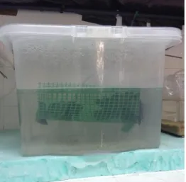
The mussels were separated in groups of 30 mussels per tank of 15 litters (2 mussels/litter) (figure 5) in a triplicate design (3 control tanks and 3 thanks contaminated with Cisplatin 100ng/l). The water was changed every two days and contamination re-established. The exposure lasted for 14 days and mussels were sampled in the beginning of the experiment and after 3, 7 and 14 days of exposure.
2.3. Dissection
10 mussels for each sampling time were dissected (Figure 6) and the gills, digestive gland and mantle frozen in liquid nitrogen and stored at -80ºC until the tissues were analysed. Shells were dried and weighted.
Figure 5: photo of one of the tanks for the experiment
2.4. Condition Index (CI)
After weighing the tissues of the mussels and the shells, the condition index was calculated as the percentage of the ratio between the wet weight of the tissues and the total weight of the organisms (tissues plus shells).
2.5. Total Proteins
Total proteins were measured in the gills and digestive gland following the assay from Bradford (1976) which relies on the binding of the dye Coomassie Blue G-250 to proteins and the absorbance measured at 595nm. Measurements were made in a microplate reader using Bovine Serum albumin as a standard. Protein concentrations are expressed as mg.g-1 wet weight tissue.
2.6. Oxidative Stress markers
2.6.1. Catalase (CAT)
CAT activity was measured from 100 μl of tissue by the decrease in absorbance at 240nm due to hydrogen peroxide (H2O2) consumption. The
assay consisted in 1.95mL phosphate buffer, 1mL H2O2 and 0.05mL
sample. The CAT activity is expressed in μmol.mg-1.total protein min-1.
2.6.2. GST
GST activity was determined in the cytosolic fraction by spectrometry at 340nm based on the method of Habig, Pabst, and Jakoby (1974). The GST activity is expressed in μmol.mg-1.total protein min-1.
2.7. Lipid Peroxidation
The tissue samples were individually homogenised on ice with 20mM TRIS-HCL buffer ant butylatedhydroxytoluene (BHT). Then it was centrifuged for 45min at 30 000 x g for 45min to precipitate the cytosolic fraction.
The lipid peroxidation was measured following the protocol of BIOXYTECH, LPO-586TM; Colorimetric Assay for Lipid Peroxidation by OxisResearchTM, a division of OXIS Health Products, Inc. In this case it was a combination of MDA with 4-hydroxyalkenals (in methanesulfonic acid). The LPO levels are expressed in MDA nmol.mg-1.total protein.
2.8. Ca2+-ATPase
Ca2+-ATPase activity in the presence of the several compounds was measured with spectrophotometry at 340 nm using coupled enzyme assay with pyruvate kinase and lactate desidrogenase following the experiment from Fraqueza et al (2013). Briefly, after the addition of the enzymes to the medium, NADP was added and then the vesicles from skeletal muscle sarcoplasmic reticulum containing the Ca –ATPase. Then, after the addition of ATP the values of the absorbance were recorded during about 1 minute and after that the ionophore was added and the decreased of the absorbance was measured during about 2 minutes. The ATPase activity and the inhibition was measured taken into consideration the decrease of the OD (Optical density) per minute in the absence (100% ) and in the presence of the compounds cyclophosphamide, tamoxifen and cisplatin (Fraqueza, et al 2012).
2.9. Statistical analysis
Data were analysed using two-way ANOVA, one way ANOVA (non-parametric ANOVA on ranks) and t-test to test differences between mussels form controls and exposed to cisplatin. The significance of the results was ascertained at p <0.05.
3. Results
3.1. Exposure Experiments
3.1.1. Condition Index (CI)
During the experiment the condition index was similar along the experiment. This imply that the physiological conditions of the organisms did not change during the experiment.
Figure 7: Condition Index (CI) of mussels from control and exposed to Cisplatin. Different letters express significant differences (p<0.05) lower case letters indicate differences within the same treatment over time and uppercase letters indicate differences within the same time between treatments.
3.2. Total Proteins
Total proteins in the gills did not significantly change between mussels from control and exposed to cisplatin indicating that cisplatin has no influence in total protein content. However in the digestive gland total proteins concentrations decreased in the tissues exposed to cisplatin compared to the controls after 3 and 14 days of exposure.
Figure 8: Total proteins in the gills and digestive gland of mussels. Different letters express significant differences (p<0.05) lower case letters indicate differences within the same treatment over time and uppercase letters indicate differences within the same time between treatments.
3.3. Oxidative Stress biomarkers
3.3.1. CAT
CAT activity did not change throughout all the exposure experiment in the gills and in the digestive gland in the controls and the exposed mussels except for the digestive gland after 14 days of exposure whose activity significantly increased as shown in figure 9-B.
Figure 9: CAT activity in tissues exposed to Cisplatin. Different letters express significant differences (p<0.05) lower case letters indicate differences within the same treatment over time and uppercase letters indicate differences within the same time between treatments.
3.3.2. GST
The exposure of the mussels to cisplatin did not cause any change in the glutathione-S-transferase activity relatively to controls in the gills and digestive gland. However in the digestive gland like for CAT levels GST activity significantly increase by the end of the exposure period (fig 10-B).
Figure 10: GST activity in tissues exposed to Cisplatin. Different letters express significant differences (p<0.05) lower case letters indicate differences within the same treatment over time and upper case letters indicate differences within the same time between treatments.
3.4. Lipid Peroxidation
In the gills, LPO has a transient variation in the gills of the mussels exposed to cisplatin compared to the controls which increased after 3 days of exposure and decreased gradually afterwards (fig 11-A). LPO in the digestive gland also increased after three days of exposure and decreased afterwards revealing a similar behaviour among tissues.
Figure 11: Lipid Peroxidation in tissues exposed to Cisplatin. Different letters express significant differences (p<0.05) lowercase letters indicate differences within the same treatment over time and uppercase letters indicate differences within the same time between treatments.
3.5. Ca2+-ATPase
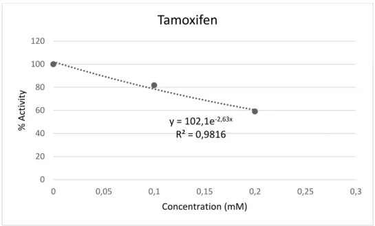
The effects of solutions containing different drugs were also studied on the activity of a calcium pump involved in the regulation of muscle contraction, the Ca2+- ATPase of sarcoplasmic reticulum. Thus, the inhibition of cisplatin, cyclophosphamide and tamoxifen in the Ca2+-ATPase are in Figs 12, 13 and 14.
Figure 12: Determination of the inhibition concentration (IC50) of the activity of Ca2+ -ATPase exposed to cisplatin.
As it can be observed from Fig 12, even for concentrations at the M range (up to 8 mM), the activity of the pump was only 40% inhibited. We estimate that for cisplatin the IC50 is about 13.7 mM. However, for cyclophosphamide much
lower concentration inhibits the calcium pump enzyme activity (Fig 13). Although more concentration should be used to determine the IC50 value, we
approximately estimate that for this drug an IC50 was near 1 μM.
Regarding tamoxifen, as can be observed from Fig 14, the IC50 was about 0.2
mM.
Figure 13: Determination of the inhibition concentration (IC50) of the activity of Ca2+ -ATPase exposed to Cyclophosphamide
y = 94,901e-0,565x R² = 0,8614 0 20 40 60 80 100 120 0 0,2 0,4 0,6 0,8 1 1,2 1,4 % Act iv ity Concentration (μM)
Cyclophosphamide
Figure 14: Determination of the inhibition concentration (IC50) of the activity of Ca2+ -ATPase exposed to Tamoxifen
Putting it all together and comparing the inhibitory capacity for these three compounds the following order of inhibition was observed : cyclophosphamide > tamoxifen > cisplatin , that is, the IC50 levels were 1.134 μM, 0.271 mM and
13.722 mM, respectively. y = 102,1e-2,63x R² = 0,9816 0 20 40 60 80 100 120 0 0,05 0,1 0,15 0,2 0,25 0,3 % Act iv ity Concentration (mM)
Tamoxifen
4. Discussion
Cisplatin is a pharmaceutical with still little research in the aquatic environment. To understand its effect in M. galloprovincialis we compared the activity of biomarkers exposed to cisplatin to biomarker activity exposed to other pharmaceuticals.
4.1. Condition Index (CI)
Cisplatin did not influence the physiological condition of the organisms during the two weeks exposure. Studies with Mytilus galloprovincialis (Gonzalez-Rey and Bebianno 2011, 2013; Rey and Bebianno 2012 and Gonzalez-Rey et al. 2014) indicated that mussels show little or no alteration in CI when exposed to other pharmaceuticals namely Ibuprofen, fluoxetine and diclofenac.
4.2. Oxidative Stress biomarkers
Both treatments did not lead to significant changes in CAT activity in the gills, this was also observed for the same species in studies with the same pharmaceuticals mentioned above (Gonzalez-Rey and Bebianno 2012; Gonzalez-Rey and Bebianno 2013; Gonzalez-Rey et al. 2014). However, in the digestive gland there was an increase in CAT activity after 2 weeks of exposure unlike in other studies were the CAT activity increase in the beginning of the exposure and then decrease after the first week ( Gonzalez-Rey et al., 2014; Gonzalez-Gonzalez-Rey & Bebianno, 2012; Gonzalez-Gonzalez-Rey & Bebianno, 2011, 2012).
In the gills both treatments did not change GST activity. However in the digestive gland, like for CAT levels, GST activity significantly increased by the end of the exposure period. In other studies, the GST decrease with the exposure time when mussels were exposed to ibuprofen, diclofenac and fluoxetine (Gonzalez-Rey et al., 2014; Gonzalez-Rey & Bebianno, 2012; Gonzalez-Rey & Bebianno, 2011, 2012, 2013) .
4.3. Lipid Peroxidation
LPO has a transient variation in the gills of the mussels exposed to cisplatin compared to the controls which increased after 3 days of exposure and decreased gradually afterwards. LPO in the digestive gland has the same variation as the controls of the gills. In other studies LPO levels in Mytilus galloprovincialis exposed to ibuprofen and fluoxetine have increased in the beginning of the exposure and decreased at the end. Except in some occasions where there occurs a slight but consistent increase during the exposure or an increase at the end of the exposure time ( Rey et al., 2014; Gonzalez-Rey & Bebianno, 2012; Gonzalez-Gonzalez-Rey & Bebianno, 2011, 2012, 2013). 4.4. Ca2+-ATPase
The inhibition concentration of 50% (IC50) for cisplatin, cyclophosphamide and
tamoxifen are 13.7 mM; 1.13 μM and 0.271 mM respectively. These values are not in agreement with the ones previously described. In fact, it was previously reported, although at different experimental conditions tamoxifen inhibits the calcium ATPase with IC50 of 5μM (Kargacin, Zenobia, Ward, Pollock, &
Kargacin, 2000). Lower levels were also reported for cyclophosphamide and cisplatin although the former drug is in fact the lower calcium pump inhibitor, as described elsewhere (Sudharsan, Mythili, Selvakumar, & Varalakshmi, 2006) Therefore, for the drugs that were tested the cyclophosphamide is the strongest inhibitor of the calcium pump.
5. Conclusions
Cisplatin, at the doses used in mussel samples, does not represent a serious environmental risk in relation to oxidative stress and physiologically for mussels. However, there is not enough information to fully understand if Cisplatin is of environmental concern for the current concentration or not. It is still necessary to better understand its impact in biodiversity at cellular and molecular levels and study other ways of exposure. Compared to tamoxifen and cyclophosphamide the cisplatin was the pharmaceutical which had less inhibition in the Ca2+-ATPase, being necessary concentration of 2.67 g/L of cisplatin in the water to attain the IC50
which is a concentration much higher than the 36-176 ng/L of environmental concentration.
Bibliography
Bonnet, J. L., Dusser, M., Bohatier, J., & Laffosse, J. (2003). Cytotoxicity assessment of three therapeutic agents, cyclosporin-A, cisplatin and doxorubicin, with the ciliated protozoan Tetrahymena pyriformis. Research in Microbiology, 154, 375–385. doi:10.1016/S0923-2508(03)00085-8
Booker, V., Halsall, C., Llewellyn, N., Johnson, A., & Williams, R. (2014). Prioritising anticancer drugs for environmental monitoring and risk assessment purposes. Science of
the Total Environment, 473-474, 159–170. doi:10.1016/j.scitotenv.2013.11.145
Bradford, M. M. (1976). A rapid and sensitive method for the quantitation of microgram
quantities of protein utilizing the principle of protein-dye binding. Analytical Biochemistry,
72, 248–254. doi:10.1016/0003-2697(76)90527-3
Cappello, T., Maisano, M., D’Agata, A., Natalotto, A., Mauceri, A., & Fasulo, S. (2013). Effects of environmental pollution in caged mussels (Mytilus galloprovincialis). Marine
Environmental Research, 91, 52–60. doi:10.1016/j.marenvres.2012.12.010
Fraqueza, G. (2013). Interação de oxometalatos de vanádio, nióbio, tungstéio e molib´rnio com a Ca2+-ATPase de retículo sarcoplasmático: um alvo de ação de fármacos. Universidade
Do Algarve. Retrieved from
http://books.google.com/books?hl=en&lr=&id=mRQbbgpMDjEC&oi=fnd&pg=PA141&d
q=Universidade+do+algarve&ots=LW1cahVSSS&sig=-SPtHq3pt1YAMMK4sOHRV8AzAOA
Fraqueza, G., Ohlin, C. A., Casey, W. H., & Aureliano, M. (2012). Sarcoplasmic reticulum calcium ATPase interactions with decaniobate, decavanadate, vanadate, tungstate and molybdate. Journal of Inorganic Biochemistry, 107(1), 82–89.
doi:10.1016/j.jinorgbio.2011.10.010
Gonzalez-Rey, M., & Bebianno, M. J. (2011). Non-steroidal anti-inflammatory drug (NSAID) ibuprofen distresses antioxidant defense system in mussel Mytilus galloprovincialis gills.
Aquatic Toxicology, 105(3-4), 264–269. doi:10.1016/j.aquatox.2011.06.015
Gonzalez-Rey, M., & Bebianno, M. J. (2012). Does non-steroidal anti-inflammatory (NSAID) ibuprofen induce antioxidant stress and endocrine disruption in mussel Mytilus
galloprovincialis? Environmental Toxicology and Pharmacology, 33(2), 361–371. doi:10.1016/j.etap.2011.12.017
Gonzalez-Rey, M., & Bebianno, M. J. (2012). Non-steroidal anti-inflammatory drug (NSAID) diclofenac exposure effects in mussel Mytilus galloprovincialis. Comparative
Biochemistry and Physiology Part A: Molecular & Integrative Physiology, 163, S34.
doi:10.1016/j.cbpa.2012.05.100
Gonzalez-Rey, M., & Bebianno, M. J. (2013). Does selective serotonin reuptake inhibitor (SSRI) fluoxetine affects mussel Mytilus galloprovincialis? Environmental Pollution, 173, 200–209. doi:10.1016/j.envpol.2012.10.018
Gonzalez-Rey, M., Mattos, J. J., Piazza, C. E., Bainy, A. C. D., & Bebianno, M. J. (2014). Effects of active pharmaceutical ingredients mixtures in mussel Mytilus galloprovincialis.
Aquatic Toxicology (Amsterdam, Netherlands), 153, 12–26.
Habig, W., Pabst, M., & Jakoby, W. (1974). The first enzymatic step in mercapturic acid formation, (22), 7130–7140.
Hann, S., Stefánka, Z., Lenz, K., & Stingeder, G. (2005). Novel separation method for highly sensitive speciation of cancerostatic platinum compounds by HPLC-ICP-MS. Analytical
and Bioanalytical Chemistry, 381, 405–412. doi:10.1007/s00216-004-2839-z
IARC. (1897). EVALUATION OF CARCINOGENIC RISKS OveraIl Evaluations of Carcinogenicity : Cancer.
Jarque, S., Prats, E., Olivares, A., Casado, M., Ramón, M., & Piña, B. (2014). Seasonal variations of gene expression biomarkers in Mytilus galloprovincialis cultured
populations: Temperature, oxidative stress and reproductive cycle as major modulators.
Science of The Total Environment, 499, 363–372. doi:10.1016/j.scitotenv.2014.08.064
Kargacin, M. E., Zenobia, A., Ward, C. a., Pollock, N. S., & Kargacin, G. J. (2000). Tamoxifen inhibits Ca2+ uptake by the cardiac sarcoplasmic reticulum. Pflugers Archiv European
Journal of Physiology, 440(4), 573–579. doi:10.1007/s004240050008
Kosjek, T., & Heath, E. (2011). Occurrence, fate and determination of cytostatic
pharmaceuticals in the environment. TrAC - Trends in Analytical Chemistry, 30(7), 1065– 1087. doi:10.1016/j.trac.2011.04.007
Lenz, K., Hann, S., Koellensperger, G., Stefanka, Z., Stingeder, G., Weissenbacher, N., … Fuerhacker, M. (2005). Presence of cancerostatic platinum compounds in hospital
wastewater and possible elimination by adsorption to activated sludge. Science of the Total
Environment, 345, 141–152. doi:10.1016/j.scitotenv.2004.11.007
Letendre, J., Chouquet, B., Rocher, B., Manduzio, H., Leboulenger, F., & Durand, F. (2008). Differential pattern of Cu/Zn superoxide dismutase isoforms in relation to tidal spatio-temporal changes in the blue mussel Mytilus edulis. Comparative Biochemistry and
Physiology - C Toxicology and Pharmacology, 148, 211–216.
doi:10.1016/j.cbpc.2008.05.012
Marigómez, I., Zorita, I., Izagirre, U., Ortiz-Zarragoitia, M., Navarro, P., Etxebarria, N., … Cajaraville, M. P. (2013). Combined use of native and caged mussels to assess biological effects of pollution through the integrative biomarker approach. Aquatic Toxicology,
136-137, 32–48. doi:10.1016/j.aquatox.2013.03.008
Rowney, N. C., Johnson, A. C., & Williams, R. J. (2009). Cytotoxic drugs in drinking water: a prediction and risk assessment exercise for the thames catchment in the United kingdom.
Environmental Toxicology and Chemistry / SETAC, 28(12), 2733–2743.
doi:10.1897/09-067.1
Sudharsan, P. T., Mythili, Y., Selvakumar, E., & Varalakshmi, P. (2006). Lupeol and its ester inhibit alteration of myocardial permeability in cyclophosphamide administered rats.
Molecular and Cellular Biochemistry, 292(1-2), 39–44. doi:10.1007/s11010-006-9171-1
Turner, a, & Mascorda, L. (2014). Particle-water interactions of platinum-based anticancer drugs in river water and estuarine water. Chemosphere, 119C, 415–422.
Van der Oost, R., Beyer, J., & Vermeulen, N. P. . (2003). Fish bioaccumulation and biomarkers in environmental risk assessment: a review. Environmental Toxicology and Pharmacology,
13(2), 57–149. doi:10.1016/S1382-6689(02)00126-6
Van der Oost, R., Porte-Visa, C., & van den Brink, N. W. (2005). Biomarkers in environmental assessment. In L. Taylor & Francis Group (Ed.), Ecotoxicological testing of marine and
freshwater ecossystems (pp. 87–137).
Vidal-Liñán, L., & Bellas, J. (2013). Practical procedures for selected biomarkers in mussels, Mytilus galloprovincialis - Implications for marine pollution monitoring. Science of the
Total Environment, 461-462, 56–64. doi:10.1016/j.scitotenv.2013.04.079
Vlahogianni, T. H., & Valavanidis, A. (2007). Heavy-metal effects on lipid peroxidation and antioxidant defence enzymes in mussels Mytilus galloprovincialis. Chemistry and Ecology,
23(5), 361–371. doi:10.1080/02757540701653285
Vyas, N., Turner, A., & Sewell, G. (2014). Platinum-based anticancer drugs in waste waters of a major UK hospital and predicted concentrations in recipient surface waters. Science of the
Total Environment, 493, 324–329. doi:10.1016/j.scitotenv.2014.05.127
Xie, H. (2012). Occurrence , Ecotoxicology , and Treatment of Anticancer Agents as Water Contaminants. Environmental and Analyical Toxicology. doi:10.4172/2161-0525.S2-002