Corresponding author: Dr. Gulcan Sahal. e-mail: [email protected] Received 22 April 2018
Accepted 7 August 2018
Major Article
Distribution of clinical isolates of
Candida
spp. and
antifungal susceptibility of high
biofilm-forming
Candida
isolates
Gulcan Sahal
[1]and Isil Seyis Bilkay
[1][1]. Biotechnology Section, Department of Biology, Hacettepe University, Ankara, Turkey.
Abstract
Introduction: The increase in the incidence of fungal infections, especially those caused by Candida albicans and other Candida
species, necessitates the understanding and treatment of Candida-associated infections. In this study, we aimed to investigate
the identification, distribution, and biofilm formation ability of different clinical Candida isolates and evaluate the distribution
and antifungal susceptibilities of high biofilm-forming (HBF) Candida isolates. Methods: For identification, carbohydrate
fermentation, carbohydrate assimilation, and ChromAgar tests were used. Biofilm formation was assessed using crystal violet
binding assay, while the susceptibility to antifungal agents was determined using ATBTM Fungus 3 test kits. Results: The majority
of Candida species were C. parapsilosis (31.3%; 31/99) and C. tropicalis (30.3%; 30/99). C. tropicalis was found to be the most
frequently isolated species among all HBF Candida species. HBF Candida isolates were more frequently isolated from vaginal
swab (35.7%; 10/28), tracheal aspirate (17.9%; 5/28), and urine (17.9%; 5/28). The majority of tested isolates were resistant to itraconazole and voriconazole, whereas no isolate was deemed resistant to 5-flucytosine. Conclusions:C. tropicalis displays the
highest biofilm formation ability among all the Candida species evaluated, and HBF Candida isolates were more frequently seen
in vaginal swab, tracheal aspirate, and urine samples. Our findings revealed that 5-flucytosine is the most efficient antifungal agent against HBF Candida isolates.
Keywords: Antifungal resistance. Biofilm formation. Candida albicans. Non-Candida albicans. Candida Species. 5-Flucytosine
INTRODUCTION
Candida species are natural colonizers of gastrointestinal and urogenital tracts and known to reside as commensals in
the oral and conjunctival flora of the healthy human body1.
These organisms are known as opportunistic pathogens that may cause various infections ranging from oral candidiasis and esophagitis to hospital-acquired blood stream infections2-4.
Although Candida albicans has been reported as the most predominant Candida species that frequently causes invasive
fungal infections, a significant increase in non-C. albicans Candida (NCAC) species such as Candida glabrata, Candida krusei, Candida tropicalis, and Candida parapsilosis in human candidiasis has also been indicated over the last decade2,4,5. The increase in the occurrences of all NCAC species as pathogens has led to improvements in diagnostic methods that can sensitively differentiate between NCAC and C. albicans5.
On the other hand, the widespread use of a broad range of medical implant devices and an increase in patients that receive immunosuppressive therapy have led to the colonization of different Candida species and various Candida infections2,6.
Biofilm formation is one of the most important reasons involved
in the transformation of Candida species into important human pathogens6. Biofilm formation is responsible for many
problems, as it avoids penetration and diffusion of various
antimicrobial agents, causes generation of biofilm cells that
have physiological and metabolic alterations, and provides a suitable environment for horizontal gene transfer mechanisms, which play an important role in antimicrobial resistance7,8. As
biofilm environments are suitable for the acquisition of new
traits via horizontal gene transfer9, investigation of the antifungal resistance of Candida isolates with biofilm formation ability
and determination of effective antifungal agents against these
isolates are necessary to prevent biofilm-associated Candida
infections. In this study, we aimed to identify different clinical
Candida isolates, determine their biofilm formation ability, and
investigate the susceptibility of high biofilm-forming (HBF)
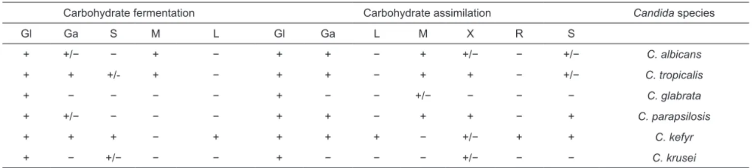
TABLE 1: Carbohydrate fermentation and carbohydrate assimilation test results of different Candida species.
Carbohydrate fermentation Carbohydrate assimilation Candida species
Gl Ga S M L Gl Ga L M X R S
+ +/− − + − + + − + +/− − +/− C. albicans
+ + +/- + − + + − + + − +/− C. tropicalis
+ − − − − + − − +/− − − − C. glabrata
+ +/− − − − + + − + + − + C. parapsilosis
+ + + − + + + + − +/− + + C. kefyr
+ − +/− − − + − − − +/− − − C. krusei
Gl: glucose; Ga: galactose: L: lactose; M: maltose; R: raffinose; S: sucrose; X: xylose. METHODS
Microorganisms
We evaluated 99 clinical Candida isolates that were randomly collected from patients treated at two different
hospitals in Ankara, Turkey, between July 2005 and March
2014. Collected isolates were inoculated into the brain heart
infusion (BHI) broth (Lab M Ltd, Lancashire, UK) media supplemented with 10% glycerol and stored at -20°C for use in
further experiments.
Identification tests
Colony morphologies and microscopic images of collected isolates were examined. By visual inspections, cells and colonies suspected to be Candida were subjected to carbohydrate fermentation, carbohydrate assimilation, and ChromAgar tests.
Carbohydrate fermentation tests
Carbohydrate fermentation tests were performed as per the method described by Bhavan10, with some modifications.
Briefly, nutrient broth media supplemented with 1% (v/v)
bromothymol blue as a pH indicator and carbohydrates such as glucose, galactose, lactose, maltose, and sucrose were separately
prepared. A total of 10μL of each Candida isolate suspended
in McFarland 0.5 standard in 5mL of saline buffer was added into 96 wells containing 100μL of different carbohydrate media. The plates were incubated at 37°C for 48h. Fermentation of any
carbohydrate was considered as positive upon the change in the
color of bromothymol blue to yellow. A total of 99 Candida
isolates were identified according to their positive/negative
carbohydrate fermentation test results, as presented in Table 14,11-15.
Carbohydrate assimilation tests
Carbohydrate assimilation tests were carried out as per the method described by Marinho et al.4, with some modifications.
Briefly, 2% (w/v) carbohydrate solutions of glucose, galactose, lactose, maltose, sucrose, xylose, and raffinose were separately
prepared and deposited onto sterile blotting paper discs prepared
from eight layers of Whatman No. 1 filter paper. Each Candida
isolate suspended in McFarland 0.5 standard in 5mL of saline buffer was inoculated onto 1% yeast nitrogen base (YNB)
agar medium (Difco™). The sterile carbohydrate discs were
placed onto the agar plates and the plates were incubated at
37°C for 48h. Assimilation of any carbohydrate was considered
as positive with a presence of a growth zone around the
carbohydrate disc. A total of 99 Candida isolates were identified
according to their positive/negative carbohydrate assimilation
test results, as presented in Table 14,11-15.
Growths on chromagar media
Each Candida isolate was inoculated into CHROMagar™
Candida medium; (CAC, Becton Dickinson, Heidelberg,
Germany), which is designed to identify different Candida
species based on their colony colors and morphologies. All
plates were incubated at 37°C for 48h and visually observed after incubation. A total of 99 Candida isolates were identified
according to their colony morphologies on CHROMAgar™
Candida medium. In CHROMAgar™ Candida medium, smooth
colonies that appear light to medium green were considered as
C. albicans; while dark blue to metallic blue smooth colonies
were considered as C. tropicalis. Pink colonies with a whitish rough border were deemed as C. krusei, whereas pink-lavender smooth colonies were considered as C. glabrata. In addition, pink-salmon smooth colonies were deemed as Candida kefyr, while white-pale pink smooth colonies were considered as C. parapsilosis4,11,15.
18S ribosomal RNA gene sequence analysis
Within 99 Candida isolates, Candida isolates that could
not be identified by the identification tests used in this study were identified by 18S ribosomal ribonucleic acid (rRNA) gene sequence analysis (RefGen Biotechnology Co. Ltd., Ankara, Turkey).
Biofilm formation on 24-well polystyrene plates
Biofilm formation abilities of 99 different Candida isolates were determined by crystal violet binding assay described by O’Toole16, with some modifications. Briefly, single yeast
colonies were picked-up from BHI agar plate and inoculated
into 10-mL BHI broth medium and incubated at 37°C overnight.
The overnight culture was 1:100 diluted into fresh BHI medium
After incubation, the medium was gently removed and the wells were gently washed with sterile distilled water. After allowing
wells to dry, each well was stained with 1% (w/v) crystal violet (Merck)/sterile distilled water for 45 min at 25°C. Excess of
crystal violet was removed by sterile distilled water and the bound crystal violet in each well was solubilized by adding
1mL of ethanol (96.6%) solution. Solubilized crystal violet from each well was read by a spectrophotometer (Shimadzu UV - 1700, Kyoto, Japan) at 560nm wavelength. According to biofilm formations, Candida isolates were classified into four
categories as follows:
• 0 ≤ OD < 0.4: non biofilm former (NBF) • 0.4 ≤ OD < 0.8: low biofilm former (LBF) • 0.8 ≤ OD < 1.2: intermediate biofilm former (IBF) • OD ≥ 1.2: high biofilm former (HBF)
The experiment was performed in triplicates.
Antifungal susceptibility tests
Antifungal susceptibilities of HBF Candida isolates [optical
density (OD) ≥ 1.60] were determined by ATBTM Fungus 3
test kits (BioMérieux®, France). Antifungal susceptibilities of 25 HBF Candida isolates against 5-flucytosine, fluconazole,
itraconazole, and voriconazole were evaluated. Briefly, all isolates were inoculated onto sabouraud dextrose agar (SDA) and incubated at 37°C for 48h. After incubation, each Candida
isolate was suspended in saline solution, and yeast cells
corresponding to a 2.0 McFarland standard were added into ATB F2 medium (yeast nitrogen base 6.7g; glucose 6.5g; asparagine 1.5g; disodium phosphate 2.5g; trisodium citrate 2.5g; potassium nitrate 5.5g; demineralized water 1,000mL; pH: 6.5-6.8). ATB F2 media with different Candida isolates were transferred into
antifungal test strips and all the test strips were incubated at 37°C
for 48h. After incubation, minimum inhibitory concentrations
(MICs) of antifungal agents were visually determined and all the isolates were classified as resistant (R), intermediate (I), or sensitive (S) according to the MIC standards constituted by the Clinical and Laboratory Standards Institute (CLSI) (M27-A3, 2008)17. Breakpoints (mg/L) for Candida spp. were as follows:
Sensitive Intermediate Resistant
5-Flucytosine ≤ 4 8 - 16 ≥ 32
Fluconazole ≤ 8 16 - 32 ≥ 64
Itraconazole ≤ 0.125 0.25 - 0.5 ≥ 1 Voriconazole ≤ 1 2 ≥ 4
Statistical analysis
Chi-square analysis was applied to estimate differences between the effects of four different antifungal agents. Bonferroni
post-hoc test was used to evaluate antifungal susceptibility and
agents with more/less significant effects were estimated. The significance level was set at 5% and the difference between the effects of each antifungal agent was considered as significant when p-value < 0.05. Statistical analysis was performed using Statistical Package for Social Sciences (SPSS) 20.0 Software (IBM Corp, New York, USA).
RESULTS
According to all phenotypic identification tests, six different
Candida species, namely, C. kefyr (n = 1), C. glabrata (n = 8), C. albicans (n = 13), C. krusei (n = 15), C. tropicalis (n = 30), and C. parapsilosis (n = 31) were identified in this study. One of the Candida isolates could not be clearly identified by phenotypical
methods and was thought to be C. tropicalis or C. krusei. The
results of 18S ribosomal RNA gene sequence analysis identified
this strain as Candida orthopsilosis.
The frequencies of different Candida species isolated in this study show that most of the Candida species were C. parapsilosis (31.3%; n = 31) and C. tropicalis (30.3%; n = 30),
followed by C. krusei (15.2%; n = 15), C. albicans (13.1%; n
= 13), C. glabrata (8.1%; n = 8), C. kefyr (1%; n = 1), and C. orthopsilosis (1%; n = 1). Furthermore, the majority of Candida
isolates were observed to be isolated from vaginal swab (49.5%;
n = 49), followed by specimens of tracheal aspirate (10.1%; n
= 10), blood (9.1%; n = 9), sputum (9.1%; n = 9), urine (8.1%; n = 8), bronchoscopic culture (5.1%; n = 5), wound (3%; n =
3), bronchial lavage (1%; n = 1), thoracentesis (1%; n = 1), eye
(1%; n = 1), synovial fluid (1%; n = 1), and catheter (1%; n =
1). The examination of the distribution of different Candida
species in different clinical specimens revealed C. tropicalis
as the most frequent Candida species isolated from tracheal aspirate and urine specimens, while most of Candida isolates
obtained from blood specimen were identified as C. albicans
(Figure 1). C. glabrata was the only Candida species isolated
from synovial fluid and thoracentesis specimens (Figure 1).
However, all isolates obtained from catheter and bronchial lavage were C. tropicalis and all isolates isolated from wound and eye specimens were C. parapsilosis (Figure 1). Among all
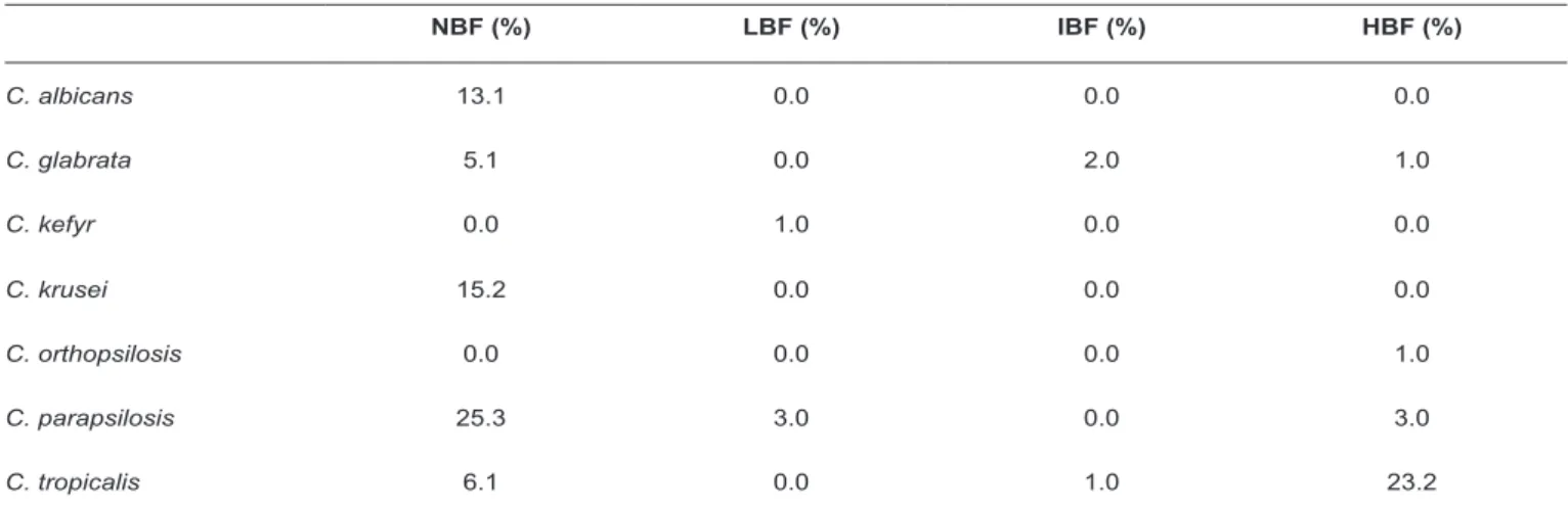
HBF Candida species such as C. tropicalis, C. parapsilosis, C. glabrata, and C. orthopsilosis, C. tropicalis was found to be the
most frequent species (23.2%; n = 23) (Table 2). However, all C. krusei isolates were found as NBF (Table 2). HBF Candida
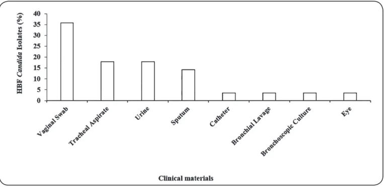
isolates were obtained from clinical samples such as vaginal swab, tracheal aspirate, urine, catheter, sputum, bronchial
lavage, bronchoscopic culture, and eye specimens (Figure 2). We evaluated the distribution of all HBF Candida isolates (n
= 28) in different clinical materials and found vaginal swab as the most frequent clinical material for HBF Candida isolate
isolation (35.7%; n = 10) (Figure 2). Furthermore, frequencies
of HBF Candida isolates obtained from tracheal aspirate (17.9%; n = 5), urine (17.9%; n = 5), and sputum (14.3% n = 4) were
higher than those of HBF Candida isolates from catheter (3.6%; n = 1), bronchial lavage (3.6%; n = 1), bronchoscopic culture
(3.6%; n = 1), and eye (3.6%; n = 1) specimens (Figure 2). The
susceptibilities of 25 HBF Candida isolates to 5-flucytosine,
fluconazole, itraconazole, and voriconazole were evaluated and all the agents were found to exhibit different effects (Chi-square test, p-value < 0.05). Most isolates were resistant to itraconazole
and voriconazole, while all were deemed sensitive to the
effect of 5-flucytosine (Figure 3). Among all the antifungal agents used in this study, 5-flucytosine was efficient against HBF Candida isolates (Bonferroni post-hoc test, p-value <
TABLE 2: Frequencies of non-biofilm-forming, low biofilm-forming, intermediate biofilm-forming, and high-biofilm forming Candida species (n = 99 isolates)*.
NBF (%) LBF (%) IBF (%) HBF (%)
C. albicans 13.1 0.0 0.0 0.0
C. glabrata 5.1 0.0 2.0 1.0
C. kefyr 0.0 1.0 0.0 0.0
C. krusei 15.2 0.0 0.0 0.0
C. orthopsilosis 0.0 0.0 0.0 1.0
C. parapsilosis 25.3 3.0 0.0 3.0
C. tropicalis 6.1 0.0 1.0 23.2
NBF: non-biofilm-forming; LBF: low biofilm-forming; IBF: intermediate biofilm-forming; HBF: high biofilm-forming; OD: optical density. *Biofilm
groups have been generated according to biofilm formation OD of tested Candida isolates given as follows: NBF: 0 ≤ OD < 0.4; LBF: 0.4 ≤ OD < 0.8;
IBF: 0.8 ≤ OD < 1.2; HBF: OD ≥ 1.2.
FIGURE 1: Distribution of different Candida species in various clinical materials (n = 99 isolates).
vitro activity against all HBF Candida isolates (n = 25) tested
(Bonferroni post-hoc test, p-value < 0.00).
DISCUSSION
In many studies, C. albicans has been regarded as the most prevalent Candida species18,19. However, the results of the present study show that C. parapsilosis and C. tropicalis
were observed in high frequencies. In addition, the frequency of C. krusei was higher than that of C. albicans, confirming
that the occurrence of NCAC species such as C. tropicalis,
C. parapsilosis, and C. krusei is increasing11,18,20 as observed in
a recent study. C. tropicalis was indicated as the most prevalent
Candida species among all Candida species isolated and is regarded as an important emerging fungal pathogen associated with high mortality rate11. In line with the results of the present
study, NCAC species are shown to be more prevalent than
C. albicans in pediatric (< 3 year) and older (> 60 year) patients
than in patients from other age groups (4-18, 19-60 years) and intensive care unit (ICU) patients21. According to other studies
FIGURE 2: Distribution of HBF Candida isolates in different clinical materials (n = 28 isolates). HBF: high biofilm-forming.
FIGURE 3: Antifungal susceptibilities of HBF Candida isolates (biofilm formation OD ≥ 1.60) (n = 25 isolates.). All antifungal agents had significantly different
effects (Chi-square test, p-value < 0.05). HBF: high biofilm-forming; OD: optical density. *Indicates that the tested HBF Candida isolates were significantly
more sensitive to 5-flucytosine than to other antifungal agents used in this study (Bonferroni post-hoc test, p-value < 0.00). **Indicates that the tested HBF Candida isolates were significantly less sensitive to voriconazole than to other antifungal agents used in this study (Bonferroni post-hoc test, p-value < 0.00).
higher than that of C. albicans, and C. parapsilosis has been indicated as a predominant pathogen of invasive candidiasis in neonates22,23.
Urine, vaginal swab, blood, indwelling biomaterial, and respiratory tract samples are found to be the most prevalent specimens for Candida isolation11,21,24. In parallel with these
findings, the majority of Candida isolates were isolated from vaginal swab specimen, followed by specimens of tracheal aspirate, blood, sputum, and urine. However, C. tropicalis was found as the most frequent Candida species in tracheal aspirate
and sputum specimens (Figure 1), contradicting the results of
previous studies on the predominance of C. albicans in lower respiratory tract specimens12,25. Themost prevalent Candida species
isolated from blood was C. albicans, confirming that C. albicans
remains the most frequent fungal species in blood specimen18.
The investigation of the distribution of Candida species in
different biofilm groups showed that C. tropicalis, which was more frequently isolated in this study, was also found as the most
prevalent HBF Candida species, whereas all other C. albicans
isolates were NBFs (Table 2). Therefore, the predominance of
C. tropicalis instead of C. albicans was thought to be related
to its enhanced biofilm formation ability24.
Applications of temporary or permanent biomaterials and medical devices in medicine have particularly led to an increase
in the incidence of biofilm-associated infections26,27. One of
catheter (Figure 2). Furthermore, HBF Candida isolates were
more frequently isolated from the clinical specimens (vaginal swab, tracheal aspirate, and urine) that were related to the body
parts that may be exposed to biomaterials such as intrauterine devices, endotracheal tubes, and urinary catheters.
The treatment of invasive fungal infections is usually carried
out with five major groups of antifungal agents, including
azoles, polyenes, allylamines, echinocandins, and pyrimidine analogues28. Fluconazole, voriconazole, and itraconazole belong
to azole class, while 5-flucytosine is a pyrimidine analogue (Figure 3). Of these, azoles that target ergosterol biosynthesis
via blockage of the enzyme lanosterol 14a-demethylase28 are the
most widely used group of antifungal agents29. A recent study evaluating the susceptibilities of different Candida species to
fluconazole, voriconazole, itraconazole, ketoconazole, and 5-flucytosine showed that the majority of Candida isolates
were sensitive to fluconazole and 5-flucytosine30. However,
5-flucytosine known to inhibit both ribonucleic acid (RNA) and deoxyribonucleic acid (DNA) synthesis after being converted to 5-fluorouracil31 was the most effective antifungal agent against
all HBF Candida species tested in the present study (Figure 3). The high resistance to fluconazole may be mainly related to the high biofilm formation ability of the tested Candida isolates.
Biofilms are known as suitable environments for horizontal gene
transfer mechanisms32. Therefore, high biofilm formation ability may play an important role in the acquisition of new antifungal resistance traits in various Candida species.
Candida tropicalis isolates that demonstrated high biofilm
formation capacity were shown to display higher rate of
resistance to fluconazole in one of the recent studies33.
In conclusion, the present study shows that C. tropicalis
displays the highest biofilm formation ability among the Candida
species evaluated. Our findings indicate high frequency of HBF
Candida isolation from clinical samples of vaginal swab, tracheal
aspirate, and urine. We also found that 5-flucytosine is the most efficient antifungal agent against HBF Candida isolates.
Acknowledgements
The authors are grateful to Abbas Yousefi Rad for the collection of clinical
specimens.
Conflict of interest
The authors declare that there is no conflict of interest.
Financial support
This study was part of a PhD and was supported by the funding received from
Scientific Research Projects Coordination Unit (grant number: FDK-2016-10821) of Hacettepe University.
REFERENCES
1. Spampinato C, Leonardi D. Candida infections, causes, targets, and resistance mechanisms: traditional and alternative antifungal
agents. Biomed Res Int. 2013;2013:1-13: 204237.
2. Sanguinetti M, Posteraro B, Lass-Flörl C. Antifungal drug resistance among Candida species: mechanisms and clinical impact. Mycoses.
2015;58(S2):2-13.
3. Hernandez S, Lo´pez-Ribot JL, Najvar LK, McCarthy DI, Bocanegra
R, Graybill JR. Caspofungin resistance in candida albicans: correlating clinical outcome with laboratory susceptibility testing of three isogenic isolates serially obtained from a patient with progressive Candida esophagitis. Antimicrob Agents Chemother.
2004;48(4):1382-83.
4. Marinho SA, Teixeira AB, Santos OS, Cazanova RF, Ferreira CAS,
Cherubini K, et al. Identification of Candida spp. by phenotypic
tests and PCR. Braz J Microbiol. 2010;41:286-94.
5. Silva S, Negri M, Henriques M, Oliveira R, Williams DW, Azeredo J. Candida glabrata, Candida parapsilosis and Candida tropicalis: biology, epidemiology, pathogenicity and antifungal resistance.
FEMS Microbiol Rev. 2012;36(2):288-305.
6. Ramage G, Martínez JP, López-Ribot JL. Candida Biofilms on
implanted biomaterials: a clinically significant problem. FEMS Yeast Res.2006;6(7):979-86.
7. Carneiro VA, dos Santos HS, Arruda FVS, Bandeira PN, Albuquerque MRJR, Pereira MO, et al. Casbane diterpene as a
promising natural antimicrobial agent against biofilm-associated
infections. Molecules.2011;16(1):190-201.
8. Donlan RM, Costerton JW. Biofilms: survival mechanisms of clinically relevant microorganisms. Clin Microbiol Rev.
2002;15(2):167-93.
9. Silva S, Rodrigues CF, Ara D, Rodrigues ME, Henriques M.
Candida species biofilms’ antifungal resistance. J Fungi (Basel).
2017;3(1). pii: E8. doi: 10.3390/jof3010008.
10. Bhavan PS, Rajkumar R, Radhakrishnan S, Seenivasan C, Kannan
S. Culture and identification of Candida albicans from Vaginal ulcer
and separation of enolase on SDS-PAGE. Int J Biol.2010;2(1):84-93.
11. Yesudhason BL, Mohanram K. Candida tropicalis as a predominant isolate from clinical specimens and its antifungal susceptibility pattern in a Tertiary Care Hospital in Southern India.J Clin Diagn
Res. 2015;9(7):DC14-6.
12. Ogba OM, Abia-Bassey LN, Epoke J, Mandor BI, Iwatt GD. Characterization of Candida species isolated from cases of lower
respiratory tract infection. World J AIDS 2013;3:201-6.
13. Lachance MA, Boekhout T, Scorzetti G, Fell JW, Kurtzman CP.
Candida Berkhout. In: Kurtzman CP, Fell JW, Boekhout T, editors.
The yeasts, a taxonomic study. Amsterdam: Elsevier; 2011. p.
987-1278.
14. Manjunath V, Vidya GS, Sharma A, Prakash MR, Murugesh. Speciation of Candida by Hicrome agar and Sugar assimilation test in both HIV infected and non-infected patients.Int J Biol Med Res.
2012;3(2):1778-82.
15. Jose NV, Mudhigeti N, Asir J, Chandrakesan SD. Detection of virulence factors and phenotypic characterization of Candida
isolates from clinical specimens. J Curr Res Sci Med.2015;1:27-31. 16. O’Toole GA. Microtiter dish biofilm formation assay. J Vis Exp
2011; (47):e2437.
17. Clinical and Laboratory Standards Institute (CLSI). Reference Method for Broth Dilution Antifungal Susceptibility Testing of
Yeasts; Approved Standard-Third Edition. Document M27-A3. Wayne, PA: CLSI; 2008. 13p.
18. Wisplinghoff H, Ebbers J, Geurtz L, Stefanik D, Major Y, Edmond MB, et al. Nosocomial bloodstream infections due to Candida Spp. in the USA: species distribution, clinical features and antifungal
19. Liu XP, Fan SR, Peng YT, Zhang HP. Species distribution and
susceptibility of Candida isolates from patient with vulvovaginal
candidiasis in Southern China from 2003 to 2012. J Mycol Med. 2014;24(2):106-11.
20. Almeida AA, Mesquita CS, Svidzinski TI, Oliveira KM. Antifungal susceptibility and distribution of Candida spp. isolates from the University Hospital in the municipality of Dourados, State of Mato
Grosso do Sul, Brazil. Rev Soc Bras Med Trop. 2013;46(3):335-9.
21. Pahwa N, Kumar R, Nirkhiwale S, Bandi A. Species distribution and drug susceptibility of candida in clinical isolates from a Tertiary
Care Centre at Indore. Indian J Med Microbiol. 2014;32(1):44-8.
22. Garzillo C, Bagattini M, Bogdanović L, Di Popolo A, Iula VD, Catania MR, et al. Risk factors for Candida parapsilosis bloodstream infection in a neonatal intensive care unit: a case-control study. Ital
J Pediatr. 2017;43(1):10.
23. Chow BDW, Linden JR, Bliss JM. Candida parapsilosis and the
Neonate: Epidemiology, Virulence and Host Defense in a Unique Patient Setting. Expert Rev Anti Infect Ther. 2012;10(8):935-46.
24. Silva AP, Miranda IM, Lisboa C, Pina-Vaz C, Rodrigues AG.
Prevalence, distribution, and antifungal susceptibility profiles of
Candida parapsilosis, C. orthopsilosis, and C. metapsilosis in a
Tertiary Care Hospital. J Clin Microbiol. 2009;47(8):2392-97. 25. Bailly S, Maubon D, Fournier P, Pelloux H, Schwebel C, Chapuis
C, et al. Impact of antifungal prescription on relative distribution and susceptibility of Candida spp. - Trends over 10 years. J Infect.
2016;72(1):103-11.
26. Junter GA, Thébault P, Lebrun L. Polysaccharide-based antibiofilm
surfaces. Acta Biomater. 2016;30:13-25.
27. Dutta Sinha S, Chatterjee S, Maiti PK, Tarafdar S, Moulik SP.
Evaluation of the role of substrate and albumin on Pseudomonas aeruginosa biofilm morphology through FESEM and FTIR studies
on polymeric biomaterials. Prog Biomater. 2017;6(1-2):27-38.
28. Mathé L, Van Dijck P. Recent Insights into Candida albicans biofilm
resistance mechanisms. Curr Genet. 2013;59(4):251-64.
29. Roemer T, Krysan DJ. Antifungal drug development: challenges,
unmet clinical needs, and new approaches. Cold Spring Harb Perspect Med. 2014;4(5):pii: a019703. doi: 10.1101/cshperspect.
a019703.
30. Razzaghi-Abyaneh M, Sadeghi G, Zeinali E, Alirezaee M,
Shams-Ghahfarokhi M, Amani A, et al. Species distribution and antifungal susceptibility of Candida spp. isolated from superficial candidiasis
in outpatients in Iran. J Mycol Med. 2014;24(2):e43-50.
31. Vermes A, Guchelaar HJ, Dankert J. Flucytosine: a review of its
pharmacology, clinical indications, pharmacokinetics, toxicity and
drug interactions. J Antimicrob Chemother. 2000;46(2):171-9. 32. Hall-Stoodley L, Stoodley P. Biofilm formation and dispersal
and the transmission of human pathogens. Trends Microbiol.
2005;13(1):7-10.
33. Deorukhkar SC, Saini S, Mathew S. Virulence factors contributing to pathogenicity of Candida tropicalis and Its antifungal