Species distribution and antifungal susceptibility
patterns of
Candida
isolates from a public tertiary
teaching hospital in the Eastern Cape Province,
South Africa
P. Mnge
1, B.I. Okeleye
1,3, S.D. Vasaikar
1,2and T. Apalata
1,2 1Division of Medical Microbiology, Department of Pathology and Laboratory Medicine, Faculty of Health Sciences,Walter Sisulu University, Mthatha, South Africa
2National Health Laboratory Services, Mthatha, South Africa 3Phytomedicine and Phytopharmacology Research Group, Department of Plant Science, University of the Free State,
Phuthaditjhaba, South Africa
Abstract
Candidaspecies are the leading cause of invasive fungal infections, and over the past decade there has been an increased isolation of drug resistantCandidaspecies. This study aimed to identify the species distribution of Candidaisolates and to determine their unique antifungal susceptibility and resistance patterns. During a cross-sectional study, 209Candidaisolates (recovered from 206 clinical samples) were collected and their species distribution was determined using ChromAgarCandida. The Vitek-2 system (Biomerieux, South Africa) was used to determine minimum inhibitory concentrations (MICs) to azoles (fluconazole, voriconazole), echinocandins (caspofungin, micafungin), polyenes (amphotericin B) andflucytosine. Four species ofCandidawere isolated, of whichC. albicanswas the most frequent, isolated in 45.4% (95/209) of the isolates, followed by
C. glabrata: 31.1% (65/209). The MICs of the different antifungal drugs varied amongst the species ofCandida. From the 130 isolates tested for MICs, 90.77% (112/130) were susceptible to all antifungal drugs and 6.9% (9/130) of the isolates were multi-drug resistant.C. dubliniensis(n=2) isolates were susceptible to all the above mentioned antifungal drugs. There was no significant difference in species distribution amongst clinical specimens and between patients’genders (P40.05). An increase in MIC values forfluconazole andflucytosine towards the resistance range was observed. To our knowledge, this is thefirst report on surveillance of Candida species distribution and antifungal susceptibility at a public tertiary teaching hospital in Eastern Cape, South Africa.
Key words: Candidaspecies; Distribution; Antifungal susceptibility; Identification; South Africa
Introduction
Candidaspecies are commensal fungi of the human gastrointestinal tract, lower genital tract and mouth cavity. Among immunocompetent individuals, Candida species have an inherently low virulence. The incidence of candi-diasis is more frequent in immunocompromised patients with impaired physiological and cellular barriers.Candida species colonize and invade host tissues wherein they cause localized to invasive systemic infections, which dis-seminate hematogenously to various organs of the body (1). Candidaspecies are the leading cause of mycoses worldwide and the fourth leading cause of invasive noso-comial bloodstream infections with significant crude mortal-ity and morbidmortal-ity rates (2–5). In South Africa, the incidence and prevalence ofCandidaspecies is not well documented,
howeverC. albicans remains the leading cause of candi-diasis worldwide (6–9). Recent epidemiological reports indi-cate a change in species distribution patterns ofCandida infections, with an increasing frequency of non-Albicans Candidaspecies such asC. glabrataandC. parapsilosis being isolated from clinical samples (10,11).
In African health care settings, amphotericin B and fluconazole are routinely used to treatCandidainfections (9,12). The changes in the epidemiology ofCandida spe-cies is in parallel with the emergence of antifungal drug resistant species. Antifungal drug resistance is associated with an uncontrolled distribution and prolonged use of antifungals to treat recurrent infections in immunocompro-mised patients (13,14). Furthermore, as it is imperative for
Correspondence: B.I. Okeleye:<[email protected]>
laboratories to provide identification to the species level, there is continued isolation of new species which are resis-tant to currently available antifungals.
The growing trend in antifungal drug resistance and emergence of new species ofCandida,poses a need for regional surveillance of antifungal drug susceptibility pro-files, sincein vitro drug susceptibility patterns are asso-ciated with therapeutic outcome. This study sought to determine the distribution andin vitrosusceptibility profiles ofCandidaspecies isolated from patients attending Nelson Mandela Academic Complex, a public tertiary teaching hospital in Mthatha, Eastern Cape.
Material and Methods
Study population and sampling strategy
A total of 209Candidaisolates (from 206 clinical sam-ples) were collected during a cross-sectional study among patients at Nelson Mandela Academic Complex in Mthatha. A standardized data collection form was used to collect information on patients’demographics (age and gender) and clinical history from medical records. Permission to collect patient’s clinical data, including laboratory informa-tion, was obtained from the hospital and laboratory man-agers. Ethical clearance was obtained from the Research Ethics Committee of Walter Sisulu University (Ethics Ref. No. 038/13).
Isolation and identification ofCandidaspecies Yeast cells isolated from clinical samples were stored in 10% glycerol (Sigma-Aldrich, South Africa) at –20°C until further use. Isolates were sub-cultured onto freshly prepared Sabaroud dextrose (SAB) agar and incubated overnight at 37°C. The germ tube test was used for the presumptive identification of Candida species. Briefly, 24-h fresh cultures were inoculated on 3 drops of human serum and incubated at 37°C. After 2.5 h, the formation of germ tubes was observed under microscopy (40
objective).
The ability of Candida species to form differentially colored colonies on chromogenic assay (ChromAgar Candida, Media-mage, South Africa) was used to identify Candidaisolates to the species level. Inocula from 24–48 h SAB-agar cultures were re-cultured onto commercially pre-pared ChromAgarCandidaplates and incubated for 48–72 h at 37°C. Intense colony coloration was observed after incu-bation and species differentiation was done according to the manufacturer’s instructions (14,15).
Antifungal susceptibility assay
The antifungal susceptibility profile ofCandidaspecies was determined using the Vitek 2 Systems Version 07.01 (Biomeriex, South Africa) following the CLSI document M27-A3 (2015) (16). The antifungal agents tested were amphotericin B,fluconazole, voriconazole, caspofungin, micafungin and flucytosine. The test was carried out
according to the manufacturer’s instructions. About 2–3 colonies of 24-h Candida cultures were inoculated into 5-mL glass tubes containing 3 mL of 10% saline, adjusted to 2 McFarland standards. Vitek 2 cards with 12-fold serial dilutions of antifungals were placed onto the test tube and loaded onto the Vitek cassette. Loaded cassettes were then placed onto the Vitek instrument and incubated for 9 to 33 h depending on the sample. Standard strains C. albicansATCC 90028 andC. parapsilosisATCC 22019 were used for quality control. The antifungal susceptibility of the isolates was interpreted as sensitive (S), interme-diate (I), and resistant (R) according to the CLSI inter-pretative breakpoints criteria (16,17). The chi-square test was used to test for significant associations between the defined variables, while ANOVA was used to test the difference between groups. SPSS (IMB, USA) version 20.0 for Windows was used for all statistical analyses.
Results
Species distribution
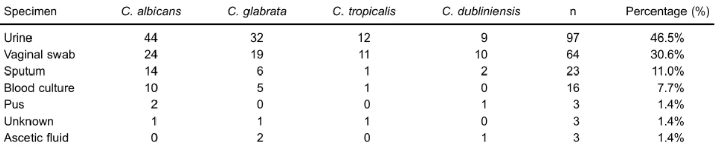
A total of 209 isolates ofCandidawere obtained from 206 clinical specimens; the highest number of isolates were from urine specimens (46.5%, n=97), followed by vaginal swabs (30.6%, n=64). The mean age of the patients was 29.7±1.97 years, ranging from 1 month to 87 years. The gender distribution of patients, based on clinical records was 148 (71.9%) females and 46 (22.3%) males. For 5.8% (12/206) isolates, the gender was not stated in clinical records.C. albicansaccounted for 45.5% (95/209) of the species isolated while 31.1% (65/209) of the spe-cies were C. glabrata, 12.4% (26/209)C. tropicalis, and C. dubliniensisaccounted for 11.0% (23/209) of the total isolates (Table 1). There was no significant difference in species distribution amongst clinical specimens (X2=36; DF=66, and P=0.99) and between patients’ genders (X2=11.964; DF=22, and P=0.958).
Antifungal susceptibility testing
The minimum inhibitory concentrations (MICs) and antifungal susceptibility ofCandidaspecies to the various antifungal drugs are summarized in Tables 2 and 3. The results are presented by species as cumulative counts of susceptible organisms at each concentration throughout the full dilution series.
Table 1.Distribution ofCandidaspecies among clinical specimens (n=209).
Specimen C. albicans C. glabrata C. tropicalis C. dubliniensis n Percentage (%)
Urine 44 32 12 9 97 46.5%
Vaginal swab 24 19 11 10 64 30.6%
Sputum 14 6 1 2 23 11.0%
Blood culture 10 5 1 0 16 7.7%
Pus 2 0 0 1 3 1.4%
Unknown 1 1 1 0 3 1.4%
Asceticfluid 0 2 0 1 3 1.4%
Table 2.Minimum inhibitory concentrations (MIC) of clinical isolates ofCandida(n=130) to various antifungal drugs.
Antifungal MIC range (mg/mL)*
0.065 0.125 0.25 0.5 1 2 4 8 16 32 64
Fluconazole
C. albicans 84 4 4 10 1 6
C. glabrata 5 3 1 2
C. tropicalis 5 2 2
C. dubliniensis 2
Voriconazole
C. albicans 96 4 1 4 1 1 2
C. glabrata 9 2
C. tropicalis 7 1 1
C. dubliniensis 2
Caspofungin
C. albicans 102 3 4 4
C. glabrata 9 2
C. tropicalis 6 2 1
C. dubliniensis 2
Micafungin
C. albicans 101 2 2 4
C. glabrata 6 3 2
C. tropicalis 6 1 2
C. dubliniensis 2 Amphotericin B
C. albicans 72 31 2 4
C. glabrata 3 6 2
C. tropicalis 6 1 2
C. dubliniensis 2
Flucytosine
C. albicans 103 2 2 2
C. glabrata 7 3 1
C. tropicalis 6 1 1 1
C. dubliniensis 2
A similar resistance pattern was observed in 2 isolates of bothC. glabrataandC. tropicalis. One isolate from each species was fully resistant to all antifungal drugs, while the other isolate was resistant to all antifungal drugs except for flucytosine at MIC X64 mg/mL in C. tropicalis and MIC of 32 mg/mL in C. glabrata. Notably, at flucytosine MIC ofp1mg/mL (susceptible), 3 (2.6%)C. albicansand 2 (18.2%)C. glabrata isolates were resistant to all other classes of antifungals. Two isolates ofC. albicanswere resis-tant to all antifungals, exceptflucytosine (MICp16mg/mL) and intermediate to voriconazole at MIC of 2mg/mL.
Discussion
The study sought to compare the species distribution pattern ofCandidaand to compare the antifungal suscep-tibility patterns of 6 antifungal drugs against the isolated species ofCandida. The most common species isolated wasC. albicans, and thisfinding is similar to previously published reports in South Africa, whereC. albicanswas the most commonly isolated species from clinical samples (9,12). In a study that determined the microbial carriage in bloodstream infections, C. albicans was responsible for 31% (21/68) of all fungal infections (18). Thefindings of this study also correlate with reports from the Neonatal Intensive Care Unit in Dr. George Mukhari Hospital, South
Africa, where C.albicanswas the most common species isolated at the unit in 2002 (9). A recent report by Makhado et al. (9) has, however, indicated a shift in the epidemiology of candidiasis withC. kruseireplacing C.albicansas the most common species isolated. AlthoughC. albicanswas the most common species isolated in this present study, the rate of non-albicansCandidaspecies varied amongst the clinical specimens and these species were isolated in more than 50% of the clinical samples.
In contrast to C. albicans being the most commonly isolated species, C. glabrata, C. krusei and C. tropicalis are also reported to be leading causes of candidiasis. The frequency of these non-albicans Candidaspecies varies throughout the world. This variation may be a result of the underlying medical condition, geographic distribution and patients’age and gender (1,19–21).C. glabrataand
C. tropicalis are the most common species isolated in neutropenic patients, in catheter related infections, and in adults, whileC. kruseihas been reported as the leading cause of candidemia. The increase in isolation of non-albicansCandidaspecies is in parallel with a decrease in the isolation ofC. albicans. (1,14,18,20)
The high frequency ofCandidaspecies isolated from vaginal and urine samples from female patients can be explained by the imbalance in vaginal microflora as a result of diabetes and vaginal estrogenization, which in turns Table 3.Antifungal susceptibility patterns ofCandidaspecies to different antifungal drugs.
Antifungal Species (n=130)
C. albicans (n=109)
C. glabrata (n=11)
C. tropicalis (n=8)
C. dubliniensis (n=2)
P
Fluconazole 0.277
S 102 (93.6%) 9 (81.8%) 6 (75%) 2 (100%)
I 2 (1.8%)
R 5 (4.6%) 2 (18.2%) 2 (25%)
Voriconazole 0.061
S 105 (96.3%) 9 (81.8%) 6 (75%) 2 (100%)
I 1 (0.9%)
R 3 (2.8%) 2 (18.2%) 2 (25%)
Caspofungin 0.065
S 105 (96.3%) 9 (81.8%) 6 (75%) 2 (100%)
R 4 (3.7%) 2 (18.2%) 2 (25%)
Micafungin 0.068
S 105 (96.3%) 9 (81.8%) 6 (75%) 2 (100%)
R 4 (3.7%) 2 (18.2%) 2 (25%)
Amphotericin B 0.068
S 105 (96.3%) 9 (81.8%) 6 (75%) 2 (100%)
R 4 (3.7%) 2 (18.2%) 2 (25%)
Flucytosine 0.0934
S 102 (93.6%) 10 (90.9%) 7 (87.5%) 2 (100%)
I 2 (1.8%)
R 5 (4.6%) 1 (9.1%) 1 (12.5%)
gives rise to vulvovaginal candidiasis (21,22). Sympto-matic vulvovaginal candidiasis (VVC) occurs in women aged 18–84 years and is associated with a significant morbidity rate. Approximately 75% of women experience one episode of VVC in their lifetime (23–25). The underlying medical conditions for the isolates were vaginal discharge, retroviral disease and pelvic inflammatory discharge. On most urine samples, the clinical diagnosis was stated as illegible thus making it difficult to ascertain if the presence of yeast in urine represents a true infection or merely colonization and contamination of the bladder (26).
The ongoing change in the epidemiology ofCandida species is in parallel with the emergence of antifungal drug resistantCandidaspecies (14). This is especially true forfluconazole, which is used as thefirst line drug treatment for hematological malignancy, HIV/AIDS and oropharyngeal candidiasis in South Africa and Africa as a whole. This, in turn, leads to the selection of less susceptible isolates, with an inherent or acquired resistance tofluconazole, especially C. albicansandC. tropicalisisolates (8,9,12,13,27). The wide-spread use of fluconazole does not only lead to selection of antifungal drug resistant species, but also to a shift from C. albicans as the leading cause of candidiasis to non-albicans species such asC. glabrataandC. tropicalisas the causative agents ofCandidainfections (14,28).
Relatively low levels of fluconazole resistance were observed in all species of Candida isolated; it is worth mentioning that the non-albicansspecies had the highest levels of resistance compared toC. albicans. Primary resistance to fluconazole is reported in C. albicans, C.tropicalisand C.krusei isolates. Multi-drug resistance was observed inC. albicans,C. glabrataandC. tropicalis isolates, indicating a major public health concern and reflecting the inappropriate use of antifungals drugs. This also supports the importance of ongoing in vitro sur-veillance and careful monitoring of antifungal treatment regimens since in vitro resistance is associated with therapeutic failure (12,14,29).
The lowest MIC (0.065mg/mL) in all species ofCandida was observed in micafungin (Table 2); this echinocandin class of antifungals is said to confer excellent fungicidal effects against Candida species. Echinocandins are not routinely used in antifungal therapy due to their hepatic effect. Flucytosine resistance, more especially inC. glabrata isolates, is reported when the drug is administered as monotherapy. It is, therefore, suggested that in order to achieve optimal flucytosine activity, the drug should be administered in combination with amphotericin B, and/or any azole and echinocandins (30–32).
The antifungal susceptibility of echinocandins and liposomal amphotericin B against Candida biofilms was investigated by Marcos-Zambrano (33). It was found that C. tropicalis biofilms had the highest level of resistance and amphotericin B was unable to reduce the metabolic activity of the biofilms. In a study conducted by Blignaut et al. (34), the South African clade ofC. albicanshad an 8.4% level of resistance to amphotericin B and the clade was found to be naturally resistant to that drug. That find-ing is supported in this study, in whichC. tropicalisisolates had a 25% level of resistance compared to C. albicans and C. glabrata.Although the results are statistically not significant, thisfinding has to be considered when ampho-tericin B is used to treat C. tropicalisinfections. This is equally important, since amphotericin B is routinely used to treatfluconazole-resistant infections (33).
There was a degree of variability in the MIC values within the same class of antifungals, for example the MIC range for micafungin was between 0.025 to 4mg/mL and caspofungin MIC ranged from 0.065 to 8 mg/mL. On the other hand, MIC values forfluconazole ranged from 1 to 64 mg/mL and for voriconazole from 0.125 to 16mg/mL. A similar pattern was also observed by Villareal et al. (35) inC. glabrataisolates. The differences in MIC values within species and classes are indicative of species-specific resistance patterns. Hence, there is a need for regional surveillance of fungal species distribution, and antifungal therapeutic regimes should be implemented based on epidemiological data and antifungal sensitivity. The low MICs and increased spectrum of activity for voriconazole and micafungin inC. albicansand non-albicansspecies suggest good clinical activity for these drugs.
The epidemiology of Candida infections in Africa, a home for new and emerging drug-resistantCandida spe-cies, is not well documented (9,14), and this study pre-sented thefirst regional surveillance in the Eastern Cape province to investigate the prevalence and antifungal susceptibility patterns ofCandidaisolates.
Acknowledgments
We would like to thank the staff of National Health Lab-oratory Services (Mthatha) for their support in sample col-lection, Mrs. N. Sibathathu for her technical assistance in conducting laboratory experiments, the National Research Foundation (South Africa) forfinancial support, and lastly the staff of Division of Medical Microbiology, Department of Pathology and Laboratory Medicine (Walter Sisulu Univer-sity) for assisting the successful completion of the study.
References
1. Quindos G. Epidemiology of candidemia and Invasive candidiasis changing face. Rev Iberoamer Micol 2013; 271: 1–7.
3. Sanglard D, Kuchler K, Fischer F, Pagani JL, Monod M, Bille J. Mechanisms of resistance to azole antifungal agents in Candida albicansisolates from AIDS patients involves spe-cific multidrug transporters. Antimicrob Agents Chemother 1995; 39: 2378–2386, doi: 10.1128/AAC.39.11.2378. 4. Bruder-Nascimento A, Camargo CH, Sugizak MF, Sadatsune
T, Montelli AC, Mondelli AL, et al. Species distribution and susceptibility profile ofCandidaspecies in a Brazilian public tertiary hospital.BMC Res Notes2010; 3: 1–5, doi: 10.1186/ 1756-0500-3-1.
5. Bhooshon S, Gayal A, Agrawa lA, Verma V. Prevalence and drug resistant ofCandida speciesin pediatrics patients in Tertiary Care Hospital, North India.J Microbiol Biomed Res2015; 1: 1–6.
6. Okonko IO, Odu NN, Kolade A, Nwanze JC. Detection and prevalence ofCandidaisolates among patients in Ibadan, Southwestern Nigeria. J Microbiol Biotechnol Res 2011; 1: 176–184.
7. Pfaller MA, Diekama DJ. Epidemiology of invasive candi-diasis: A persistent public health problem.Clin Microbiol Rev 2007; 20: 133–163, doi: 10.1128/CMR.00029-06.
8. Owatade FJ, Gulube Z, Ramla S, Patel M. Antifungal sus-ceptibility ofCandida albicansisolated from the oral cavities of patients with HIV infection and cancer. South African Dental J2016; 71: 8–11.
9. Makhado NA, Ismal F, Dango Y, Chephe TJH, Hoosen AA, Chabeleng M. Antifungal susceptibility profile of yeast isolates from sterile sites at a public teaching hospital in South Africa.South African J Infect Dis2014;29: 97–100. 10. Nnadi NE, Ayanbimpe GM, Scordino F, Okolo MO, Enwean
IB, Crisea G, et al. Isolation and molecular characteriza-tion ofC. Africanafrom Jos, Nigeria.Med Mycol2012: 50: 765–767, doi: 10.3109/13693786.2012.662598.
11. Chow JK, Golan Y, Ruthezer R, Karchmer AW, Carmeli Y, Lichtenberg D, et al. Factors associated with candidemia caused by non-albicansspecies andCandida albicans in the intensive care unit.Clin Infect Dis2008; 46:1206–1213, doi: 10.1086/529435.
12. Abrantes PMDS, McArthur CP, African CWJ. Multi-drug resistance (MDR) oralCandidaspecies isolated from HIV-positive patients in South Africa and Cameroon.Diag Micro-biol Infect Dis2014; 79: 222–227, doi: 10.1016/j.diagmicro bio.2013.09.016.
13. Hajjeh RA, Sofar AN, Harrison LH, Lyon GM, Hartington-Skaggs BA, Mirza SA, et al. Incidence of bloodstream infec-tions due toCandidaspecies andin vitrosusceptibilities of isolates collected from 1998 to 2000 in a population-based active surveillance program. J Clin Microbiol 2004; 42: 1519–1527, doi: 10.1128/JCM.42.4.1519-1527.2004. 14. Efushnie AM, Oduyebo O, Osuagwu CS, Koenig B. Species
distribution and antifungal susceptibility ofCandidaIsolates from pregnant women in a tertiary hospital in nigeria.African J Clin Exper Microbiol 2016; 17: 183–189, doi: 10.4314/ ajcem.v17i3.5.
15. ChromAgar: The Chromogenic Media Pioneer. NT-EXT-001. Version 7.
16. Wayne PA. Reference method for broth dilution antifungal susceptibility testing of yeasts. Clin Lab Standards Inst 2008; 3: M27-A3.
17. Behzadi P, Behzadi E, Ranjbar R. Urinary tract infections andC. albicans.Centr Eur J Eurol2015; 68: 96–101.
18. Dramowski A, Cotton MF, Rabie H, Whitelaw A. Trends in paediatric bloodstream infections at a South African referral hospital.BMC Paediatrics 2015; 15: 33–44, doi: 10.1186/ s12887-015-0354-3.
19. Olaniran O, Adefusi OF, Idowu OJ, Oladipo OA, Afolayan DO, Aderibigbe I, et al. Isolation and evaluation Candida species of among pregnant women in Obafemi Awolowo University teaching hospital, ile-ife. Nigeria.J. Clin Microbiol Case Reports2015; 1: 1–6.
20. Fortún J, Martin-Dávila P, Pedrosa EG, Pintado V, Cobo T, et al. Emerging trends in candidemia: A higher incidence but a similar outcome. J. Infect 2012, 65: 64–70, doi: 10.1016/j. jinf.2012.02.011.
21. Yapur N. Epidemiology and risk factors for invasive candidiasis. Ther Clin Risk Manag 2014; 10: 95–105, doi: 10.2147/TCRM.S40160.
22. Mendling W, Brasch J. Guideline vulvovaginal candidosis (2010) of the German society for gynecology and obstetrics, the working group for infections and infectimmunology in gynecology and obstetrics, the germansociety of dermatol-ogy, the board of german dermatologists and the german speaking mycological society. Mycoses 2012; 55: 1–13, doi: 10.1111/j.1439-0507.2012.02185.x.
23. Shrivastav VK, Shukla D, Shrivastav A, Jana AM. Pre-valence of vaginal candidiasis in diabetic women of Madhya Pradesh, India. Int J Current Microbiol Appl Sci 2015; 4: 834–846
24. Apalata T, Carr WH, Sturm WA, Longo-Mbenza B, Moodley P. Factors associated with symptomatic vulvovaginal candidiasis: A study among women attending a primary healthcare clinic in Kwazulu-Natal, South Africa.Ann Med Health Sci Res2014; 3: 410–416, doi: 10.4103/2141-9248. 133470.
25. Oyewole OA, Okoliegbe IN, Alkhalil S, Isah P. Prevalence of vaginal candidiasis among pregnant women attending Federal University of Technology, Minna, Nigeria, Bosso Clinic.Res J Pharm Biol Chel Sci2013; 4: 113–120. 26. Nucci M, Colombo AL. Emergence of resistantCandidain
neutropenic patients.Braz J Infect Dis2002; 6: 124–128, doi: 10.1590/S1413-86702002000300004.
27. Sebti A, Kiem TE, Peili D, Cathurvedi V, Wong M, Doney A, et al.Candida dubliniensisat a cancer.Clin Infect Dis2001; 32: 1034–1038, doi: 10.1086/319599.
28. Perlin DS, Shor E, Zhao Y. Update on antifungal drug resistance.Curr Clin Micro Rpt2015; 2: 84–95, doi: 10.1007/ s40588-015-0015-1.
29. Mulu A, Kassu A, Anagaw B, Moges B, Gelaw A et al. Frequent detection of ‘azole’ resistant Candida species among late presenting AIDS patients in northwest Ethiopia.BMC Infect Dis 2013; 13: 1–10, doi: 10.1186/1471-2334-13-82.
30. Mukherjez P, Wang M. Antifungal drug resistance: signifi -cance and mechanisms.Informa Healthcare2010: 63–86. 31. Charlier C, Sissy CEL, Bachelier-Bassi S, Scemla A,
Quesne G, Sitterlé E, et al. Acquiredflucytosine resistance during combination therapy with caspofungin andflucytosine forCandida glabratacystitis.Antimicrob Agents Chemother 2016; 60: 662–665, doi: 10.1128/AAC.02265-15.
33. Marcos-Zambrano LJ, Gómez-Perosanz M, Escribano P, Zaragoza O, Bouza E, Guinea J. Echinocandin-resistant yeast species: biofilm production and anti-biofilm activity of echinocandins and liposomal amphotericin B. Antimicrob Agents Chemother2015:1–28.
34. Blignaut E, Molepo J, Pujol C, Soll DR, Pfaller MA. Clade-related amphotericin B resistance among South African
Candida albicansisolates.Diagn Microbiol Infect Dis2005; 53: 29–31, doi: 10.1016/j.diagmicrobio.2005.03.013. 35. Villareal NC, Fothergill AW, Kelly C, et al. Candida