Vol.60: e17160385, January-December 2017 http://dx.doi.org/10.1590/1678-4324-2017160385
ISSN 1678-4324 Online Edition
BRAZILIAN ARCHIVES OF BIOLOGY AND TECHNOLOGY A N I N T E R N A T I O N A L J O U R N A L
Production of Antimicrobial Metabolites by
Streptomyces
lavendulocolor
VHB-9 Isolated from Granite Mines
Hima Bindu B.S.S.N
1, Vijayalakshmi Muvva
1*, Rajesh Kumar Munaganti
1, Krishna
Naragani
1, Saidulu Konda
2, Kumar Reddy Dorigondla
21 Department of Botany and Microbiology, Acharya Nagarjuna University, Guntur-522510, A.P, India. 2 Organic Chemistry Division-II (CPC), c Organic Chemistry Division-I (Natural products Laboratory),
CSIR-Indian Institute of Chemical Technology, Hyderabad-500007, A.P, India.
ABSTRACT
The actinobacterial strain Streptomyces lavendulocolor VHB-9 was isolated from granite mine soil samples of Khammam district, Telangana state, India. The strain was identified based on detailed microorphological, cultural and phylogenetic analysis. Bioactive guided isolation of the secondary metabolites of the strain was carried out by growing the strain in optimized medium (0.5% lactose, 0.5% peptone, 0.05% K2HPO4, 0.2% CaCO3 with pH
adjusted to 7.0). Separation and purification of the active fractions from the crude ethyl acetate extract was carried out by silica gel column chromatography and resulted in the isolation of two active fractions. Structural elucidation of the two (B2 and B3) active compounds was carried out by FT-IR, Mass and NMR spectroscopy and were identified as Bis (7-methyloctyl) phthalate and (Z)-3-aminoacrylic acid. The antimicrobial activity of the bioactive compounds produced by S. lavendulocolor VHB-9 was expressed in terms of minimum inhibitory concentration against opportunistic pathogenic bacteria and fungi. Both fractionsexhibited good antimicrobial potential against the bacteria and fungi tested.
Key words:Granite,Streptomyces lavendulocolor, spectroscopy, Bis (7-methyloctyl) phthalate, (Z)-3-aminoacrylic acid.
*
INTRODUCTION
Natural products are the leading sources of novel biomolecules being used in the pharmaceutical and nutraceutical industries since their inception. Majority of the natural products currently used in the market as therapeutic agents or as health supplements are derived from the terrestrial organisms including plants, animals and microorganisms. Microbial secondary metabolites are the important sources of natural compounds with potential bioactivities being used in different fields such as medicine, agriculture and others. The search for novel natural products with useful pharmacological activities often includes the isolation of actinomycetes, such as
Streptomyces species, from soil samples. 1-2
A vast majority of these compounds are derived from the single actinomycete genus
Streptomyces, raising the intriguing possibility that additional chemically prolific taxa await their discovery. They are broadly used in the human therapy, veterinary, agriculture, scientific research and in countless other areas. The diversified metabolic machinery of streptomycetes can generate an infinite variety of secondary metabolites which include dark-brown to black melanoid pigments that play important ecological roles in the immediate environment. 3-4 Some Streptomyces
species in the rhizosphere protects plants against root pathogens 5-6, which could be important in the early stages of plant establishment.7 In addition, Streptomycetes can facilitate mycorrhiza 8-9, which can lead to the establishment and survival of colonizing plants by aiding in nutrition. Inturn, the rhizosphere of plants influences the species composition and diversity of Streptomyces strains.
Given the importance of the Streptomyces as a source of pharmaceuticals, exploration of the natural environment with the aim of discovering novel species in this genus is important. In addition, characterization of the physiological and genotypic features of members of this genus will broaden our understanding of the behaviour of these organisms in various ecosystems. Recent progress in genome sequencing methods has led to the discovery that Streptomyces spp. have the potential to produce a diverse array of secondary metabolites. Furthermore, genomic data have given rise to new taxonomic parameters that can be used for species classification, such as the average nucleotide identity (ANI) of common genes and the percentage of conserved DNA. Comparison of DNA-DNA hybridization (DDH) and ANI values has shown that an ANI of 95–96% correlates well with the current bacterial species boundary of 70% DDH similarity.
In an attempt to isolate and identify the rare species of Streptomyces with the potential to produce bioactive compounds, we have isolated strain VHB-9 from granite mine soil samples of Kammam district, Telangana state, India. Studies on identification by polyphasic approach and antimicrobial profile against various opportunistic pathogenic bacteria are reported in the presented study.
MATERIALS AND METHODS
Site and sample collection
Soil samples used in the present study were randomly collected from granite mines of Mudhigonda, Khammam District, Telangana state, India. Three independent replicates of samples from different sites were collected and brought to the laboratory in sterilized containers. Samples were homogenized by sieving through a 5-mm screen sieve and air dried for 2-5 days at room temperature (30 ± 2°C). The air dried soil samples were pre treated with CaC03 (10:1w/w) and incubated at 37ºC for
Isolation and screening for antagonistic activity
Soil sample (5g) was transferred to Erlenmeyer flask containing 45 ml of sterile distilled water. Then, 10-fold serial dilutions were made and 200 µl of each dilution was spread on toYeast extract malt extract dextrose agar (YMD) medium. 11 The medium was adjusted to pH 7.0 and supplemented with 25 μg/mL Secnidazole and 25 μg/mL Streptomycin to reduce the fungal and bacterial contamination respectively and incubated at 30±2oC for 7 days. Actinobacterial colonies based on their morphology were picked out and purified on YMD agar slants.12 The actinobacterial strains were then screened for the antagonistic potential against bacteria and fungi.13 One predominant strain, VHB-9 was selected for further studies based on its potential to exhibit high antagonistic activity among the 25 different strains screened for biological activity.
Identification of the potent strain VHB-9 by polyphasic approach
Taxonomic characterization of the strain VHB-9 was carried out by cultural, morphological, physiological and biochemical characteristics along with genomic analysis. The microscopic characterization was carried out by slide culture method 14 taking into account the nature of mycelium, color and spore arrangement. 15 The morphological characteristics were assessed using scanning electron microscopy (SEM: Model- JOELJSM 5600, Japan) of 4-day old culture grown on YMD medium at various magnifications. The strain was grown on nine different media to observe the cultural characteristics such as colour of aerial mycelium, substrate mycelium, pigment production and spore formation.16 Hydrolysis of starch and H2S production
was performed.17 Physiological characteristics such as the effect of pH (5-9), temperature (20-45°C) and salinity on the growth of the strain were analyzed. The susceptibility of the strain to different antibiotics was also determined by paper disc method.18
Molecular Identification DNA extraction
The genomic DNA used for the Polymerase Chain Reaction (PCR) was prepared from the colonies grown on YMD agar for 3 days. The total genomic DNA from the strain was extracted by employing the DNA purification Kit (Pure Fast® Bacterial Genomic DNA purification kit, Helini Bio molecules, India) according to the manufacturer protocol.
Amplification of 16S rDNA
The 16S rDNA fragment was amplified using Actino specific forward Primer -GCCTAACACATGCAAGTCGA-3' and actino specific reverse primer - 5'-CGTATTACCGCGGCTGCTGG-3'. Conditions of the PCR were standardized with initial denaturation at 94 °C for 3 min followed by 30 cycles of amplification (Denaturation at 94 °C for 60 sec, annealing temperature of 55 °C for 60 sec and extension at 72°C for 60 sec) and an addition of 5 min at 72°C as final extension. The amplification reactions were carried with a total volume of 50 µL in a Gradient PCR (Eppendorf, Germany). Each reaction mixture contained 1µL of DNA, 1 µL of
10 p mol forward 16S Actino specific primer
Sequencing and phylogenetic analysis
PCR product sequencing was performed by dideoxy chain termination method using 3100-Avant Genetic Analyzer (Applied Bio systems, USA). The sequences thus obtained were analyzed for homology using BLASTN (Entrez Nucleotide database). The deduced 16S rDNA sequence was compared with the sequences in GenBank (http://www.ncbi. nlm.nih.gov/) using the Basic Local Alignment Search Tool (BLAST) then aligned with the related reference sequences retrieved from GenBank databases using the Clustal W method. Phylogenetic and molecular evolutionary analyses were conducted using MEGA (Melecular Evolutionary Genetic Analysis) version 6.0.19
Nucleotide Sequence accession number
The partial 16S rDNA sequence of the strain VHB-9 is registered in the Gen Bank database.
Growth pattern of the strain
Growth pattern of the strain was determined by inoculating the culture into 250 ml flasks containing 100 ml YMD broth and incubated at 30 °C on a rotary shaker at 180 rpm. The flasks were harvested at 24 h interval and growth was determined by taking the dry weight of biomass and expressed as mg/100mL. The culture filtrates obtained after separating the biomass were extracted with ethyl acetate and antimicrobial activity of crude solvent extract was determined by agar well diffusion method.
Extraction of metabolites and antimicrobial assay
The antimicrobial activity of the strain was determined by agar well diffusion assay. Seed culture at the rate of 10 % was transferred to YMD broth (Fermentation medium) and fermentation was carried out at 30°C for 96 h under agitation at 180 rpm. The crude extract recovered from the filtrate by solvent extraction method using ethyl acetate was concentrated using rotavac and the residue thus obtained was used to determine antimicrobial activity. Ethyl acetate itself was used as negative control. 80μl of the crude extract and 80μl of negative control were poured in to separate wells. The standard antibiotic disc was placed on the agar surface as positive control. For each bacterial strain, controls were maintained with pure solvent. Plates were incubated at 37 °C for 48 h. and inhibition zones (in mm) were measured after 24-48 h. Experiment was carried out in triplicates for each test organism and the mean values were computed.
Test organisms
Bacteria: Staphylococcus aureus (MTCC 3160), Lactobacillus casei, Bacillus megaterium (NCIM 2187), Proteus vulgaris (ATCC 6380), Pseudomonas aeruginosa (ATCC 9027) and Escherichia coli (ATCC 9027).
Fungi: Aspergillus niger, Fusarium solani F. oxysporum and Candida albicans
(MTCC 183).
Extraction purification and Identification of active compounds
For the production of bioactive compounds, seed broth was prepared by culturing S. lavendulocolor on ISP-2 medium and incubated on a rotary shaker (180 rpm) at 30°C. After 72h of incubation, the seed culture @ rate of 10% was transferred to the optimized fermentation medium (production medium) consisting of 0.5% lactose, 0.5% peptone, 0.05% K2HPO4, 0.2% CaCO3 with pH adjusted to 7.0. The culture
acetate and concentrated to dryness in a rotavac. The crude dark brown residue (3.0g) thus obtained was applied to a silica gel G column (25Χ 5 cm, Silica gel, Merck, Mumbai, India) for the isolation and purification of bioactive compounds. The separation of crude extract was conducted via gradient elution system of hexane: ethyl acetate. The eluent was run over the column and small volumes of eluent collected in test tubes were analyzed via thin-layer chromatography (TLC) using silica gel plates (Silica gel, Merck, Mumbai, India) with hexane: ethyl acetate solvent system. Compounds with identical retention factors (Rf) were combined and assayed for antimicrobial activity against Gram positive (B. megaterium), Gram negative (E. coli) bacteria and yeast (C. albicans) by using agar well diffusion assay.20 Two different fractions (B2 and B3) were collected at gradient solvent system of Hexane: Ethyl acetate (60-40v/v, 50-50v/v). These two different fractions (B2- 160 mg and B3-140 mg) were purified by re-silica gel column chromatography (22 X 2.5 cm, Silica gel 100; Merck). The chemical structures of the purified compounds were elucidated on the basis of FTIR, ESI-MS, 1H NMR and 13C NMR.
Biological assay
Minimum inhibitory concentration
The antimicrobial spectra of the bioactive principles produced by the strain were determined in terms of minimum inhibitory concentration (MIC) by using the agar plate diffusion assay. Muller-Hinton agar and Sabouraud dextrose agar media were prepared to grow the bacteria and fungi, respectively. Dilutions of the bioactive principles and the reference drug were prepared in DMSO at concentrations ranging from 0 to 1000 µg/ml and were added to cups. The Petri dishes were inoculated with 1.5×10⁴ colony forming units (cfu/ml) and incubated at 37 °C for 24-48 h for bacteria and 48-72 h at 28 °C for fungi.21 The lowest concentration of the bioactive compounds exhibiting significant antimicrobial activity against the test microbes was taken as the MIC of the compound.
Test organisms
MIC of the bioactive principles produced by the strain was determined against several pathogenic bacteria and fungi.
Bacteria:
Staphylococcus aureus (MTCC 3160), B. megaterium (NCIM 2187), Bacillus subtilis (ATCC 6633), Serratia marcesens (MTCC 1457), Xanthomonas campestris
(MTCC 2286), Proteus vulgaris (MTCC 7299), P. aeruginosa (ATCC 9027), E. coli
(ATCC 35218), Enterococcus faecalis (MTCC 439), Streptococcus mutans (MTCC 497), L. casei (MTCC 1423) and L. acidophilus (MTCC 495).
Fungi:
C. albicans (ATCC 10231), Aspergillus niger (ATCC 1015), A. flavus (ATCC 9643), Fusarium solani (MTCC 4634), F. oxysporum (MTCC 3075) and Penicillium citrinum (MTCC 6489).
RESULTS AND DISCUSSION
Morphological and micro morphological observation of the strain by SEM studies revealed that the strain exhibited spiral arrangement of the spores (Fig.1).
Fig.1: Scanning electron microscopic photograph of Streptomyces lavendulocolor VHB-9 Identification of the potent strain
The cultural characteristics of the strain grown on 9 different culture media are presented in table-1. The strain VHB-9 exhibited good growth on Tryptone yeast extract agar (ISP-1), Yeast extract malt extract dextrose agar (ISP-2), Starch-inorganic salts (ISP-4) agar, Peptone yeast extract agar (ISP-6), Nutrient agar and Czapek-Dox agar. The growth was moderate on Oat meal agar (ISP-3) and Glycerol asparagine agar (ISP-5) while it was poor on tyrosine agar (ISP-7). The color of aerial mycelium was grayish white on 1, 2, 4 and white on 3, ISP-6, ISP-7 while it was cream on ISP-5, NAM and Czapek-Dox agar. The color of substrate mycelium varied from pale yellow to yellow on ISP-1, ISP-5 and ISP-6 where as light brown to brown on ISP-4, NAM, ISP-2, ISP-3 and Czapek-Dox agar. The strain did not produce any pigment on any medium tested.
Physiological and biochemical characteristics of the strain VHB-9 are recorded in table-2. The strain could grow over a wide range of pH of 4-10 with optimum being at pH 7. The temperature range for growth was recorded between 20 and 40ºC with the optimum being 30ºC. The strain is positive for catalase production, indole, methyl red and citrate utilization but negative for Vogues-Proskauer, urease production and hydrogen sulfide production (Table-2). The enzymatic profile of the strain was interesting as it could produce a broad range of commercially important extracellular enzymes like amylase, cellulase, protease, pectinase and lipase (Table-2). The strain was resistant to streptomycin, rifampicin, furazolidone and amikacin and sensitive to Ampicillin, Penicillin, Vancomycin, Tetracycline, Ciprofloxacin, Nalidixic acid, Kanamycin and Doxycyclin (Table-2). Utilization of carbohydrate plays a crucial role in the taxonomic characterization of actinomycetes as suggested by Pridham et al. (1958) 15. The strain exhibited good growth with glucose and lactose as carbon sources while growth was moderate with sucrose, maltose, galactose, fructose and sorbitol compared to starch and cellulose (Table-2).
Phylogenetic study of the strain
Fig. 2 Maximum Parsimony tree based on partial 16S rRNA gene sequence showing relationship between Streptomyces strain VHB-9 and related members of the genus Streptomyces. The numbers at the nodes indicate the level of bootstrap support based on Maximum parsimony analysis of 1000 resampled datasets; only values above 50% are given.
Table 1- Cultural Characteristics of the actinomycete strainVHB-9
* AM- Aerial mycelium ** SM- Substrate mycelium
Table 2- Physiological and Biochemical characteristics of strain VHB-9
Physiological and biochemical characteristics
Response
Gram reaction + Antibiotic sensitivity Result
Production of melanin pigment - Ampicillin (30)* S
Range of temperature for growth 20-40oC Streptomycin (30) R Optimum temperature for growth 30oC Rifampicin (30) R
Name of the Medium Growth AM* SM** Pigmentation
Tryptone yeast-extract agar (ISP-1) Good Greyish white
Pale Yellow Nil
Yeast extract malt extract dextrose agar (ISP-2)
Good Greyish
white
Brown Nil
Oat-meal agar (ISP-3) Moderate White Brown Nil
Inorganic salts starch agar (ISP-4) Good Greyish white
Light Brown Nil
Glycerol asparagine agar (ISP-5) Moderate Cream Yellow Nil
Peptone yeast extract iron agar medium (ISP-6)
Good White Pale Yellow Nil
Tyrosine agar (ISP-7) Poor White Dark Brown Nil
Nutrient agar Good Cream Light Brown Nil
Range of pH for growth 4 to 10 Penicillin (10) S
Optimum pH for growth 7 Vancomycin (30) S
NaCl tolerance 7% Tetracycline (30) S
Biochemical characters Ciprofloxacin (5) S
Catalase production + Nalidixic acid (30) S
Urease production - Kanamycin (30) S
Hydrogen sulfide production - Doxycyclin (30) S
Methyl red test + Furazolidone (50) R
Voges Proskauer test - Amikacin (30) R
Indole production + * (µg/disc)
Citrate utilization + S – Sensitive; R – Resistant Enzymatic Activity Result
Amylase P
Cellulase P
Pectinase P
L-Asparaginase N
Protease P
RNase N
DNase N
Lipase P
Utilization of carbon sources Growth*
Fructose ++
Sucrose ++
Sorbitol ++
Lactose +++
Galactose +++
Maltose ++
Glucose +++
Cellulose +
Starch +
* Growth of the strain measured as dry weight of the mycelium ‘+ + +’ - good growth; ‘+ +’ - moderate growth;
‘+’ - weak growth; ‘-‘indicates negative/no growth;
Growth Pattern and antimicrobial profile of Streptomyces lavendulocolor VHB-9
bioactive compounds produced by the strain VHB-9 after four days of incubation exhibited maximum antimicrobial activity against the test microorganisms (Table- 3). The metabolites collected from four day old culture of Streptomyces griseus
exhibited good antifungal activity.22 Similarly the extracts of four day old cultures of
Streptomyces psammoticus 23, Streptomyces tendae TKVL_ 333 24 and Nocardia levis MK-VL_113 25 were active against test bacteria and fungi.
Fig. 3: Growth pattern of the strain Streptomyces lavendulocolor VHB-9
Isolation, purification and structural elucidation of the active metabolites
The separation of crude extract was conducted via gradient elution with hexane: ethyl acetate. The eluent was run over the column and small volumes of eluent collected in test tubes were analyzed with thin-layer chromatography (TLC) using silica gel plates (Silica gel, Merck, Mumbai, India) with hexane: ethyl acetate solvent system. Compounds with identical retention factors (Rf) were combined and assayed for antimicrobial activities. The flowchart showing the isolation and purification of bioactive fractions is illustrated in Figure-6.
Among the different fractions collected, fractions (B2-160 mg and B3-140 mg) collected at gradient solvent system of Hexane: Ethyl acetate (60-40v/v, 50-50v/v) were purified by re-silica gel column chromatography (22 × 2.5 cm, Silica gel 100; Merck). The chemical structures of the purified compounds were elucidated on the basis of FTIR, ESI-MS, 1H NMR and 13C NMR. Compound B2 appeared as white solid, which was soluble in CHCl3, MeOH, DCM and DMSO. The IR absorption
maxima Vmax at 2929, 2860, 2860, 1730 and 1666 cm-1 suggested the presence of functional groups like carbonyl group (Fig.S1). In ESIMS, the compound showed molecular ions at m/z = 419 [M+1] inferring the molecular weight of C26H42O4
[M+1]+ (Fig.S2). The proton NMR of the compound displayed proton signals at δ 7.71 (2H, dd, J= 7.39, 2.26 Hz), 7.53 (2H, dd, J= 7.39, 2.26 Hz), 4.22 (4H, Q,
J=7.17 Hz), 1.72-1.65 (2H, m), 1.48-1.28 (10H, m), 0.93(6H, d, J= 7.4Hz), 0.90 (6H, d, J= 7.5Hz) (Fig.S3). 13C NMR depicted peaks at δ 167.7, 132.4, 130.8, 128.7, 68.1, 38.7, 30.3, 28.8, 23.7, 22.9, 14.0, 10.9 (Fig.S4). Based on these spectral data, the active fraction B2 was identified as Bis (7-methyloctyl) phthalate (Fig. 4). This is the first report of this compound from the strain VHB-9.
164.5, 140.6. (Fig.S8). Based on these spectral data, the active compound B3 was identified as (Z)-3-aminoacrylic acid (Fig. 5). This is the first report of this compound from the genus Streptomyces.
Fig. 4 Molecular structure of bis (7-methyloctyl) phthalate (B2)
Fig. 5 Molecular structure of (Z)-3-aminoacrylic acid (B3)
Biological assay
Minimum inhibitory concentration
Minimum inhibitory concentrations of compounds B2 and B3 obtained from the strain against different microorganisms including bacteria and fungi in terms of MIC are shown in table-4. Compound B2 is more effective than B3. B. megaterium is highly sensitive to the compounds followed by S. aureus, B. subtilis, L. acidophilus
and S. mutans among the Gram positive bacteria. P. aeruginosa is highly sensitive to the compounds followed by S. marcescens, E. faecalis, X. campestris, E. coli and P. vulgaris among the Gram negative bacteria. Among the fungi tested, A. niger was found to be sensitive when compared to others. MIC values of bis (7-methyloctyl) phthalate, (Z) 3-aminoacrylic acid and tetracycline against the test bacteria varied from 35-70 μg/ml, 50-90 μg/ml, and 25-50 μg/ml respectively. For fungi, these values ranged from 35-65 μg/ml for bis (7-methyloctyl) phthalate, 70-85 μg/ml for (Z) 3-aminoacrylic acid, 5-10 μg/ml for Carbendazim and 50 μg/ml for Griseofulvin. B2 and B3 showed good activity against C. albicans. Bis (7-methyloctyl) phthalate is good at inhibiting C. albicans than Griseofulvin.
Table 3- Antibacterial and antifungal activity of Streptomyces lavendulocolor VHB-9
Test organism
Zone of inhibition
(mm)
Bacteria:
Table 4- MIC values of the bioactive compounds produced by Streptomyces lavendulocolor VHB-9
Test organism
MIC (µg/ml)
B2 B3 Positive
control #
Bacteria
MIC* (µg/ml)
Bacillus megaterium 35 50 30
Bacillus subtilis 40 65 40
Serratia marcescens 50 75 25
Xanthomonas
campestris 55 60 40
Proteus vulgaris 70 90 50
Pseudomonas
aeruginosa 45 60 40
Escherichia coli 55 70 25
Enterococcus faecalis 55 85 25
Streptococcus mutans 50 60 30
Lactobacillus casei 45 70 25
Lactobacillus
acidophilus 35 70 25
Staphylococcus
aureus 45 55 25
Yeast
Candida albicans 45 70 50
Fungi
Aspergillusniger 35 70 5
A.flavus 50 75 10
Fusarium oxysporum 60 80 10
Fusarium solani 50 70 10
Penicillium citrinum 65 85 10 * MIC: Minimum Inhibitory Concentration
#Positive control: Tetracycline against bacteria, Griseofulvin against yeast and Carbendazim against fungi.
B2: bis (7-methyloctyl) phthalate; B3: (Z) 3-aminoacrylic acid
CONCLUSIONS
In the present study, a potent Streptomyces strain, VHB-9 was isolated on ISP-2 agar medium as a predominant strain from soil collected at granite mines. Screening of the strain for antagonistic activity revealed its potential to inhibit several opportunistic pathogens in vitro. The strain was identified as S. lavendulocolor
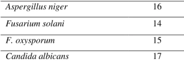
Aspergillus niger 16 Fusarium solani 14
F. oxysporum 15
by polyphasic taxonomy. An attempt was also made to characterize antimicrobial metabolites by culturing the strain in a large volume of the medium. Extraction, followed by bioactivity- guided isolation and purification of a crude ethylacetate extract, revealed the presence of Bis (7-methyloctyl) phthalate and (Z)-3-aminoacrylic acid. This is the first report on these compounds isolated from S. lavendulocolor VHB-9.
ACKNOWLEDGEMENTS
One of the authors HB thanks the Department of Botany and Microbiology for providing the laboratory facilities.
REFERENCES
1. Ritacco FV, Haltli B, Janso JE, Greenstein M, Bernan VS. Dereplication of Streptomyces soil isolates and detection of specific biosynthetic genes using an automated ribotyping instrument. JIndian Microbiol Biotechnol. 2003; 30:472-479.
2. Sembiring L, Goodfellow M. Ecological Approach to Unravel Streptomycete Diversity as an Unsurpassed Source of Natural Bioactive Products. Microbiol Indones. 2008; 2:49-56. 3. Challis GL, Hopwood DA. Synergy and contingency as driving forces for the evolution of
multiple secondary metabolite production by Streptomyces species. Proc Natl Acad Sci. 2003; 100:14555-61.
4. Fajardo A, Martınez JL. Antibiotics as signals that trigger specific bacterial responses.
Curr Opin Microbiol. 2008; 11:161-7.
5. Madigan, M.T., Martinko, J.M. In: Brock biology of microorganisms. 11th ed. New Jersey:Pearson Education, Upper Saddle River; 2006.
6. Schrey SD, Tarkka MT. Friends and foes: Streptomycetes as modulators of plant disease and symbiosis. Antonie Van Leeuwenhoek. 2008; 94:11-9.
7. Kurtboke DI, Neller RJ, Bellgard SE. Mesophilic actinomycetes in natural and reconstructed sand dune vegetation zones of Fraser Island. Australia. Microb Ecol. 2007; 54:332-40.
8. Ames BN. Mycorrhiza development in onion in response to inoculation with chitin decomposing actinomycetes. New Phytol. 1989; 112:423-427.
9. Riedlinger J, Schrey SD, Tarkka MT, Hampp R, Kapur M, Fiedler HP. Auxofuran, a novel metabolite that stimulates the growth of fly agaric, is produced by the mycorrhiza helper bacterium Streptomyces strain AcH 505. Appl Environmental Microbiol. 2006; 72:3550-3557.
10.Munaganti RK, Naragani K, Muvva VL. Antimicrobial Profile of Rhodococcus erythropolis VL-RK_05 isolated from Mango Orchards. Int J Pharm Sci Res. 2015; 6(4):1805-12.
11.Munaganti RK, Muvva VL, Indupalli MD. Studies on optimization of L-asparaginase production by Arthrobacter kerguelensis VL-RK_09 isolated from Mango orchards. Int J Pharm Pharm Sci. 2015; 7:112-115.
12.Naragani K, Rajesh Kumar M, Vijayalakshmi M. Antimicrobial Potential of Streptomyces violaceoruber VLK-4 Isolated from South Coast of Andhra Pradesh, India. Int J Pharm Sci Rev Res. 2014; 25:125-9.
13.Atta HM, Bayoumi R, El-Sehrawi M, Aboshady A, Al-Huminay A. Biotechnological application for producing some antimicrobial agents by actinomycetes isolates from Al-Khurmah Governorate. Eur J Appl Sci. 2010; 2:98-107.
14.Kavitha A, Vijayalakshmi M, Sudhakar P, Narasimha G. Screening of Actinomycete strains for the production of antifungal metabolites. Afr J Microbiol Res. 2010; 4:27-32. 15.Pridham TG, Anderson P, Foley C, Lindenfelser L, Hesseltine CW, Benedict RGA.
16.Pridham TG, Lyons. Methodologies for Actinomyctetales with reference to Streptomycetes. In: Actinomycete Taxonomy. Diatz A, Thayer DW. Eds. Sim special publication No.6, Arligton, VA; 1980. p. 153-224.
17.Gordon RE. Some criteria for the recognition of Nocardia madura (Vincent) Blanchord. J Gen Microbiol. 1966; 45:355-64.
18.Cowan ST. Cowan and Steel’s manual for the identification of medical bacteria.
Cambridge, Univ. Press: 2 nd. Edition; 1974
19.Tamura K, Peterson D, Peterson N, Stecher G, Nei M, Kumar S. MEGA5: Molecular evolutionary genetics analysis using maximum likelihood, Evolutionary distance, and maximum parsimony methods. Mol Biol Evol. 2011; 28:2731-9.
20.Cappuccino JG, Sherman N. Microbiology, Laboratory manual. Person education, INC, New Delhi. 2004; 282-283.
21.Khan ZK. In vitro and In vivo Screening Techniques for Bioactivity Screening and Evaluation. In: Proceedings of the International Workshop UNIDO-CDRI, 1997, 210. 22.Otani T, Sugimoto, Y, Aoyagi Y, Igarashi Y, Furumai T, Saito N, Yamada Y, Asao T,
Oki T. New Cdc 25B tyrosine phosphate inhibitors, nocardiones A and B, produced by Nocardia sp. TP-A0248: Taxonomy, fermentation, isolation, structural elucidation and biological properties. J Antibiot. 2000; 53: 337-344.
23.Sujatha P, Bapiraju KV, Ramana T. Studies on a new marine Streptomycete BT-408 producing polyketide antibiotic SBR-22 effective against methicillin resistant Staphylococcus aureus. Microbiol Research. 2005; 160: 119-126.
24.Kavitha A, Vijayalakshmi, M. Production of Amylases by Streptomyces tendae TK-VL_333. Int J Curr Res. 2010; 10:110-114.
25.Kavitha A, Vijayalakshmi M. Cultural parameters affecting the production of bioactive metabolites by Nocardia Levis MK-VL-113. J Appl Sci Res. 2009;5:2138-2147.