STREPTOMYCES MALACHITOFUSCUS CTF9
Imran Sajid1, 2; Khaled A. Shaaban2; Shahida Hasnain1*
¹Department of Microbiology and Molecular Genetics, University of the Punjab, Quaid-e-Azam Campus, Lahore 54590, Pakistan;
²Institute of Organic and Biomolecular Chemistry, University of Göttingen, Tammannstrasse 2, D-37077 Göttingen
Submitted: September 04, 2010; Returned to authors for corrections: October 19, 2010; Approved: January 13, 2011.
ABSTRACT
An indigenous Streptomyces isolate CTF9, exhibiting promising antifungal activity against Mucor miehei
and Candida albicans in pre-screening studies, was investigated by cultivation in a 50-L fermenter and by subsequent isolation, purification, and structure elucidation of the active metabolites. Based on the
morphological, biochemical, and physiological characterization, as well as the 16S rRNA gene sequence, the
isolate CTF9 was identified as Streptomyces malachitofuscus. Using a series of chromatographic techniques, two pure compounds were isolated from the obtained extracts after the fermentation of the isolate CTF9. The
isolated compounds were identified as phenylacetic acid and indolyl-3-lactic acid by mass spectrometry
(MS) and NMR analysis. The culture optimization studies revealed that the isolate CTF9 can use a variety of
low-cost carbon and nitrogen sources to generate the maximum quantity of industrially important
metabolites at an elevated temperature of 35°C and at a pH 7.8.
Key words: Phenylacetic acid, Indolyl-3-lactic acid, indigenous Streptomyces sp. CTF9
INTRODUCTION
Actinomycetes are the major producers of
pharmaceuticals, agricultural pesticides, and veterinary
medicine because of their ability to produce antibiotics. Among
the actinomycetes, the genus Streptomyces has continued to provide a larger number and wider variety of new antibiotics
than any other genus, suggesting that a substantial number of
Streptomyces species or strains with novel antibiotic productivity exist in nature. These bacteria produce about 75%
of commercially and medically useful antibiotics (3, 18).
Moreover, approximately 60% of the antibiotics developed for
agricultural use are isolated from Streptomyces spp. (26). Fungal pathogens have been the major culprits for significant
agricultural losses due to the plant diseases that they cause;
they are also the causal agents of many medically important
diseases. Therefore, currently, there is an immense need for
antifungal agents. Several members of the streptomycetes
family have been reported as potential biocontrol agents and as
the potent producers of antifungal compounds (16, 24),
indicating that these filamentous bacteria possess a vast
potential for producing antifungal metabolites. In search for
antifungal agents, Streptomyces strains have been isolated from various types of soils, including rice paddy, lake mud and
water, deciduous forest, tropical forest, wasteland, and cave
soils (6, 8, 11, 23, 25, 29). Their natural habitat soil is
nutritionally, biologically and physically complex and variable,
demanding their fast adaptation. As a consequence, they are
able to perform a broad range of metabolic processes and to
produce an immense diversity of bioactive secondary
metabolites.
In our search for bioactive secondary metabolites from
indigenous Streptomyces, more than 100 active Streptomyces
strains were isolated from the soil samples collected from
saline agricultural farmlands in our area (Punjab, Pakistan) and
were screened for antimicrobial activity and the metabolites
that they produced (21). The isolate Streptomyces sp. CTF9
exhibited promising antifungal activity against Candida
albicans and Mucor miehei, suggesting it as a promising strain to be further evaluated by preparative screening for the
isolation, purification and structure elucidation of active
metabolites. The microscopic, morphological, biochemical and
physiological characterization strongly suggests that the isolate
belongs to the genus Streptomyces. Its 16S rRNA gene sequence shows a maximum similarity (99%) to Streptomyces malachitofuscus; the 1426-bp sequence of the 16S rRNA gene of the isolate Streptomyces sp. CTF9 is available at GenBank, with the accession number of EU294138. Here, we reported the
fermentation, isolation, purification, and structure elucidation
of the active antifungal metabolites produced by the isolate
Streptomyces sp. CTF9, along with the optimization of the production of these metabolites under different media
compositions and culture conditions.
MATERIALS AND METHODS
Cultivation of the Strain Streptomyces sp. CTF9
The Streptomyces sp. CTF9 was cultivated in a 50-L fermenter (Biostat U) (working volume: 30 L) using M2
medium (malt extract 10 g, yeast extract 4 g, and glucose 4 g in
1 L of distilled water). The fermenter was filled with 27 L of
water mixed with the corresponding quantities of M2 medium
components. The medium was sterilized by keeping the
fermenter in the sterilization mode for 30 minutes at 121°C.
After the air supply was turned on, a stirring motor and water
circulation pumps were subsequently turned on. The acid (2 N
HCl), base (2 N NaOH) and antifoam (1% Niax/70% ethanol)
were filled in their respective jars and were connected to the
fermenter. The pH electrode was sterilized using 70% ethanol
and was adjusted The preculture was prepared by inoculating
the isolate CTF9 from well-grown plates into 12 x 250 mL of
M2 medium in 1-L Erlenmeyer flasks (total volume: 3 L). The
flasks were incubated at 28°C on a linear shaker for 4 days;
10% of the preculture was used to inoculate the fermenter. The
growth parameters were adjusted as follows: temperature 28°C,
pH 6.5 ± 1.5, and aeration 1.8 m3/h. Fermentation was
conducted for 5 days.
Extraction and purification of the active compounds
After harvesting the dark brown culture, the broth was
filtered over celite using a filter press (Schenk Niro 212 B40)
to separate the mycelium from the liquid phase. The mycelial
cake was extracted using ethyl acetate (3 times) and acetone (1
time). The water phase was extracted by adsorption on
Amberlite XAD-16 resin in a large-size glass column (100 × 5
cm) and was eluted with methanol. The solvents (ethyl acetate,
acetone, and methanol) containing bioactive metabolites were
dried using a rotary evaporator (Rotavapor R152) yielding 5.64 g of crude extracts.
Both mycelia and water-phase extracts were then collected,
based on the characterization using TLC. The crude extract
(5.64 g) was fractionated on a silica gel column (Kieselgel 60,
70 ~ 230 mesh, Merck, Darmstadt, Germany, 30 g, 1.5 × 50
cm) using a CH2Cl2: MeOH gradient (0, 5, 10, 20, 30, 40, and
50%, vol/vol) and was divided into two fractions. Fraction I
was characterized as fat (1.85 g) using TLC and spraying
reagents. Fraction II was further purified by silica gel column
preparative TLC and Sephadex LH-20 (25 ~ 100 mesh,
Pharmacia Fine Chemicals, Uppsala, Sweden) in a column (86
× 2.0 cm) and was eluted with CH2Cl2: 40% MeOH, yielding
colorless solids of phenylacetic acid (1; 25.7 mg) and
indolyl-3-lactic acid (3; 12.8 mg).
Structure determination of the purified compounds
Mass spectra: The purified fractions were analyzed by EI MS at 70 eV with Varian MAT 731, Varian 311A, AMD-402,
ESI MS using a Quattro Triple Quadruple mass spectrometer
Finigan MAT-Incos 50, MS LCQ (Finnigan) and
ESI-HRMS using perfluorokerosene as standard.
Nuclear Magnetic Resonance (NMR) Spectroscopy:1H NMR spectra were measured by Varian Unity 300 (300 MHz),
Bruker AMX 300 (300 MHz), and Varian Inova 500 (499.8
MHz). Coupling constants (J) in Hz. 13C NMR spectra were measured by Varian Unity 300 (75.5 MHz) and Varian Inova
500 (125.7 MHz). Chemical shifts (δ) were measured, relative
to the internal control tetramethylsilane.
Production optimization
The six different media A, B, C, D, E and F used in the study were as follows: A: malt extract 10 g, yeast extract 4 g, and glucose 4 g in 1 L of DH2O, B: soya been fat 20 g and
mannitol 20 g in 1 L of DH2O, C: glucose anhydrous 10 g,
peptone 2 g, yeast extract 1 g, and meat extract 1 g in 1 L of
DH2O, D: yeast extract 40 g, glucose 5 g, and CaCl2 45 g in 1 L
of DH2O, E: trypton 10 g, yeast extract 5 g, NaCl 10 g, and
glucose 5 g in 1 L of DH2O, F: glucose 21 g, fish flour 5 g,
flour 10 g, MgSO4 0.5 g, NaCl 1 g, CaCl2 0.5 g, and trace
elements 10 ml in 1 L of DH2O, (trace element stock solution:
FeSO4 0.2 g, COCl2 0.04 g, CaCl2 0.04 g, manganese(II)
chloride 0.04 g, zinc sulphate 0.08 g, and sodium borate 0.08 g
in 1 L of DH2O). In addition to different media compositions,
the following four sets of culture conditions were applied:
CC1: pH 6.5, incubation temperature of 28°C, shaking at 95
rpm, for 7 days. CC2: pH 6.5, incubation temperature of 35°C,
shaking at 110 rpm, for 7 days. CC3: pH 7.8, incubation
temperature of 28°C, shaking at 95 rpm, for 7 days. CC4: pH
7.8, incubation temperature of 35°C, shaking at 110 rpm, for 7
days.
For each of the six media, 3 L were prepared and
dispensed into 12 1-L flasks (12 x 250 mL culture broth). The
flasks were grouped according to the proposed culture
conditions; the pH in each flask was individually adjusted, and
the flasks were sterilized by autoclaving and were inoculated
with Streptomyces sp. CTF9 from well-grown plates. The inoculated flasks were incubated on linear shakers under the
proposed culture conditions (CC1, CC2, CC3 and CC4). The
strains were harvested after five days, the culture broth in each
flask was freeze dried (Christ Alpha 2-4 D) and was
individually extracted (3 x) with ethyl acetate. Ethyl acetate
was evaporated on a rotary evaporator (Heidolph Laborota
4000), and the crude extracts were obtained. The crude extracts
were weighed to determine the quantity of metabolites
produced under different cultural conditions. Subsequently, the
crude extracts were individually tested against the test
organisms Candida albicans and Mucor miehei to determine the quantity of the active antifungal metabolites per unit
volume of each extract. The results were statistically analyzed
using the analysis of variance (ANOVA) test (Duncan’s
multiple range test) by SPSS version 10 (P=0.001).
RESULTS
Purification and structure elucidation of the active compounds
The bulk fermentation was performed in a 50-L fermenter
(Biostat U) (working volume: 30 L) using M2 medium. After
harvesting, cell separation and solvent extraction, 5.64 g of the
crude extract was obtained from both the mycelium and the
liquid phase. The components of the crude extract were
purified using silica gel column, preparative TLC and gel
identified as phenylacetic acid and indolyl-3-lactic acid by 1H, 13
C NMR and mass spectrometry and by comparison to the
published data.
Phenylacetic acid (1)
Compound 1 was isolated as a colorless, UV-absorbing
solid and stained yellow on TLC by anisaldehyde/sulphuric
acid spraying reagent. According to its chromatographic
properties and spectroscopic data, compound 1 was identified
as phenylacetic acid. The result was further confirmed by
searching in AntiBase (14) and by comparing its MS and NMR
data to those of an authentic sample (19). p
-Hydroxyphenylacetic acid (2) and phenylacetic acid (1) were
isolated from Taraxacum roots as natural products, by Power
and Browning (20) and as fungal metabolites from
Ophiostoma crassivaginata (2). Phenyl acetic acid is widespread in higher plants, fungi, e.g.,Aspergillus niger (19), and bacteria. In microbial antibiotic production, compound 1
was added to the culture of Penicillium sp. to increase the production of penicillin G (parasticin) (9, 10).
R
OH
O
1: R = H; 2: R = OH
Phenylacetic acid (1): Colorless solid (25.7 mg), UV-absorbing, stained yellow by the anisaldehyde/sulphuric acid
spraying reagent. – Rf = 0.29 (CH2Cl2/3% MeOH).– 1H NMR (300 MHz, CDCl3): δ= 9.50 (brs, 1H, OH), 7.30-7.20 (m, 5H,
Ar-H), 3.62 (s, 2H, CH2) (Figure 1a). – 1H NMR (300 MHz,
Acetone-d6): δ= 10.80 (brs, 1H, OH), 7.50-7.30 (m, 5 H, Ar-H), 3.72 (s, 2H, CH2) (Figure 1b). –
13
C NMR (50 MHz,
CDCl3): δ = 178.3 (CO), 133.2 (Cq), 129.3 (2CH), 128.6
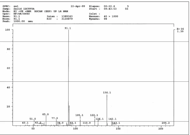
(2CH), 127.3 (CH), 41.1 (CH2) (Figure 2). – EI MS (70 eV):
m/z (%) = 136 ([M]+., 32), 91 (100), 65 (12) (Figure 3).
Figure 1b. 1H NMR (Acetone-d6, 300 MHz) of phenylacetic acid
Figure 3. EI mass spectra of phenylacetic acid
Indolyl-3-lactic acid (3)
Compound 3 was isolated as a colorless, UV-absorbing
solid and stained violet on TLC by anisaldehyde/sulphuric
acid. The 1H NMR spectrum of compound 3 exhibited two
broad 1H singlet’s of acidic protons at δ 10.75 and 8.31. In
addition, five aromatic protons distinctive for a 3-substituted
indole moiety were observed, four of which at δ 7.72 (dd), 7.38
(dd), δ 7.20 (td), and 7.10 (td) were characteristic for a
1,2-disubstituted aromatic ring. The fifth proton at δ 7.11 showed a
small long-range coupling constant (3J = 0.8 Hz). In the aliphatic region at δ 4.55, a dd signal of an oxygenated methine
was displayed, forming an ABX system with a methylene
group at δ 3.36 and δ3.20, possibly adjacent to a sp2
carbon.
The EI mass spectrum fixed the molecular weight of 3 as
205 Dalton. Furthermore, the mass ion (m/z 205) was fragmented, giving a base peak at m/z 130, characteristic for an
indolyl-3-methylene ion. This result confirmed the presence of
a hydroxy acetic acid fragment (HO-CH-COOH) and, hence, a
hydroxypropionic acid residue at the position 3 of the indole
skeleton. A search in AntiBase using the spectroscopic data
resulted in indolyl-3-lactic acid (3) as the only matched
structure. This identification was confirmed by comparing the
data to the authentic spectra (13). Compound 3 is frequently
found in bacteria, fungi and yeast (14) andexhibits activity
against Candida albicans.
N
OH
O
OH
H
1 3 5
7
1'
2' 3'
N
OH
H
1 3 4
7
1' 2'
4
N
OH
O
H
1 3 4
7
5
Indolyl-3-lactic acid (3): C11H11NO3 (205.2), colorless, moderately polar, UV-absorbing solid (12.8 mg), stained violet
by anisaldehyde/sulphuric acid and heating. – Rf = 0.35
(CHCl3/5%MeOH). – 1H NMR (CDCl3, 300 MHz): δ = 10.75
(s br, 1H, NH), 8.31 (s br, 1H, OH), 7.72 (dd, 3J = 7.9 Hz, 4J = 1.1 Hz, 1H, 4-H), 7.38 (dd, 3J = 8.3 Hz, 4J = 1.1 Hz, 1H, 7-H), 7.20 (td, 3J = 7.2 Hz, 4J = 1.2 Hz, 1H, 5-H), 7.10 (td, 3J = 7.2 Hz, 4J = 1.1 Hz, 1H, 6-H), 7.11 (d, 3J = 0.8 Hz, 1H, 2-H), 4.55 (m, 1H, 2’-H), 3.36 (dd, 2JAB = 11.1 Hz, 3JAX = 4.2 Hz, 1H, 2’-CHA), 3.26 (dd, 3J = 6.1 Hz, 1.1, 1H, 2'-CHB). – 1H NMR (DMSO-d6, 300 MHz): δ = 10.70 (s br, 1H, NH), 7.55 (dd, 3J = 7.9 Hz, 4J = 1.1 Hz, 1H, 4-H), 7.32 (dd, 3J = 8.3 Hz, 4J = 1.1 Hz, 1H, 7-H), 7.11 (d, 3J = 0.8 Hz, 1H, 2-H), 7.05 (td, 3J = 7.2 Hz, 4J = 1.2 Hz, 1H, 5-H), 6.92 (td, 3J = 7.2 Hz, 4J = 1.1 Hz, 1H, 6-H), 4.45 (s br, 1H, OH), 3.75 (m, 1H, 2’-H), 3.15 (dd, 2J
AB = 11.1 Hz, 3JAX = 4.2 Hz, 1H, 2’-CHA), 2.65 (dd, 3J = 6.1 Hz, 1.1, 1H, 2’-CHB) (Figure 4). – EI MS (70 eV): m/z (%) = 205 ([M]+., 18), 130 ([M- (HO-CH-COOH)]+., 100) (Figure 5).
Figure 5. EI MS spectra of indolyl-3-lactic acid
Impact of different media compositions and culture conditions on the yield of the active metabolites produced by Streptomyces sp. CTF9
The different media compositions (A to F) exhibited
significantly distinct impacts on the yield of the active
metabolites of the isolate Streptomyces sp. CTF9. Under the CC1 culture conditions, the media E and F exhibited highly
significant impacts on the yield of the metabolites, compared to
the other culture media A, B, C and D (Table 1). Under the
CC2 culture conditions, the medium D showed significant
impact on the growth of CTF9, followed by the medium F,
inducing the second highest yield. Under the CC3 culture
conditions, the medium F showed the most significant increase
in the weight of the crude extracts, in addition, a significant,
but lesser, impact on the yield was also observed in the media
E and D. Under the CC4 culture conditions, the medium D
yielded the maximum quantity of the crude extracts, and the
second highest yielding was the medium F, which yielded a
significantly higher quantity of the crude extracts, compared to
the media A, B, C and E (Table 1).
Similarly, different culture conditions (CC1 to CC4)
exhibited significantly different impacts on the growth of the
strain Streptomyces sp. CTF9 and the yield of its active metabolites. In case of the media A and B, significant effects
on the yield were observed under the CC4 culture conditions,
compared to the other culture conditions, i.e., CC1, CC2 and CC3 (Table 1). In case of the medium C, the impacts of the
comparable but were significantly different from those of the
other culture conditions. A significant increment in yield, to the
maximum level, was obtained in the medium D, when the
strain was cultured under the CC2 conditions. The relative
significance of the other three culture conditions, in terms of
their effects on the yield in the medium D, was
CC4>CC3>CC1. In case of the media E and F, the CC3
conditions significantly increased the yield of the crude
extracts, to the maximum level, compared to all the other
culture conditions (CC1, CC2 and CC4; Table 1).
Table 1. Comparison of the weight of the crude extracts/250 ml culture broth of the strain CTF9 for six media compositions and four sets of culture conditions (expressed as the mean of three replicates ± S.E)
Different media compositions Culture
conditions
Med A Med B Med C Med D Med E Med F
CC1 22.65ey
±0.805 26.61dy ±0.352 25.75dx ±1.014 96.66cz ±.0.476 166.68ax ±0.644 151.50by ±0.231
CC2 32.593fx
±1.273 51.61cx ±1.951 34.41ew ±0.231 173.06aw ±0.669 37.32dy ±1.544 161.05bx ±1.187
CC3 13.793ez
±0.453 26.62dy ±0.473 26.61dx ±0.929 106.36cy ±1.509 171.63bw ±0.918 183.50aw ±1.097
CC4 64.27cw
±0.274 58.90dw ±1.674 36.55fw ±0.199 150.87ax ±0.502 40.08ey ±1.319 127.29bz ±1.207
Values designated by a, b, c, d, e, and f are for six different media compositions, Values designated by w, x, y, and z are for four different culture conditions, Values designated with the same alphabets are not significantly different, Duncan’s (1955) multiple range test at the significance level = 0.001 (Med A: malt extract 10 g, yeast extract 4 g, and glucose 4 g in 1 L of DH2O, Med B: soya been fat 20 g and
mannitol 20 g in 1 L of DH2O, Med C: glucose anhydrous 10 g, peptone 2 g, yeast extract 1 g, and meat extract 1 g in 1 L of DH2O, Med D:
yeast extract 40 g, glucose 5 g, and CaCl2 45 g in 1 L of DH2O, Med E: trypton 10 g, yeast extract 5 g, NaCl 10 g, and glucose 5 g in 1 L of
DH2O, Med F: glucose 21 g, fish flour 5 g, flour 10 g, MgSO4 0.5 g, NaCl 1 g, CaCl2 0.5 g, and trace elements 10 ml in 1 L of DH2O, (trace
elements stock solution: FeSO4 0.2 g, COCl2 0.04 g, CaCl2 0.04 g, manganese(II) chloride 0.04 g, zinc sulphate 0.08 g, and sodium borate 0.08
g in 1 L of DH2O) (CC1: pH 6.5, the incubation temperature of 28°C, shaking at 95 rpm, for 7 days CC2: pH 6.5, the incubation temperature of
35°C, shaking at 110 rpm, for 7 days CC3: pH 7.8, the incubation temperature of 28°C, shaking at 95 rpm, for 7 days CC4: pH 7.8, the incubation temperature of 35°C, shaking at 110 rpm, for 7 days)
Impact of different media compositions and culture conditions on the antimicrobial activity of the strain Streptomyces sp. CTF9
Antimicrobial activity of the extracts was determined
using two fungal test strains (Candida albicans and Mucor miehei) to assess the relative concentration of the active metabolites in a crude extract. The different media
compositions (A to F) induced comparable antimicrobial
responses in an individual set of culture conditions; however,
minor variations were observed. In case of the CC1 culture
conditions, the optimal response, in respect to antimicrobial
activity, was observed in the crude extracts from the media B
and D (Table 2). Under the CC2 culture condition, the medium
E induced a significant antimicrobial activity against Candida albicans, whereas the medium C induced a significant antimicrobial activity against Mucor miehei. Under the CC3 culture conditions, the media B, D and F induced the most
significant antimicrobial activity against both test organisms. In
case of the CC4 culture conditions, the optimal antimicrobial
activity against both test organisms was seen in the crude
extracts from the media A and E (Table 2).
Similarly, different culture conditions (CC1 to CC4) also
induced comparable antimicrobial responses in a specific
the medium A, the CC4 and CC2 culture conditions showed
significant impacts on the antimicrobial activity against both
test organisms, compared to the CC1 and CC3 culture
conditions (Table 2). In the medium B, the CC3 culture
conditions showed a significant impact on the antimicrobial
activity, compared to the other culture conditions. In the
medium C, the optimal antimicrobial response was observed
under the CC2 culture conditions. In the medium D, the culture
conditions CC1, CC2 and CC4 showed comparable impacts on
the antimicrobial activity against both test organisms; however,
compared to CC3, the antimicrobial activity against Candida albicans was weaker, whereas the antimicrobial activity against
Mucor miehei was stronger. In the medium E, the impacts of the culture conditions CC4 and CC2 on antimicrobial activity
were comparable and were more significant, compared to those
of CC1 and CC3, against both test organisms. In case of the
medium F, the extracts from the culture conditions CC3 and
CC4 exhibited significantly higher antimicrobial activity
against both test organisms, compared to those from CC1 and
CC2 (Table 2).
Table 2. Comparison of the antimicrobial activity of the strain CTF9 at six media compositions and four sets of culture conditions (expressed as the mean of three replicates ± S.E)
Antimicrobial activity against test organisms Zone of inhibition (mm)
Candida albicans Mucor miehei
Media / Culture
conditions CC1 CC2 CC3 CC4 CC1 CC2 CC3 CC4
Med A 0 cy
±0.000
17.6 bx ±0.333
0 cy ±0.000
19.6 aw ±0.333
0 dx ±0.000
12 cw ±0.577
0 dx ±0.000
13 aw ±0.000
Med B 13 ax
±0.000
0 ez ±0.000
16.6 aw ±0.333
11 cy ±0.000
10.6 bx ±0.333
0 dy ±0.00
12 bw ±0.000
11 bx ±0.000
Med C 0 cx
±0.000
11.6 cw ±0.333
0 cx ±0.000
0 dx ±0.000
0 dy ±0.000
14.6 aw ±0.333
11 cx ±0.000
0 cy ±0.000
Med D 11 bx
±0.000
10.6 dx ±0.333
12 bw ±0.000
11 cx ±0.000
12.6 aw ±0.333
13 bw ±0.000
11 cx ±0.000
13 aw ±0.000
Med E 0 cx
±0.000
19.6 aw ±0.333
0 cx ±0.000
20 aw ±0.000
9.6 cx ±0.333
13 bw ±0.000
0 dy ±0.000
13 aw ±0.000
Med F 0 cy
±0.000
0 ey ±0.000
12.6 bx ±0.333
16.6 bw ±0.333
0 dy ±0.000
0 dy ±0.000
15.6 aw ±0.333
11 bx ±0.000
Values designated by a, b, c, d, e, and f are for six different media compositions, Values designated by w, x, y, and z are for four different culture conditions, Values designated with the same letters are not significantly different, Duncan’s (1955) multiple range test at a significance level = 0.001 (Med A: malt extract 10 g, yeast extract 4 g, and glucose 4 g in 1 L of DH2O, Med B: soya been fat 20 g and mannitol 20 g in 1 L
of DH2O, Med C: glucose anhydrous10 g, peptone 2 g, yeast extract 1 g, and meat extract 1 g in 1 L of DH2O, Med D: yeast extract 40 g,
glucose 5 g, and CaCl2 45 g in1 L of DH2O, Med E: trypton 10 g, yeast extract 5 g, NaCl 10 g, and glucose 5 g in 1 L of DH2O, Med F: glucose
21 g, fish flour 5 g, flour 10 g, MgSO4 0.5 g, NaCl 1 g, CaCl2 0.5 g, and trace elements 10 ml in 1 L of DH2O, (trace elements stock solution:
FeSO4 0.2 g, COCl2 0.04 g, CaCl2 0.04 g, manganese(II) chloride 0.04 g, zinc sulphate 0.08 g, and sodium borate 0.08 g in 1 L of DH2O) (CC1:
pH 6.5, incubation temperature of 28°C, shaking at 95 rpm, for 7 days CC2: pH 6.5, incubation temperature of 35°C, shaking at 110 rpm, for 7 days CC3: pH 7.8, incubation temperature of 28°C, shaking at 95 rpm, for 7 days CC4: pH 7.8, incubation temperature of 35°C, shaking at 110 rpm, for 7 days)
DISCUSSION
The present work describes the fermentation, isolation,
purification and structure elucidation of the antifungal agents
phenylacetic acid (1) and indolyl-3-lactic acid(3) from a newly
isolated cotton field isolate Streptomyces sp. CTF9 in addition
to the culture optimization to examine the appropriate medium
components and culture conditions for the maximum yield of
these commercially important compounds. Phenylacetic acid, a
deamination product of phenylalanine, has been known for its
pervasive applications in agriculture, pharmaceutical, and
effect on the growth and development of different cereal crops
(22). Wightman and Lighty (28) found that phenylacetic acid
(1) acts as a natural auxin in the shoots of higher plants, such as
barley, corn, tobacco, and tomato. The in vitro and in vivo
antifungal activity of phenylacetic acid has been reported by
many studies (7, 11, 15), suggesting that it is a potential
agroactive and biocontrol agent. In microbial antibiotic
production, phenylacetic acid is added as a precursor to the
culture of Penicillium chrysogenum to increase the production of penicillin G (9). Phenylacetic acid is also used in perfumes
at low concentrations.
Indolyl compounds, including tryptophol (4) and
indole-3-carboxylic acid (5) (12, 27), are frequently isolated from
bacteria and are characterized by their specific color reactions
with anisaldehyde/sulphuric acid (violet) and Ehrlich’s
(orange) spraying reagents. Compound 5 is known as a fungal
metabolite from Lasiodiplodia theobromae (1). From the newly isolated cotton field Streptomyces sp. CTF9, indolyl-3-lactic acid (3) (12.8 mg) was obtained as colorless solid, showing the
same physico-chemical and chromatographic properties as 4
and 5. Compound 3 is often isolated from bacteria, fungi and
yeast (14) and exhibits the antimicrobial activity against
Candida albicans.
Streptomyces can grow on a variety of media and under different culture conditions; however, it is usually helpful to
identify the optimal medium, temperature, pH and aeration of
the interesting strains for large-scale cultivation. The nutrients
and culture conditions play an important role in the onset and
intensity of secondary metabolism. To achieve high product
yields, it is a prerequisite to identify a proper production
medium to be used for an efficient fermentation process (17).
The medium composition is usually related to the biosynthesis
of antibiotics (4). The role of the medium is to provide
nutrients and precursors to the culture (5). Six media
compositions (A, B, C, D, E, and F), along with four sets of
culture conditions (CC1, CC2, CC3 and CC4), were examined
to determine the ability of the Streptomyces sp. CTF9 to use
different carbon and nitrogen sources and to identify the
optimal culture conditions for the maximum yields of
phenylacetic acid and indolyl-3-lactic acid. The cultural
optimization studies revealed that different media compositions
and cultural conditions have significant impacts on the growth
and antimicrobial activity of the strain CTF9: it exhibited
promising growth on the culture media D, E and F; however,
the maximum yields of the crude extracts were obtained from
the medium D under the CC2 culture conditions (Table 1) and
from the media E and F under the culture conditions CC1 and
CC3 (Table 1). The similar impacts of different media on the
growth demonstrate the ability of the strain CTF9 to use
various nitrogen sources (including yeast extract, peptone, fish
flour, etc.) and different carbon sources supplemented in these media. The impacts of other media, such as A, B and C, on the
growth of the strain CTF9 was demonstrated by the low yield
of the crude extracts, compared to those of the media D, E and
F (Table 1). Different quantities of the crude extracts were
obtained from the same medium under different culture
conditions. In the media A, B, C and D, the culture conditions
CC2 and CC4 showed significant impacts on the growth,
compared to the culture conditions CC1 and CC3, whereas in
the media E and F, the culture conditions CC1 and CC3
showed a significant impact on the growth, compared to CC2
and CC4, indicating the ability of the strain CTF9 to grow in a
broad range of temperatures (28 to 35°C) and pH values (6.5 to
7.8). Studies were also conducted on the antimicrobial activity
of the crude extracts of the strain CTF9, obtained from
different media under different culture conditions, against the
test organisms Candida albicans and Mucor miehei. In general, different media and culture conditions induced similar
activities against the test organisms with minor variations,
suggesting that the proportion of the active components in the
unit weight of the crude extracts is similar. Based on the
extensive investigations on different media compositions and
culture conditions, we demonstrate that the maximum yield of
(phenylacetic acid and indolyl-3-lactic acid) can be obtained by
culturing the strain on the media D, E or F, at 35°C and pH 7.8.
ACKNOWLEDGMENTS
We thank Prof. Dr Hartmut Laatsch (Institute of Organic
and Biomolecular Chemistry, University of Gottingen,
Germany) for providing the facilities for fermentation, MS and
NMR analysis. The financial support of this work by a grant
from the Higher Education Commission (HEC) of Pakistan
under IRSIP is gratefully acknowledged.
REFERENCES
1. Aldridge, D.C.; Galt, M.S.; Giles, D.; Turner, W.B. (1971). Metabolites of Lasiodiplodia theobromae. J. Chem. Soc., C, 1623-1627.
2. Ayer, W.A.; Trifonov, L.S. (1995). Phenolic and polyketide metabolites of the aspen blue stain fungus Ophiostoma crassivaginata.
Phytochemistry., 38, 371-372.
3. Berdy, J. (2005). Bioactive microbial metabolites: A personal view. J. Antibiot., 58(1), 1-26.
4. Chatterjee, S.; Vining, L.C. (1981). Nutrient utilization in actinomycetes: Induction of -glucosidases in Streptomyces venezualae. Can. J. Microbiol., 27, 639-645.
5. Demain, A.L.; Aharonowitz, Y.; Martin, J.F. (1983). Metabolic control of secondary biosynthetic pathways, In: Vining, L.C. (ed). Biochemistry and genetic regulation of commercially important antibiotics, Addison-Wesley, Reading. p. 49-71.
6. Hayakawa, M.; Ishizawa, K.; Nonomura, H. (1988). Distribution of rare actinomycetes in Japanese soils. J. Ferment. Technol., 66, 367–373. 7. Hwang, B.K; Lim, S.W.; Kim, B.S.; Lee, J.Y.; Moon, S.S. (2001).
Isolation and In Vivo and In Vitro Antifungal Activity of Phenylacetic Acid and Sodium Phenylacetate from Streptomyces humidus. Appl. Environ. Microbiol. 67, 3739–3745.
8. Jiang, C.L.; Xu, L.H. (1996). Diversity of aquatic actinomycetes in lakes of the middle plateau, Yunnan, China. Appl. Environ. Microbiol., 62, 249–253.
9. Kato, K. (1953). Further notes on penicillin-nucleus. J. Antibiot., 4, 184-185.
10. Kato, K. (1953). Occurrence of penicillin-nucleus in culture broths. J. Antibiot., 6, 130–136.
11. Kim, B.S.; Lee, J.Y.; Hwang, B.K. (1998). Diversity of actinomycetes antagonistic to plant pathogenic fungi in cave and sea-mud soils of Korea. J. Microbiol., 36, 86–92.
12. Kradolfer, P.; Niederberger, P.; Huetter, R. (1982). Tryptophan degradation in Saccharomyces cerevisiae: characterization of two aromatic aminotransferases. Arch. Microbiol. 133, 242-248.
13. Kuang-Ren, C.; Turksen, S.; Umran, E.; Timmer, L. W.; Ueng, P. P. (2003). Indole derivatives produced by the fungus Colletotrichum acutatum causing lime anthracnose and post bloom fruit drop of citrus.
FEMS Microbiol. Letters., 226, 23-30.
14. Laatsch, H. (2007). AntiBase 2007, A Data Base for Rapid Structural Determination of Microbial Natural Products, 2007 and annual updates, Chemical Concepts, Weinheim, Germany; see http://wwwuser. gwdg. de/~ucoc/laatsch/AntiBase. htm.
15. Lee, J.Y.; Kim, H.S.; Kim, K.D.; Hwang, B.K.(2004). In Vitro Anti-Oomycete Activity and In Vivo Control Efficacy of Phenylacetic Acid Against Phytophthora capsici. Plant Pathol. J., 20, 177-183.
16. Lu, C.G.; Liu, W.C.; Qiu, J.Y.; Wang, H.M.; Liu, T.; Liu, D.W. (2008). Identification of an antifungal metabolite produced by a potential biocontrol actinomyces strain A01. Braz. J. Microbiol., 39, 701-707. 17. Macedo, J.A.; Sette, L.D.; Sato, H.H. (2007). Optimization of medium
composition for transglutaminase production by a Brazilian soil
Streptomyces sp. Electr. J. Biotechnol., 10 (4), 618-626.
18. Miyadoh, S. (1993). Research on antibiotic screening in Japan over the last decade: a producing microorganisms approach. Actinomycetol., 9, 100-106.
19. Nair, M. G.; Burke, B. A. (1988). A new fatty acid methyl ester and other biologically active compounds from Aspergillus niger.
Phytochemistry, 27, 3169-73.
20. Power, F.B.; Browning, H.J. (1912). Constituents of Taraxacum Root. J. Chem. Soc., 101, 2411-2429.
21. Sajid, I.; Yao, C.B.; Shaaban, K.A.; Hasnain, S.; Laatsch, H. (2009). Antifungal and antibacterial activities of indigenous Streptomyces
isolates from saline farmlands: prescreening, ribotyping and metabolic diversity. World J. Microbiol. Biotechnol., 25, 601–610.
22. Sarwar, M.; Frankenberger, W.T. (1995). Fate of L-phenylalanine in soil and its effect on plant growth. Soil Sci., 59, 1625–1630.
23. Shomura, T. (1993). Screening for new products of new species of
Dactylosporangiumand other actinomycetes. Actinomycetol., 7, 88–98. 24. Soares A. C. F.; Sousa C. D. S.; Garrido M. D. S.; Perez J. O.; Almeida
N. S. (2006). Soil streptomycetes with in vitro activity against the Yam pathogens Curvularia eragrostides and Colletotrichum gloeosporioides. Braz. J. Microbiol. 37:456-461.
25. Suzuki, K.; Nagai, K.; Shimizu,Y.; Suzuki, Y. (1994). Search for actinomycetes in screening for new bioactive compounds.
Actinomycetol., 8, 122–127.
27. Turner, W. B.; Aldridge, D. C.; Galt, M.; Giles, D. (1971). Metabolites of Lasiodiplodia theobromae . J. Chem. Soc. (C), 1623-1627.
28. Wightman, F.; Lighty, D.L. (1982). Identification of phenylacetic acid as
a natural auxin in the shoot of higher plants. Physiol, Plant., 55, 17–24. 29. Xu, L.H.; Li, Q.R.; Jiang, C.L. (1996). Diversity of soil actinomycetes in
Yunnan, China. Appl. Environ. Microbiol., 62, 244–248.