Printed version ISSN 0001-3765 / Online version ISSN 1678-2690 http://dx.doi.org/10.1590/0001-3765201720160725
www.scielo.br/aabc | www.fb.com/aabcjournal
Melipona mondury
produces a geopropolis with antioxidant,
antibacterial and antiproliferative activities
TÁSSIA L.A. DOS SANTOS1
, RAPHAEL F. QUEIROZ2
, ALEXANDRA C.H.F. SAWAYA3
, BEGOÑA GIMENEZ-CASSINA LOPEZ3, MILENA B.P. SOARES4,5, DANIEL P. BEZERRA4, ANA CAROLINA
B.C. RODRIGUES4
, VANDERLÚCIA F. DE PAULA6
and ANA MARIA WALDSCHMIDT1
1
Departamento de Ciências Biológicas, Universidade Estadual do Sudoeste da Bahia, Rua José Moreira Sobrinho, s/n, Jequiezinho, 45208-091 Jequié, BA, Brazil
2
Departamento de Ciências Naturais, Universidade Estadual do Sudoeste da Bahia, Estrada do Bem Querer, Km 04, s/n, 45031-900 Vitória da Conquista, BA, Brazil
3
Departamento de Biologia e Ciências Farmacêuticas, Instituto de Biologia, Universidade Estadual de Campinas, Rua Monteiro Lobato, 255, Cidade Universitária, 13083-862 Campinas, SP, Brazil
4
Instituto Gonçalo Moniz, Fundação Oswaldo Cruz/IGM-FIOCRUZ-BA, Rua Waldemar Falcão, 121, Candeal, 40296-710 Salvador, BA, Brazil
5
Centro de Biotecnologia e Terapia Celular, Hospital São Rafael, Avenida São Rafael, 2152, São Marcos, 41253-190 Salvador, BA, Brazil
6
Departmento de Química e Ciências Exatas, Universidade Estadual do Sudoeste da Bahia, Rua José Moreira Sobrinho, s/n, Jequiezinho, 45208-091 Jequié, BA, Brazil
Manuscript received on October 26, 2016; accepted for publication on March 3, 2017
ABSTRACT
Geopropolis is a special type of propolis produced by stingless bees. Several pharmacological properties have been described for different types of geopropolis, but there have been no previous studies of the geopropolis from Melipona mondury. In this study, we investigated the antioxidant, antibacterial, and antiproliferative activities of M. mondury geopropolis, and determined its chemical profile. The antioxidant
activity was determined using in vitro ABTS•+
, •DPPH, and β-carotene/linoleic acid co-oxidation methods. The antibacterial activity was determined using a microdilution method with Pseudomonas aeruginosa, Staphylococcus aureus, and methicillin-resistant S. aureus. The antiproliferative effect was determined in tumor cell lines using the Alamar Blue assay. The chemical profile was obtained using UHPLC-MS and UHPLC-MS/MS. The butanolic fraction had the highest concentration of phenolic compounds and more potent antioxidant properties in all assays. This fraction also had bacteriostatic and bactericidal effects against all bacterial strains at low concentrations, especially S. aureus. The hexane fraction had the highest antiproliferative potential, with IC50 values ranging from 24.2 to 46.6 µg/mL in HL-60 (human promyelocytic leukemia cell) and K562 (human chronic myelocytic leukemia cell), respectively. Preliminary chemical analysis indicates the presence of terpenes and gallic acid in the geopropolis. Our results indicate the therapeutic potential of geopropolis from M. mondury against inflammatory, oxidative,
infectious, and neoplastic diseases.
Key words: antibacterial, antioxidant, antiproliferative, geopropolis, Melipona mondury.
Correspondence to: Raphael Ferreira Queiroz E-mail: rfqueiroz@uesb.edu.br
INTRODUCTION
Geopropolis is a mixture of resin, wax, and soil, and it is an uncommon type of propolis produced by native stingless bees from the Meliponini tribe, which are widely distributed in tropical and subtropical areas worldwide (Sforcin and Bankova 2011, Souza et al. 2014). The geopropolis from
Melipona mondury (
Hymenoptera: Apidae:
Meliponini)
was investigated in the present study because this bee species is distributed extensively in Brazil, especially in Bahia state in the northeastof the country (Melo 2003). M. mondury is found
exclusively in warm and humid climates in about 24% of the municipalities of Bahia state (Souza et al. 2012). In addition to their importance as efficient pollinators of native flora, stingless bees yields products such as honey, pollen, and geopropolis, which are sources of food, medicine, and income for the rural population (Melo 2003).
Studies of different types of geopropolis have demonstrated their significant antinociceptive (Souza et al. 2014), anti-inflammatory (Franchin et al. 2013, Campos et al. 2015), antioxidant (Souza et al. 2014, Campos et al. 2014, 2015), immunomodulatory (Liberio et al. 2011), antibacterial (Liberio et al. 2011, Cunha et al. 2013, Campos et al. 2014, 2015), and antiproliferative (Cunha et al. 2013, Campos et al. 2014, 2015) activities. Their chemical compositions are extremely complex and their diversity depends on the flora visited and the season (Marcucci 1995, Bankova et al. 2000, Sforcin and Bankova 2011). Indeed, numerous potential bioactive metabolites have been reported previously in geopropolis, such as phenylpropanoids, flavonoids (Souza et al. 2013), gallotannins, ellagitannins (Dutra et al. 2014), benzophenones (Cunha et al. 2013, 2016), coumarins (Cunha et al. 2016) terpenes, and gallic acid (Bankova et al. 2000, Souza et al. 2013, Dutra et al. 2014). These findings have encouraged
the exploration of M. mondury geopropolis as a
potential source of novel bioactive compounds. The biological activities and chemical compositions of geopropolis have been reported for many different stingless bee species (Liberio et al. 2011, Franchin et al. 2013, Cunha et al. 2013, Campos et al. 2014, 2015). However, there have been no previous pharmacological and chemical
analyses of geopropolis from M. mondury.
Therefore, we performed the first study of the in
vitro antioxidant, antibacterial, and antiproliferative
activities of geopropolis from M. mondury, as well
as determining the chemical profile of its ethanolic extract and fractions. This study obtained reliable information, which may add value to this natural product.
MATERIALS AND METHODS
GEOPROPOLIS SAMPLE AND FRACTIONATION
Fresh samples of M. mondury geopropolis were
collected in Nova Ibiá city, Bahia state, Brazil
(13°48’36’’ S, 39°37’32’’ W) during August
2015. The geopropolis (100 g) was extracted three times with absolute ethanol for 72 h and filtered to obtain the ethanolic extract of geopropolis (EEGP = 10 g). EEGP was fractionated via liquid-liquid partitioning with hexane, ethyl acetate, and butanol solvents in order of increasing polarity to obtain hexane (HFGP = 0.5 g), ethyl acetate (EAFGP = 1.6 g) and butanol fractions of geopropolis (BFGP 0.5 g).
TOTAL PHENOLIC CONTENTS
for 30 min at 37°C before measuring the absorbance at 765 nm. A calibration curve was obtained with gallic acid (0.5, 5, 10, 15 and 25 µg) and the results were expressed as µg gallic acid equivalent (GAE) per mg of sample.
DETERMINATION OF ANTIOXIDANT ACTIVITIES
The antioxidant activities of geopropolis samples were evaluated using three well-known methods: •
DPPH (2,2–diphenyl-1-picrylhydrazyl) free radical scavenging (Piccinelli et al. 2004),
ABTS•+
(2,2’-azino-bis(3-ethylbenzothiazoline-6-sulphonic acid cation radical) decolorization (Re et
al. 1999), and β-carotene-linoleic acid cooxidation
(Miller 1971, with minor modifications) assays.
In the DPPH assay, the samples (0-100 µg/
mL) were incubated to 2 mL of an ethanolic
solution of •DPPH (70 µM). After 20 min at 25°C,
the absorbance was measured at 517 nm. In the
ABTS•+ assay, we first prepared ABTS radical
cation by reacting ABTS salt (7 mM) concentration with potassium persulfate (2.45 mM) and allowing the mixture to stand in the dark at room temperature
for 16 h before use. The stock ABTS•+ solution
was diluted with ethanol (absorbance of 0.7 at 734 nm) and incubated to samples (0-100 µg/mL) in a final volume of 2 mL. The absorbance was measured at 734 nm after 7 min of incubation. In both experiments, the values were expressed as the concentration of sample necessary to reduce 50% of the free radicals (IC50).
In the β-carotene-linoleic acid cooxidation assay, a stock solution of β-carotene/linoleic acid was initially prepared by dissolving 2 mg of β-carotene in 100 µL of chloroform. Ten microliters of β-carotene solution were mixed to 40 mg of linoleic acid and 530 µL of Tween 40. The chloroform was rotaevaporated and aerated distilled water was added to the mixture until an initial absorbance of 0.65 at 470 nm. Two milliliters of β-carotene/linoleic acid emulsion were mixed with
samples at 74 µg/mL. The reactions were incubated at 50°C for 2h before taking another absorbance reading. The data were expressed as the percentage inhibition of β-carotene bleaching by geopropolis extract or fraction (% IO).
In all assays gallic acid and trolox were used as positive controls.
DETERMINATION OF ANTIBACTERIAL ACTIVITY
Bacterial strains
The bacterial strains used in this study were
Staphylococcus aureus ATCC 29213, Pseudomonas aeruginosa ATCC 27853 and methicillin-resistant
Staphylococcus aureus (MRSA). The CA-MRSA strain was cultured at the Microbiology Laboratory in the Multidisciplinary Institute of Health, Federal of University of Bahia. This strain was isolated from raw human milk (LC2). Phenotypic and genotypic tests, identification
of the nuc gene (Brakstad et al. 1992), detection
of the mecA gene (Perez-Hoth et al. 2001), and
chromosomal cassette typing mec (ssmec) (Boye
et al. 2007) were conducted to identify the strains.
The isolated LC2 was positive for the nuc gene and
mecA as well as ssmec, and thus typed as IV, which characterized it as a CA-MRSA strain.
Susceptibility testing
The antibacterial activities of EEAGP and other fractions were examined by determining the minimum inhibitory concentration (MIC) and minimum bactericidal concentration (MBC) according to the Institute of Clinical and Laboratory Standards (CLSI 2006, Cunha et al. 2013). To
determine the MIC, 5 × 10⁵ CFU/mL diluted in brain
of EEGP or fraction that allowed no visible growth after incubation with 0.01% resazurin dye (Sigma-Aldrich) for 60 min at room temperature. MBC was determined by sub-culturing 10 µL of each incubated well that had a concentration higher than the MIC on Müller-Hinton agar. The MBC was then treated as the lowest concentration of each sample with no visible colony growth on agar plates. Tetracycline and ceftriaxone were used as positive controls for Gram-positive and -negative bacteria, respectively.
ANTIPROLIFERATIVE ASSAY
Cells
Antiproliferative effects were determined in tumor cell lines: B16-F10 (mouse melanoma), HepG2 (human hepatocellular carcinoma), K562 (human chronic myelocytic leukemia), and HL-60 (human promyelocytic leukemia). All of the cell lines were kindly provided by Hospital A.C. Camargo (São Paulo, SP, Brazil) and maintained in Roswell Park Memorial Institute-1640 (RPMI-1640; Gibco) medium supplemented with 10% fetal bovine serum (Cultilab), 2 mM l-glutamine
(Vetec Química Fina), and 50 µg/mL gentamycin
(Novafarma). All cell lines were cultured in cell
culture flasks at 37°C in 5% CO2 and subcultured
every 3–4 days to maintain exponential growth (Rodrigues et al. 2015)
Peripheral blood mononuclear cells (PBMCs) were isolated from healthy, non-smoker donors using a Ficoll-Paque density gradient (GE Healthcare). PBMCs were washed and resuspended
at a concentration of 0.3 × 10⁶ cells/mL in RPMI
1640 medium supplemented with 20% fetal bovine serum, 2 mM glutamine, and 50 µg/mL gentamycin
at 37°C with 5% CO2. Next, the cells were
incubated with a T-lymphocyte-specific mitogen, concanavalin A (10 µg/mL, Sigma Chemical Co.), for 24 h before the experiments to obtain a culture
of human lymphoblasts (Berthold 1981, Brown and Lawce 1997). In all experiments, cell viability was determined using the Trypan blue assay (>90%).
The Research Ethics Committee of the Oswaldo Cruz Foundation (Salvador, Bahia, Brazil) approved the experimental protocol (#031019/2013). All blood donors signed a written informed consent form to participate in the study.
In vitro antiproliferative activity assay
Cell viability was quantified using the Alamar Blue assay, as described previously by Ahmed et al. (1994) with minor modifications (Rodrigues et al.
2015). Non-tumor and tumor cells were placed in
96-well plates (7 x 10⁴ cells/mL for adherent cells
or 3 x 10⁵ cells/mL for suspended cells in 100 µL of
medium) and incubated with EEGP or its fractions
(0.39–50 µg/mL) for 72 h at 37°C under a CO2 (5%)
atmosphere. Cell viability was quantified based on the ability of living cells to reduce Alamar Blue dye (52 µg/mL; Sigma Aldrich) to a red resorufin product where the absorbance was measured at 570 and 600 nm (DTX-880, Beckman Coulter) after 2h
at 37°C under CO2 (5%) atmosphere. Doxorubicin
(purity 95%, doxorubicin hydrochloride,
Laboratory IMA) was used as the positive control (0.08–5 µg/mL).
CHEMICAL ANALYSIS
conditions, followed by column re-equilibration. The ESI conditions were: capillary = 3.00 kV, cone = 30 V, source temperature = 150°C, desolvation temperature = 350°C, and collision energy = 30 V,
with data acquisition between m/z 100 and 1000.
The (ESI(-)-MS) fingerprints were obtained by direct injection in duplicate using 0.01 mL of EEGP and the fractions (1 mg/mL) by flow insertion with a solvent mixture comprising methanol:water (90:10 v/v) at flow rate of 0.1 mL/min. The components of geopropolis samples were putatively identified
by comparing their m/z values and fragmentation
patterns with previous reports.
STATISTICAL ANALYSIS
Data were expressed as the mean ± standard
deviation or IC50 values based on three independent
experiments. Significant differences (p < 0.05)
were detected by one-way ANOVA with Tukey’s post-test using GraphPad 4.00.
RESULTS AND DISCUSSION
The chemical composition of propolis is very complex and closely related to its biological activity (Bankova et al. 2000, Sforcin and Bankova 2011, Cunha et al. 2016). Phenolic compounds, terpenes, and aromatic acids are considered the main bioactive constituents of propolis (Bankova et al. 2000, Souza et al. 2013, Dutra et al. 2014). The total phenolic compounds and antioxidant
activities of geopropolis samples produced by M.
mondury are shown in Table I. BFGP contained the
highest amount of phenolic (303.1 ± 0.1 µgGAE /
mg), which differed significantly from that in the
other fractions (p < 0.05). HFGP contained a low
concentration of phenolics (GAE 40.1 ± 0.4 µg/mg), as expected. In our study, the amount of reducing compounds was much higher than that found by
Souza et al. (2013) in M. subnitida geopropolis
from Paraiba state, Brazil (ranging from 25.6 ± 0.5 to 115.8 ± 0.8 µg GAE/mg). This suggests that the
bee species and/or its habitat influence the chemical composition of geopropolis (Campos et al. 2015).
Three different methods were employed to determine the antioxidant properties of the
geopropolis: scavenging of DPPH• and ABTS•+
radicals, and inhibition of β-carotene bleaching. All samples exhibited some radical scavenging activity
against DPPH• and ABTS•+ (Table I). BFGP was
more effective fraction at reducing DPPH· and
ABTS•+ (IC
50 ~ 2.2 and 0.9 µg/mL, respectively).
BFGP (74 µg/mL) inhibited β-carotene bleaching at around 78.6%, which was similar to that with the same concentration of trolox standard. Unexpectedly, the hexane fraction had weak activity in the β-carotene/linoleic acid bleaching
assay. Nevertheless, the antioxidant profile of M.
mondury geopropolis was higher compared with
that of M. subnitida geopropolis, which is known as
Jandaíra in northeastern Brazil (Souza et al. 2013). Previously, it has been suggested that reducing compounds such as phenolics are responsible for the antioxidant activity of geopropolis (Souza et al. 2013, 2014, Campos et al. 2014, Dutra et al. 2014), which may explain why the BFGP had the highest antioxidant activity in all the assays.
Infectious diseases are important causes of morbidity and mortality among humans, especially
in developing and poor countries. S. aureus and P.
aeruginosa are particularly important because they are often associated with nosocomial infections, and they have increased resistance to many clinically available antibiotics, thereby stimulating the search for alternative treatments (Cinegaglia et al. 2013, Sampaio et al. 2013). Geopropolis has also been proposed as an alternative for prevention and/or treatment of infectious diseases because it has broad antimicrobial activities. In fact, several types of geopropolis have antibacterial properties (Liberio et al. 2011, Cunha et al. 2013, Campos et al. 2014, 2015).
According to this study, M. mondury
Gram-positive and -negative bacteria (Table II).
EEGP exhibited bactericidal activity against P.
aeruginosa, a Gram-negative bacillus (MIC =
MBC = 250 μg/mL). BFGP inhibited the growth
of S. aureus, CA-MRSA, and P. aeruginosa at concentrations of 5–10, 250, and 500 μg/mL, respectively. The MBC values showed that BFGP had bactericidal activities at concentrations of
25 and 1000 μg/mL against S. aureus and P.
aeruginosa, respectively. The MIC and MBC values for BFGP against all bacterial strains were comparable to those of standard antibiotics, such as ceftriaxone (MIC = 3 μg/mL and MBC = 10 μg/ mL) and tetracycline (MIC = MBC = 3 μg/mL), considering that BFGP is a complex sample. The
MIC values of EAFGP against S. aureus,
CA-MRSA, and P. aeruginosa were 25, 500, and 250
μg/mL, respectively. The EAFGP had a bactericidal
effect only on S. aureus at 1000 µg/mL. EEGP and
its fractions had no bactericidal effects on CA-MRSA at the test concentrations (MIC > 1000 μg/mL). These results demonstrate that all of the samples were bactericidal agents (MIC/MBC ≤ 4), except for EAFGP (MBC/MIC = 40), according to French (2006).
Previously, Campos et al. (2014) reported
MIC and MBC values ˃ 1000 µg/mLagainst S.
aureus and E. coli for an ethanolic extract of M. orbignyi geopropolis collected in Mato Grosso do Sul state, Brazil. Velikova et al. (2000) also
demonstrated that M. quadrifasciata geopropolis
had antibacterial activities against S. aureus and E.
coli. Campos et al. (2015) found that the ethanolic
extract of Tetragonisca fiebrigi geopropolis
(known as Jataí) had antibacterial effects on two
strains of S. aureus (MIC = 0.55–0.65 mg/mL and
MBC ˃ 1000 µg/mL) and P. aeruginosa (MIC
and MBC ˃ 1000 µg/mL). In these studies, it was
suggested that prenylated benzophenones (Cunha et al. 2013), diterpene kaurenoic acid (Velikova et al. 2000), cinnamic acid (Campos et al. 2015),
and p-coumaric acid (Campos et al. 2015) in the
different types of geopropolis were responsible for their antibacterial properties.
The ethanolic extract of geopropolis from M.
scutellaris (known as Uruçu verdadeira) and its hexane fraction had strong antibacterial activities
against S. aureus and S. aureus MRSA strains (MIC
= 6.25–12.5 μg/mL and MBC = 25–50 μg/mL), but
no activity against P. aeruginosa. By contrast, our
TABLE I
Total phenolics and antioxidant activities of the Melipona mondury geopropolis extract and fractions.
Sample Total Phenolics µg GAE/mg •
DPPH IC50 (µg/mL) ABTS•+
IC50 (µg/mL) β-carotene-linoleic acidcooxidation (% IO)
EEGP 144.4 ± 0.01a
6.91 ± 017a
5.96 ± 0.08a
27.99 ± 0.2a
HFGP 40.12 ± 0.4b
20.22 ± 0.2b
20.51 ± 0.15b
4.7 ± 0.001b
EAFGP 140.9 ± 0.28a
6.58 ± 0.04c
5.5 ± 0.028c
26.45 ± 0.4a
BFGP 303.1 ± 0.14c
2.23 ± 0,05d
0.87 ± 0.003d
78.57 ± 0.3c
Trolox - 1.54 ± 0.01e
1.20 ± 0.035e
83.69 ± 0.7d
Gallic acid - 1.45 ± 0.01e
1.08 ± 0.04d,e
54.7 ± 0.4e
BFGP had similar MIC and MBC values against S. aureus to the hexane fraction of M. scutellaris
geopropolis (Cunha et al. 2013). It should be noted that we determined all the MIC values only after incubating with resazurin for 60 min because shorter incubation periods overestimated the results (data not shown). Unfortunately, the incubation time employed by Cunha et al. (2013) was not stated.
Propolis has also been reported to have cytotoxic effects against several tumor cell lines (Cunha et al. 2013, 2016, Cinegaglia et al. 2013,
Campos et al. 2015). Thus, we evaluated the in
vitro antiproliferative potential of M. mondury
geopropolis in four cell tumor lines: B16-F10, HepG2, HL-60, and K562 (Table III). EEGP had antiproliferative activities against tumor cells with
IC50 values ranging from 33.48 to 48.67 µg/mL
using HL-60 and HepG2. HFGP had IC50 values
ranging from 24.24 to 46.62 µg/mL using HL-60
and K562. EAFGP had IC50 values ranging from
29.79 to 45.90 µg/mL using B16-F10 and HepG2. BFGP was not tested because it exhibited no antiproliferative activity at 50 µg/mL in preliminary screening using HepG2 and HL-60 cells. According
TABLE II
Minimum inhibitory concentration (mic) and minimum bactericidal concentration (mbc) values for the geopropolis ethanolic extract and its fractions.
Bacterial strains EEGPa
HFGPa
EAFGPa
BFGPa
MIC MBC MIC MBC MIC MBC MIC MBC
Staphylococcus aureus ATCC 29213 250 1000 500 b
25 1000 5–10 25
Staphylococcus aureus (CA-MRSA) b b b b
500 b
250 b
Pseudomonas aeruginosa ATCC 27853 250 250 1000 b
250 b
500 1000
EEGP = ethanolic extract of geopropolis, HFGP = hexane fraction of geopropolis, EAFGP = ethyl acetate fraction of geopropolis, BFGP = butanol fraction of geopropolis. a
concentration at µg/mL. b
values >1000 µg/mL.
TABLE III
In vitro antiproliferative activities of Melipona mondury geopropolis ethanolic extract and its fractions.
Samples
IC50 values (µg/mL)
B16-F10 HepG2 HL-60 K562 Human
lymphoblast
EEGP 35.70
32.60–39.11
48.67 37.93–62.46
33.48 30.48–36.78
46.93
35.96–61.25 >50
HFGP 28.00
25.54–30.71
39.41 36.46–42,60
24.24 21.54–27.27
46.62
35.95–60.45 >50
EAFGP 29.79
26.74–33.19
45.90 40.89–51.53
35.57 31.71–39.89
42.19 35.14–50.65
47.67 41.40–54.89
DOX 0.20
0.17–0.24
0.19 0.15–0.25
0.18 0.16–0.21
0.29 0.23–0.37
to Suffness and Pezzuto (1990), extracts with IC50 values <30 µg/mL are promising cytotoxic agents for use against neoplastic cells. In addition, EEGP and HFGP had no cytotoxic effects on non-tumor cells (human lymphocyte T) whereas EAFGP
had an IC50 value of 47.67 µg/mL in lymphoblast
cells. Therefore, HFGP is a promising source for the isolation and identification of antiproliferative compounds.
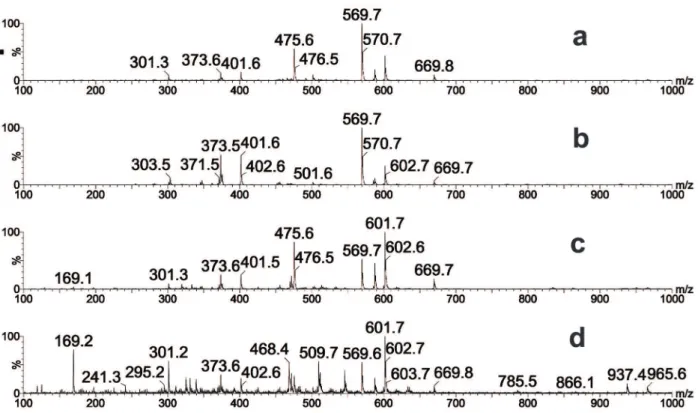
To determine the chemical profile of M.
mondury geopropolis, the EEGP and its fractions were analyzed by direct infusion negative ion mode electrospray ionization mass spectrometry
(ESI(-)-MS). The most abundant anions [M-H]–in all
samples were at m/z 169, 475, 569, and 601. We also
observed other less intense ions (m/z 301, 373, 401,
and 569) in the raw extract and fractions (Figure 1). We then performed chromatographic analysis using UHPLC-MS and obtained the UHPLC-MS/ MS spectra of selected peaks based on the main ions in the fingerprints (Table IV).
The ion at m/z 169 and its fragments (Figures
2a and b) were characteristic of gallic acid based on comparisons with previous studies (Abdel-Hameed et al. 2013, Saldanha et al. 2013). In addition, this
compound was previously identified in Myrcia
bella, which is a source of resin for M. mondury
(Z.S. Lopes et al., unpublished data).
The ion at m/z 301 and its fragments (Figures
2c and d) were compatible with E/Z communic
acid isomers, which were identified previously in
some types of Brazilian propolis and Araucaria
heterophylla resins (Marcucci et al. 2008). E/Z
communic acid is a diterpene (Barrero et al. 2012) with several known pharmacological properties, including antimicrobial, antitumoral, anti-inflammatory, and antioxidant activities (Velikova et al. 2000, Popova et al. 2009, Barrero et al. 2012, Campos et al. 2015).
Previous studies of propolis produced by native bees from different Brazilian regions found ions at
m/z 373 (Figures 2e and f) and 401 as diagnostic
of terpenes with acid groups present in Schinus
terebinthifolius resin (e.g., Sawaya et al. 2006,
2007, 2009). M. mondury geopropolis contained
the same ions, which suggests that this bee also
collects resin from S. terebinthifolius (e.g., Sawaya
et al. 2007, 2009). This plant occurs naturally on the coastline of Brazil, in the remaining areas of the Atlantic Forest (Carvalher-Machado et al. 2008),
which is an area inhabited by M. mondury (Melo
2003, Souza et al. 2012).
Finally, the ions at m/z 601, 475, and 569
(Figure 3) could not be identified, although they
were present in all the samples produced by M.
mondury tested in this study.
UHPLC-MS has been used widely for analyzing propolis (Bankova et al. 2000, Sawaya et al. 2011, Novak et al. 2014, Wali et al. 2015, Ristivojević et al. 2015, Sagi et al. 2016), but it has limitations in terms of the identification of uncommon substances because there is no database for making comparisons among geopropolis samples. Therefore, the unambiguous characterization of the compounds corresponding
to the ions at m/z 601, 475, and 569 may be possible
after their isolation and further analysis using other spectrometric methods (e.g., NMR), in addition to MS experiments.
In this study, for the first time, we demonstrated
that the geopropolis produced by M. mondury has
antioxidant, antibacterial, and antiproliferative properties, thereby indicating the therapeutic potential of this natural product for the prevention and/or treatment of inflammatory, oxidative, and infectious diseases as an unexplored source of new antiproliferative compounds. The chemical composition of geopropolis appears to be partially polar because its activities were concentrated in
the butanol fraction. Therefore, M. mondury
Figure 1 - ESI-MS fingerprints of M. mondury geopropolis extract and fractions determined in the negative ion mode. a) EEGP: ethanolic extract; b) HFGP: hexane fraction; c) EAFGP: ethyl acetate fraction; d) BFGP: butanol fraction.
TABLE IV
Retention times and ms/ms fragments of main ions in M. mondury geopropolis determined by uhplc-esi(-)-ms/ms in the negative ion mode.
Retention time (min) [M-H]– Major fragments (m/z)
0.7 169 125, 106
3.9 301 220, 205, 109
2.7 373 329
1.5 475 407, 390, 347, 335
3.4 475 407, 399
4.8 475 399
1.8 569 501
1.7 601 550, 491, 109
ACKNOWLEDGMENTS
This study was supported by grants from Fundação de Amparo à Pesquisa do Estado da Bahia (FAPESB) (RED038/2014), Conselho Nacional de Desenvolvimento Científico e Tecnológico (CNPq) (462401/2014-6; 308575/2013-0), and Programa de Formação de Recursos Humanos - Petrobras
(PRH-PB 211). The authors are grateful to Prof. Dr. Lucas Miranda and Hellen Braga Martins MSc from
the Multidisciplinary Institute of Health, Federal of
University of Bahia, for generously providing the
methicillin-resistant S. aureus (MRSA) strain. The
authors also wish to thank Gleiza M. Costa for help
Figure 2 - Extracted ion UHPLC-MS chromatograms for (a) m/z 169, (c) m/z 301, and (e)m/z 373. MS/MS fragments for ions (b)
REFERENCES
ABDEL-HAMEED ES, BASAID SA AND SALMAN MS. 2013. Characterization of the phytochemical constituints of Taif Rose and its antioxidant and anticancer activities. Biomed Res Int 2013: 345465.
AHMED SA, GOGAL RM AND WALSH JE. 1994. A new rapid and simple non-radioactive assay to monitor and determine the proliferation of lymphocytes an alternative to [3H] thymidine incorporation assay. J Immunol Meth 170: 211-224.
BANKOVA V, CASTRO SL AND MARCUCCI MC. 2000. Propolis: recent advances in chemistry and plant origin. Apidologie 31: 3-15.
BARRERO AF, HERRADOR MM, ARTEAGA P, ARTEAGA JF AND ARTEAGA AF. 2012. Communic acids: occurrence, properties and uses as chirons for the synthesis of bioactive compounds. Molecules 17: 1448-1467. BERTHOLD F. 1981. Isolation of human monocytes by ficoll
density gradient centrifugation. Blut 3: 367-371.
BRAKSTAD O, KJETILL A AND MAELND JA. 1992. Detection of Staphylococcus aureus by polymerase chain reaction amplification of the nuc Gene. J Clin Microbiol 30: 1654-1660.
BROWN MG AND LAWCE HJ. 1997. Peripheral blood cytogenetic methods. In: Barch MJ, Knutsen T and Spurbeck JL (Eds), The AGT Cytogenetics Laboratory Manual. Lippincott-Raven Publishers, Philadelphia, 3nd ed., p. 77-171.
BOYE K, BARTELS MD, ANDERSEN IS, MOLLER JA AND WESTH H. 2007. A new multiplex PCR for easy screening of methicillin–resistant Sthaphylococcus aureus
SCC mec types I-V. Clin Microbiol Infect 13: 725-727. CAMPOS JF, SANTOS UP, MACORINI LF, MELO AM,
BALESTIERI JB, PAREDES-GAMERO EJ, CARDOSO CA, SOUZA KP AND SANTOS EL. 2014. Antimicrobial, antioxidant and cytotoxic activities of propolis from
Melipona orbignyi (Hymenoptera, Apidae). Food Chem Toxicol 65: 374-380.
CAMPOS JF, SANTOS UP, ROCHA PS, DAMIÃO MJ, BALESTIERI JBP, CARDOSO CAL, PAREDES-GAMERO EJ, ESTEVINHO LM, SOUZA KP AND SANTOS EL. 2015. Antimicrobial, antioxidant, anti-inflammatory, and cytotoxic activities of propolis from the stingless bee Tetragonisca fiebrigi (Jataí). Evid Based Complement Alternat Med 2015: 296186.
CARVALHER-MACHADO SC, ROSAS EC, BRITO FA, HERINGE AP, OLIVEIRA RR, KAPLAN MA AND HENRIQUES MG. 2008. The anti-allergic activity of the acetate fraction of Schinus terebinthifolius leaves in IgE induced mice paw edema and pleurisy. Int Immunopharmacol 8: 1552-1560.
CINEGAGLIA NC, BERSANO PRO, ARAÚJO MJAM, BÚFALO MC AND SFORCIN JM. 2013. Anticancer effects of geopropolis produced by stingless bees on canine osteosarcoma cells in vitro. Evid Based Complement Alternat Med 2013: 737386.
CLSI - CLINICAL AND LABORATORY STANDARDS INSTITUTE. 2006. Methods for dilution antimicrobial susceptibility tests for bacteria that grow aerobically; approved standard. CLSI, Wayne, PA, USA, CLSI document M07-A7, 7.
CUNHA MG, FRANCHIN M, GALVÃO LCC, RUIZ ALTG, CARVALHO JE, IKEGAKI M, ALENCAR SM, KOO H AND ROSALEN PL. 2013. Antimicrobial and antiproliferative activities of stingless bee Melipona scutellaris geopropolis. BMC Complement Alternat Med 13: 23.
CUNHA MG, ROSALEN PL, FRANCHIN M, DE ALENCAR SM, IKEGAKI M, RANSOM T AND BEUTLER JA. 2016. Antiproliferative constituints of geopropolis from the bee Melipona scutellaris. Planta Med 82: 190-194. DUTRA RP, ABREU BV, CUNHA MS, BATISTA MCA,
TORRES LMB, NASCIMENTO FRF, RIBEIRO MNS AND GUERRA RNM. 2014. Phenolic acids, hydrolyzable tannins, and antioxidant activity of geopropolis from the stingless bee Melipona fasciculata Smith. J Agric Food Chem 62: 2549-2557.
FRANCHIN M, CUNHA MG, DENNY C, NAPIMOGA MH, CUNHA TM, KOO H, ALENCAR SM, IKEGAKI M AND ROSALEN PL. 2013. Bioactive fraction of geopropolis from Melipona scutellaris decreases neutrophils migration in the inflammatorlimy process: involvement of nitric oxide pathway. Evid Based Complement Alternat Med 2013: 907041.
FRENCH GL. 2006. Bactericidal agents in the tratament of MRSA infections- the potential role of daptomycin. J Amtimicrob Chemother 58: 1107-1117.
LIBERIO SA ET AL. 2011. Antimicrobial activity against oral pathogens and immunomodulatory effects and toxicity of geopropolis produced by the stingless bee Melipona fasciculata Smith. BMC Complement Alternat Med 11: 108.
MARCUCCI MC. 1995. Propolis: chemical composition, biological properties and therapeutic activity. Apidologie 26: 83-99.
MARCUCCI MC, SAWAYA ACHF, CUSTÓDIO AR, PAULINO N AND EBERLIN MN. 2008. Chapter 3: HPLC and ESI-MS typification: new approaches for natural therapy with Brazilian própolis. In Scientific Evidence of the Use of propolis in Ethnomedicine. Transworld Research Network, Kerala, India, p. 33-54.
espécies. Apoidea Neotropical. Homenagem aos 90 anos de Jesus Santiago Moure 2003: 85-92.
MILLER HE. 1971. A simplified method for the evaluation of antioxidants. J Am Oil Chem Soc 48: 91.
NOVAK EM, SILVA MSC, MARCUCCI MC, SAWAYA ACHF, LÓPEZ BGC, FORTES MAHZ, GIORGI RR, MARUMO KT, RODRIGUES RF AND MARIA DA. 2014. Antitumoral actitivity of Brazilian red propolis enriched with xanthochymol and formononetin: an in vitro
and in vivo study. J Funct Foods 11: 91-102.
PEREZ-HOTH E, CLAVERIE-MARTÍN F, VILLAR J AND MÉNDEZ-ÁLVAREZ S. 2001. Multiplex PCR for simultaneous identification of Sthaphylococcus aureus and detection of methicillin. J Clin Microbiol 39: 4037-4041. PICCINELLI AL, DE SIMONE F, PASSI S AND RASTRELLI
L. 2004. Phenolic constituents and antioxidant activity of
Wendita calysina leaves (burrito), a folk Paraguayan tea. J Agric Food Chem 52: 5863-5868.
POPOVA MP, CHINOU IB, MAREKOV IN AND BANKOVA VS. 2009. Terpenes with antimicrobial activity from Cretan propolis. Phytochem 70: 1262-1271.
RE R, PELEGRINI N, PROTEGGENTE A, PANNALA A, YANG M AND RICE-EVANS C. 1999. Antioxidant activity applying an improved ABTS radical cation decolorization assay. Free Radic Biol Med 26: 1231-1237. RISTIVOJEVIĆ P, TRIFKOVIĆ J, GAŠIĆ U, ANDRIĆ
F, NEDIĆ N, TEŠIĆ Ž AND MILOJKOVIĆ-OPSENICA D. 2015. Ultrahigh-performance Liquid Chromatography and Mass Spectrometry (UHPLC–LTQ/Orbitrap/MS/ MS) study of phenolic profile of serbian poplar type propolis. Phytochem Anal 26: 127-136.
RODRIGUES AC, BOMFIM LM, NEVES SP, MENEZES LRA, DIAS RB, SOARES MB, PRATA APN, ROCHA CAG, COSTA EV AND BEZERRA DP. 2015. Antitumor properties of the essential oil from the leaves of Duguetia gardneriana. Planta Med 81: 798-803.
SAGI S, AVULA B, MASOODI MH , FAROOQ WALI A AND IA KHAN. 2016. Identification and quantification of chemical constituents from Indian bee propolis extracts using HPTLC and UHPLC-DAD-MS. Planta Med 83: 5. SALDANHA LL, VILEGAS W AND DOKKEDAL AL.
2013. Characterization of Flavonoides and phenolic acids in Myrcia bella Cambess. Using a FIA-ESI-IT-MS and HPLC-PAD-ESI-IT-MS combined with NMR. Molecules 18: 8402-8416.
SAMPAIO CPL, DIAS IM, FARIA FM AND OLIVEIRA MVM. 2013. Principais bactérias causadoras de infecção hospitalar. Rev Dig Buenos Aires 10: 182.
SAWAYA ACHF, CALADO JCP, SANTOS LC, MARCUCCI MC, AKATSU IP, SOARES AEE, ABDELNUR PV, CUNHA IBS AND EBERLIN MN. 2009. Composition
and antioxidant activity of propolis from three species of
Scaptotrigona stingless bees. JAAS 1: 37-42.
SAWAYA ACHF, CUNHA IBS AND MARCUCCI MC. 2011. Analytical methods applied to diverse types of Brazilian propolis. Chem Cent J 5: 27.
SAWAYA ACHF, CUNHA IBS, MARCUCCI MC, AIDAR DS, SILVA ECA, CARVALHO CAL AND EBERLIN MN. 2007. Electrospray ionization mass spectrometry fingerprinting of propolis of native Brazilian stingless bees. Apidologie 38: 93-103.
SAWAYA ACHF, CUNHA IBS, MARCUCCI MC, RODRIGUES RFO AND EBERLIN MN. 2006. Brazilian propolis of Tetragonisca angustula and Apis mellifera. Apidologie 37: 398-407.
SFORCIN JM AND BANKOVA V. 2011. Propolis: is there a potential for the development of new drugs? J Ethnopharmacol 133: 253-260.
SLINKARD K AND SINGLETON VL. 1977. Total phenol analyses: automation and comparison with manual methods. Am J Enol Vitic 28: 49-55.
SOUZA HAC, VIANA MVC, ALVES RMO, PEREIRA DV AND WALDSCHMIDT AM. 2012. Distribution of
Melipona mondury Smith1863 (Hymenoptera: Apidae, Meliponini) from state of Bahia. Semana Entomológica da Bahia. Magistra, Cruz das Almas 24: 99-104.
SOUZA SA, CAMARA CA, SILVA SEM AND SILVA TMS. 2013. Composition and antioxidant activity of geopropolis collected by Melipona subnitida (Jandaíra) bees. Evid Based Complement Alternat Med 2013: 801383.
SOUZA SA, DIAS TLMF, SILVA TMG, FALCÃO RA, ALEXANDRE-MOREIRA MS, SILVA EMS, CAMARA CA AND SILVA TMS. 2014. Chemical composition, antinociceptive and free radical-scavenging activities of geopropolis from Melipona subnitida Ducke (Hymenoptera: Apidae: Meliponini). Sociobiology 61: 560-565.
SUFFNESS M AND PEZZUTO JM. 1990. Assays related to cancer drug discovery. In: Hostettmann, Keditor. Methods in Plant Biochemistry: Assays for Bioactivity. Academic Press: London, p. 71-133.
VELIKOVA M, BANKOVAA V, TSVETKOVAB I, KUJUMGIEVB A AND MARCUCCIC MC. 2000. Antibacterial ent-kaurene from Brazilian propolis of native stingless bees. Phytotherapie 71: 693-696.