Article
Chemical Profile and Antioxidant, Anti-Inflammatory,
Antimutagenic and Antimicrobial Activities of
Geopropolis from the Stingless Bee
Melipona orbignyi
Helder Freitas dos Santos1, Jaqueline Ferreira Campos1, Cintia Miranda dos Santos1, JoséBenedito Perrella Balestieri1, Denise Brentan Silva2, Carlos Alexandre Carollo2, Kely de Picoli Souza1, Leticia Miranda Estevinho3,4and Edson Lucas dos Santos1,*
1 Research group on Biotechnology and Bioprospecting Applied to Metabolism (GEBBAM),
Federal University of Grande Dourados, Rodovia Dourados Itahum, Km 12, 79804-970 Dourados, MS, Brazil; helderspk@gmail.com (H.F.d.S.); jcampos_bio@yahoo.com.br (J.F.C.);
cinty.santos94@gmail.com (C.M.d.S.); josebalestieri@ufgd.edu.br (J.B.P.B.); kelypicoli@gmail.com (K.d.P.S.)
2 Laboratory of Natural Products and Mass Spectrometry, Federal University of Mato Grosso do Sul,
Cidade Universitária, 79070-900 Campo Grande, MS, Brazil; denisebrentan@gmail.com (D.B.S); carloscarollo@gmail.com (C.A.C.)
3 Polytechnic Institute of Bragança, Agricultural College of Bragança, Campus Santa Apolónia,
E 5301-855 Bragança, Portugal; leticia@ipb.pt
4 Centre of Molecular and Environmental Biology, Biology Department, Minho University,
Campus de Gualtar, 4710-057 Braga, Portugal
* Correspondence: edsonsantosphd@gmail.com; Tel.: +55-67-3410-2210
Academic Editor: Maurizio Battino
Received: 4 April 2017; Accepted: 27 April 2017; Published: 3 May 2017
Abstract:Geopropolis is a resin mixed with mud, produced only by stingless bees. Despite being popularly known for its medicinal properties, few scientific studies have proven its biological activities. In this context, the objective of this study was to determine the chemical composition and antioxidant, anti-inflammatory, antimutagenic and antimicrobial activities of theMelipona orbignyi geopropolis. The hydroalcoholic extract of geopropolis (HEGP) was prepared and its chemical composition determined by high performance liquid chromatography coupled to diode array detector and mass spectrometry (HPLC-DAD-MS). The antioxidant activity was determined by the capture of free radicals and inhibition of lipid peroxidation in human erythrocytes. The anti-inflammatory activity was evaluated by the inhibition of the hyaluronidase enzyme and the antimutagenic action was investigated inSaccharomyces cerevisiaecolonies. The antimicrobial activities were determined against bacteria and yeasts, isolated from reference strains and hospital origin. The chemical composition of HEGP included flavonoids, derivatives of glycosylated phenolic acids and terpenoids. HEGP showed high antioxidant activity, it inhibited the activity of the inflammatory enzyme hyaluronidase and reduced the mutagenic effects inS. cerevisiae. In relation to the antimicrobial activity, it promoted the death of all microorganisms evaluated. In conclusion, this study reveals for the first time the chemical composition of the HEGP of M. orbignyi and demonstrates its pharmacological properties.
Keywords: HPLC-DAD-MS 1; phenolic compounds 2; lipid peroxidation 3; hyaluronidase 4; mutation 5; microorganisms 6
1. Introduction
Geopropolis is an apicultural product used in popular medicine for the treatment of digestive, respiratory, visual, female fertility, and dermatosis problems, in addition to being antiseptic and
immunostimulatory [1,2]. In Brazil, it is used by indigenous communities in the Amazon region [3] and in the northeast of the country [4].
This material is exclusively produced by stingless bees (Hymenoptera, Apidae, and Meliponinae) [5–7] found in the Neotropical region [8]. To produce geopropolis, bees add their mandibular secretions and waxes to the exudates collected from plant materials and mix mud into the result, conferring unique characteristics to this product [7]. This material is used in the beehive to seal cracks, provide mechanical protection and prevent excessive entry of air [9].
In recent years, some scientific reports have described the therapeutic properties of the geopropolis produced by different species of stingless bees, such as antimicrobial [5], anticancer [2], antioxidant [7,10], anti-inflammatory [6,11], gastroprotective [12] and antiviral [13] activities.
Regarding its chemical composition, the presence of phenylpropanoids, flavonoids [10], phenolic acids, hydrolysable tannins [7], triterpenes, saponins [14] and alkaloids [13] have been described, compounds that may be related to the biological properties of this bee product. Despite the growing interest in the pharmacological properties of the geopropolis produced by theMelipona, the data in the literature remain very scarce compared to those on the propolis of theApis melliferaspecies. In the Chemical Abstract database, only 35 studies involving geopropolis were found, while more than 4000 papers studied the chemical composition and biological properties ofApispropolis [15].
Melipona orbignyiis among the species of bees that produce the geopropolis (Guérin, 1844),
popularly known in Brazil as Manduri-de-Mato-Grosso [9]. This species is found in South America, specifically in Argentina, Bolivia, Paraguay and Brazil, in the latter being restricted to the states of Mato Grosso and Mato Grosso do Sul [8]. In addition to geopropolis, other bee products are also produced by this species, such as honey and propolis. Recently, Campos et al. [16] described the chemical composition and biological activities of the propolis produced byM. orbignyi, in the only study thus far reported on the products generated by this species.
In this context, the objective of this study was to investigate the chemical composition and antioxidant, anti-inflammatory, antimutagenic and antimicrobial properties of the geopropolis produced byM. orbignyi.
2. Results
2.1. Chemical Analysis
2.1.1. Determination of Phenolic Compounds and Flavonoids
The content of phenolic compounds present in the M. orbignyi geopropolis extract was 121±0.6 mg expressed in mg of gallic acid equivalents (GAE)/g of hydroalcoholic extract of
geopropolis (HEGP), and the total flavonoid content was 19.9±1.1 mg expressed in mg of quercetin
equivalent (QE)/g HEGP.
2.1.2. Chemical Composition of HEGP by High Performance Liquid Chromatography Coupled to Diode Array Detector and Mass Spectrometry (HPLC-DAD-MS)
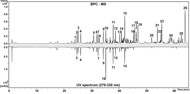
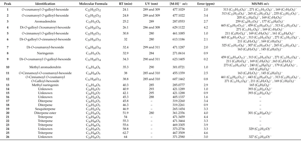
The glycosylated phenolic acid derivatives consisted of galloyl, cinnamoyl and coumaroyl substituents exhibiting two or three O-phenolic acids in the structures. The peaks of 1, 2, 4, 5, 6, 9 and 12 showed a fragment ion at m/z 169 (C7H5O5)− in negative ion mode, confirming the presence of galloyl in the structures. In addition, compounds 4, 6, 9 and 12 also showed product ions at m/z 313 (C13H13O9)− and 163 (C9H7O3)− related to the ions [M-H-coumaroyl-H2O]−and [M-H-galloyl-hexosyl]−, respectively. All this spectral information taken together allowed the putative identification of the compounds asO-coumaroylO-galloyl-hexoside (1), O-coumaroyl O-galloyl-hexoside (2), di-O-galloyl O-coumaroyl-hexoside (4), O-cinnamoyl O-galloyl-hexoside (5), di-O-galloyl O-cinnamoyl-hexoside (6), di-O-coumaroyl-hexoside (7), di-O-coumaroylO-galloyl-hexoside (9),O-cinnamoylO-coumaroyl-hexoside (10) andO-cinnamoyl O-coumaroylO-galloyl-hexoside (12). All the data were compatible with data previously published for these compounds [22].
The compounds at the end of the chromatogram did not absorb in the UV region and had a molecular constitution typical of terpene derivatives; they belong to the classes of sesquiterpenes (compound19), diterpenes (compounds17,18and20) and triterpenes (21,22,23and25), which are common in propolis and geopropolis [16,23–25]. Due to the huge variety in the skeleton arrangement of these classes and the lack of systematic information about fragmentation by ESI in the literature, these compounds were not completely identified.
− − − −
–
–
Figure 1.Base peak chromatogram in negative ion mode and UV spectrum in the range of 270–300 nmTable 1.Compounds identified fromMelipona orbignyigeopropolis extract by high performance liquid chromatography coupled to diode array detector and mass spectrometry (HPLC-DAD-MS).
Peak Identification Molecular Formula RT (min) UV (nm) [M-H]−m/z Error (ppm) MS/MSm/z
1 O-coumaroylO-galloyl-hexoside C22H22O12 24.1 289 and 309 477.1029 2.0 313 (C13H13O9)−, 271 (C11H11O8)−, 169 (C7H5O5)−
2 O-coumaroylO-galloyl-hexoside C22H22O12 24.8 289 and 309 477.1022 3.4 313 (C13H13O9)
−, 265 (C
13H13O6)−, 235 (C12H11O5)−,
205 (C11H9O4)−, 169 (C7H5O5)−
3 Aromadendrin C15H12O6 25.2 289 287.0553 2.7 259 (C14H11O5)−, 177 (C10H9O3)−
4 Di-O-galloylOcoumaroyl-hexoside C29H26O16 26.1 286 and 308 629.1122 4.2 465 (C20H17O13)
−, 459 (C22H19O11)−, 313 (C13H13O9)−, 271 (C11H11O8)−, 169 (C7H5O5)−
5 O-cinnamoylO-galloyl-hexoside C22H22O11 30.8 280 461.1085 1.0 211 (C9H7O6)−, 169 (C7H5O5)−, 161 (C10H9O2)−
6 Di-O-galloylO-cinnamoyl-hexoside C29H26O15 32 280 613.1186 2.1 465 (C20H17O13)
−, 313 (C
13H13O9)−, 271 (C11H11O8)−,
211 (C9H7O6)−, 169 (C7H5O5)−
7 Di-O-coumaroyl-hexoside C24H24O10 32.4 299 and 311 471.1287 2.0 325 (C15H17O8)
−, 307 (C15H15O7)−, 265 (C13H13O6)−, 163 (C9H7O3)−, 145 (C9H5O2)−
8 Naringenin C15H12O5 32.9 284 271.0614 0.9 –
9 Di-O-coumaroylO-galloyl-hexoside C31H28O14 34.3 290 and 311 623.1405 0.2 459 (C22H19O11)
−, 313 (C13H13O9)−, 271 (C11H11O8)−, 211 (C9H7O6)−, 169 (C7H5O5)−,163 (C9H7O3)−
10 Methyl aromadendrin C16H14O6 35.3 290 301.0721 1.0 273 (C15H13O5)
−, 240 (C14H8O4)−, 179 (C8H3O5)−, 165 (C8H5O4)−
11 O-Cinnamoyl-O-coumaroyl-hexoside C24H24O9 38 285 and 310 455.1359 2.5 163 (C9H7O3)−, 145 (C9H5O2)−
12 O-CinnamoylO-Galloyl-hexosideO-coumaroyl C31H28O13 38.8 285 and 310 607.1462 0.8 461 (C22H21O11)
−, 443 (C22H19O10)−, 313 (C13H13O9)−, 271 (C11H11O8)−, 211 (C9H7O6)−, 169 (C7H5O5)−
13 Methyl naringenin C16H14O5 40.3 285 285.0777 2.9 165 (C8H5O4)−
14 Unknown C24H22O7 40.9 293 421.1289 1.0 393 (C23H21O6)−
15 Unknown C24H22O7 42.1 295 421.1288 0.9 393 (C23H21O6)−
16 Unknown C24H22O6 45.3 288 405.1337 1.6 –
17 Diterpene C20H32O3 45.8 – 319.2260 3.4 –
18 Diterpene C20H32O3 46.3 – 319.2261 0.9 –
19 Sesquiterpene C15H22O4 46.9 – 265.1454 3.3 –
20 Diterpene ester C22H34O4 51.9 280 361.2356 4.0 301 (C20H29O2)−
21 Triterpene C30H48O4 54 – 471.3459 4.4 –
22 Triterpene C30H48O4 55.3 – 471.3464 3.3 –
23 Triterpene C30H46O4 55.6 – 469.3305 3.9 –
24 Unknown C24H38O3 58.8 – 373.2736 3.3 329 (C23H37O)−
25 Triterpene C31H48O3 62.7 – 467.3509 4.6 –
26 Unknown C24H36O3 64 – 371.2580 3.0 327 (C23H35O)−
2.2. Antioxidant Activity
2.2.1. Capture of Free Radicals DPPH•and ABTS•+
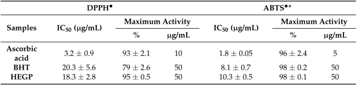
HEGP inhibited 50% of the free radicals (IC50) at a concentration approximately six times higher than the ascorbic acid control, in both the DPPH•radical capture assay and the ABTS•+. In turn, it presented results similar to the reference antioxidant BHT in both trials (Table2).
Table 2.IC50and maximum activity of reference antioxidants and treatments with HEGP.
DPPH• ABTS•+
Samples IC50(µg/mL)
Maximum Activity
IC50(µg/mL)
Maximum Activity
% µg/mL % µg/mL
Ascorbic
acid 3.2±0.9 93±2.1 10 1.8±0.05 96±2.4 5 BHT 20.3±5.6 79±2.6 50 8.1±0.7 98±0.2 50
HEGP 18.3±2.8 95±0.5 50 10.3±0.5 98±0.1 50 Values are means±standard error of the mean (SEM).
2.2.2. Hemolytic Activity and Inhibition of Oxidative Hemolysis
In this assay, the hemolytic activity of HEGP and its ability to protect erythrocytes against hemolysis induced by the oxidizing agent 2,2′-azobis (2-methylpropionamidine) dihydrochloride (AAPH) were evaluated. When erythrocytes were incubated only with HEGP, in the absence of AAPH, no hemolysis was observed throughout the experimental period at the concentrations evaluated (Figure2).
• •+
• •+
• •+
′
–125 μg/mL), incubated with erythrocytes
−
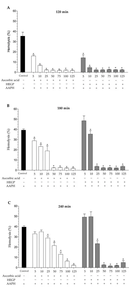
In evaluating the extract’s ability to protect erythrocytes against AAPH
– concentrations of 25 and 50 μg/mL, the extract reduced hemolys
26.7% ± 4.9% (25 μg/mL) and 45.7% ± 8.0% (50 μg/mL) (Figure 3A– 0
10 20 30 40 50 60
5 10 25 50 75 100 125
Ascorbic acid HEGP AAPH
_
5 10 25 50 75 100 125 _
5
+ + + + + + +
_ _ _ _ _ _ _
_ _ _ _ _ _ _ +_ _+ _+ _+ +_ +_ +_
_ _ _ _ _ _ _
_ Control
H
em
o
ly
si
s
(%
)
Figure 2.Hemolytic activity of ascorbic acid and HEGP (5–125µg/mL), incubated with erythrocytes and 0.9% NaCl solution for 240 min, without the presence of AAPH. The control consists of erythrocytes incubated only with 0.9% NaCl solution.−: absence; +: presence.
In evaluating the extract’s ability to protect erythrocytes against AAPH-induced hemolysis, HEGP was able to inhibit oxidative hemolysis for up to 240 min of incubation (Figure3A–C). At concentrations of 25 and 50µg/mL, the extract reduced hemolysis by 40.9%±8.0% and 93.2%±0.8%, respectively,
–125 μg/mL) incubated with
−
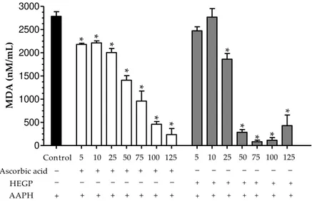
2.2.3. Dosage of Malondialdehyde
HEGP demonstrated the concentration-dependent reduction of MDA levels and presented better activity than the ascorbic acid control (Figure4). At the concentration of 50µg/mL, the extract
reduced MDA levels by 89.75%±2.1%, while the ascorbic acid control presented a reduction of only
49.45%±3.5% under the same conditions.
4). At the concentration of 50 μg/mL, the extract
–125 μg/mL) for 240 min. The control consists of erythrocytes incubated only with AAPH − – 0 500 1000 1500 2000 2500 3000 * * * * * * * * * * * * Ascorbic acid HEGP AAPH
5 10 25 50 75 100 125 5 10 25 50 75 100 125
+ + + + + + + + + + + + + + + + + + + + + + + + + + + + + _ _ _ _ _ _ _ _ _ _ _ _ _ _ _ _ Control M D A (n M /m L )
Figure 4.Concentration of malondialdehyde (nM/mL) in erythrocytes incubated with ascorbic acid and HEGP (5–125µg/mL) for 240 min. The control consists of erythrocytes incubated only with AAPH solution (50 mM). *p< 0.05 when the treated groups were compared to the AAPH control group. −: absence; +: presence.
2.3. Anti-Inflammatory Activity
The anti-inflammatory property of HEGP was assessed indirectly by its inhibition of the activity of the hyaluronidase enzyme. HEGP exhibited a concentration-dependent inhibition of enzyme activity, demonstrating inhibition of 35.6%±2.4% at the concentration of 75 mg/mL (Figure5).
4). At the concentration of 50 μg/mL, the extract
–125 μg/mL) for 240 min. The control consists of erythrocytes incubated only with AAPH − – 0 10 20 30 40 50
0.20 0.39 0.78 1.56 3.13 6.25 12.5 25.0 50.0 75.0
Concentration (mg/mL) Concentration (mg/mL) In h ib it io n ( % )
Figure 5.Percentage inhibition of hyaluronidase enzyme activity by HEGP (0.2–75 mg/mL).
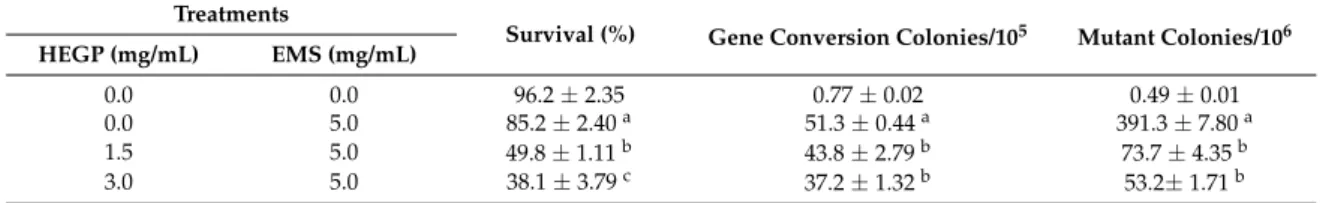
2.4. Antimutagenic Activity
HEGP was able to reduce the survival ofS. cerevisiaeD7 (ATCC 201137) by 48.1%±1.1% and
incubated only with the mutagen EMS, there was an increase in the conversion of genes and in the quantity of mutant colonies. When incubated with HEGP in the presence of EMS, 14.6%±5.4%
(1.5 mg/mL) and 27.5%±2.5% (3.0 mg/mL) reductions of gene conversion were observed. In addition,
HEGP was able to reduce the amount of mutant colonies by 81.1% ± 1.1% (1.5 mg/mL) and
86.3%±0.4% (3.0 mg/mL) (Table3).
Table 3.Effect of the hydroethanolic extract of geopropolis ofMelipona orbignyi(HEGP) on the survival percentage of yeast cellsSaccharomyces cerevisiae(diploid line D7 ATCC 201137), conversion of genes and mutant colonies.
Treatments
Survival (%) Gene Conversion Colonies/105 Mutant Colonies/106
HEGP (mg/mL) EMS (mg/mL)
0.0 0.0 96.2±2.35 0.77±0.02 0.49±0.01
0.0 5.0 85.2±2.40a 51.3±0.44a 391.3±7.80a
1.5 5.0 49.8±1.11b 43.8±2.79b 73.7±4.35b
3.0 5.0 38.1±3.79c 37.2±1.32b 53.2±1.71b
a, b, c: Means with different superscripts are significantly different for each attribute (p< 0.05). Values are means±SEM. EMS, Ethyl methanesulfonate.
2.5. Antimicrobial Activity
HEGP demonstrated antimicrobial activity against all evaluated microorganisms. Gram-positive bacteria were more sensitive to the action of the extract than gram-negative bacteria. The inhibition observed against the evaluated microorganisms followed the sequence:S. aureus>E. faecalis>E. coli> P. aeruginosa>C. neoformans>C. albicans. HEGP also showed action against all microorganisms resistant to antimicrobial drugs, at concentrations similar to the reference strains. In addition to inhibitory activity, HEGP showed bactericidal and fungicidal activity against all microorganisms evaluated in this study, ranging from 8.50± 0.28 mg/mL forS. aureusATCC 6538™ to 36.1±0.50 mg/mL for
amphotericin B-resistantC. albicansESA 97 from biological fluid (Table4).
Table 4.Minimum inhibitory concentration (MIC), minimum bactericidal concentration (MBC), and minimum fungicidal concentration (MFC) for HEGP fromM. orbignyi.
Microorganisms HEGP (mg/mL) Gentamicin (µg/mL)
Gram-positive bacteria MIC MBC MBC
Staphylococcus aureusATCC 6538™ 6.13±0.10 8.50±0.28 2.00±0.28
S. aureusESA 175 Methicillin-resistant 6,42±0.46 8.75±0.62 2.67±0.16
S. aureusESA 159 Methicillin-resistant 6.92±0.30 9.08±0.58 2.50±0.28
Enterococcus faecalisATCC 43300™ 7.08±0.58 10.5±0.22 2.83±0.30
E. faecalisESA 201 Vancomycin-resistant 8.17±0.44 10.9±0.30 3.25±0.14
E. faecalisESA 361 Vancomycin-resistant 9.08±0.08 11.2±0.38 3.33±0.16
Gram-negative bacteria
Escherichia coliATCC 29998™ 10.5±0.82 13.2±0.52 4.58±0.30
E. coliESA 37 Cephalosporins-resistant 11.2±0.32 13.8±0.72 4.67±0.22
E. coliESA 54 Cephalosporins-resistant 11.5±0.76 13.7±0.90 4.92±0.08
Pseudomonas aeruginosaATCC 15442™ 12.9±0.50 16.5±0.30 5.00±0.28
P.aeruginosaESA 22 Imipenem-resistant 13.3±0,60 17.0±1.08 6.17±0.16
P.aeruginosaESA 23 Imipenem-resistant 13.5±1.22 17.7±1.02 6.50±0.28
Microorganisms HEGP (mg/mL) Amphotericin-B (µg/mL)
Fungi MIC MFC MFC
Cryptococcus neoformansATCC 32264 ™ 19.3±0.60 25.0±1.04 0.13±0.07
C. neoformansESA 211 Amphotericin-B resistant 20.9±0.79 26.0±1.04 0.25±0.14
C. neoformansESA 105 Amphotericin-B resistant 21.4±1.10 26.1±1.58 0.38±0.22
Candida albicansATCC 10231™ 23.0±1.06 34.0±1.12 0.29±0.16
C.albicansESA 100 Amphotericin-B resistant 23.7±1.29 35.0±1.23 0.14±0.08
C.albicansESA 97 Amphotericin-B resistant 24.4±1.83 36.1±0.50 0.25±0.14
3. Discussion
Geopropolis is a beehive material of complex composition, produced specifically by species of stingless bees of the Neotropical region and popularly known for its therapeutic properties. However, despite the great diversity of species of bees capable of generating this product, few studies have investigated and described its chemical and pharmacological properties.
In this study, when investigating the chemical composition of M. orbignyi HEGP, different compounds were identified from the ones found in geopropolis produced by other species of stingless bees, such as derivatives of glycosylated phenolic acids. The chemical composition of geopropolis from theM. fasciculataandM. scutellarisbees presented great differences from the composition observed in this work for theM. orbignyispecies, including benzophenones, hydrolyzable tannins (gallotannins and ellagitannins) and gallic and ellagic acid substances [7,26]. These differences in chemical composition suggest thatM. orbignyiuses different plant sources of raw material to produce its geopropolis and/or that the production process significantly changes the composition of this material, as the genetic variability of bee species may influence this chemical composition [21]. Thus, these bioproducts from these stingless bees are unique, increasing the possibilities of identifying new bioactive compounds with pharmacological properties.
The total phenol content observed for HEGP was similar to the value obtained for the geopropolis ofM. scutellaris(127±1.9 mg GAE/g of extract) but higher than that forM. fasciculata(47.78±0.04 mg
GAE/g of extract) and that forM. interruptandM. seminigra(419 to 4378µg GAE/g of extract) [7,11,21].
HEGP demonstrated potent antioxidant activity, being able to inhibit the free radicals DPPH•and ABTS•+. The ethanolic extracts from the geopropolis ofM. interruptandM. seminigraexhibited almost 40 and 30 times higher total phenol content than those from HEGP (121±0.6 mg GAE/g HEGP),
but the antioxidant activities of DPPH•were 10±0.5 and 26.3±3.9µg/mL, respectively [21]. This result demonstrates the great antioxidant potency of the compounds of HEGP, even though it had a lower total phenol content, its antioxidant activity in DPPH•was 18.3±2.8µg/mL. In addition, as the chemical compositions of these geopropolis types and HEGP were different, especially with respect to the glycosylated derivatives of phenolic acids, this result demonstrates the importance of the structural characteristics of the constituents for this activity. The phenolic acids and flavonoids have hydroxyl groups in their rings [7], conferring the ability to stabilize unpaired electrons, which are typical of free radicals, thus acting as antioxidants [27].
In addition, HEGP presented in vitro antioxidant action in a human biological model by inhibiting lipid peroxidation in erythrocytes induced by an oxidizing agent. The extract showed better results than the standard antioxidant ascorbic acid, in terms of both anti-hemolytic action and the reduction of MDA levels, exhibiting a greater protective effect. This result can be attributed to the synergistic action of its compounds, including the different types of phenolic constituents, which offer excellent cellular protection against lipid peroxidation, acting as hydrogen donor agents, singlet oxygen reducers and superoxide radicals, antioxidant enzyme activators and metal chelators [27,28]. Some authors classify glycosylated derivatives of phenolic acids as a new type of hydrolysable tannin, similar to the ones described in plants such asBalanophora harlandii (Balanophoraceae) [29]. Among the phenolic compounds identified in the HEGP, tannins are described as acting as metal ion chelators [30] and have many hydroxyl groups in their phenolic rings, promoting the antioxidant activity of these compounds [31].
In addition, the terpenes constitute another class of compounds that display important antioxidant action. Triterpenes, one of the terpene subtypes identified in HEGP, are described as acting to capture free radicals, including superoxide anion and hydroxyl radical, and acting to protect against the lipid peroxidation process [32]. In addition, they are capable of modulating the activity of antioxidant enzymes, such as superoxide dismutase, catalase and glutathione peroxidase [33]. Mechanisms similar to the ones described for sesquiterpenes [34], compounds also identified in HEGP.
biomolecules, such as nucleic acids, proteins and DNA [35]. This process is often related to the development of neurodegenerative diseases such as Alzheimer’s and Parkinson’s, as well as cancer, diabetes, atherosclerosis [36] and chronic inflammation [37].
Inflammation is due to the action of several mediators that promote vascular and cellular events [38]. Inflammatory cells produce mediators, such as arachidonic acid metabolites, cytokines and chemokines, which aid in the production of reactive species [37]. Under normal physiological conditions, these oxidative and immunological processes aid in the response against stressors and pathogens [39]. However, the constant activation of these processes may lead to an increased risk of more serious diseases [37,39], such as diabetes [40] and obesity [41].
Natural products have been the objects of studies for the development of new anti-inflammatory drugs [6]. Among them, geopropolis has presented satisfactory results, such as the geopropolis extract of Melipona scutellaris, which promoted anti-inflammatory action through the inhibition of IL-1B and TNF-α[11] and decreased neutrophil migration in vivo [6]. In this study, HEGP demonstrated
anti-inflammatory activity by inhibiting the enzyme hyaluronidase, reducing the degradation of hyaluronic acid. This polysaccharide performs important biological functions in the organism, such as cell proliferation, differentiation and tissue repair [42]. According to Pascoal et al. [43], the degradation of hyaluronic acid by the enzyme hyaluronidase can lead to bone loss, pain and inflammation.
Sesquiterpenes are among the compounds with anti-inflammatory potential reported in HEGP; they inhibit the pro-inflammatory transcription factors NF-κB and STAT3 [44]. In addition,
phenolic compounds are described as anti-inflammatory agents because they modulate the expression of cytokines [45] and pro-inflammatory enzymes [46], such as nitric oxide synthase and cyclooxygenase-2 [47]. Of the phenolic compounds present in HEGP, aromadendrin exerts anti-inflammatory action by reducing the release of inflammatory factors MCP-1 and IL-8 in normal keratinocytes stimulated with interferon and histamine [48]. In addition, methyl aromadendrin (4′-methyl ether), a compound also present in HEGP and commonly found in green propolis produced byApis mellifera, can reduce reactive species produced by neutrophil metabolism during the inflammatory process [49].
The reactive species generated during the process of chronic inflammation may contribute to the onset of mutagenesis and carcinogenesis [50]. In this way, the identification of compounds with antioxidant and antimutagenic properties may have great therapeutic importance. Substances with such activities have been found in many natural products, such as plants [51] and bee products [42].
In addition to antioxidant properties,M. orbignyiHEGP demonstrated antimutagenic potential. In this study, the extract decreased the survival of yeastS. cerevisiae, which may be related to its fungicidal action. However, it was able to reduce the frequency of gene conversion and the amount of mutant colonies induced by the mutagenic agent EMS. Some studies suggest that the antimutagenic activity ofApis melliferapropolis is related to its antioxidant activity [52]. Phenolic compounds and terpenes are well known for their antioxidant properties as well as for acting as antimutagenic agents. In addition, their intake is related to the reduction of cancer risks [53]. Diterpenes are described as being capable of repairing DNA errors, also related to their anticancer activity [54].
Another biological activity presented by HEGP was antimicrobial action, including effectiveness against hospital strains resistant to antimicrobial drugs, such as methicillin-resistant Staphylococcus aureusgram-positive bacteria, cephalosporin-resistantEscherichia coligram-negative bacteria, and amphotericin B-resistant Candida albicans yeast. Currently, the intensive use of antimicrobial drugs, both for therapeutic and prophylactic purposes, has resulted in the development of multiresistant strains, becoming a worldwide problem [55].
Among the natural substances that present antimicrobial action, phenolic compounds act through the permeabilization of the microbial cytoplasmic membrane, inhibition of the synthesis of nucleic acids of gram-negative and gram-positive bacteria [59], inhibition of the synthesis of ATP and interruption of electron transport [60]. In addition to these mechanisms, flavonoids form complexes with proteins through hydrogen bonds, and their antimicrobial activity is related to the inhibition of microbial adhesins, enzymes and protein transport [28].
Among the flavonoids present in HEGP, flavanone naringenin promotes the inhibition of bacterial cytoplasmic membrane function, reducing its motility [59], even against methicillin-resistant Staphylococcus aureus[28]. In addition, glycosylated phenolic derivatives are present in the extract and are described by some authors as new types of hydrolysable tannins, which exert antimicrobial action by breaking the permeability of the membrane of different microbial strains [61].
4. Materials and Methods
4.1. Preparation of the Hydroethanolic Extract of Geopropolis
Samples of geopropolis were obtained in Mato Grosso do Sul, Brazil (22◦13′12′′S–54◦49′2′′W). The hydroethanolic extract of geopropolis (HEGP) was prepared in the proportions of 240 mL of 70% ethanol for each 80 g of powder geopropolis. The solution was then kept shaking in a closed container for 24 h at room temperature. Subsequently, the extract was filtered, concentrated in a rotary evaporator (Gehaka, São Paulo, SP, Brazil) at 40◦C and lyophilized to obtain the dry extract. The yield of HEGP was 3.49%, and the final freeze-dried was stored at−20◦C protected from light.
4.2. Determination of Phenolic Compounds and Flavonoids
4.2.1. Phenolic Compounds
The concentration of phenolic compounds present in HEGP was determined by the Folin–Ciocalteu colorimetric method. For this purpose, 0.5 mL of HEGP (100µg/mL) was added
to 2.5 mL of the Folin–Ciocalteau reagent and 2.0 mL of 14% sodium carbonate (Na2CO3) solution. Absorbance reading was performed at 760 nm after 2 h of incubation at room temperature in the dark. To produce the calibration curve, gallic acid (0.4–16µg/mL) was used as a standard. The average of
three readings was used to determine the content of phenolic compounds expressed in mg of GAE/g of HEGP.
4.2.2. Total Flavonoids
To verify the concentration of flavonoids present in the extract, 0.5 mL of HEGP (100µg/mL)
was mixed with 4.5 mL of methanolic solution of aluminum chloride hexahydrate 2% (AlCl3·6H2O) and incubated for 30 min at room temperature in the dark. After this period, absorbance reading was performed at 415 nm. To produce the calibration curve, quercetin (0.4–16µg/mL) was used as a
standard. The average of three readings was used to determine the flavonoid content expressed in mg of QE/g of HEGP.
4.3. HPLC-DAD-MS Analyses
The HEGP (1 mg/mL) was injected into an ultra-fast liquid chromatograph UFLC (LC-20AD, Shimadzu) coupled to a diode array detector operating at 240–800 nm and a mass spectrometer with electrospray ionization (ESI) and the analyzers quadrupole–Time-of-Flight (QTOF) (micrOTOF-Q III, Bruker Daltonics) monitoring betweenm/z120 and 1200 in negative and positive ion mode, equipped with a C-18 column (Kinetex, 150×2.2 mm id, 2.6µm) and using an oven temperature of 50◦C.
washing and reconditioning of the column (8 min). The flow rate was 0.3 mL/min, and the injection volume was 1µL.
4.4. Antioxidant Activities
4.4.1. DPPH•Free Radical Capture Activity
For this experiment, 200 µL of the HEGP was mixed with 1800 µL of 0.11 mM
2,2-diphenyl-1-picrylhydrazyl (DPPH•) radical solution in 80% ethanol. The final concentrations of the extract ranged from 0.1 to 200µg/mL. The mixture was incubated at room temperature in
the dark for 30 min. The absorbance was measured at 517 nm in a T 70 UV/VIS spectrophotometer (PG Instruments Limited, Leicestershire, UK). Ascorbic acid and butylated hydroxytoluene (BHT) were used as reference antioxidants. As control, 200µL of the solvent (80% ethanol) was incubated
with 1800µL of the DPPH•solution. Three independent experiments were performed in triplicate.
The percent inhibition was calculated from the control using the following equation:
Capture activity of DPPH•(%) = (1−Abs
sample/Abscontrol)×100 (1)
4.4.2. ABTS•+Free Radical Capture Activity
The 2,2′-azino-bis(3-ethylbenzothiazoline-6-sulphonic acid) (ABTS•+) radical was prepared from a mixture of 5 mL of ABTS (7 mM) and 88µL of potassium persulfate (140 mM) and incubated for 12–16 h
at room temperature in the dark. After this period, the ABTS•+radical solution was diluted in absolute ethanol until an absorbance of 0.70±0.05 at 734 nm was obtained in a T 70 UV/VIS spectrophotometer
(PG Instruments Limited, Leicestershire, UK). Then, 20µL of HEGP was added to 1980µL of the
ABTS•+radical. The final concentrations of the extract ranged from 0.1 to 200µg/mL. The mixture was incubated at room temperature in the dark for 6 min. The absorbance was evaluated at 734 nm in a T 70 UV/VIS spectrophotometer (PG Instruments Limited, Leicestershire, UK). Ascorbic acid and BHT were used as positive controls. As control, 20µL of the solvent (80% ethanol) was incubated
with 1980µL of the ABTS•+radical. Two independent experiments were performed in triplicate.
The percentage of ABTS•+radical inhibition was calculated according to the following equation:
Inhibition of the radical ABTS•+(%) = ((Abscontrol−Abssample)/Abscontrol)×100 (2)
4.4.3. Antioxidant Assays in Human Erythrocytes
Preparation of the Erythrocyte Suspensions
Human erythrocyte assays were performed after receiving approval from the Research Ethics Committee (ComitêdeÉtica em Pesquisa, CEP) of the University Center of the Grande Dourados (Centro
Universitário da Grande Dourados, UNIGRAN), Brazil (CEP process number 123/12). Peripheral blood
from healthy donors was collected and placed in tubes containing sodium citrate. The tubes were then centrifuged at 700×gfor 10 min, and the blood plasma and leukocyte layer were discarded. The erythrocytes were washed three times with 0.9% sodium chloride (NaCl) solution, and then, a 10% erythrocyte suspension in 0.9% NaCl solution was prepared for the inhibition assays of oxidative hemolysis and malondialdehyde, determined using the method described by Campos et al. [62].
Inhibition of Oxidative Hemolysis
The protective effect of HEGP was evaluated according to the method described by Campos et al. [55]. Erythrocytes were pre-incubated at 37◦C for 30 min in the presence of different concentrations of HEGP (5–125µg/mL). After this period, the samples received only 0.9% NaCl
and ascorbic acid was used as a positive control at the same concentrations as HEGP. The treatments were incubated at 37◦C with periodic homogenization. The hemolysis content was determined after 120, 180 and 240 min of incubation. Samples were centrifuged at 400×g/10 min, and the supernatant was transferred to tubes containing 0.9% NaCl solution to perform the reading at 540 nm in a T 70 UV/VIS spectrophotometer (PG Instruments Limited, Leicestershire, UK). As a control of total hemolysis, human erythrocytes were incubated with distilled water. Three independent experiments were performed in duplicate. The percentage of hemolysis was calculated according to the following equation:
Hemolysis (%) = (Abssample/Abstotal hemolysis)×100 (3)
Dosage of Malondialdehyde (MDA)
The ability of HEGP to inhibit the lipid peroxidation process was investigated in human erythrocytes incubated with the oxidizing agent AAPH, and the MDA concentration was determined as described by Campos et al. [55]. For this experiment, the erythrocytes were pre-incubated at 37◦C for 30 min with different concentrations of HEGP (5–125µg/mL). Erythrocytes incubated with ethanol
at the final concentration of 1% were used as a negative control, and ascorbic acid was used as a positive control at the same concentrations as HEGP. Then, AAPH solution (50 mM) was added and the samples incubated at 37◦C for 4 hours with periodic homogenization. After this time, the samples were centrifuged at 700×g/10 min, and 0.5 mL of the supernatant was mixed with 1 mL of 10 nmol thiobarbituric acid (TBA) dissolved in 75 mM potassium phosphate monobasic buffer at pH 2.5. As a standard control, 0.5 mL of MDA solution (20µM) was incubated with 1 mL of TBA. Samples were
maintained at 96◦C for 45 min. After cooling the solution, 4 mL ofn-butyl alcohol was added, followed by centrifugation at 1600×g/5 min. The supernatant from the samples was analyzed at 532 nm in a T 70 UV/VIS spectrophotometer (PG Instruments Limited, Leicestershire, UK). Two independent experiments were performed in duplicate. The MDA levels of the samples were expressed in nmol/mL, obtained by the following formula:
MDA(nmol/mL) = ABSsample ×
20
×220.32
ABSstandardMDA
(4)
4.5. Anti-Inflammatory Activity—Hyaluronidase Assay
Inhibition of the activity of the hyaluronidase enzyme was determined using the method described by Silva et al. [63] The reaction mixture consisted of 50µL of HEGP (0.2–75 mg/mL) and 50µL of
the enzyme hyaluronidase (350 units) (type IV-S: bovine tests, Sigma, St. Louis, MO, USA) incubated at 37◦C for 20 min. Then, 1.2µL of calcium chloride solution (2.5×10³M) was added to activate the enzyme and kept at 37◦C for 20 min. After this period, 0.5 mL of hyaluronic acid sodium salt substrate (0.1 M) was added and the solution incubated at 37◦C for 40 min. Subsequently, 0.1 mL of potassium tetraborate (0.8 M) was added and the solution incubated at 100◦C for 3 min. The solution was then cooled, and 3 mL ofp-dimethylaminobenzaldehyde was added, followed by incubation at 37◦C for 20 min. Finally, the absorbance was measured at 585 nm in a T 70 UV/VIS spectrophotometer (PG Instruments Limited, Leicestershire, UK), using distilled water as a control. The experiments were performed in triplicate. The percentage of hyaluronidase enzyme inhibition was calculated according to the following equation:
Inhibition of the enzyme hyaluronidase (%) = ((Abscontrol−Abssample)/Abscontrol)×100 (5)
4.6. Antimutagenic Activity
conversions at the tryptophan locus and reverse mutations at the isoleucine locus, before experimental use. Culture cells with low spontaneous gene conversion and low reverse mutation frequency were cultured in liquid medium at 28 ◦C until they reached the stationary growth stage. They then underwent sedimentation and were resuspended in sterile potassium phosphate buffer (0.1 M), pH 7.4, until a final value of 2×10⁸cells/mL was obtained. The mutagenic agent ethyl methanesulfonate
(EMS) (5 mg/mL) was added together with HEGP, evaluated at concentrations of 1.5 and 3.0 mg/mL. The mixture was incubated with stirring for 2 h at 37◦C. After this period, the cells were plated in complete and selective medium to determine yeast survival, tryptophan converters and isoleucine reversers. The experiments were performed in triplicate.
4.7. Antimicrobial Activity
The antimicrobial activity of HEGP was investigated in microorganisms collected from biological fluids at the Hospital Center and identified in the Microbiology Laboratory of Escola Superior Agrária (ESA) de Bragança, Portugal. Reference strains were obtained from the authorized
ATCC distributor (LGC Standards SLU, Barcelona, Spain). The microorganisms used were Staphylococcus aureus ATCC 6538™, methicillin-resistant S. aureus ESA 175, methicillin-resistant S. aureus ESA 159, Enterococcus faecalis ATCC 43300™, vancomycin-resistant E. faecalis ESA 201, vancomycin-resistantE. faecalisESA 361,Escherichia coliATCC 29998™, cephalosporin-resistantE. coli ESA 37, cephalosporin-resistantE. coliESA 54,Pseudomonas aeruginosaATCC 15442, imipenem-resistant P. aeruginosaESA 22, imipenem-resistantP. aeruginosaESA 23,Cryptococcus neoformansATCC 32264, amphotericin B-resistantC. neoformansESA 211, amphotericin B-resistant C. neoformansESA 105, Candida albicans ATCC 10231™, amphotericin B-resistant C. albicans ESA 100 and amphotericin B-resistantC. albicansESA 97.
The microorganisms were stored in Muller-Hinton medium supplemented with 20% glycerol at 70◦C prior to experimental use. The inoculum was then prepared by dilution of the cell mass in 0.85% NaCl solution, adjusted to 0.5 on the MacFarland scale, as confirmed by spectrophotometric reading at 580 nm for bacteria and 640 nm for yeast. Antimicrobial assays were performed as described by Silva et al. [63] using Nutrient Broth (NB) for bacteria or Yeasts Peptone Dextrose (YPD) for yeast in microplates (96 wells). The HEGP was diluted in dimethylsulfoxide (DMSO) and transferred to the first well, followed by serial dilution. The inoculum was added to all wells (104Colony Forming Units (CFU)/mL), and the plates were incubated at 37◦C for 24 h for bacteria and at 25◦C for 48 h for yeast. Gentamicin and amphotericin B were used as antibacterial and antifungal controls, respectively. A negative control (medium only), a positive control (inoculated medium) and a DMSO control (DMSO with inoculated medium) were performed in each experiment. After the incubation period, the antimicrobial activity was detected by the addition of 20µL of 2,3,5-triphenyl-2H-tetrazoliumchloride (TTC) solution (5 mg/mL). The minimum inhibitory concentration (MIC) was defined as the lowest concentration of HEGP that visibly inhibited the growth of microorganisms, as indicated by TTC staining, which marks viable cells.
To determine the minimum bactericidal concentration (MBC) and minimum fungicidal concentration (MFC), 20µL of the last well where growth was observed and from each well where no
color changes were seen was seeded in NB or YPD and incubated for 24 h at 37◦C for bacteria growth and 48 h for yeast growth. The lowest concentration that did not result in growth (<10 CFU/plate) after this subculture process was considered the MBC or MFC. The experiments were performed in triplicate, and the results were expressed in mg/mL.
4.8. Statistical Analyses
The data are shown as the mean±SEM and were analyzed for statistically significant differences
5. Conclusions
In conclusion, this study demonstrates the presence of bioactive compounds in the hydroethanolic extract ofM. orbignyigeopropolis, as well as its antioxidant, anti-inflammatory, antimutagenic and antimicrobial activities.
Acknowledgments: This work was supported by grants from Foundation to Support to Fundação de Apoio ao Desenvolvimento do Ensino, Ciência e Tecnologia do Estado de Mato Grosso do Sul (FUNDECT, Brazil), Coordenação de Aperfeiçoamento de Pessoal de Nível Superior (CAPES, Brazil), Conselho Nacional de Desenvolvimento Científico e Tecnológico (CNPq, Brazil) and PRODER, (24.073 –Â, Portugal).
Author Contributions: Helder Freitas dos Santos, Jaqueline Ferreira Campos, Cintia Miranda dos Santos, JoséBenedito Perrella Balestieri, Leticia Miranda Estevinho, Denise Brentan Silva, Carlos Alexandre Carollo, Kely de Picoli Souza and Edson Lucas dos Santos conceived and designed the experiments; Helder Freitas dos Santos, Jaqueline Ferreira Campos, Cintia Miranda dos Santos, Denise Brentan Silva, Carlos Alexandre Carollo, Leticia Miranda Estevinho, Kely de Picoli Souza and Edson Lucas dos Santos performed the experiments; Helder Freitas dos Santos, Jaqueline Ferreira Campos, Cintia Miranda dos Santos, JoséBenedito Perrella Balestieri, Leticia Miranda Estevinho, Denise Brentan Silva, Carlos Alexandre Carollo, Kely de Picoli Souza and Edson Lucas dos Santos analyzed the data; Carlos Alexandre Carollo, Kely de Picoli Souza, Leticia Miranda Estevinho and Edson Lucas dos Santos contributed reagents/materials/analysis tools; and Helder Freitas dos Santos, Jaqueline Ferreira Campos, Cintia Miranda dos Santos, JoséBenedito Perrella Balestieri, Leticia Miranda Estevinho, Denise Brentan Silva, Carlos Alexandre Carollo, Kely de Picoli Souza and Edson Lucas dos Santos wrote the paper. All authors have read and approved the final manuscript.
Conflicts of Interest:The authors declare no conflict of interest.
Abbreviations
AAPH 2,2′-Azobis (2-methylpropionamidine) dihydrochloride
ABTS•+ 2,2′-Azino-bis(3-ethylbenzothiazoline-6-sulphonic acid)
ATCC American Type Culture Collection
BHT Butylated hydroxytoluene
DPPH• 2,2-Diphenyl-1-picrylhydrazyl
EMS Ethyl methanesulfonate
ESA Escola Superior Agrária
GAE Gallic acid equivalents
HEGP Hydroalcoholic extract of geopropolis
MDA Malondialdehyde
MBC Minimum bactericidal concentration
MIC Minimum inhibitory concentration
MFC Minimum fungicidal concentration
M. orbignyi Melipona orbignyi
NaCl Sodium chloride
QE Quercetin equivalents
SEM Standard error of the mean
TBA Thiobarbituric acid
TTC 2,3,5-Triphenyl-2H-Tetrazoliumchloride
References
1. Freitas, M.O.; Ponte, F.A.; Lima, M.A.S.; Silveira, E.R. Flavonoids and triterpenes from the nest of the stingless beeTrigona spinipes.J. Braz. Chem. Soc.2008,19, 532–535. [CrossRef]
2. Bartolomeu, A.R.; Frión-Herrera, Y.; da Silva, L.M.; Romagnoli, G.G.; de Oliveira, D.E.; Sforcin, J.M. Combinatorial effects of geopropolis produced byMelipona fasciculataSmith with anticancer drugs against human laryngeal epidermoid carcinoma (HEp-2) cells.Biomed. Pharmacother.2016,81, 48–55. [CrossRef] [PubMed]
3. Souza, R.C.; Paiva, L.K.O.Y.J.; Aguiar, L.; Plácido, F.; Oliveira, M. Valor nutricional do mel e pólen de abelhas sem ferrão da região amazônica.Acta Amazon.2004,34, 1–4. [CrossRef]
5. Cunha, M.G.; Franchin, M.; Galvão, L.C.D.C.; Bueno-Silva, B.; Ikegaki, M.; Alencar, S.M.; Rosalen, P.L. Apolar bioactive fraction ofMelipona scutellarisgeopropolis onStreptococcus mutansbiofilm.Evid.-Based Complement. Altern. Med.2013,2013, 256287. [CrossRef] [PubMed]
6. Franchin, M.; Cunha, M.G.; Denny, C.; Napimoga, M.H.; Cunha, T.M.; Bueno-Silva, B.; Alencar, S.M.; Ikegaki, M.; Rosalen, P.L. Bioactive fraction of geopropolis fromMelipona scutellarisdecreases neutrophils migration in the inflammatory process: Involvement of nitric oxide pathway. Evid.-Based Complement. Altern. Med.2013,2013, 907041. [CrossRef] [PubMed]
7. Dutra, R.P.; Abreu, B.V.B.; Cunha, M.S.; Batista, M.C.A.; Torres, L.M.B.; Nascimento, F.R.F.; Ribeiro, M.N.S.; Guerra, R.N.M. Phenolic acids, hydrolyzable tannins, and antioxidant activity of geopropolis from the stingless beeMelipona fasciculataSmith.J. Agric. Food Chem.2014,62, 2549–2557. [CrossRef] [PubMed] 8. Moure’s bee catalogue. Available online:http://moure.cria.org.br(accessed on 11 November 2016). 9. Nogueira-Neto, P. Arquitetura dos ninhos. InVida e criação de abelhas indígenas sem ferrão; Urna Edição
Nogueirapis: Nogueirapis São Paulo, Brazil, 1997; pp. 47–60.
10. Souza, S.A.; Camara, C.A.; Silva, E.M.S.; Silva, T.M.S. Composition and antioxidant activity of geopropolis collected byMelipona subnitida(Jandaíra) bees.Evid.-Based Complement. Altern. Med.2013,2013, 801383. 11. Franchin, M.; Cunha, M.G.; Denny, C.; Napimoga, M.H.; Cunha, T.M.; Koo, H.; Alencar, S.M.; Ikegaki, M.;
Rosalen, P.L. Geopropolis fromMelipona scutellarisdecreases the mechanical inflammatory hypernociception by inhibiting the production of IL-1βand TNF-α.J. Ethnopharmacol.2012,143, 709–715. [CrossRef] [PubMed] 12. Ribeiro-Junior, J.A.; Franchin, M.; Cavallini, M.E.; Denny, C.; Alencar, S.M.; Ikegaki, M.; Rosalen, P.L.
Gastroprotective effect of geopropolis fromMelipona scutellarisis dependent on production of nitric oxide and prostaglandin.Evid.-Based Complement. Altern. Med.2015,2015, 459846. [CrossRef] [PubMed]
13. Coelho, G.R.; Mendonça, R.Z.; Vilar, K.D.S.; Figueiredo, C.A.; Badari, J.C.; Taniwaki, N.; Namiyama, G.; Oliveira, M.I.; Curti, S.P.; Silva, P.E.; et al. Antiviral action of hydromethanolic extract of geopropolis from Scaptotrigona posticaagainst antiherpes simplex virus (HSV-1).Evid.-Based Complement. Altern. Med.2015, 2015, 296086. [CrossRef] [PubMed]
14. Dutra, R.P.; Nogueira, A.M.C.; Marques, R.R.D.O.; Costa, M.C.P.; Ribeiro, M.N.S. Pharmacognostic evaluation of geopropolis ofMelipona fasciculataSmith from Baixada Maranhense, Brazil.Rev. Bras. Farmacogn.2008,18, 557–562. [CrossRef]
15. Chemical Abstract Service database. Available online:https://scifinder.cas.org(accessed on 15 January 2017). 16. Campos, J.F.; Santos, U.P.; Macorini, L.F.B.; Melo, A.M.M.F.; Balestieri, J.B.P.; Gamero, E.J.P.; Cardoso, C.A.L.; Souza, K.P.; Santos, E.L. Antimicrobial, antioxidant and cytotoxic activities of propolis fromMelipona orbignyi (Hymenoptera, Apidae).Food Chem. Toxicol.2014,65, 374–380. [CrossRef] [PubMed]
17. Markham, K.R. Ultraviolet-visible absorption spectroscopy. In Techniques of Flavonoid Identification; Academic Press: London, UK, 1982; pp. 36–51.
18. Cuyckens, F.; Claeys, M. Mass spectrometry in the structural analysis of flavonoids.J. Mass Spectrom.2004, 39, 1–15. [CrossRef] [PubMed]
19. Justino, G.C.; Borges, C.M.; Florencio, M.H. Electrospray ionization tandem mass spectrometry fragmentation of protonated flavone and flavonol aglycones: A re-examination.Rapid. Commun. Mass Spectrom.2009,23, 237–248. [CrossRef] [PubMed]
20. Bankova, V.; Christov, R.; Marcucci, C.; Popov, S. Constituents of Brazilian geopropolis.Z. Naturforsch. C 1998,53, 402–406.
21. Costa da Silva, E.C.; Muniz, M.P.; Nunomura, R.C.S.; Nunomura, S.M.; Zilse, G.A.C. Phenolic constituents and antioxidant activity of geopropolis from two species of Amazonian stingless bees.Quim. Nov.2013,36, 628–633. 22. Wang, J.B.; Qin, Y.; Kong, W.J.; Wang, Z.W.; Zeng, L.N.; Fang, F.; Jin, C.; Zhao, Y.L.; Xiao, X.H. Identification of the antidiarrhoeal components in official rhubarb using liquid chromatography–tandem mass spectrometry. Food Chem.2011,129, 1737–1743. [CrossRef]
23. Walker, P.; Crane, E. Constituents of propolis.Apidologie1987,18, 327–334. [CrossRef]
24. Patricio, E.F.L.R.A.; Cruz-Lopez, L.; Maile, R.; Tentschert, J.; Jones, G.R.; Morgan, E.D. The propolis of stingless bees: Terpenes from the tibia of threeFrieseomelittaspecies.J. Insect Physiol.2002,48, 249–254. [CrossRef] 25. Marquez Hernandez, I.; Cuesta-Rubio, O.; Campo Fernandez, M.; Perez, A.R.; Porto, R.M.O.; Piccinelli, A.L.;
26. Cunha, M.G.; Rosalen, P.L.; Franchin, M.; Alencar, S.M.; Ikegaki, M.; Ransom, T.; Beutler, J.A. Antiproliferative constituents of geopropolis from the beeMelipona scutellaris.Planta Med.2016,82, 190–194. [CrossRef] [PubMed] 27. Carocho, M.; Ferreira, I.C. A review on antioxidants, prooxidants and related controversy: Natural and synthetic compounds, screening and analysis methodologies and future perspectives.Food Chem. Toxicol. 2013,51, 15–25. [CrossRef] [PubMed]
28. Kumar, S.; Pandey, A.K. Chemistry and biological activities of flavonoids: An overview.Sci. World J.2013, 2013, 162750. [CrossRef] [PubMed]
29. Wang, W.; Zeng, S.F.; Yang, C.R.; Zhang, Y.J. A new hydrolyzable tannin fromBalanophora harlandiiwith radical-scavenging activity by scavenging activity.Helv. Chim. Acta2009,92, 1817–1822. [CrossRef] 30. Karama´c, M. Chelation of Cu (II), Zn (II), and Fe (II) by tannin constituents of selected edible nuts.Int. J.
Mol. Sci.2009,10, 5485–5497. [CrossRef] [PubMed]
31. Figueroa-Espinoza, M.C.; Zafimahova, A.; Alvarado, P.G.M.; Dubreucq, E.; Poncet-Legrand, C. Grape seed and apple tannins: Emulsifying and antioxidant properties.Food Chem.2015,178, 38–44. [CrossRef] [PubMed] 32. Ramachandran, S.; Prasad, N.R. Effect of ursolic acid, a triterpenoid antioxidant, on ultraviolet-B
radiation-induced cytotoxicity, lipid peroxidation and DNA damage in human lymphocytes. Chem. Biol. Interact.2008,176, 99–107. [CrossRef] [PubMed]
33. Ramachandran, S.; Prasad, R.; Pugalendi, K.V.; Menon, V.P. Modulation of UVB-induced oxidative stress by ursolic acid in human blood lymphocytes.Asian J. Biochem.2008,3, 11–18.
34. Shoaib, M.; Shah, I.; Ali, N.; Adhikari, A.; Tahir, M.N.; Shah, S.W.; Ishtiaq, S.; Khan, J.; Khan, S.; Umer, M.N. Sesquiterpene lactone! A promising antioxidant, anticancer and moderate antinociceptive agent from Artemisia macrocephalajacquem.BMC Complement. Altern. Med.2017,17, 27. [CrossRef] [PubMed]
35. Valko, M.; Rhodes, C.J.; Moncol, J.; Izakovic, M.; Mazur, M. Free radicals, metals and antioxidants in oxidative stress-induced cancer.Chem. Biol. Interact.2006,160, 1–40. [CrossRef] [PubMed]
36. Lobo, V.; Patil, A.; Phatak, A.; Chandra, N. Free radicals, antioxidants and functional foods: Impact on human health.Pharmacogn. Rev.2010,4, 118–126. [CrossRef] [PubMed]
37. Reuter, S.; Gupta, S.C.; Chaturvedi, M.M.; Aggarwal, B.B. Oxidative stress, inflammation, and cancer: How are they linked?Free Radic. Biol. Med.2010,49, 1603–1616. [CrossRef] [PubMed]
38. Mackay, C.R. Moving targets: Cell migration inhibitors as new anti-inflammatory therapies.Nat. Immunol. 2008,9, 988–998. [CrossRef] [PubMed]
39. Rawdin, B.J.; Mellon, S.H.; Dhabhar, F.S.; Epel, E.S.; Puterman, E.; Su, Y.; Burke, H.M.; Reus, V.I.; Rosser, R.; Hamilton, S.P.; et al. Dysregulated relationship of inflammation and oxidative stress in major depression. Brain Behav. Immun.2013,31, 143–152. [CrossRef] [PubMed]
40. Agil, A.; Reiter, R.J.; Jiménez-Aranda, A.; Ibán-Arias, R.; Navarro-Alarcón, M.; Marchal, J.A.; Adem, A.; Fernández-Vázquez, G. Melatonin ameliorates low-grade inflammation and oxidative stress in young Zucker diabetic fatty rats.J. Pineal Res.2013,54, 381–388. [CrossRef] [PubMed]
41. Fernández-Sánchez, A.; Madrigal-Santillán, E.; Bautista, M.; Esquivel-Soto, J.; Morales-González,Á.; Esquivel-Chirino, C.; Durante-Montiel, I.; Sánchez-Rivera, G.; Valadez-Veja, C.; Morales-González, J.A. Inflammation, oxidative stress, and obesity.Int. J. Mol. Sci.2011,12, 3117–3132. [CrossRef] [PubMed] 42. Kogan, G.; Šoltés, L.; Stern, R.; Gemeiner, P. Hyaluronic acid: A natural biopolymer with a broad range of
biomedical and industrial applications.Biotechnol. Lett.2007,29, 17–25. [CrossRef] [PubMed]
43. Pascoal, A.; Rodrigues, S.; Teixeira, A.; Feás, X.; Estevinho, L.M. Biological activities of commercial bee pollens: Antimicrobial, antimutagenic, antioxidant and anti-inflammatory. Food Chem. Toxicol. 2014,63, 233–239. [CrossRef] [PubMed]
44. Formisano, C.; Sanna, C.; Ballero, M.; Chianese, G.; Sirignano, C.; Rigano, D.; Millán, E.; Muñoz, E.; Taglialatela-Scafati, O. Anti-inflammatory sesquiterpene lactones fromOnopordum illyricumL. (Asteraceae), an Italian medicinal plant.Fitoterapia2017,116, 61–65. [CrossRef] [PubMed]
45. Terra, X.; Montagut, G.; Bustos, M.; Llopiz, N.; Ardèvol, A.; Bladé, C.; Fernández-Larrea, J.; Pujadas, G.; Salvadó, J.; Arola, L.; et al. Grape-seed procyanidins prevent low-grade inflammation by modulating cytokine expression in rats fed a high-fat diet.J. Nutr. Biochem.2009,20, 210–218. [CrossRef] [PubMed] 46. Soobrattee, M.A.; Neergheen, V.S.; Luximon-Ramma, A.; Aruoma, O.I.; Bahorun, T. Phenolics as potential
47. Costa, G.; Francisco, V.; Lopes, M.C.; Cruz, M.T.; Batista, M.T. Intracellular signaling pathways modulated by phenolic compounds: Application for new anti-inflammatory drugs discovery.Curr. Med. Chem.2012,19, 2876–2900. [CrossRef] [PubMed]
48. Venditti, A.; Serrilli, A.M.; Rizza, L.; Frasca, G.; Cardile, V.; Bonina, F.P.; Bianco, A. Aromadendrine, a new component of the flavonoid pattern ofOlea europaeaL. and its anti-inflammatory activity.Nat. Prod. Res. 2012,27, 1–10.
49. Simões-Ambrosio, L.M.; Gregório, L.E.; Sousa, J.P.; Figueiredo-Rinhel, A.S.; Azzolini, A.E.; Bastos, J.K.; Lucisano-Valim, Y.M. The role of seasonality on the inhibitory effect of Brazilian green propolis on the oxidative metabolism of neutrophils.Fitoterapia2010,81, 1102–1108. [CrossRef] [PubMed]
50. Choudhari, S.K.; Chaudhary, M.; Gadbail, A.R.; Sharma, A.; Tekade, S. Oxidative and antioxidative mechanisms in oral cancer and precancer: A review.Oral Oncol.2014,50, 10–18. [CrossRef] [PubMed] 51. Cano-Lamadrid, M.; Marhuenda-Egea, F.C.; Hernández, F.; Rosas-Burgos, E.C.; Burgos-Hernández, A.;
Carbonell-Barrachina, A.A. Biological activity of conventional and organic pomegranate juices: Antioxidant and antimutagenic potential.Plant Foods Hum. Nutr.2016,71, 375–380. [CrossRef] [PubMed]
52. Fernandes, F.H.; Guterres, Z.R.; Garcez, W.S.; Lopes, S.M.; Corsino, J.; Garcez, F.R. Assessment of the (anti) genotoxicity of brown propolis extracts from Brazilian Cerrado biome in aDrosophila melanogastermodel. Food Res. Int.2014,62, 20–26. [CrossRef]
53. Cai, Y.; Luo, Q.; Sun, M.; Corke, H. Antioxidant activity and phenolic compounds of 112 traditional Chinese medicinal plants associated with anticancer.Life Sci.2004,74, 2157–2184. [CrossRef] [PubMed]
54. Islam, M.T.; Mata, A.M.O.F.; Aguiar, R.P.S.; Paz, M.F.C.J.; Alencar, M.V.O.B.; Ferreira, P.M.P.; Melo-Cavalcante, A.A.C. Therapeutic potential of essential oils focusing on diterpenes. Phytother. Res. 2016,30, 1420–1444. [CrossRef] [PubMed]
55. Campos, J.F.; Santos, U.P.; Rocha, P.S.; Damião, M.J.; Balestieri, J.B.P.; Cardoso, C.A.L.; Gamero, E.J.P.; Estevinho, L.M.; Picoli Souza, K.; Santos, E.L. Antimicrobial, antioxidant, anti-inflammatory, and cytotoxic activities of propolis from the stingless beeTetragonisca fiebrigi(Jataí).Evid.-Based Complement. Altern. Med. 2015,2015, 296186. [CrossRef] [PubMed]
56. Borges, A.; Ferreira, C.; Saavedra, M.J.; Simoes, M. Antibacterial activity and mode of action of ferulic and gallic acids against pathogenic bacteria.Microb. Drug Resist.2013,19, 256–265. [CrossRef] [PubMed] 57. Laxminarayan, R.; Duse, A.; Wattal, C.; Zaidi, A.K.; Wertheim, H.F.; Sumpradit, N.; Vlieghe, E.; Hara, G.L.;
Gould, I.M.; Goossens, H.; et al. Antibiotic resistance—The need for global solutions.Lancet Infect. Dis.2013, 13, 1057–1098. [CrossRef]
58. Hawkey, P.M.; Jones, A.M. The changing epidemiology of resistance. J. Antimicrob. Chemother. 2009,64, i3–i10. [CrossRef] [PubMed]
59. Cushnie, T.P.T.; Lamb, A.J. Antimicrobial activity of flavonoids.Int. J. Antimicrob. Agents2005,26, 343–356. [CrossRef] [PubMed]
60. Vattem, D.A.; Lin, Y.T.; Labbe, R.G.; Shetty, K. Phenolic antioxidant mobilization in cranberry pomace by solid-state bioprocessing using food grade fungusLentinus edodesand effect on antimicrobial activity against select food borne pathogens.Innov. Food Sci. Emerg. Technol.2004,5, 81–91. [CrossRef]
61. Ekambaram, S.P.; Perumal, S.S.; Balakrishnan, A. Scope of hydrolysable tannins as possible antimicrobial agent.Phytother. Res.2016,30, 1035–1045. [CrossRef] [PubMed]
62. Campos, J.F.; Castro, D.T.H.; Damião, M.J.; Torquato, H.F.V.; Paredes-Gamero, E.J.; Carollo, C.A.; Estevinho, L.M.; Picoli Souza, K.; Santos, E.L. The chemical profile ofSenna velutina leaves and their antioxidant and cytotoxic effects.Oxid. Med. Cell Longev.2016,2016, 1–12. [CrossRef] [PubMed]
63. Silva, J.C.; Rodrigues, S.; Feás, X.; Estevinho, L.M. Antimicrobial activity, phenolic profile and role in the inflammation of propolis.Food Chem. Toxicol.2012,50, 1790–1795. [CrossRef] [PubMed]