http://www.uem.br/acta ISSN printed: 1679-9275 ISSN on-line: 1807-8621
Acta Scientiarum
Doi: 10.4025/actasciagron.v36i1.16557
Somatic embryogenesis in anthurium (
Anthurium andraeanum
cv.
Eidibel) as affected by different explants
Marcos Vinícius Marques Pinheiro1*, Fabrina Bolzan Martins2, Ana Claudia Ferreira da Cruz1, Ana Cristina Portugal Pinto de Carvalho3, Evelyn Jardim de Oliveira1 and Wagner Campos Otoni1
1
Laboratório de Cultura de Tecidos Vegetais, Departamento de Biologia Vegetal, Universidade Federal de Viçosa, Campus Universitário, Avenida P.H. Rolfs, s/n, 36570-000, Viçosa, Minas Gerais, Brazil. 2Instituto de Recursos Naturais, Universidade Federal de Itajubá, Itajubá, Minas Gerais, Brazil. 3Laboratório de Cultura de Tecidos e Genética Vegetal, Embrapa Agroindústria Tropical, Fortaleza, Ceará, Brazil. *Author for
correspondence. E-mail: [email protected]
ABSTRACT. This study establishes a protocol for the induction of somatic embryogenesis in Anthurium andraeanum cv. Eidibel. The experiment was arranged in a completely randomized 5 x 5 x 5 factorial design using five explant types (whole leaves, half leaves; petiole; nodal segments and root segments) from in vitro plantlets; five auxins: indole-3-acetic acid (IAA), α-naphthaleneacetic acid (NAA), indole-3-butyric acid (IBA), 2,4-dichlorophenoxyacetic acid (2,4-D), and 4-amino-3,5,6-trichloropicolinic acid (Picloram); at five concentrations (0, 2.5, 5, 7.5 and 10 μM), with five replications using five Petri dishes. The cultures were maintained in a growth room at 25 ± 2ºC in the dark. The explant type was investigated for the induction of somatic embryogenesis in anthurium cv. Eidibel, and nodal segments were shown to be the most suitable explant for this process. After 60 days in culture, the highest number of embryogenic calli was recorded for the nodal segments cultured in NAA (5, 7.5 and 10 μM), 2,4-D (10 μM) and Picloram (7.5 and 10 μM). The histological analysis confirmed the presence of embryos with established polarization, procambium, ground meristem and protoderm in the nodal segments cultured in Pierik medium containing 10 μM NAA. After the conversion of the somatic embryos into plantlets, these plantlets were acclimatized transferred to in vivo conditions and grown into normal plants.
Keywords: auxins, nodal segments, somatic embryos.
Embriogênese somática em antúrio (
Anthurium andraenum
cv. Eidibel) afetada por
diferentes explantes
RESUMO. O objetivo foi estabelecer um protocolo para indução de embriogênese somática em Anthurium andraeanum cv. Eidibel. O experimento foi arranjado em delineamento inteiramente casualizado, com esquema fatorial 5 x 5 x 5, cinco explantes (folha inteira e seccionada ao meio; pecíolo; segmento nodal e de raiz); cinco auxinas (AIA, ANA, AIB, 2,4-D e Picloram) em cinco concentrações (0,0; 2,5; 5,0; 7,5 e 10 μM) e cinco repetições, consistindo de cinco placas de Petri, mantidas em sala de crescimento a 25 ± 2 ºC, no escuro. Após 60 dias de cultivo, a maior produção de calos embriogênicos foi registrada nos segmentos nodais cultivados na presença de ANA (5; 7,5 e 10 μM), 2,4-D (10 μM) ou Picloram (7,5 e 10 μM). As análises histológicas confirmaram a presença de embriões com polaridade definida, procâmbio, meristema fundamental e protoderme em segmentos nodais cultivados em meio Pierik com 10 μM ANA. Depois da conversão dos embriões somáticos, as plantas foram aclimatizadas e transferidas para as condições in vivo, com crescimento normal das mesmas.
Palavras-chave: auxinas, segmentos nodais, embriões somáticos.
Introduction
Anthurium Schott is a large neotropical genus belonging to the Araceae family. The majority of the 600 species included in this genus are ornamental. These plants are grown for their brightly colored flower spathes and ornamental leaves. Many of the plants are herbaceous epiphytes native to the warm regions of tropical America, and approximately 130
species are found in Brazil. Anthurium andraeanum
Linden has been used widely in floriculture and landscaping, and among the tropical ornamentals, it is the second most traded species in the world, surpassed only by orchids (CASTRO et al., 2004; TOMBOLATO et al., 2004).
variations in the color, shape and inflorescence size. On the other hand, asexual propagation is achieved by clump division or the use of single node cuttings along with the division of suckers or offshoots, which allows the spread of pests and diseases and limits the number of available plants (HAMIDAH et al., 1997; TOMBOLATO et al., 2004; VIÉGAS et al., 2007). Therefore, in vitro propagation has been used extensively as an important method for the mass production of A. andraeanum clones allowing for the uniformity of the plants (FUZITANI; NOMURA, 2004; MAIRA et al., 2010).
The anthurium has been propagated via indirect organogenesis from various explants, such as leaves (ATAK; ÇELIK, 2009; BEYRAMIZADE et al., 2008; NHUT et al., 2006; PIERIK, 1975; PIERIK et al., 1974; PUCHOOA, 2005; TENG, 1997; VIÉGAS et al., 2007; YU et al., 2009), petioles (KUEHNLE; SUGII, 1991; YU et al., 2009), axillary buds (KUNISAKI, 1980), fruits (SANTOS et al., 2005), seeds (MAIRA et al., 2010; SCHIAVINATO et al., 2008), roots (CHEN et al., 1997) or anthers (WINARTO et al., 2010, 2011a and b). However, traditionally, young leaf sections have been used to establish calli and the further differentiation of adventitious shoot buds in vitro (ATAK; ÇELIK, 2009; NHUT et al., 2006; PIERIK, 1975; PIERIK et al., 1974; TOMBOLATO; QUIRINO, 1996).
Currently, the anthurium species are propagated in vitro using somatic embryogenesis from leaves (BAUTISTA et al., 2008; DUQUENNE et al., 2007; HAMIDAH et al., 1997; KUEHNLE et al., 1992; TE-CHATO et al., 2006), petioles (FITCH et al., 2011; HAMIDAH et al., 1997; KUEHNLE et al., 1992), nodal and internodal segments and root explants (FITCH et al., 2011).
Somatic embryogenesis has a number of advantages over other techniques because its high level of automation (bioreactors) allows large-scale plant production and the reduction of labor input which reduces the cost per unit produced significantly. In addition, the synchronization of somatic embryo development can be achieved, resulting in a high level of clonal uniformity and genetic conformity, which makes somatic embryogenesis an essential method for the improvement of the majority of economically important plants (LU et al., 2011; NINKOVIĆ et al., 2010; PINTO et al., 2011; SIVANESAN et al., 2011; VON ARNOLD et al., 2002; YANG et al., 2010; YEUNG, 1995). The method is also useful for various applications, such as artificial seed production, germplasm conservation (cryopreservation), and transgenic plant development (BAKHSHAIE et al., 2010; FITCH et al., 2011; KONIECZNY et al., 2010; QUIROZ-FIGUEROA et al., 2006).
In this study, somatic embryogenesis in
A. andraeanum cv. Eidibel is induced to evaluate the morphogenetic responses relative to the explant source and the type and concentration of auxin. In addition, the development of the embryogenic calli produced is examined histologically.
Material and methods
Experimental site and plant material
The Agronomic Institute of Campinas (IAC) provided A. andraeanum of cv. Eidibel plantlets that were established in vitro from the indirect organogenesis of a young leaf-derived callus. These
in vitro regenerated plantlets were multiplied by the successive subculture of single-bud nodal segments every four weeks. The explants, five in total, were inoculated into 220-mL glass jars containing 30 mL of Pierik medium (PIERIK, 1976) supplemented with 4.44 μM BA and 0.54 μM NAA; the pH was adjusted to ± 5.7 prior to autoclaving and the medium was solidified using 6.5 g L-1 agar (Merck®,
Germany). The cultures were maintained in a growth chamber at 25 2°C, 36 μmol m-2 s-1
irradiance and a 16-hour photoperiod.
Embryogenic cultures
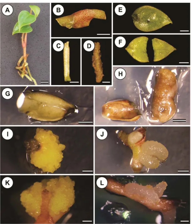
Single-bud nodal segments (Figure 3B), petiole (Figure 3C), root segments without the apex (Figure 3D), and whole leaves (Figure 3E) and half leaves sectioned perpendicular to the midrib (Figure 3F) were obtained from the in vitro A. andraeanum cv. Eidibel (Figure 3A) clones. The explants measured approximately 1.0 cm in length, and the leaf explants were measured based on the midrib length. The leaf explants were cultured with the adaxial surface facing up, whereas the petiole and the nodal and root segments were inoculated on to the medium horizontally. The explants were placed in 90 x 15 mm Petri dishes containing 25 mL of Pierik medium supplemented with 20 g L-1 sucrose and 6.5 g L-1
agar, the pH was adjusted to 5.8, and the medium was autoclaved at 121ºC for 15 minutes. The medium was supplemented with different concentrations (0, 2.5, 5, 7.5, 10 μM) of the auxins IAA, IBA, NAA, 2,4-D and Picloram. The cultures were maintained in a growth chamber at 25 ± 2ºC in the dark for 60 days.
Statistical analysis
(0, 2.5, 5, 7.5 and 10 μM) plus five control plates (consisting of the five explants types cultured on the same medium but lacking growth regulators), with five replications. Each replication consisted of five Petri dishes containing nine explants per dish, totaling 45 explants per treatment. After 60 days, the morphogenetic responses were evaluated for the production of the somatic embryogenesis of shoots and roots. The data were processed and analyzed using SAS software (SAS, 2001).
The homogeneity, normality and independence of the data were assessed using Bartlett, Kolmogorov-Smirnov and Durbin Watson’s tests, respectively. The count data, such as the occurrence of embryogenic calli, shoots and roots were transformed to x0.5 and were examined by analysis of variance. The means were compared using Tukey’s test at a 5% probability level.
A regression analysis related to the growth regulator concentrations in which the quantitative data were significant by Tukey’s test for the production of embryogenic calli was performed. The significance of the regression was assessed using the F test, whereas the significance of the coefficients was assessed using the ‘t’ test. The regression adjustment was performed using the adjusted determination coefficient (R ²%). The data applied to the adjustment of regressions were transformed as x0.5. The regression analyses were performed using the statistical software SISVAR (FERREIRA, 2003).
Histological analyses
The histochemical analyses of the embryogenic calli were performed using double staining with acetocarmine (2%) and Evan’s blue (0.1%) (DURZAN, 1988), and Lugol solution (0.1%) was used for starch detection (JENSEN, 1962).
The anatomical aspects of the somatic embryogenesis were assessed by fixing the samples in Karnovsky (1965) solution (2.5% glutaraldehyde, 4% paraformaldehyde, 3% sucrose, and 5 mM CaCl2
in 0.1 M cacodylate buffer) under refrigeration. The samples were dehydrated using a series of increasing concentrations of ethanol solutions (10 to 95%) and were embedded in methacrylate resin (Historesin Leica® Instruments, Heidelberg, Germany). The
thin sections (5-8 μm) were cut using a rotary automatic microtome (RM 2155, Leica Microsystems Inc., Deerfield, USA) equipped with disposable steel blades and were stained in toluidine blue, pH 4.0 (O’BRIEN; McCULLY, 1981), for 10 minutes. After drying, the glass slides were mounted in Permount® synthetic resin (Daigger, USA).
The structural analysis and photography were performed using an Olympus AX70TRF microscope (Olympus Optical, Japan) equipped with a U-Photo camera system (Spot Insightcolour 3.2.0, Diagnostic Instruments Inc., USA). The remaining photographs were taken using an Olympus SZX stereo microscope with a coupled with a camera system (Olympus E-330).
Results and discussion
Data analysis
The induction of somatic embryogenesis in
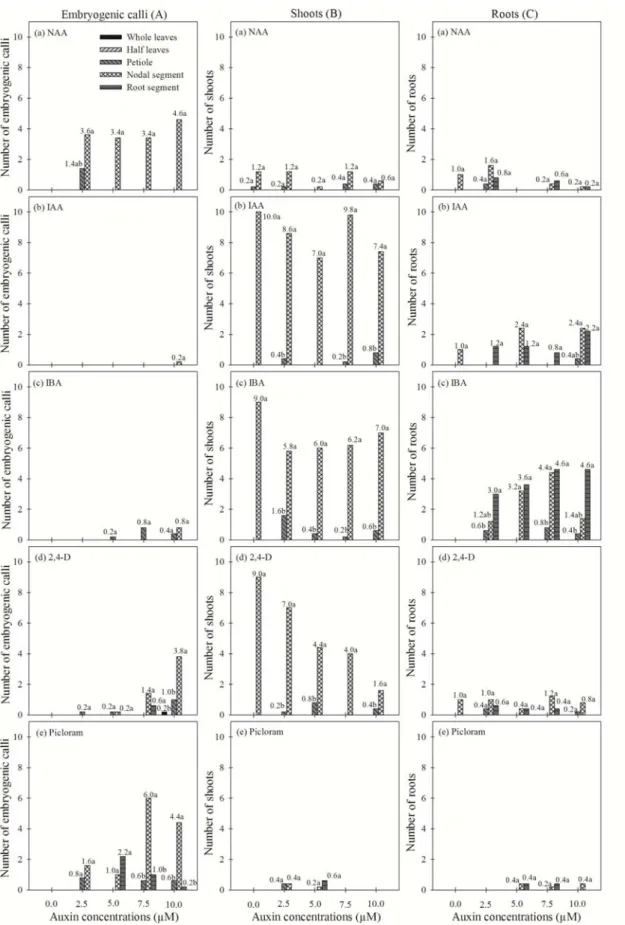
A. andraeanum cv. Eidibel is affected by three main factors: the source of the explants and the type and concentration of the auxin. In this study, we observed differences in the morphogenetic responses among the explants tested regarding the development of embryogenic calli, shoots and roots. The most competent explants for the induction of embryogenic calli were derived from the petioles and the nodal segments; the nodal segments were the most responsive (Figure 4A). In contrast, the whole leaf, sectioned leaf and root segments did not produce calli with embryogenic capacities, becoming oxidized after five weeks in culture. However, the root explants produced roots, even after oxidization.
The growth responses under the in vitro
conditions can vary with the explant type, and genotype is a major factor influencing the development and the physiology of explants in tissue culture (KRISHNARAJ; VASIL, 1995). Vasudevan and Van Staden (2011) reported that shoot-tip explants of leopard orchid (Ansellia Africana Lindl.) responded better in terms of shoot length and leaf number than nodal segments. In anthurium, the callus formation was significantly affected by the genotype, and the type and color of callus obtained from each cultivar differed (TE-CHATO et al., 2006).
The analysis of variance showed a significant triple interaction for the sources of the variation (explant type and the type and concentration of the auxin), using the F test for the independent variables.
Although the Kolmogorov-Smirnov and Bartlett’s tests were significant for shoot production, the Durbin Watson’s test was not significant at a 5% significance level. For the variables for embryogenic callus and root production, all tests were significant at 5% significance level. Thus, because at least two of the assumptions were not fulfilled, the data for all the variables were transformed to x0.5, and the analyses were rerun on the transformed data at 5% significance level.
There was no induction of somatic embryogenesis, or the induction was relatively low, using the media supplemented with IAA and IBA, regardless the concentration, for all explant types tested. The nodal segment was the most responsive explant type for embryogenic callus induction, showing the highest means on media containing NAA, Picloram and 2,4-D (Figure 1A; Table 1). I contrast, Kuehnle et al. (1992) demonstrated that using a modified half-strength MS medium (MURASHIGE; SKOOG, 1962) supplemented with 6.79 μM 2,4-D and 2.33 μM 6-furfuryl aminopurine (kinetin), the somatic embryo production was higher in the leaf explants compared with the petioles from A. andraeanum
hybrids.
For calli production affected by NAA, the cubic model (R2 = 99.75%) for the regression
displayed the greater adjustment of transformed data for the callus yield, whereas the quadratic (R2 = 99.75%) and linear model (R2 = 84.69%)
fitted better to the callus yield as affected by 2,4-D and Picloram, respectively (Figure 2). With the exception of the regression coefficient b1 for 2,4-D, all the remaining regression coefficients were significant (p < 0.05). The regression analysis indicated that the maximum average values for the induction of embryogenic calli were achieved at 10.0 μM NAA, 2,4-D and Picloram (Figure 2A, B and C, respectively).
We observed that the highest frequencies of embryogenic callus induction from nodal segments occurred only on medium supplemented with NAA (5, 7.5 and 10 μM), 2,4-D (10 μM) and Picloram (7.5 and 10 μM) (Figure 1Aa, Ad and Ae; Table 1). Biologically, 10
μM NAA (4.6) was more efficient at establishing embryogenic cultures from nodal segments of
A. andraeanum cv. Eidibel (Figures 1Aa, 2A and 4G), despite the higher numbers of embryogenic calli obtained using 7.5 μM Picloram (6.0) (Figure 1Ae; Table 1). NAA was also used for the induction of somatic embryogenesis in transformed explants of Medicago truncatula on medium supplemented with 10 μM NAA and 4
μM BA (NOLAN et al., 2009). In Eucalyptus camaldulensis, lower levels of 2,4-D (0.45, 2.26, 4.52 μM) induced loose and whitish calli, whereas at higher concentrations (9.05 μM), the callus was hard (PRAKASH; GURUMURTHI, 2010). However, the same authors reported that a friable nodular callus was formed on basal medium containing NAA (10.75 or 16.13 μM).
Prakash and Gurumurthi (2010) reported that the frequency of callus induction and the type of callus generated varied depending on the explant and type and level of auxin used. In this study, after 45 days in culture, the whole leaves plated on medium containing 10 μM 2,4-D produced a yellowish-white callus mass in only one of the nine explants plated (Figures 1Ad and 2B; Table 1). Different results were reported in a study by Bautista et al. (2008), demonstrating reporting that leaf segments of A. andraeanum var. Lambada produced calli after 30 days in culture. Hamidah et al. (1997), testing three explant types (leaves, petioles and roots) of A. scherzerianum, also observed that leaf explants were more responsive than petioles and roots after 45 days in culture. Yu et al. (2009) tested leaf and petiole explants of A. andraeanum cv. Valentino and observed higher callus production in the leaf explants 30 days after inoculation. In these studies, the petioles produced calli at both ends of the explants (HAMIDAH et al., 1997; YU et al., 2009), which is similar to our observations for petiole and nodal segments of the cultivar Eidibel (Figure 4B).
NAA (µM)
Num
b
er
o
f em
b
ry
o
ge
n
ic
ca
ll
i
0 1 2 3
Observed averages Estimated averages R2 = 99.75% Y = 0.7142 + 0.8478X - 0.1654X2 + 0.0096X3
(A)
2.5 5.0 7.5 10.0
0.0
I I I I
2,4-D (µM)
R2 = 99.61% Y = 0.7302 - 0.0841X + 0.0211X2
(B)
2.5 5.0 7.5 10.0
0.0 I I I I
I I
Picloram (µM) R2 = 84.69% Y = 0.8041 + 0.1691X
2.5 5.0 7.5 10.0
0.0 I I I I
(C)
I
Figure 2. Regression analysis for embryogenic calli as affected by auxins concentrations (0.0; 2.5; 5.0; 7.5; 10.0) (A) NAA; (B) 2,4-D and (C) Picloram from transformed data.
According to Lema-Rumińska (2011), 2,4-D in the culture medium did not influence the ploidy levels of regenerated cactus (Copiapoa tenuissima
Ritt. forma monstruosa) tissue. However, the prolonged maintenance of embryogenic calli on medium containing 2,4-D may induce epigenetic changes affecting the embryogenic potential of the cultures. It has been shown that somatic embryos become auxin-habituated when maintained in the presence of this growth regulator for long periods, leading to the loss of maturation capacity and the ability of the embryos to convert into plants. Accordingly, Pan et al. (2010) determined that 2,4-D also exerts a negative effect on the growth and development of plant tissues and organs cultured in vitro. Moreover, the authors suggest that 2,4-D inhibits somatic embryo initiation at least in part, by inducing osmotic stress in callus cells. Cangahuala-Inocente et al. (2007) demonstrated that in a somatic embryogenesis system, the reduction in the conversion rates of somatic embryos may be caused by the residual effects of the 2,4-D used in the induction phase and that these effects can be minimized by replacing the auxin with Picloram.
Picloram, at concentrations of 7.5 and 10 μM, induced higher frequencies of embryogenic calli in nodal segments than in the other explants (Figures 1Ae and 2C; Table 1). Picloram has been used successfully to induce embryogenic callus formation in peach palm (Bactris gasipaes), macaw palm (Acrocomia aculeata), and spring snowflake (Leucojum vernum L.) among other species (MOURA et al., 2008, 2009, 2010; PTAK, 2010; STEINMACHER et al., 2007). Picloram also induces somatic embryogenesis in date palm (Phoenix dactylifera L.), although a high frequency of hyperhydricity and oxidation was observed in these cultures (OTHMANI et al., 2009).
The highest means of shoot production means were obtained from the nodal segments, which were higher than the shoot production means in the other explant types in most treatments. This superiority is likely due to the proliferation of shoots from the pre-existing axillary buds, even when grown in medium in the absence of growth regulators. The highest shoot production occurred in the nodal segments inoculated on medium supplemented with IAA and IBA (Figures 1Bb, Bc and 3J; Table 1). However, in medium supplemented with 2,4-D, a reduction in the shoot production rate was observed with increasing concentrations of this auxin (Figure 1Bd; Table 1). In media supplemented with Picloram and NAA, the shoot production was low compared with the other treatments (Figure 1Ba and Be; Table 1).
In most treatments, the highest mean of root production was observed in the nodal segments and in the root explants themselves. IBA was more effective at in producing roots than the other auxins. The highest means were achieved at concentrations of 5 and 7.5 μM IBA for nodal segments and 2.5, 5, 7.5 and 10 μM IBA for the root explants (Figure 1Cc; Table 1). The highest means for media supplemented with IAA were recorded at concentrations of 5 and 10 μM for the nodal segments and 2.5, 5 and 10 μM for the root explants. The explants cultured on medium containing NAA, 2,4-D or Picloram (Figure 1Ca, 1Cd and 1Ce; Table 1) demonstrated low root production compared to the treatments with IBA or IAA. Therefore, root production was lower in the treatments that stimulated the formation of embryogenic calli, i.e., with the addition of NAA, 2,4-D and Picloram.
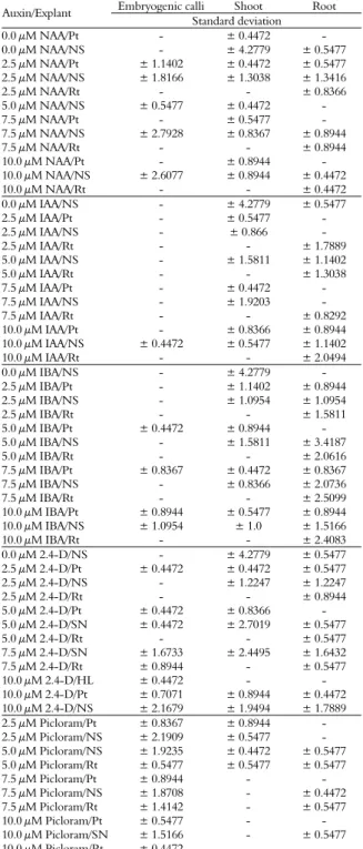
Table 1. Standard deviation of embryogenic calli, shoots and roots from different sources of explants of Anthurium andraeanum cv. Eidibel, as affected by auxin type and concentration, after 60 days of in vitro culture.
Embryogenic calli Shoot Root
Auxin/Explant
Standard deviation
0.0 μM NAA/Pt - ± 0.4472 -
0.0 μM NAA/NS - ± 4.2779 ± 0.5477
2.5 μM NAA/Pt ± 1.1402 ± 0.4472 ± 0.5477
2.5 μM NAA/NS ± 1.8166 ± 1.3038 ± 1.3416
2.5 μM NAA/Rt - - ± 0.8366
5.0 μM NAA/NS ± 0.5477 ± 0.4472 -
7.5 μM NAA/Pt - ± 0.5477 -
7.5 μM NAA/NS ± 2.7928 ± 0.8367 ± 0.8944
7.5 μM NAA/Rt - - ± 0.8944
10.0 μM NAA/Pt - ± 0.8944 -
10.0 μM NAA/NS ± 2.6077 ± 0.8944 ± 0.4472
10.0 μM NAA/Rt - - ± 0.4472
0.0 μM IAA/NS - ± 4.2779 ± 0.5477
2.5 μM IAA/Pt - ± 0.5477 -
2.5 μM IAA/NS - ± 0.866 -
2.5 μM IAA/Rt - - ± 1.7889
5.0 μM IAA/NS - ± 1.5811 ± 1.1402
5.0 μM IAA/Rt - - ± 1.3038
7.5 μM IAA/Pt - ± 0.4472 -
7.5 μM IAA/NS - ± 1.9203 -
7.5 μM IAA/Rt - - ± 0.8292
10.0 μM IAA/Pt - ± 0.8366 ± 0.8944
10.0 μM IAA/NS ± 0.4472 ± 0.5477 ± 1.1402
10.0 μM IAA/Rt - - ± 2.0494
0.0 μM IBA/NS - ± 4.2779 -
2.5 μM IBA/Pt - ± 1.1402 ± 0.8944
2.5 μM IBA/NS - ± 1.0954 ± 1.0954
2.5 μM IBA/Rt - - ± 1.5811
5.0 μM IBA/Pt ± 0.4472 ± 0.8944 -
5.0 μM IBA/NS - ± 1.5811 ± 3.4187
5.0 μM IBA/Rt - - ± 2.0616
7.5 μM IBA/Pt ± 0.8367 ± 0.4472 ± 0.8367
7.5 μM IBA/NS - ± 0.8366 ± 2.0736
7.5 μM IBA/Rt - - ± 2.5099
10.0 μM IBA/Pt ± 0.8944 ± 0.5477 ± 0.8944
10.0 μM IBA/NS ± 1.0954 ± 1.0 ± 1.5166
10.0 μM IBA/Rt - - ± 2.4083
0.0 μM 2.4-D/NS - ± 4.2779 ± 0.5477
2.5 μM 2.4-D/Pt ± 0.4472 ± 0.4472 ± 0.5477
2.5 μM 2.4-D/NS - ± 1.2247 ± 1.2247
2.5 μM 2.4-D/Rt - - ± 0.8944
5.0 μM 2.4-D/Pt ± 0.4472 ± 0.8366 -
5.0 μM 2.4-D/SN ± 0.4472 ± 2.7019 ± 0.5477
5.0 μM 2.4-D/Rt - - ± 0.5477
7.5 μM 2.4-D/SN ± 1.6733 ± 2.4495 ± 1.6432
7.5 μM 2.4-D/Rt ± 0.8944 - ± 0.5477
10.0 μM 2.4-D/HL ± 0.4472 - -
10.0 μM 2.4-D/Pt ± 0.7071 ± 0.8944 ± 0.4472
10.0 μM 2.4-D/NS ± 2.1679 ± 1.9494 ± 1.7889
2.5 μM Picloram/Pt ± 0.8367 ± 0.8944 -
2.5 μM Picloram/NS ± 2.1909 ± 0.5477 -
5.0 μM Picloram/NS ± 1.9235 ± 0.4472 ± 0.5477 5.0 μM Picloram/Rt ± 0.5477 ± 0.5477 ± 0.5477
7.5 μM Picloram/Pt ± 0.8944 - -
7.5 μM Picloram/NS ± 1.8708 - ± 0.4472
7.5 μM Picloram/Rt ± 1.4142 - ± 0.5477
10.0 μM Picloram/Pt ± 0.5477 - -
10.0 μM Picloram/SN ± 1.5166 - ± 0.5477
10.0 μM Picloram/Rt ± 0.4472 - -
The concentration and type of auxin used directly influences rhizogenesis. Different auxins can induce rooting and the most commonly used are IBA, IAA and NAA. Other auxins, including 2,4-D (CHEN et al., 2010; LEMA-RUMIŃSKA, 2011; YANG et al., 2010; ZHANG et al., 2010; SIVANESAN et al., 2011) and Picloram (MOURA
et al., 2008, 2009, 2010; OTHMANI et al., 2009; PTAK, 2010; STEINMACHER et al., 2007), are important growth regulators that are used in the majority of embryogenic cell and tissue culture systems in most plant species. NAA is commonly used in isolation media, although when added at concentrations above a few tenths of a milligram, this auxin tends to stimulate callus formation in a manner similar to that observed in this study for the cultivar Eidibel.
Morpho-histology
The whole leaf, sectioned leaf and root explants, regardless of the type and concentration of auxin, did not adequately form embryogenic calli or shoots. Many of these explants became yellowish and/or were oxidized after five weeks in culture (Figure 3G-H), the swelling of the buds, the development of roots in nodal segments (Figure 3J) and the proliferation of roots in root segments (Figure 3L). After five weeks in culture, the was swelling and oxidation of nodal segments and petiole explants was observed, leading to the formation of pale yellow and semi-friable primary calli with radial growth (Figure 3I and K).
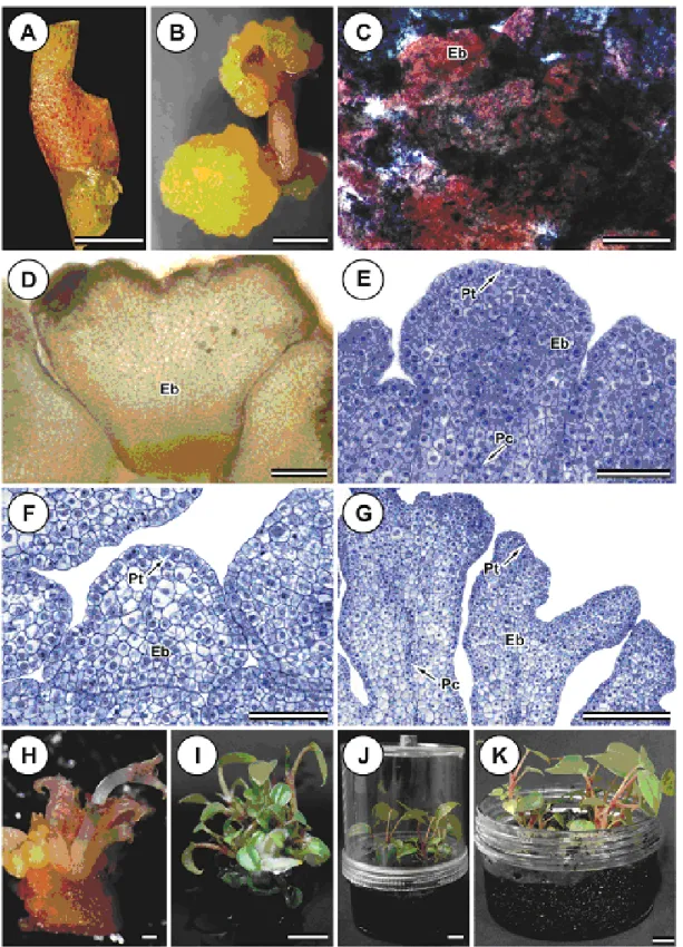
The histochemical analyses confirmed the embryogenic nature of the calli (Figure 4B) by double staining with acetocarmine and Evans blue. The calli reacted strongly with the acetocarmine, revealing their embryogenic characteristics (Figure 4C). The Lugol test revealed large quantities of starch grains, corroborating that the calli are embryogenic structures (Figure 4D).
Embryo polarity can be affected by an increase in endogenous auxin levels caused by high concentrations of exogenous auxin in the culture medium (FISCHER; NEUHAUS, 1996). Filippi et al. (2001) induced embryogenic calli from banana (Musa spp.) explants on medium supplemented with 5 and 10 μM Picloram; however the embryos were not functional, demonstrating that the quality of embryos produced is essential for their successful conversion into plants.
The genotype, explant type, growth phase, and the type and concentration of exogenous growth regulators markedly affected the acquisition of embryogenic competence in Acca sellowiana
(GUERRA et al., 2001). Our study corroborates this finding, considering that the induction of embryogenic cultures in A. andraeanum cv. Eidibel varied with the explant type and the concentration and type of growth regulator.
Figure 3. Embryogenic calli from Anthurium andraeanum cv. Eidibel explants cultured on Pierik medium, after 60 days in culture. (A) In vitro plantlet used as source of explants. (B) Nodal segment. (C) Petiole explant. (D) Root segment. (E) Whole leaf. (F) Sectioned leaf. (G) Whole leaf becoming oxidized. (H) Sectioned leaf becoming oxidized. (I) Petiole forming embryogenic calli on Pierik medium with 2.5 μM NAA. (J) Nodal segment forming roots and shoots on Pierik medium with 7.5 μM IBA. (K) Nodal segment forming embryogenic calli on Pierik medium with 10
Conclusion
In this study, we developed a highly efficient protocol for the induction of somatic embryogenesis in anthurium (Anthurium andraeanum cv. Eidibel) using nodal segments from in vitro established plantlets that were cultured in Pierik medium supplemented with 10 μM NAA. This auxin resulted in a well-defined procambium, a clear delimitation of the protoderm layer and signs of polarization, which are important features for further maturation of the somatic embryo and the effective conversion into plants. This protocol is useful not only for large-scale vegetative propagation but also for genetic transformation or synthetic seed production and can be combined with other biotechnological approaches.
Acknowledgements
The authors would like to thank the Coordination of Improvement of Higher Education Personnel (CAPES) for the research scholarship to M.V.M.P., FAPEMIG (Grant number CRA-APQ-01451-12 to W.C.O.) and the Agronomic Institute (IAC) for kindly providing the in vitro clones of anthurium cv. Eidibel (IAC 0-11) used in this research.
References
ATAK, Ç.; ÇELIK, Ö. Micropropagation of Anthurium andraeanum from leaf explants. Pakistan Journal of Botany, v. 41, n. 3, p. 1155-1161, 2009.
BAKHSHAIE, M.; BABALAR, M.; MIRMASOUMI, M.; KHALIGHI, A. Somatic embryogenesis and plant regeneration of Lilium ledebourii (Baker) Boiss., an endangered species. Plant Cell, Tissue and Organ Culture, v. 102, n. 2, p. 229-235, 2010.
BAUTISTA, N. D. R.; PEÑALVER, D. A.; RODRÍGUEZ, R. B.; CHIU, W. C.; LÓPEZ, R. C.; TERRY, F. J.; PERALTA, M. P.; MARTÍNEZ, O. G. Embriogénesis somática en (Anthurium andraeanum Lind.) variedad ‘Lambada’. Revista de Sociedad, Cultura y Desarrollo Sustentable, v. 4, n. 1, p. 135-149, 2008. BEYRAMIZADE, E.; AZADI, P.; MII, M. Optimization of factors affecting organogenesis and somatic embryogenesis of Anthurium andreanum lind. ‘Tera’. Propagation of Ornamental Plants, v. 8, n. 4, p. 198-203, 2008.
CANGAHUALA-INOCENTE, G. C.; DALVESCO, L. L.; STEINMACHER, D.; TORRES, A. C.; GUERRA, M. P. Improvements in somatic embryogenesis protocol in Feijoa (Acca sellowiana (Berg) Burret): induction, conversion and synthetic seeds. Scientia Horticulturae, v. 111, n. 3, p. 228-234, 2007.
CASTRO, A. C.; RESENDE, L. V.; GUIMARÃES, W. N. R.; LOGES, V. Uso de técnicas moleculares em estudo de diversidade genética em antúrio. Revista Brasileira de Horticultura Ornamental, v. 10, n. 1/2, p. 6-9, 2004.
CHEN, A. H.; YANG, J. L.; NIU, Y. D.; YANG, C. P.; LIU, G. F.; YU, C. Y.; LI, C. H. High-frequency somatic embryogenesis from germinated zygotic embryos of Schisandra chinensis and evaluation of the effects of medium strength, sucrose, GA3, and BA on somatic embryo
development. Plant Cell, Tissue and Organ Culture, v. 102, n. 3, p. 357-364, 2010.
CHEN, F.; KUEHNLE, A. R.; SUGII, N. Anthurium roots for micropropagation and Agrobacterium tumefaciens mediated gene transfer. Plant Cell, Tissue and Organ Culture, v. 49, n. 1, p. 71-74, 1997.
DUQUENNE, B.; EECKHAUT, T.; WERBROUCK, S.; HUYLENBROECK, J. Effect of enzyme concentrations on protoplast isolation and protoplast culture of Spathiphyllum and Anthurium. Plant Cell, Tissue and Organ Culture, v. 91, n. 2, p. 165-173, 2007.
DURZAN, D. J. Process control in somatic polyembryogenesis. In: HALLGREN, J. E. (Ed.). Frans symposium department of forest genetics and plant physiology. Swedish: University of Agricultural Sciences, 1988. p. 147-186.
FERREIRA, D. F. Programa de análises estatísticas (Statistical Analysis Software) e planejamento de experimentos. SISVAR 5.0 (Build 67), 2003. Available from: <http://www.dex.ufla.br/~danielff/softwares.htm>. Access on: May 30, 2012.
FILIPPI, S. B.; APPEZZATO-DA-GLÓRIA, B.; PINHEIRO, A.; RODRIGUEZ, M. Histological changes in banana explants, cv. Nanicão (Musa spp., Group AAA), submitted to different auxins for induction of somatic embryogenesis. Revista Brasileira de Botânica, v. 24, n. 4, p. 595-602, 2001.
FISCHER, C.; NEUHAUS, G. Influence of auxin on the establishment of bilateral symmetry in monocots. The Plant Journal, v. 9, n. 5, p. 659-669, 1996.
FITCH, M. M. M.; LEONG, T. C. W.; HE, X.; MCCAFFERTY, H. R. K.; ZHU, Y. J.; MOORE, P. H.; GONSALVES, D.; ALDWINCKLE, H. S.; ATKINSON, H. J. Improved transformation of anthurium.
Hortscience, v. 46, n. 3, p. 358-364, 2011.
FUZITANI, E. J.; NOMURA, E. S. Produção de mudas
in vitro. Revista Brasileira de Horticultura
Ornamental, v. 10, n. 1/2, p. 14-17, 2004.
GUERRA, M. P.; DAL VESCO, L. L.; DUCROQUET, J. P. H.; NODARI, R. O.; REIS, M. S. Somatic embryogenesis in Feijoa sellowiana: genotype response, auxinic shock and synthetic seeds. Revista Brasileira de Fisiologia Vegetal, v. 13, n. 2, p. 117-128, 2001.
HAMIDAH, M.; KARIM, A. G. A.; DEBERGH, P. Somatic embryogenesis and plant regeneration in Anthurium scherzerianum. Plant Cell, Tissue and Organ Culture, v. 48, n. 3, p. 189-193, 1997.
JENSEN, W. A. Botanical histochemistry: principles and practice. San Francisco: WH Freeman, 1962.
KARNOVSKY, M. J. A formaldehyde–glutaraldehyde fixative of high osmolarity for use in electron microscopy.
KONIECZNY, R.; PILARSKA, M.; TULEJA, M.; SALAJ, T.; ILNICKI, T. Somatic embryogenesis and plant regeneration in zygotic embryos of Trifolium nigrescens (Viv.). Plant Cell, Tissue and Organ Culture, v. 100, n. 2, p. 123-130, 2010.
KRISHNARAJ, S.; VASIL, I. K. Somatic embryogenesis in herbaceous monocots. In: THORPE, T. A. (Ed.). In vitro embryogenesis in plants. Dordrecht: Kluwer Academic Press, 1995. p. 155-203.
KUEHNLE, A. R.; CHEN, F.; SUGII, N. Somatic embryogenesis and plant regeneration in Anthurium andraeanum hybrids. Plant Cell Reports, v. 11, n. 9, p. 438-442, 1992.
KUEHNLE, A. R.; SUGII, N. Callus induction and plantlet regeneration in tissue cultures of Hawaiian anthuriums. Hortscience, v. 26, n. 7, p. 919-921, 1991. KUNISAKI, J. T. In vitro propagation of Anthurium andreanum Lind. Hortscience, v. 15, n. 4, p. 508-509, 1980.
LEMA-RUMIŃSKA, J. Flow cytometric analysis of somatic embryos, shoots, and calli of the cactus Copiapoa tenuissima Ritt. forma monstruosa. Plant Cell, Tissue and Organ Culture, v. 106, n. 3, p. 531-535, 2011.
LU, J.; VAHALA, J.; PAPPINEN, A. Involvement of ethylene in somatic embryogenesis in Scots pine (Pinus sylvestris L.). Plant Cell, Tissue and Organ Culture, v. 107, n. 1, p. 25-33, 2011.
MAIRA, O.; ALEXANDER, M.; VARGAS, T. E. Micropropagation and organogenesis of Anthurium andreanum Lind cv Rubrun. In: JAIN, S. M.; OCHATT, S. J. (Ed.). Protocols for in vitro propagation of ornamental plants, methods in molecular biology. Totowa: Humana Press Edition, 2010. p. 3-14.
MOURA, E. F.; MOTOIKE, S. Y.; VENTRELLA, M. C.; DE SÁ JÚNIOR, A. Q.; CARVALHO, M. Somatic embryogenesis in macaw palm (Acrocomia aculeata) from zygotic embryos. Scientia Horticulturae, v. 119, n. 4, p. 447-454, 2009.
MOURA, E. F.; VENTRELLA, M. C.; MOTOIKE, S. Y. Anatomy, histochemistry and ultrastructure of seed and somatic embryo of Acrocomia aculeata (Arecaceae). Scientia Agricola, v. 67, n. 4, p. 399-407, 2010.
MOURA, E. F.; VENTRELLA, M. C.; MOTOIKE, S. Y.; DE SÁ JÚNIOR, A. Q.; CARVALHO, M.; MANFIO, C. E. Histological study of somatic embryogenesis induction on zygotic embryos of macaw palm (Acrocomia aculeata (Jacq.) Lodd. ex Martius). Plant Cell, Tissue and Organ Culture, v. 95, n. 2, p. 175-184, 2008.
MURASHIGE, T.; SKOOG, F. A revised medium for rapid growth and bio-assays with tobacco tissue cultures.
Physiologia Plantarum, v. 15, n. 3, p. 473-497, 1962. NHUT, D. T.; NGUYEN, D.; VY, N. N. H.; KHUE, C. D.; KHIEM, D. V.; VINH, D. N. Impact of Anthurium spp. genotype on callus induction derived from leaf explants, and shoot and root regeneration capacity from callus. Journal of Applied Horticulture, v. 8, n. 2, p. 135-137, 2006.
NINKOVIĆ, S.; DJORDJEVIĆ, T.; VINTERHALTER, B.; UZELAC, B.; CINGEL, A.; SAVIĆ, J.; RADOVIĆ, S.
Embryogenic responses of Beta vulgaris L. callus induced from transgenic hairy roots. Plant Cell, Tissue and Organ Culture, v. 103, n. 1, p. 81-91, 2010.
NOLAN, K. E.; KURDYUKOV, S.; ROSE, R. J. Expression of the Somatic Embryogenesis Receptor-Like Kinase1 (SERK1) gene is associated with developmental change in the life cycle of the model legume Medicago truncatula. Journal of Experimental Botany, v. 60, n. 6, p. 1759-1771, 2009.
O’BRIEN, T. P.; McCULLY, M. E. The study of plant structure principles and select methods. Melbourne: Termarcarphi Pty Ltd, 1981.
OTHMANI, A.; BAYOUDH, C.; DRIRA, N.; MARRAKCHI, M.; TRIFI, M. Somatic embryogenesis and plant regeneration in date palm Phœnix dactylifera L., cv. Boufeggous is significantly improved by fine chopping and partial desiccation of embryogenic callus. Plant Cell, Tissue and Organ Culture, v. 97, n. 1, p. 71-79, 2009. PAN, Z.; ZHU, S.; GUAN, R.; DENG, X. Identification of 2,4-D-responsive proteins in embryogenic callus of Valencia sweet orange (Citrus sinensis Osbeck) following osmotic stress. Plant Cell, Tissue and Organ Culture, v. 103, n. 2, p. 145-153, 2010.
PIERIK, R. L. M. Callus multiplication of Anthurium andraeanum Lindl. in liquid media. Netherlands Journal of Agricultural Science, v. 23, p. 299-302, 1975. PIERIK, R. L. M. Anthurium andraeanum Lindl. plantlets produced from callus tissues cultivated in vitro.
Physiologia Plantarum, v. 37, n. 1, p. 80-82, 1976. PIERIK, R. L. M.; LEEUWEN, P. V.; RIGTER, G. C. M. Regeneration of leaf explants of Anthurium andraeanum Lindl. in vitro. Netherlands Journal of Agricultural Science, v. 27, p. 221-226, 1979.
PIERIK, R. L. M.; STEEGMANS, H. H. M.; VAN DER MEYS, J. A. J. Plantlet formation on calllus tissues of Anthurium andraeanum Lind.. Cientia Horticulturae, v. 2, n. 2, p. 193-198, 1974.
PINTO, D. L. P.; ALMEIDA, A. M. R.; RÊGO, M. M.; SILVA, M. L.; OLIVEIRA, E. J.; OTONI, W. C. Somatic embryogenesis from mature zygotic embryos of commercial passionfruit (Passiflora edulis Sims) genotypes.
Plant Cell, Tissue and Organ Culture, v. 107, n. 3, p. 521-530, 2011.
PRAKASH, M. G.; GURUMURTHI, K. Effects of type of explant and age, plant growth regulators and medium strength on somatic embryogenesis and plant regeneration in Eucalyptus camaldulensis. Plant Cell, Tissue and Organ Culture, v. 100, n. 1, p. 13-20, 2010.
PTAK, A. Somatic embryogenesis in in vitro culture of Leucojum vernum L. In: JAIN, S. M.; OCHATT, S. J. (Ed.). Protocols for in vitro propagation of ornamental plants, methods in molecular biology. London: Human Press, 2010. p. 223-233.
Embryo production through somatic embryogenesis can be used to study cell differentiation in plants. Plant Cell, Tissue and Organ Culture, v. 86, n. 3, p. 285-301, 2006. SANTOS, M. R. A. D.; TIMBÓ, A. L. D. O.; CARVALHO, A. C. P. P. D.; MORAIS, J. P. S. Callus induction and plant regeneration from Anthurium andraeanum Lindl. fruits. Plant Cell Culture and Micropropagation, v. 1, n. 2, p. 77-79, 2005.
SAS-Statistical Analisys System. The SAS-System for Windows: release 8.0. Cary: Statistical Analisys System Institute Corporation, 2001.
SCHIAVINATO, Y. O.; NOGUEIRALUCON, T.; TOMBOLATO, A. F. C.; BARBOSA, W.; VEIGA, R. F. D. A. Micropropagação de Anthurium plowmannii Croat.
Plant Cell Culture and Micropropagation, v. 4, n. 1, p. 15-20, 2008.
SIVANESAN, I.; LIM, M.; JEONG, B. Somatic embryogenesis and plant regeneration from leaf and petiole explants of Campanula punctata Lam. var. rubriflora Makino. Plant Cell, Tissue and Organ Culture, v. 107, n. 2, p. 365-369, 2011.
STEINMACHER, D. A.; KROHN, N. G.; DANTAS, A. C. M.; STEFENON, V. M.; CLEMENT, C. R.; GUERRA, M. P. Somatic embryogenesis in peach palm using the thin cell layer technique: induction, morpho-histological aspects and AFLP analysis of somaclonal variation. Annals of Botany, v. 100, n. 4, p. 699-709, 2007.
TE-CHATO, S.; SUSANON, T.; SONTIKUN, Y. Cultivar, explant type and culture medium influencing embryogenesis and organogenesis in Anthurium spp.
Songklanakarin Journal of Science and Technology, v. 28, n. 4, p. 717-722, 2006.
TENG, W. Regeneration of Anthurium adventitious shoots using liquid or raft culture. Plant Cell, Tissue and Organ Culture, v. 49, n. 2, p. 153-156, 1997.
TOMBOLATO, A. F. C.; QUIRINO, E. A. Multiplicação in vitro de novas seleções de Anthurium andraeanum Lindl. Revista Brasileira de Horticultura Ornamental, v. 2, n. 1, p. 37-46, 1996.
TOMBOLATO, A. F. C.; UZZO, R. P.; CASTRO, A. C. R.; SAKAI, M.; SAES, L. A. Recursos genéticos e melhoramento do antúrio (Anthurium andraeanum Linden) no IAC–APTA. Revista Brasileira de Horticultura Ornamental, v. 10, n. 1-2, p. 1-5, 2004.
VASUDEVAN, R.; VAN STADEN, J. Cytokinin and explant types influence in vitro plant regeneration of Leopard Orchid (Ansellia africana Lindl.). Plant Cell, Tissue and Organ Culture, v. 107, n. 1, p. 123-129, 2011.
VIÉGAS, J.; ROCHA, M. T. R. D.; FERREIRA-MOURA, I.; ROSA, D. L. D.; SOUZA, J. A. D.; CORRÊA, M. G. S.; SILVA, J. A. T. D. Anthurium andraeanum (Linden ex André) culture: in vitro and ex vitro.
Floriculture and Ornamental Biotechnology, v. 1, n. 1, p. 61-65, 2007.
VON ARNOLD, S.; SABALA, I.; BOZHKOV, P.; DYACHOK, J.; FILONOVA, L. Developmental pathways of somatic embryogenesis. Plant Cell, Tissue and Organ Culture, v. 69, n. 3, p. 233-249, 2002.
WINARTO, B.; MATTJIK, N. A.; SILVA, J. A. T. D.; PURWITO, A.; MARWOTO, B. Ploidy screening of anthurium (Anthurium andreanum Linden ex André) regenerants derived from anther culture. Scientia Horticulturae, v. 127, n. 1, p. 86-90, 2010.
WINARTO, B.; RACHMAWATI, F.; PRAMANIK, D.; TEIXEIRA DA SILVA, J. Morphological and cytological diversity of regenerants derived from half-anther cultures of anthurium. Plant Cell, Tissue and Organ Culture, v. 105, n. 3, p. 363-374, 2011a.
WINARTO, B.; RACHMAWATI, F.; TEIXEIRA DA SILVA, J. New basal media for half-anther culture of Anthurium andreanum Linden ex André cv. Tropical. Plant Growth Regulation, v. 65, n. 3, p. 513-529, 2011b. YANG, J. L.; SEONG, E. S.; KIM, M. J.; GHIMIRE, B. K.; KANG, W. H.; YU, C. Y.; LI, C. H. Direct somatic embryogenesis from pericycle cells of broccoli (Brassica oleracea L. var. italica) root explants. Plant Cell, Tissue and Organ Culture, v. 100, n. 1, p. 49-58, 2010.
YEUNG, E. C. Structural and developmental patterns in somatic embryogenesis. In: THORPE, T. A. (Ed.). In vitro embryogenesis in plants. Dordrecht: Kluwer Academic Publishers, 1995. p. 205-247.
YU, Y.; LIU, L.; LIU, J.; WANG, J. Plant regeneration by callus-mediated protocorm-like body induction of Anthurium andraeanum Hort. Agricultural Sciences in China, v. 8, n. 5, p. 572-577, 2009.
ZHANG, N.; FANG, W.; SHI, Y.; LIU, Q.; YANG, H.; GUI, R.; LIN, X. Somatic embryogenesis and organogenesis in Dendrocalamus hamiltonii. Plant Cell, Tissue and Organ Culture, v. 103, n. 3, p. 325-332, 2010.
Received on March 28, 2012. Accepted on July 16, 2012.
License information: This is an open-access article distributed under the terms of the Creative Commons Attribution License, which permits unrestricted use, distribution, and reproduction in any medium, provided the original work is properly cited.