Maio de 2016
Ana Catarina Novais Belinha
Implication of amino acid metabolism in
monocyte differentiation
O impacto do metabolismo dos aminoácidos na
diferenciação de monócitos
Dissertação de Mestrado
Mestrado em Ciências da Saúde
Trabalho efetuado sob a orientação de
Doutor Ricardo Silvestre
ii
DECLARAÇÃO Nome: Ana Catarina Novais Belinha
Endereço eletrónico: abelinha@ecsaude.uminho.pt Telefone: +351 914958979
Número do Bilhete de Identidade: 14312366
Título da dissertação: Implication of amino acid metabolism in monocyte differentiation Orientadores: Doutor Ricardo Silvestre
Ano de conclusão: 2016
Designação Ramo de Conhecimento do Mestrado: Ciências da Saúde
DE ACORDO COM A LEGISLAÇÃO EM VIGOR, NÃO É PERMITIDIDA A REPRODUÇÃO DE QUALQUER PARTE DESTA TESE.
Universidade do Minho, 18 de Maio de 2016
iii
The work presented in this dissertation was done in the Microbiology and Infection Research Domain of the Life and Health Sciences Research Institute (ICVS), School of Health Sciences, University of Minho, Braga, Portugal (ICVS/3B’s – PT Government Associate Laboratory, Braga/Guimarães, Portugal).
v
A
GRADECIMENTOS
A realização deste trabalho não teria sido possível sem o apoio e dedicação de algumas pessoas que têm sido fundamentais quer no meu desenvolvimento pessoal, como profissional. Durante o ultimo ano e meio, muitas foram as pessoas que me ajudaram a atingir os meus objetivos, protegendo-me e fazendo-me perceber qual o caminho a seguir. Desta forma, é com imensa gratidão que vos agradeço por estarem ao meu lado.
Começo por agradecer ao meu orientador Ricardo Silvestre, por todo o empenho e motivação na orientação do meu trabalho, mas, mais do que isso, por todo o apoio que me deu, e continua a dar, tanto a nível pessoal como profissional, que decerto me fez crescer e tornar-me melhor de dia para dia. Obrigada pela confiança depositada no meu trabalho e por todos os momentos de compreensão e disponibilidade fornecidos para me ajudar.
Agradeço também à minha amiga e fiel companheira Inês Mesquita, por me fazer seguir sempre em frente, sem nunca desistir de mim e fazendo-me lutar para ser cada vez melhor. Sem o seu apoio e amizade nada disto seria possível. Agradeço imenso todo o interesse e amparo na realização do meu trabalho e, muito mais do que isso, por estar sempre ao meu lado.
Não poderia deixar de agradecer também à Joana Gaifem, por toda a ajuda que me disponibilizou, mesmo quando achava não ser possível. Obrigada Joana por me teres recebido tão bem no laboratório e por continuares ao meu lado quando preciso de ti.
Agradeço ao meu eterno amigo Henrique Machado. Apesar de teres abraçado um projeto novo, ajudaste-me imenso a superar as minhas dificuldades. Obrigado Henrique por todos os momentos em que me apoiaste e me fizeste acreditar que eu conseguia atingir todos os meus objetivos, sem nunca desistir. Agradeço-te sinceramente por todas as conversas científicas e informais, em que me fizeste olhar as coisas por uma perspetiva diferente. E não poderia deixar de agradecer à Filipa, claro, por todos os bons momentos dentro e fora do laboratório que passamos antes de ela sair.
vi
Agradeço imenso a todas as outras pessoas do laboratório que, sem dúvida, fizeram com que o meu trabalho no laboratório fosse mais fácil e divertido. Obrigada Alexandra, Bruno, Cláudia, Nuno, Alice, Rita, Diogo, Pedro, Isabel, Miguel, George, Andreia, Joana, Ângela, Olga e a todos os outros que por cá passaram e tive o prazer de conhecer, especialmente à minha querida Patrícia que, apesar de estar longe, continua a fazer parte de nós.
Em especial, quero também agradecer à Margarida por todas as conversas motivadoras, que me inspiraram e fizeram mais fácil a realização deste trabalho; E também à Diana e Patrícia por me acompanharem durante estes dois anos mestrado, fazendo deles muito mais encantadores.
Agradeço também, muito francamente, à Cristina Cunha e Belém Marques por todo o apoio que me deram tanto a nível técnico como pessoal. Sem dúvida que o apoio delas era indispensável para este trabalho, mas, mais do que isso, agradeço-lhes por todo o amparo que me deram tanto dentro como fora do laboratório.
Quero também dizer um muito obrigado ao Professor Gil Castro, Fernando Rodrigues, Egídio Torrado e Agostinho Carvalho, por toda a ajuda científica que me deram. A opinião deles foi sem dúvida muito importante para o avanço deste trabalho e para me fazer compreender algumas questões. À Professora Paula Ludovico agradeço toda a ajuda fornecida na concretização desta tese.
Aos meus pais e ao meu irmão quero agradecer pelo apoio incansável que me têm dado e pela força proporcionada para atingir todos os meus sonhos.
Aos meus amigos Daniela Sousa, João Rodrigues, Marta Miranda, Luís Tavares, Rafaela Teixeira e Ana Köch quero agradecer por, mesmo sem me verem há meses, manterem o contacto e amizade que é imprescindível para o meu dia-a-dia.
Por fim, aos meus “roomies” Carla Gonçalves e Gustavo Teixeira quero agradecer pela paciência que têm comigo todos os dias, tomando sempre muito bem conta de mim, sem deixando esquecer claro, a Mónica Sá, Sara Alves e Ana Ferreira, que mesmo não estando presentes todos os dias me transmitem o seu apoio para nunca desistir.
ix
A
BSTRACT
Monocytes are circulating blood leukocytes that arise from the bone marrow and are responsible for a wide array of homeostatic functions upon recruitment to different tissues. These phagocytes are able to mount an anti-microbial response, they are responsible for the production of a large array of cytokines and, importantly, they represent a pool of myeloid precursors that ultimately may originate tissue macrophages and dendritic cells. Recently, several reports have shed light on the relation between metabolism and immune function, which has been vastly explored in groundbreaking research on the field of immunometabolism. The development of a specific immune response relies on the accurate utilization of certain nutrients, thus determining distinct metabolic phenotypes and consequently distinct effector functions. Although several reports state the importance of the metabolic environment for the correct function of macrophages, little is known about the impact of amino acids during monocyte differentiation.
We are interested in studying the metabolic requirements of human CD14+ monocytes derived from
peripheral blood mononuclear cells during differentiation in macrophage populations. The analysis of amino acid catabolism/anabolism by these populations, using high performance liquid chromatography, revealed distinct metabolic needs, depending on the differentiation profile. In order to assess the contribution of non-essential amino acids during differentiation, we cultivated the purified monocytes in conditioned media, where we selectively depleted L-aspartate. Moreover, the role of L-aspartate as a modulator of monocyte differentiation was assessed for several functional characteristics of macrophages such as phenotypical markers and other effector functions by flow cytometry. We observed that the differential availability of this amino acid during monocyte-to-macrophage differentiation impacts their final fate regarding function, thus showing a role for amino acid metabolism in the modulation of macrophage response.
xi
R
ESUMO
Os monócitos são leucócitos circulantes originários da medula óssea responsáveis por várias funções homeostáticas após recrutamento para diferentes tecidos. Estes fagócitos são capazes de desenvolver uma resposta anti-microbiana, produzir diversas citoquinas pró-inflamatórias e representam uma reserva de precursores mieloides, que podem eventualmente originar macrófagos tecidulares e células dendríticas.
Recentemente, diversos trabalhos expuseram a relação entre o metabolismo e as funções imunes, um conceito que tem vindo a ser aprofundado na área do imunometabolismo. O desenvolvimento de uma resposta imune específica depende da correta utilização de certos nutrientes, o que por sua vez dita a existência de diferentes fenótipos metabólicos e, consequentemente, funções efetoras distintas. Apesar de diversos estudos demonstrarem a importância do microambiente metabólico para o desenvolvimento de uma função adequada dos macrófagos, pouco se sabe acerca do papel dos aminoácidos durante a diferenciação de monócitos. Este trabalho tem como objetivo estudar os requisitos metabólicos de
monócitos humanos CD14+, derivados de células mononucleares do sangue periférico, durante a sua
diferenciação em populações macrofágicas. A análise do catabolismo/anabolismo de aminoácidos, através de cromatografia liquída de alta performance, revelou diferentes necessidades metabólicas, consoante o perfil de diferenciação induzido nos macrófagos. De forma a entender a contribuição dos aminoácidos não essenciais durante este processo, os monócitos foram purificados e cultivados em meios condicionados em que o L-aspartato foi seletivamente depletado. O papel deste aminoácido como modulador da diferenciação de monócitos foi avaliado através do estudo de várias características funcionais dos macrófagos, como os marcadores fenotípicos e outras funções efetoras, que foram avaliados por citometria de fluxo. Neste estudo foi possível observar que a biodisponibilidade deste aminoácido durante a diferenciação dos monócitos em macrófagos tem um impacto no seu perfil final, nomeadamente em termos de função, o que demonstra um novo papel do metabolismo de aminoácidos na modulação da resposta dos macrófagos.
xiii
T
ABLE OF CONTENTS
ABSTRACT ... ix
RESUMO ... xi
INDEX ... xv
LIST OF ABBREVIATIONS ... xvii
INTRODUCTION ... 1
1. Impact of cellular metabolism on immune function ... 3
2. Monocyte and Macrophage development ... 5
2.1. Monocytes: origin, development and fates ... 5
2.2. Monocyte Differentiation and Macrophage Polarization ... 8
3. Immunometabolism ... 10
3.1. Overview of macrophage metabolism ... 10
3.2. Metabolic reprogramming and macrophage polarization ... 13
3.2.1. M1-macrophage polarization ... 14
3.2.2. M2-macrophage polarization ... 17
3.3. Amino Acid metabolism and Macrophage function ... 19
AIMS ... 23
MATERIAL AND METHODS ... 27
1. Biological Samples ... 29
2. Culture medium ... 29
3. Generation of monocyte-derived macrphages and dendritic cells... 29
4. Flow cytometry analysis ... 30
5. Cytokine production evaluation by Enzyme-Linked Immunosorbent Assay (ELISA) ... 31
6. Amino Acid quantification by High Performance Liquid Chromatography (HPLC) ... 31
7. Glucose and Lactate Quantification by High Performance Liquid Chromatography (HPLC) ... 32
xiv
RESULTS ... 35
1. Metabolic and phenotypical characterization of human macrophages and dendritic cells derived from CD14+ blood monocytes ... 37
1.1. Amino acid metabolism evaluation in monocyte differentiation ... 37
1.2. Carbohydrate metabolism during monocyte differentiation ... 40
1.3. Phenotypical evaluation of macrophages and dendritic cells after monocyte differentiation ... 42
2. Metabolic and phenotypical characterization of human macrophages derived from CD14+ blood monocytes in conditioned media ... 44
2.1. Amino acid metabolism evaluation during monocyte differentiation in conditioned media ... 44
2.2. Evaluation of carbohydrate metabolism during monocyte differentiation in conditioned media ... 46
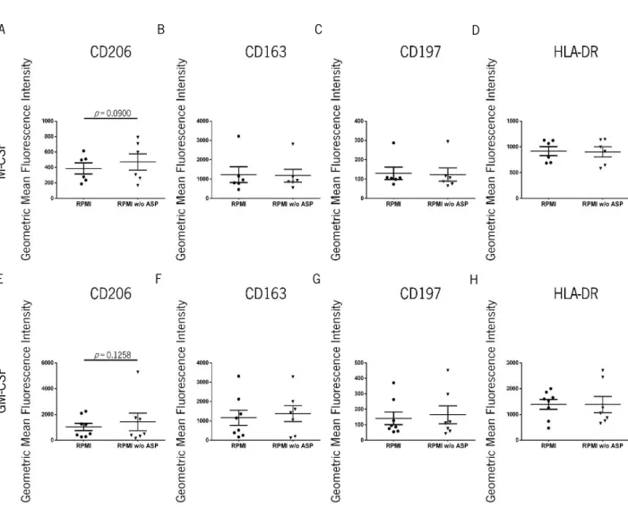
2.3. Phenotypical evaluation of macrophages after monocyte differentiation in conditioned media ... 47
3. Functional characterization of human monocyte-derived macrophages differentiated in conditioned media... 49
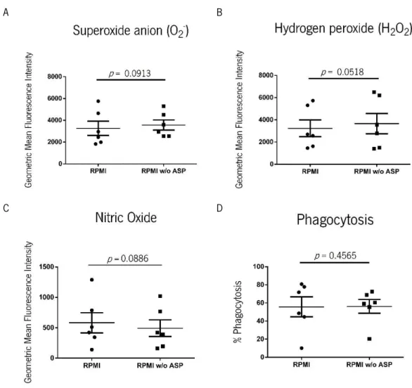
3.1. Effector function evaluation of macrophages differentiated in conditioned media ... 49
3.2. Assessment of effector functions of macrophages differentiated in conditioned media after stimulation with LPS ... 53
DISCUSSION AND FUTURE PERSPECTIVES ... 59
1. Metabolic and phenotypical characterization of human macrophages and dendritic cells derived from CD14+ blood monocytes ... 61
2. Metabolic and phenotypical characterization of human macrophages derived from CD14+ blood monocytes in conditioned media ... 66
3. Functional characterization of human monocyte-derived macrophages differentiated in conditioned media... 69
CONCLUSION ... 75
SUPPLEMENTARY DATA ... 81
xv
INDEX
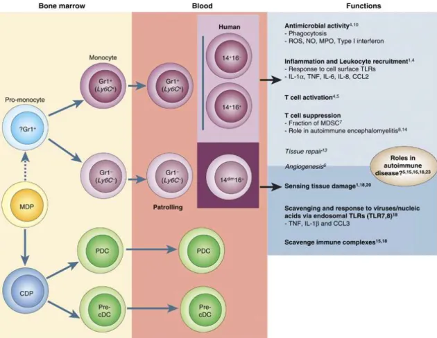
Figure 1 Development of blood monocytes from hematopoietic precursor cells. Page 6
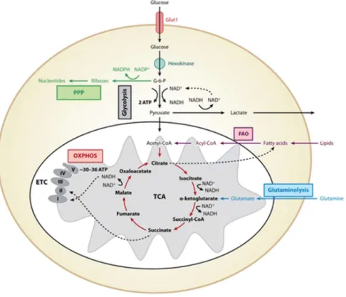
Figure 2 Major metabolic pathways of immune cells. Page 12
Figure 3 Metabolic profile of an M1 macrophage. Page 15
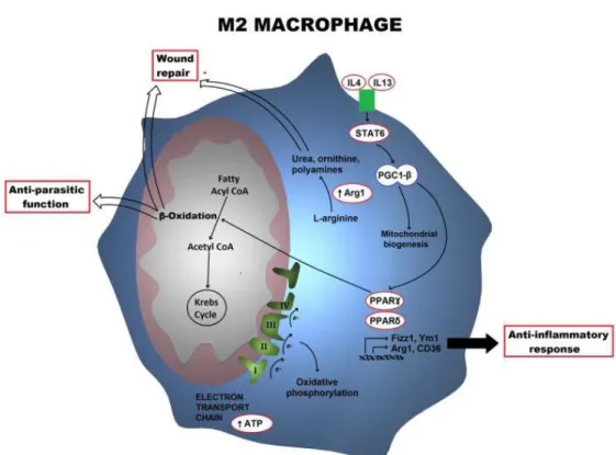
Figure 4 Metabolic profile of an M2 macrophage. Page 18
Figure 5 Characterization of amino acids metabolism by human Mφ and DCs
derived from CD14+ peripheral blood monocytes.
Page 38
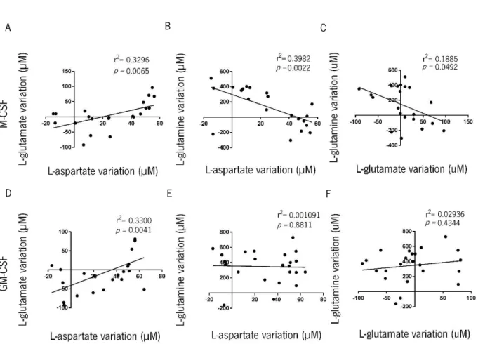
Figure 6 Correlation of amino acids metabolic profiles of human Mφ derived
from CD14+ peripheral blood monocytes.
Page 39
Figure 7 Characterization of glucose consumption and lactate production by
Mφ and DCs derived from CD14+ peripheral blood monocytes.
Page 41
Figure 8 Phenotypical characterization of surface markers of human Mφ and
DCs derived from CD14+ peripheral blood monocytes.
Page 43
Figure 9 Characterization of amino acids metabolism by human Mφ derived
from CD14+ peripheral blood monocytes in conditioned media.
Page 45
Figure 10 Characterization of glucose consumption and lactate production by
human Mφ derived from CD14+ peripheral blood monocytes in
conditioned media.
Page 47
Figure 11 Phenotypical characterization of human Mφ derived from CD14+
peripheral blood monocytes in conditioned media.
Page 48
Figure 12 Effector function evaluation measuring cytokine production levels after
human monocyte differentiation into Mφ in conditioned media. Page 50
Figure 13 Effector function evaluation of human Mφ differentiated in conditioned
media.
Page 51
Figure 14 Mitochondrial evaluation of human Mφ differentiated in conditioned
media.
xvi
Figure 15 Effector function evaluation measuring cytokine production levels after human monocyte differentiation into Mφ in conditioned media upon stimulation with LPS.
Page 54
Figure 16 Effector function evaluation of human Mφ differentiated in conditioned
media after stimulation with LPS.
Page 55
Figure 17 Mitochondrial evaluation of human Mφ differentiated in conditioned media after stimulation with LPS.
Page 56
Figure 18 Graphical conclusion of main results of this work. Page 78
Supplementary
figure 1 Gating strategy of Flow cytometry analysis. Page 83
Supplementary figure 2 Supplementary figure 3
Amino acids consumption and production profiles upon human monocyte differentiation.
Correlations between amino acids consumption and production profiles in human monocyte-derived DCs.
Page 84 Page 85
Supplementary
figure 4 Correlations between amino acids consumption and production profiles in human monocyte-derived Mφ. Page 86 Supplementary
figure 5 Characterization of other amino acids metabolism by human monocytes-derived Mφ in conditioned media in the presence of
M-CSF.
Page 87
Supplementary
figure 6 Characterization of other amino acids metabolism by human monocytes-derived Mφ in conditioned media in the presence of
GM-CSF.
xvii
L
IST OF ABBREVIATIONS
7-AAD, 7-aminoactinomycin D Acetyl-CoA, Acetyl-coenzyme A
AMPK, Adenosine monophosphate-activated protein kinase APC, Allophycocyanin
Arg, Arginase
ASAT, Aspartate aminotransferase ATP, Adenosine triphosphate
BMDM, Bone marrow-derived macrophages CCL, Chemokine (C-C motif) ligand
CCR2, C-C chemokine receptor type 2 CD, Cluster of differentiation
CDP, Common dendritic cell precursor CLR, C-type lectin receptor
CMP, Common myeloid progenitor
CO2, Carbon dioxide
CSF, Colony-stimulating factor
CSF-1R, Colony-stimulating factor -1 receptor CX3CR1, CX3C chemokine receptor 1
DAF-FM, 4-amino-5-methylamino-2',7'-difluorofluorescein DAMPs, Damage-associated molecular patterns
DC, Dendritic cell DHE, Dihydroethidium DHR, Dihydrorhodamine
ECAR, Extracellular acidification rate
ELISA, Enzyme-linked immunosorbent assay EMP, Erythrocyte-myeloid progenitor
xviii
FAO, Fatty acid oxidation FAS, Fatty acid synthesis FBS, Fetal bovine serum
FcγRIII, Fc fragment of IgG receptor III FITC, Fluorescein isothiocyanate GLUT, Glucose transporter
GM-CSF, Granulocyte-macrophage colony-stimulating factor GMP, Granulocyte macrophage progenitor
G6PD, Glucose-6-phosphate dehydrogenase GSH, Glutathione
H2O2, Hydrogen peroxide
HEPES, 4-(2-hydroxyethyl)-1-piperazineethanesulfonic acid HIF-1α, Hypoxia-inducible factor-1 alpha
HLA-DR, Human leucocyte antigen – antigen D related HPLC, High performance liquid chromatography HSC, Hematopoietic stem cell
IDO1, Indoleamine 2, 3-dioxygenase 1 IFN, Interferon
IL, Interleukin
iNOS, inducible Nitric oxide synthase LPS, Lipopolysaccharide
Mφ, Macrophage
M-CSF, Macrophage colony-stimulating factor MDM, Monocyte-derived macrophages MDP, Macrophage/DC progenitors MDSC, Myeloid-derived suppressor cells MEM, Minimum Essential Medium mTOR, mammalian Target of rapamycin mTORC1, mTOR complex 1
xix
NAD+, Nicotinamide adenine dinucleotide
NADH, Nicotinamide adenine dinucleotide hydrogen
NADP+, Nicotinamide adenine dinucleotide phosphate
NADPH, Nicotinamide adenine dinucleotide phosphate hydrogen NAO, Nonyl acridine orange
NF-κB, Nuclear factor- κB NK, Natural killer
NLR, NOD-like receptor NO, Nitric oxide
O2-, Superoxide anion
OPA, ortho-phthalaldehyde
OXPHOS, Oxidative phosphorylation
PAMPs, Pathogen-associated molecular patterns PBMCs, Peripheral blood mononuclear cells PBS, Phosphate-buffered saline
PDC, Plasmacytoid dendritic cells PE-Cy7, Phycoerythrin-cychrome 7 PHD, Prolyl hydroxylase
PI, Propidium iodide
PI3K/Akt, Phosphatidylinositide 3-kinase/Protein kinase B PGC-1β, PPARγ-coactivator-1 beta
PPAR, Peroxisome proliferator-activated receptor PPP, Pentose-phosphate pathway
Pre-cDC, pre-classical dendritic cells PRRs, Pattern recognition receptors RLR, RIG-I-like receptor
ROS, Reactive oxygen species
RPMIc, Roswell Park Memorial Institute complete RPMI w/o ASP, RPMI without L-aspartate
xx
RT, Room temperature
SLE, Systemic lupus erythematous SOD, Superoxide dismutase
STAT, Signal Transducer and Activator of Transcription TAM, Tumor-associated macrophages
TCA, Tricarboxylic acid TF, Transcription factor Th, T helper
TLR, Toll like-receptor
TMRE, Tetramethylrhodamine ethyl ester perchlorate TNF, Tumor necrosis factor
1
3
1. The impact of cellular metabolism on immune function
The immune system is a crucial player on the homeostatic maintenance of any organism. It comprises adaptive and innate immune responses responsible for the protection of organisms
against exogenous and endogenous pathogens or danger signals1. The adaptive system requires
several days to mount a specific effective humoral and cellular response (characterized by T and B cells), which will recognize a particular antigen leading to the development of immunological
memory2. While humoral responses are characterized by the production of specific antibodies by B
cells, cellular responses are mediated by T cells after recognition of specific antigens delivered by antigen presenting cells. Adaptive immunity can provide long-time protection due to an acquired response that prepares the immune system for future challenges. On the other hand, the innate immune system, composed by proteins and cells involving phagocytes (dendritic cells (DCs), monocytes, macrophages (Mφ), neutrophils) or natural killer (NK) cells, is characterized by very
rapid responses uponrecognition of features common to pathogens or cellular damage3,4. The typical
inducers of innate immunity are pathogen-associated molecular patterns (PAMPs) and damage-associated molecular patterns (DAMPs), which instruct the initiation of effector responses through the activation of pattern recognition receptors (PRRs) such as “Toll-like” receptors (TLRs), “nucleotide-binding oligomerization domain receptors (NOD)-like” receptors (NLRs), C-type lectin Receptors (CLRs) or “retinoic acid inducible gene 1 (RIG-I)-like” receptors (RLRs). The recognition of intracellular or extracellular danger signals by these receptors allows the induction of distinct intracellular signaling to develop an efficient immune response. The combination of innate and adaptive responses allows the formation of a complex and robust immune response capable of eliminating the pathogen or recovering from the endogenous damage that originated a particular danger signal.
Although innate immunity lacks the specificity of adaptive immunity, being this interpreted as antigen-specific, recent evidences have been changing the paradigm that only adaptive immunity can
build immunological memory5. Indeed, both “innate immune memory” and “trained immunity” have
been coined to characterize the innate immunological memory of past insults. The concept of trained immunity implies that monocytes and/or Mφ exhibit memory characteristics orchestrated by
4
epigenetic reprogramming, which mediate protective effects after a second signal or encounter with a pathogen. As an example, β-glucan was describe to train monocytes to produce increased levels of
pro-inflammatory cytokines after contact with a secondary and distinct stimulus6. This enhanced
response confers protection against some infections such as by Candida albicans7. Importantly, the
modulation of these functional programs may be used as a cellular and molecular basis for vaccine
development8. In opposition, monocytes can undergo a distinct functional program during
inflammatory conditions known as tolerance. This term has been described as the unresponsiveness state of cells to repeated or prolonged stimulus such as lipopolysaccharide (LPS). The LPS tolerance leads to inhibition of TLR signaling and consequent to a decreased production of inflammatory
cytokines9. Interestingly, exclusive epigenetic signatures have been considered responsible for the
distinct phenotype between trained and tolerant monocytes10. The trained and tolerant cells display
distinct metabolic requirements, which will fulfill different energetic needs. If in one hand, naïve and tolerant cells use preferentially fatty acid β-oxidation and oxidative phosphorylation (OXPHOS) as
energy sources, trained monocytes rely mostly on enhanced glycolysis and fermentation11. Associated
with distinct metabolic choices and epigenetic signatures, different effector functions are also associated with these subsets. Although the epigenetic and metabolic reprograming of these cells may render groundbreaking and innovative mechanisms for the development of protection against infections and mucosal tolerance, trained immunity programs may also originate maladaptive states,
as in what happens during immune paralysis in sepsis5. During acute inflammation, as seen in septic
conditions, leukocytes shift towards fermentation, with upregulation of glycolytic genes involved in the mammalian target of rapamycin-Hypoxia-inducible factor-1 alpha (mTOR-HIF-1α) pathway and
increased secretion of lactate and nicotinamide adenine dinucleotide (NAD+) production. However,
tolerant leukocytes (either induced in vitro through LPS exposure or extracted from septic patients),
enter a state of immunometabolic paralysis11. This later stage of the disease is characterized by a
shift to an anti-inflammatory stage, ultimately triggered to induce tissue repair but consequently
leading to a premature inhibition of host defense pathways. These cellsshowed a defect in glycolytic
and oxidative metabolism which is, on the contrary, characterized by an impairment of all major metabolic pathways, as seen by the decreased utilization of lipids and a diminished activation of
5
2. Monocyte and Macrophage development
2.1. Monocytes: origin, development and fates
Circulating monocytes are a heterogeneous population of cells that initiate the maturation process in the blood and, when recruited to the sites of inflammation, undergo further maturation
and activation12. The heterogeneity of this population is associated with different origins and fates13. In
the bone marrow, blood monocytes and DCs are originated from hematopoietic stem cells (HSCs) and this differentiation includes the transition to a common myeloid progenitor (CMP), which will subsequently generate granulocyte macrophage progenitor (GMP) and then the
macrophage/dendritic cell progenitors (MDP)14. Afterwards, MDP gives rise to monocytes or to the
common DC precursors (CDPs) (Figure 1)15. Thus, MDPs give rise to distinct types of blood
monocytes characterized by the surface expression of Ly6C (glycosylphosphatidylinositol-anchored molecule) in mice or CD14 in humans. These cells are able to leave bone marrow to circulate in the blood vessels and then give rise to Mφ or to inflammatory DCs upon entrance in tissues. Upon recruitment, monocytes populate different tissues as Mφ displaying significant heterogeneity in
terms of phenotype, homeostatic turnover and function15. If monocytes have not been recruited to
tissues after two days in circulation, they die and are removed from the blood. Indeed, this short
half-life has suggested these myeloid precursors for optimal replenishment of tissue Mφ and DCs16.
However, recent studies have shown that tissue-resident Mφ can self-maintain independently of
HSCs and thus do not depend on monocytes originated from bone marrow myeloid precursors17,18,19.
These resident Mφ originate from yolk-sac-derived erythrocyte-myeloid progenitors (EMPs) distinct from HSCs. The vast majority of adult tissue-resident Mφ in liver (Kupffer cells), brain (microglia), epidermis (Langerhans cells) and lung (alveolar Mφ) originate from the EMP in the yolk sac that
migrate to the fetal liver during development20. Tissue-resident Mφ promote tissue homeostasis and
present different characteristics and transcriptional profiles depending on the tissue21. For example,
red-pulp Mφ in spleen regulate iron recycling, promoting red blood renewal; alveolar Mφ regulate
pulmonary surfactant replenishment; osteoclasts promote bone regeneration22. Although these recent
findings changed the initial paradigm that bone marrow progenitors were entirely responsible for replenishment of tissue Mφ through monocyte differentiation, it also reinforces the idea that Mφ are
6
a heterogeneous population, which display distinct functional programs depending on several factors, as the differentiation factors present in the microenvironment and epigenetic signatures originated though development.
Figure 1. Development of blood monocytes from hematopoietic precursor cells. Hematopoietic stem cells produce monocytes and dendritic cells precursors (MDP) in the bone marrow via a myeloid committed precursor. MDPs give rise to monocytes, possibly through a pro-monocyte (dashed line), and pre-classical dendritic cells (Pre-cDC) and plasmacytoid dendritic cells (PDC) via a common dendritic cell precursor (CDP). In the mouse, two monocyte subsets Ly6C+ and Ly6C− leave the bone marrow to enter the circulation. The corresponding human monocyte subsets are shown
in the light and dark purple boxes: inflammatory murine Ly6C+ and two human CD14+ subsets; and murine Ly6C− and
human CD14dim monocyte subsets. Known subset specific functions are shown in bold in the light and dark grey boxes,
while suspected functions are in italics. CD14+ monocytes respond to cells surface TLRs and are involved in inflammation
and leukocyte recruitment, while CD14dim monocytes sense tissue damage and respond to viruses and nucleic acids via
endosomal TLRs (TLR7 and TLR8). Monocytes may play an important role in autoimmune disease: patrolling mouse Ly6C− and human CD14dim via the production of tumor necrosis factor (TNF), interleukin (IL)-1β and chemokine (C-C
motif) ligand 3 (CCL3) after recognition of nucleic acids, while murine Ly6C+ and human CD14+ monocytes can either
7
Circulating monocytes are heterogeneous blood leukocytes responsible for a wide array of
homeostatic functions24,25. They constitute 10% of all leukocytes present in a healthy human blood and
approximately 4% of blood leukocytes in mice. During the process of development there is a clear definition of different subsets of these monocytes. In humans, peripheral blood monocytes are characterized based on their morphology and expression of different antigenic markers. The expression of CD14 (a surface marker involved in innate immune response to LPS) and CD16 (the Fc fragment of IgG receptor III - FcγRIII) surface markers on blood monocytes allow the discrimination
and identification of three functional subsets of human monocytes26. The classic monocytes,
CD14++CD16− represent approximately 90% of the human monocyte blood population under
physiological conditions and are characterized by a higher phagocytic activity as suggested by the high levels of CD36 and CD163 scavenger receptors. This subset also present an optimal antimicrobial activity as demonstrated by the increased production of reactive oxygen species (ROS) and nitric oxide (NO) in response to bacteria. These monocytes also have an important role in leukocyte recruitment expressing high levels of C-C chemokine receptor 2 (CCR2) and low levels of CXC3 chemokine receptor 1 (CX3CR1). In opposition, the remaining 10% of human monocytes are characterized by the expression of CD16 marker, an important molecule for cytokine production. This
population is sub-divided in the intermediate subset (CD14+CD16+) that expresses normal levels of
CD14 marker and the non-classical subset (CD14lowCD16++), which presents lower levels of CD1426.
Depending on the phenotypic expression levels of CD14, these two monocyte subsets play different functions in circulation. Both subsets expressed more TLRs 2, 4, 5, co-stimulatory molecules CD80, CD86 and Human Leukocyte Antigen – antigen D Related (HLA-DR) than the classical subset,
suggesting their role in antigen presentation. Yet, while CD14lowCD16+ monocytes present
‘inflammatory’ features on activation and have high antigen presenting capability, CD14+CD16+
intermediate monocytes appear to be transitional monocytes between classical and non-classical
subsets displaying both phagocytic and inflammatory functions27. Moreover, the expression of high
levels of CX3CR1 and low levels of CCR2, in opposite to classical monocytes, are characteristic of non-classic monocytes, being associated to the patrolling of the endothelium of blood vessels
functions in steady state28. Some studies have also suggested that the expansion of the CD16+ pool of
monocytes as an indicator of inflammatory diseases, being the main producers of tumor necrosis
8
inflammation such as sepsis both classical and non-classical subsets increases, in a chronic inflammatory context as systemic lupus erythematous (SLE), only the non-classical subset is
expanded27.
The monocyte heterogeneity is conserved among mammals, as these different monocyte
subsets have already been reported for other species30,31. For instance, in mice, circulating monocytes
can be separated in two distinct subsets, according to their expression of Ly6C31. Ly6Chi CX3CR1low
CCR2+ CD62L+ (L-selectin) monocytes present a gene expression profile that resembles the one
observed in human classical monocytes. They comprise approximately 40% of the blood monocyte population, presenting higher migratory properties and being the main source of Mφ and DCs. On
contrary, Ly6Clo CX3CR1hi CCR2- CD62L-, that resemble non-classical monocytes, represent 60% of
monocytes and consist in a patrolling and long-lived cell population that maintain the homeostasis of
endothelium32,31. As the human subset, Ly6Chi monocytes, when recruited to inflamed tissues,
undergo activation and produce TNF-α, NO and ROS, and are able to stimulate effector T-cell
proliferation33. However, it has been described that these cells may also have an important role in the
suppression of T-cell activation34. Furthermore, Ly6Clo monocytes have also been associated with
autoimmune diseases by the release of IL-1β and TNF-α in response to nucleic acids and viruses35.
Similar to what happens with human CD16+ monocyte subsets, these cells also patrol the
endothelium and protect the vasculature from tissue damage and infection and may be involved in
the sensing of dying or infected cells through TLR736.
Overall, the identification of these subsets and the understanding of their developmental program allow us to study their function in a physiological context and associate them with a specific
fate mapping18.
2.2. Monocyte Differentiation and Macrophage Polarization
Blood monocytes and DCs, derived from CD34+ myeloid progenitor cells in the bone marrow,
are always being replenished by the bone marrow precursors, even during adulthood37. These cells
are regulated by colony-stimulating factors (CSFs) that mediate the survival, proliferation,
9
stimulating factor (M-CSF, also known as CSF-1) and granulocyte–macrophage
colony-stimulating factor (GM-CSF) are the predominant promoters of monocyte differentiation39. The CSFs
are distributed differently on myeloid cell populations40 and are capable of activate monocytic and
macrophagic lineages41,42.
M-CSF is ubiquitously produced by several tissues and controls Mφ frequency and number
in many tissues43. M-CSF and IL-34 growth factors instruct cells to leave the bone marrow and to
circulate in the bloodstream44. During this developmental stage, monocytes and their precursors
express CSF-1 receptors (CSF-1R) that bind to M-CSF. The level of CSF-1R expression increases from hematopoietic precursors to monocytes and Mφ and the maturation process is controlled by the presence of M-CSF being its removal of circulation deleterious for monocyte proliferation,
differentiation and survival45. GM-CSF plays also an important role in the Mφ and DCs network, since
it presents low basal circulating levels that are often elevated during immune/inflammatory
conditions46. GM-CSF receptors are also expressed on CD34+ progenitor cells and maturation of these
progenitors into monocytic and granulocytic lineages leads to an increase in the expression of these
receptors38,47. GM-CSF induces growth, differentiation, and survival of Mφ, granulocyte, erythrocyte
and megakaryocyte cells from bone marrow progenitors and modulates the functions of mature
effector cells such as neutrophils, Mφ and DCs38.
Although monocyte subsets present different functions and have distinct fates, the monocyte and later Mφ plasticity allows them to respond to a variety of environment signals and undergo
distinct differential profiles to maintain homeostasis48. Consequently, Mφ mature and become
polarized to a particular specialized functional phenotype, usually clustered under the umbrella of the
canonical pro-inflammatory M1 and anti-inflammatory M2 subsets49. These in vitro phenotypes are a
clear reflection of Mφ function specialization. M1-Mφ (classically activated Mφ) result from the exposure to classical activating signals such as interferon-gamma (IFN-γ) and TLR ligands (e.g. LPS). These Mφ respond to further stimulation by secreting TNF-α, IL-12 and IL-6 and through the upregulation of surface expression of co-stimulatory molecules as CD86. This phenotype is associated with killing and degradation of intracellular pathogens due to the production of large amounts of NO and ROS. In the opposite extreme, M2-Mφ (alternatively activated Mφ) are induced
10
by certain anti-inflammatory cytokines as IL-4 and IL-13 and are associated to tissue remodeling, helminth infection clearance, tumor progression, adaptive immunity regulation and modulation of
inflammation50,51,52. These Mφ also produce IL-10 and have an increased expression of scavenger and
mannose receptors. In a similar manner, monocyte differentiation in response to M-CSF or GM-CSF
growth factors leads to Mφ with distinct phenotypes53,54. Human monocytes are usually differentiated
in monocyte-derived Mφ (MDM) using M-CSF (M-MDM) or GM-CSF (GM-MDM) as models for tissue Mφ39,55. GM-CSF in combination with IL-4 is also often used to induce the differentiation of human
and mouse monocytes into DCs56. However it has been shown that the transcriptome analysis of
these GM-MDM is more similar to Mφ than to DCs57. GM-MDM have been referred as M1-like Mφ
and characterized by the production of pro-inflammatory cytokines. They exhibit potent microbicidal properties, thus promoting a strong IL-12-mediated T helper 1 (Th1) responses. On the other hand, M-MDM have been shown to have a repertoire of M2-like transcriptome with production of anti-inflammatory cytokines with acquisition of certain functions that support T helper 2 (Th2)-associated
effector functions and resolution of inflammation55. A similar profile has been associated to the
polarization phenotypes of murine bone marrow-derived Mφ (BMDM)58,59. BMDM, after differentiation
in the presence of M-CSF (M-BMDM), are an excellent model for studying M2-Mφ function and
signaling60. Nevertheless, upon differentiation with GM-CSF (GM-BMDM) it originates a cluster of cells
that display similarities with DCs but, like in humans, they have a transcriptome similar to M1-Mφ58.
Hence, a comparative analysis of M-CSF- and GM-CSF-treated human and murine Mφ populations is essential to fully understand the function of these CSFs in MØ polarization and acquisition of
biological functions, particularly in inflammatory diseases61.
3. Immunometabolism
3.1 . Overview of macrophage metabolism
Growing evidences suggest that the polarization of Mφ is associated with a metabolic and bioenergetic reprogramming. Furthermore, the adopted metabolic pathways are a clear reflection of
Mφ function and phenotype62. The accurate control of the involved metabolic pathways, which allow
11
functioning of Mφ. During the different stages that include development, proliferation, differentiation and the achievement of a particular effector function, immune cells demand an appropriate bioenergetic supply. For that to happen harmoniously, a crosstalk between the extracellular signals sensed by the immune system and the uptake and catabolism/anabolism of nutrients (glucose,
amino acids, and fatty acids) is essential63. Upon a stimulus, the modulation of certain metabolic
pathways is intrinsically connected to the regulation of cellular bioenergetics, thus supporting the development of an immune response. While before activation Mφ maintain a basal metabolic state,
after a response to extracellular signals they undergo an extensive metabolic reprogramming 63. Yet,
the anabolic and catabolic choices strictly depend on the type of stimulus received and are associated with the establishment of an effector response. During this process, the acquired substrates and their intracellular fate will be crucial for the synthesis of adenosine triphosphate (ATP), to feed the synthesis of macromolecules such as RNA, DNA, proteins or even lipids and
glucose and to maintain the control of redox state of the cell (Figure 2)63. During the glycolytic
process, the uptake of glucose via glucose transporters (GLUT) nourish distinct metabolic pathways: anabolic pathways, such as the pentose-phosphate pathway (PPP) that generates riboses (for RNA or DNA synthesis) and nicotinamide adenine dinucleotide phosphate (NADPH) (for fatty acid synthesis (FAS) and the respiratory burst of phagocytes) or the glycogen synthesis for increasing the energetic storages; and catabolic pathways, committed to the production of pyruvate, ATP and nicotinamide adenine dinucleotide (NADH). Under normoxic conditions, pyruvate may be used in the Tricarboxilic Acid (TCA) cycle for the formation of Acetyl-Coenzyme A (acetyl-CoA), which via the electron transport chain of OXPHOS leads to the production of ATP and co-factors important for other metabolic pathways. Instead, in hypoxic conditions, the pyruvate is reduced to lactate, leading to the acidification of the extracellular environment. The Warburg effect, present mainly in cancer cells, is characterized by the production of ATP through high rates of glycolysis and subsequent production of lactate by fermentation. Even in the presence of oxygen, the pyruvate oxidation rate in mitochondria
is low in comparison to the fermentative rate64. Similarly to what happens with carbohydrates, amino
acids and fatty acids also can enter the TCA cycle and be catabolized to acetyl-CoA that replenishes the OXPHOS and allows ATP production. Besides its role in the synthesis of several macromolecules, acetyl-CoA is also involved in posttranslational modifications (namely acetylation) of histones and
12
use of non-essential amino acids as glutamine provides a reinforcement of the TCA cycle and the PPP. Two anaplerotic reactions are used to fulfill the TCA cycle: the first converts pyruvate to oxaloacetate via pyruvate carboxylase, whereas the second converts glutamate to α-ketoglutarate via glutamate dehydrogenase.
Figure 2. Major metabolic pathways of immune cells. Glucose enters the cells preferably through the GLUT1 and is phosphorylated to G-6-P by hexokinases. During glycolysis, G-6-P is metabolized to pyruvate, reducing NAD+ to
NADH and generating two ATP. In hypoxia, pyruvate is reduced to lactate, restoring NAD+ levels in the cell. In normoxia,
pyruvate is metabolized to acetyl-CoA, which is oxidized in the TCA cycle to generate NADH. In the redox reactions of OXPHOS, electrons are sequentially transferred to generate a H+ gradient across the inner mitochondrial membrane,
which drives the synthesis of ATP. In contrast to glycolysis, mitochondrial OXPHOS is a highly efficient form of generating ATP, yielding ∼30–36 ATP per molecule of glucose. Three additional pathways are shown; the PPP, glutaminolysis, and FAO. G-6-P is the entry point for PPP, which generates riboses for nucleotide synthesis. During this process, NADP+ is
reduced to NADPH, forming the critical cofactor required for ROS production via the NADPH oxidase system in neutrophils and Mφ. During glutaminolysis, glutamine is metabolized to glutamate and subsequently to α-ketoglutarate, which then enters the TCA cycle. The fate of glutamine depends on the activation state of the immune cell; it can either be oxidized completely to generate ATP or used to replenish the metabolic intermediates of TCA cycles, which are diverted for macromolecule biosynthesis. The FAO yields acetyl-CoA, which enter the TCA cycle and OXPHOS pathways to generate ATP. (Additional abbreviations: GLUT1, Glucose transporter 1; G-6-P, glucose 6-phosphate; ATP, adenosine triphosphate; TCA, Tricarboxylic acid; ETC, electron transport chain; FAO, fatty acid oxidation; ROS, reactive oxygen species; NAD+, nicotinamide adenine dinucleotide; NADH, nicotinamide adenine dinucleotide hydrogen; OXPHOS,
oxidative phosphorylation; PPP, pentose-phosphate pathway; FAO, fatty acid oxidation; NADP+, nicotinamide adenine
13
Moreover, the glycolysis and TCA cycle pathways also provide intermediates for the biosynthesis of non-essential amino acids. For instance, the glycolysis intermediates 3-phosphoglycerate and pyruvate are used as precursors for the synthesis of serine, cysteine, glycine, and alanine, whereas the TCA cycle intermediates oxaloacetate and α-ketoglutarate are used to
synthesize aspartate, asparagine, proline, and arginine63. The biosynthetic reactions obtain energy by
the continuum replenishment of the TCA cycle and glycolysis metabolic pathways66. Their existence in
different immune metabolic pathways display a role in regulatory immune networks to control
proliferation and immune cells function67,68.
Importantly, another homeostatic control of cell is the redox state. The reduction of pyruvate
to lactate allows the generation of NAD+ that is essential to sustain the redox state of the cell. The
balance NAD+/NADH is maintained by a control of regulated pathways as TCA cycle and glycolysis. In
mitochondria, the malate-aspartate shuttle and NADP+-dependent malate dehydrogenase also allows
the balance of NADP+/NADPH, which is required for generation of ROS. The regulation of these
distinct metabolic pathways on MØ permits the efficient polarization into distinct phenotypes that are associated to different metabolic requirements.
3.2. Metabolic reprogramming and macrophage polarization
The control of nutrient metabolism and the maintenance of cellular redox state are essential for a homeostatic performance of immune cells. The sensing of extracellular signals through cell-surface receptors (as TLRs) leads to the activation of several metabolic regulators, such as the phosphatidylinositide 3-kinase/Protein kinase B (PI3K/Akt)/mTOR pathways, to initiate the required metabolic adaptations. The mTOR complex 1 (mTORC1) integrates the received signals and sense
amino acids availability in order to control its import69,70. Nevertheless, the antagonism of 5'-adenosine
monophosphate-activated protein kinase (AMPK) and mTOR activity appears to play a significant role in the regulation of the intermediary metabolism as bioenergetic sensors of cells. AMPK is modulated by hormonal and nutrient signals that in turn regulate food intake and energy availability, being a bioenergetic sensor essential in the regulation of cellular energy homeostasis. The role of this kinase in sensing energetic breaks lead to activation of catabolic pathways to obtain energy. On the other
14
side, AMPK inhibits mTOR, leading to inhibition of anabolic processes and to regulation of this
intracellular nutrient sensor that controls protein synthesis, cell growth and metabolism71.
Consequently, an equilibrium between these nutrient sensors allow nutrients to trigger metabolic
switches that prime immune cells for their cell fate functions63. Genetic perturbations that prime or
inhibit immune cell metabolism have a profound effect on their ability to undergo activation. For instance, deletion of c-Myc in mature T cells lead to inhibition of glycolysis and glutaminolysis
pathways, which are crucial for the establishment of an effective proliferation process by these cells63.
Besides, reprogramming of cellular metabolism is required for the proper polarization and functions
of activated Mφ, as previously explained72. Recent evidences have begun to illustrate the
mechanisms by which metabolism influences the functional phenotype acquired by Mφ under
physiological and pathological conditions62. The metabolic variations impact the different polarization
profiles adopted by Mφ that involve a continuum of functional phenotypes, canonically characterized
by the in vitro M1 pro-inflammatory and M2 anti-inflammatory Mφ. The conception of metabolic
requirements and effector functions of these Mφ are essential to understand the distinct phenotypes adopted by these cells (Figures 3 and 4).
3.2.1. M1-macrophage polarization
The induction of M1-Mφ upon activation by TLRs and pro-inflammatory cytokines, as IFN-γ,
is associated with a pro-inflammatory and microbicidal phenotype73. In mice, one of the hallmarks of
M1 polarization is the IFN-γ dependent expression of high levels of inducible nitric oxide synthase
(iNOS) that leads to the production of high quantities of NO74. M1-Mφ present a metabolic
reprogramming associated with a high glycolytic rate involving an increase in glucose consumption
with consequent high levels of lactate secretion (Figure 3)75. The fermentation of glucose is the
preferred metabolic choice, since it allows these Mφ to rapidly meet their high energy requirements. Accordingly, the glucose transporter GLUT1 is the rate-limiting transporter on pro-inflammatory
15
Figure 3. Metabolic profile of an M1 macrophage. Classically activated Mφ present a fermentative program that results in lactate production and increased levels of intermediates of the TCA cycle. The HIF-1α transcription factor becomes activated and drive production of pro-inflammatory cytokines. The key functional consequences are intracellular pathogen killing, mostly through the production of ROS and NO, and inflammation, which occurs via cytokine production. G6P, glucose-6-phosphate; F6P, fructose-6-phosphate; R5P, ribulose-5-phosphate; S7P, sedoheptulose phosphate; NO, nitric oxide; ROS, reactive-oxygen species; HIF-1α, hypoxia-inducible factor alpha. (Adapted from77).
During phagocytosis, there is also an increase in glycolysis that may be explained by an induction of PI3K/Akt/mTOR pathway. The high glycolytic rate that M1-Mφ adopt allows their survival under hypoxic conditions given their lower necessity on oxygen-dependent mitochondrial ATP
production functions78. Although glycolysis is less efficient in ATP production, the rapid production of
ATP by cells allows the quick replenishment of energetic pools, which is important for cellular
homeostasis64. Indeed, these high glycolytic rates practiced by Mφ permit a production of
16
shown that classically activated Mφ present an increased activity of hexokinase and glucose-6-phosphate dehydrogenase (G6PD), which suggests a metabolic shift between glycolysis and PPP in
these Mφ79, essential for the synthesis of nucleic acids. In addition, the biosynthesis of other
macromolecules such as lipids is also important for the membranes restructuration and, consequently, for the establishment of an effective immune response. In parallel, there is a decrease in turnover rates of TCA cycle, which are revealed by an accumulation of citrate and succinate
intermediates80,81. For instance, in LPS-stimulated Mφ, the mitochondrial membrane potential is
reduced to allow the cells to survive and maintain their function during an immune response82. It has
also been shown that LPS upregulates the cationic amino acid transporter of L-arginine in these Mφ and the consequent NO production leads to a suppression of cellular oxidative metabolism by
inhibition of electron transport chain enzymes83. The suppression of mitochondrial functions and this
accumulation of intermediaries such as succinate in the cytosol leads to a stabilization of hypoxia-inducible factor alpha (HIF-1α) that binds to IL-1β promoter and activates the sustained production
of high levels of IL-1β, TNF-α and IL-6 by Mφ81. On the other hand, the stabilization of HIF-1α by
inhibition of prolyl hydroxylase (PHD) protein, in the presence of succinate in cytosol, also leads to a
higher glycolytic rate that is responsible for inducing ROS production81,76. The elevated GLUT1-driven
glucose metabolism increases PPP that leads to the production of NADPH involved in ROS production. Lipidomics has also contributed to the understanding of the plasticity of lipid metabolism during Mφ activation. In that sense, citrate is another TCA cycle intermediate that accumulates upon LPS stimulation, resulting in its transport to the cytosol, where it serves as a precursor for fatty acid and phospholipid synthesis. In cytosol occurs the expansion of the Endoplasmic Reticulum and Golgi
compartments to accommodate the increased secretory demand of M1 Mφ80. During Mφ response
to TLRs, fatty acids may increase intracellular nuclear factor- κB (NF-κB) signaling. It has been shown that, in response to TLR4 stimulation, Mφ suffer lipid remodeling of glycerolipids,
glycerophospholipids and prenols, which may impact its functions84. Therefore, the metabolic
pathways chosen by immune cells contributes with rapid energy and reducing equivalents, which are crucial for their effector functions.
17
3.2.2. M2-macrophage polarization
In contrast, the metabolic shift observed during M2 polarization is associated with low glycolytic rates while relying on fatty acid oxidation and oxidative metabolism for energy production
(Figure 4)85. Cell-intrinsic lysosomal lipolysis has been considered essential in degradation of external
triacylglycerol substrates via the scavenger receptor CD36, having a critical role in M2-activation86.
However, endogenous lipid metabolism also contributes to Mφ phagocytosis by regulating membrane fluidity for the process. Nevertheless, M2-Mφ still present oxidative glucose metabolism through pyruvate oxidation in mitochondria to provide sustained energy for tissue remodeling and repair. This shift to oxidative metabolism allows the Mφ to meet the bioenergetics demands for long-term activation. In mice, M2-Mφ are characterized by the hallmark expression of arginase (Arg) 1dependent of signal transducer and activator of transcription (STAT) 6 and peroxisome
proliferator-activated receptor (PPAR)49,87, in particular due to a significant upregulation of PPARγ, a genetic
sensor of fatty acids88. The latter plays an important role in Mφ lipid homeostasis since their deletion
in Mφ impairs their ability to induce oxidative metabolism89. In response to IL-4 signal,
PPARγ-coactivator-1β (PGC-1β) has been shown to induce fatty acid oxidation and mitochondrial biogenesis
in Mφ90. Indeed, expression of PGC-1β primes Mφ for alternative activation and strongly inhibits
pro-inflammatory immune response. Moreover, human Mφ present high levels of PPARα responsible for
the regulation of cholesterol efflux91,87. PPARα also induces apoptosis and downregulates the
expression of metalloproteinase in alternatively activated Mφ92. Besides, PPARδ controls an
inflammatory switch through its association and disassociation with transcriptional repressors93.
Moreover, IL-4-activated M2-Mφ are characterized by the high expression of mannose, scavenging and galactose receptors. These Mφ also present an efficient phagocytic activity and an
increased production of polyamines by arginase pathway94. In contrast to what happens in
M1-stimulated Mφ, HIF-2α activation is detected in M2-Mφ, which leads to an induction of the
expression of Arg1 and consequent suppression of NO production95. A significant induction of mTOR
pathway inhibits M2 polarization, suggesting this molecule is also closely involved in the regulation of
18
Figure 4. Metabolic profile of an M2 macrophage. Alternatively activated Mφ trigger a metabolic program including the electron transport chain as well as fatty acid β-oxidation, which is orchestrated by STAT6 and PGC-1β. Arg1 also drives the production of polyamines and ornithine. The key functional consequences are tissue repair and anti-parasitic responses. PGC-1β, Peroxisome proliferator-activated receptor gamma coactivator 1-beta; Arg1, arginase 1. (Adapted from77).
Overall, the plasticity related to metabolic modulation is exemplified by the blockage of oxidative metabolism, which prevents M2 polarization and reverts it to a M1 phenotype. Similarly,
forcing oxidative metabolism in M1-Mφ potentiates the M2 phenotype75. Thus, the metabolic changes
occurring during Mφ polarization appear to play an essential role in the determination of the
activation and effector status of cells97. Although the key metabolic differences between differentially
activated Mφ are currently accepted, the mechanisms and players responsible for orchestrating these different profiles at the molecular level remain largely unknown.
19
3.3. Amino Acid metabolism and Macrophage function
Amino acid metabolism is largely associated with the establishment of an effective immune response. The molecular pathways involved in amino acid catabolism have shown to be important in the regulation of innate and adaptive immune responses during physiological and pathological
conditions68. Nevertheless, the consequence of alterations in amino acid catabolism or anabolism
remains unclear. Contrarily to essential amino acids, non-essential amino acids can be synthetized by the cells of the organism. Yet, immune cells are auxotrophs for several of these nonessential
amino acids98. Glutamine and asparagine represent nonessential amino acids required by immune
system to regulate immune responses. For instance, T cells present an obligatory requirement for
high levels of glutamine for achieve an effective proliferation99.Besides, asparagine auxotrophy has
also been described to be involved in cancer cell metabolism and the modulation of this amino acid may prove itself as a groundbreaking therapy against cancer cells. In fact, asparaginase, the enzyme responsible for catalyzing asparagine in aspartate, is already in clinical trials for several neoplastic
diseases and it has been shown to inhibit tumor growth100. Due to the importance of these nutrients
in several cell types, some studies intend to use catabolic enzymes for in vivo depletion of these
indispensable amino acids101,102.
The presence of amino acids may also be important for the development of effector functions. Regarding essential amino acids, leucine has been shown to activate mTORC1, thus
leading to metabolic alterations in cell growth functions103. The mechanism of mTOR activation may
be different in the presence of leucine, leading to an effective nutrient sensing mechanism essential
for catabolism of cells103. In addition, another essential amino acid involved in immunometabolic
regulation is tryptophan. It is a L-arginine-derived metabolite involved in regulation of immunosuppressive activity of myeloid-derived suppressor cells (MDSCs). Through activation of indoleamine 2, 3-dioxygenase 1 (IDO1), induced by TNF-α and IFN-γ stimulation, Mφ suppress T cell
proliferation, thus decreasing inflammation104. Moreover, tryptophan is important for the de novo
synthesis of NAD+. The rate-limiting enzyme IDO1 converts L-tryptophan in N-formylkynurenine, a
biosynthetic precursor of NAD+, which in homeostatic levels assures an adequate environment for
immune cells105. In spite of its known importance in the maintenance of redox cell homeostasis, NAD+
20
phagocytes106. It has also been shown that Mφ are able to block tumor growth through the
consumption of arginine, thus increasing competition for this semi-essential amino acid107. However,
a dual effect of L-arginine has been described in the context of Mφ polarization. Arginine is imported
by activated Mφ through cation transporters as SLC7A2108 and then can be metabolized by two
enzymes involved in its metabolism: iNOS and Arg1. iNOS metabolizes L-arginine leading to the
production of NO and L-citruline and Arg1 uses L-arginine to the production of ornithine and urea109.
Arg1 and iNOS compete with each other for arginine, however, the expression of these two enzymes is associated with different Mφ-polarized functions. Arg1 has an important role in the urea cycle and the deletion of this gene originates a lethal phenotype in M2-Mφ. In opposition, deletion of Arg2, the mitochondrial isoform of the enzyme, only leads to defects in biosynthesis of other amino acids such
as glutamate, proline and citruline110. Overall, metabolic enzymes such as IDO1 and Arg1 that
regulate amino acids availability are linked to the control of immunological mechanisms.
Regarding non-essential amino acids, glutamine, the most abundant amino acid in
circulation, is important for the establishment of an effective immune response by Mφ111. It is crucial
for the development of an M2-like phenotype but it has no major effects in M1-Mφ profile112.
Depletion of glutamine has been described to decrease the expression of genes associated to M2 polarization, as chemokine (C-C motif) ligand (CCL) 22 gene. Additionally, glutamine shows an
important role in inducing anaplerotic reactions that fulfill the TCA cycle113. Glutamine-driven
glutamate is converted to α-ketoglutarate via glutamate dehydrogenase, which is an essential intermediate that completes TCA cycle contributing to the mitochondrial production of ATP. However, aspartate also contributes to anaplerosis by its conversion in oxaloacetate and consequent entry in the TCA cycle for activation of malate-aspartate shuttle, which transfers electrons to the inner mitochondrial membrane and produce ATP in the electron transport chain. Despite its importance in the production of ATP in mitochondria and biosynthesis of other amino acids, less is known about the effects of aspartate in immunometabolism. The induction of TCA cycle or, in contrast, the blockage of this amino acid catabolism may shift the metabolism in order to command cells to adopt different functions. Although the activation pathways involved in amino acids metabolism remain unclear in immune cells, new insights about amino acid metabolism may lead us to develop new
21
strategies that will allow a further understanding of their biological importance in immunoregulatory pathways.
23
25
Monocytes are cells of the innate immune system essential in the regulation of homeostatic functions upon recruitment to the tissues. The monocyte differentiation is an immune process with associated bioenergetic costs, being the utilization of certain nutrients indispensable for the accurate development of effector cells. However, how the acquisition of distinct metabolic phenotypes is handled during monocyte differentiation remains unclear. Moreover, less is known about the nutrients required during the differentiation process and in the acquisition of effective functional phenotypes. Accordingly, our main goals are to characterize the amino acid and carbohydrate metabolism requirements of human monocytes during the differentiation process and to understand how an altered amino acid metabolism may impact the acquisition of a metabolic and functional phenotype. Overall, our final objective is to deplete L-aspartate from the differentiation medium and assess how the differential availability of this amino acid during monocyte-to-macrophage differentiation impacts their metabolic and functional phenotype, thus showing a role for amino acid metabolism in the modulation of macrophage response.
27
29
1. Biological samples
All experiments were conducted using buffy coats from healthy blood donors (n=23) supplied
by Hospital de Braga. Ethical approval reference SECVF014/2015.
2. Culture medium
RPMI 1640 without amino acids (US Biological Life Sciences) was supplemented with 10% of heat-inactivated dialyzed FBS, Penicillin (100 U/mL), streptomycin (100 mg/mL) and 10 mM HEPES (all from Gibco, Life Technologies). Complete RPMI (RPMIc) was finalized by the addition of essential (MEM Amino Acids Solution, Gibco, Life Technologies) and non-essential amino acids suitable for cell culture: 2 mM L-Glutamine, 128 µM L-Alanine, 378 µM L-Asparagine, 136 µM L-Glutamic Acid, 133 µM Glycine, 174 µM L-Proline, 285 µM L-Serine and 150 µM L-Aspartic Acid (all from Sigma Aldrich).
3. Generation of monocyte-derived macrophages and dendritic cells
Human peripheral blood mononuclear cells (PBMCs) were obtained from buffy coats using a density gradient centrifugation step (400g, 30 min at RT, Multifuge 3 S-R, Heraeus) with Histopaque-1077 density solution (ratio 1:1) (Sigma Aldrich). Monocyte isolation was performed by immunomagnetic separation using anti-CD14-labelled microbeads (Miltenyi Biotec) according to themanufacturer’s instructions. In vitro differentiation into Mφ and DCs was performed during 7 days at
37ºC, 5% CO2. Isolated CD14+ monocytes were washed in PBS, counted using a Neubauer chamber
and cultured at a concentration of 5x10⁵/mL in RPMIc with or without aspartate. Mφ were
differentiated by cultivating monocytes in the presence of 20 ng/mL of GM-CSF or M-CSF, while DCs were obtained by cultivating monocytes with 20 ng/mL of IL-4 plus 20 ng/mL of GM-CSF (Peprotech). At day 4, the media was renewed using similar concentrations of each differentiating
factor. At the 7th day of differentiation, Mφ were stimulated with 10 ng/mL lipopolysaccharide (LPS)
30
supernatants were stored at -20ºC for posterior analysis and the cells were recovered for phenotypic analysis and effector function evaluation.
4.
Flow Cytometry Analysis
The phenotypical and functional characterization of monocyte-derived Mφ and LPS-stimulated Mφ was performed by flow cytometry (Supplementary figure 1). For the assessment of the phagocytic activity, fluorescent yellow-green latex beads (1.0 μm mean particle size) (Sigma-Aldrich) were added to Mφ cultures at a ratio 1:10 (Mφ /beads) and incubated for 4 hours at 37ºC,
5% CO2. Mφ were washed three times with FACS buffer (PBS with 2% FBS) to remove
non-phagocytized or adherent latex beads and acquired at the flow cytometer. Mφ with no incubation with the fluorescent beads were used as control. Mitochondrial mass and membrane potential were assessed by staining Mφ with 2.5 µM of 10 nonyl acridine orange (NAO) during 30 minutes at 37ºC and 400 nM of tetramethylrhodamine ethyl ester perchlorate (TMRE) during 15 minutes at 37ºC, respectively. Mφ intracellular nitric oxide was quantified by staining with 5 µM of 4-Amino-5-Methylamino-2',7'-Difluorofluorescein Diacetate (Daf-FM diacetate) during 60 minutes at 37ºC. Total
intracellular Mφ superoxide anion (O2-) and hydrogen peroxide (H2O2) production was evaluated with
0.1 µM Dihydroethidium (DHE) and 10 µg/mL Dihydrorhodamine 123 probe (DHR 123), respectively, during 10 minutes at 37ºC (all from Molecular Probes, Life Technologies). For the
phenotypical characterization, 2.5x10⁵ Mφ were stained for surface antigens during 30 minutes at
4ºC in the dark with the following monoclonal antibodies: PE-cy7 mouse anti-human CD163 1:25 (clone GHI/61), APC mouse anti-human CD206 (MMR) 1:25 (clone 15-2), Brilliant Violet 711 mouse anti-human CD197 (CCR7) 1:12.5 (clone G043H7) and Pacific Blue mouse anti-human HLA-DR 1:25 (clone 307633) (all from Biolegend). Subsequently, cells were washed FACS buffer to remove unbound antibodies and resuspended in FACS buffer prior to flow cytometric analysis. Single stainings for each fluorochrome was performed in unstimulated cells for fluorescence compensation and unstained cells were used as auto fluorescence control. Cell viability was assessed by 7-aminoactinomycin D (7-AAD) staining labeling Mφ prior to analysis. In acquisition, cells were selected on the basis of forward scatter/side scatter values disregarding the dead cells. Data was