CLINICS 2010;65(7):739-42
Copyright © 2010 CLINICS – This is an Open Access article distributed under the terms of the Creative Commons Attribution Non-Commercial License (http://creativecommons. org/licenses/by-nc/3.0/) which permits unrestricted non-commercial use, distribution, and reproduction in any medium, provided the original work is properly cited.
LETTER TO THE EDITOR
I Department of Surgical Oncology, Hospital Erasto Gaertner, Liga
Pa-ranaense de Combate ao Câncer - Curitiba/PR, Brazil.
II Department ofPathology Oncology, Hospital Erasto Gaertner, Liga
Para-naense de Combate ao Câncer - Curitiba/PR, Brazil.
III Department of Skin Cancer and Melanoma,Hospital Erasto Gaertner, Liga
Paranaense de Combate ao Câncer - Curitiba/PR, Brazil. Email: [email protected]
Tel.: 55 41 3361-5000
ECCRINE POROCARCINOMA (MALIGNANT ECCRINE POROMA): A SERIES OF EIGHT
CHALLENGING CASES
doi: 10.1590/S1807-59322010000700014
Murilo de Almeida Luz,I Daniel Cury Ogata,II Marcos Flávio Gomes Montenegro,III Luciano José Biasi,III Leandro Carvalho
RibeiroIII
INTRODUCTION
Eccrine porocarcinoma (EP), a particular malignant sweat gland tumor, represents only 0.005% of epithelial cutaneous neoplasms. The irst reported case was attributed
to Pinkus and Mehregan,1 and since then, only a few
subsequent studies have been presented. Among these reported series, the majority describe aggressive tumor behavior.2-5
In this study, we present the features and outcomes of eight fully documented cases (one of the 10 largest single-center studies ever published). Although we have previously published a detailed case showing bone invasion and lymph node metastasis,6 the current study focuses on the
complexities of diagnosis and the overall clinical features that render this ailment dificult to accurately diagnose.
PATIENTS AND METHODS
Between 1990 and 2005, three surgeons at a single center evaluated and treated eight patients with conirmed EP. The study was approved by the Institutional Ethics Board Committee (number 1519/07). Relevant clinical data were assessed. The TNM classiication system for cutaneous carcinomas was applied in each case. A surgical pathologist with expertise in cutaneous malignancies reviewed all of the hematoxylin and eosin (H&E)-stained slides.
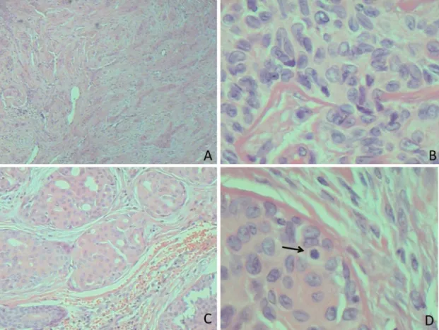
The diagnosis was established based on the presence of malignant cell clusters that showed an invasive architectural pattern, ductal and eccrine differentiation and cytological pleomorphisms (Figure 1). The ductal differentiation varied from small intracytoplasmic lumens to mature well-established ducts that were lined with a thin eosinophilic layer. No tumor displayed granular cells or decapitated lumens, thus ruling out the possibility of apocrine differentiation. The criteria used to deine invasion included tumor desmoplasia and/or the presence of irregular dermis-iniltrating cell clusters. Other observed features included basaloid and clear-cell patterns as well as squamous differentiation.
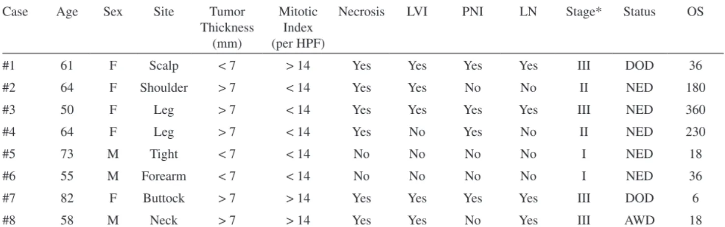
The parameters evaluated concerning prognostic analysis were the following: lymphovascular invasion, perineural invasion, necrosis and the mitotic index. The mitotic index was analyzed qualitatively, and the cutoff for high or low index was 14 mitotic cells per high-power ield (HPF). Vertical growth was assessed from the granular layer to the deepest site of invasion, as noted in the Breslow classiication. Any tumor presenting vertical growth greater than 7 mm was considered thick.
RESULTS
740
CLINICS 2010;65(7):739-42 Eccrine porocarcinoma: a challenging diagnosis
Luz MA et al.
Copyright © 2010 CLINICS
months. The mean size of the primary lesion was 48 mm, ranging from 10 mm to 100 mm. Five patients (62.5%) had infected lesions at the initial examination. The clinical and pathological characteristics upon admission are shown in table 2.
None of the tumors were classiied as pure “in situ” neoplasia. Only a single case contained all the morphological criteria within the incisional biopsy. Three cases reported an initial suspicion of EP; however, the absence of ductal differentiation prevented a deinitive diagnostic conclusion at that time.
Four cases had inaccurate diagnosis at irst biopsy. A basaloid cell pattern was present in the specimen analysis of one case, and the absence of ductal differentiation led to the incorrect diagnosis of basal cell carcinoma. Another tumor showed extensive clear-cell components, leading the pathologist to overlook the ductal differentiation and report the inal diagnosis as squamous cell carcinoma of the clear-cell subtype. Those two cases were clearly deined as EP after surgical resection because suficient material for the proper analysis of ductal differentiation was obtained. A third case yielded only a small amount of tissue, and the
Figure 1 - Eccrine Porocarcinoma – Histology. A. diffuse iniltrative pattern, B. atypical elongated cells and central groove, C. duct formation, D. mitotic igure (arrow)
Table 1 - Pathological report from the initial biopsy for eight cases with an eventual deinitive eccrine porocarcinoma diagnosis.
Patient Primary Lesion Size (mm) Diagnosis after Initial Biopsy
#1 65 Excisional biopsy as initial treatment
#2 50 Ulcerated basal cell carcinoma
#3 35 Ulcerated epithelial neoplasm suggestive of porocarcinoma
#4 50 Round cell undifferentiated neoplasm
#5 20 Clear cytoplasm cell carcinoma (suggestive of porocarcinoma)
#6 10 Adnexal skin carcinoma (suggestive of porocarcinoma)
#7 100 Malignant eccrine poroma
741
CLINICS 2010;65(7):739-42 Eccrine porocarcinoma: a challenging diagnosis
Luz MA et al.
Copyright © 2010 CLINICS
analysis was thus impaired. As a consequence, an incorrect diagnosis of “undifferentiated” small round-cell neoplasm was reported. Deinitive surgery gave better-quality tissue for an accurate analysis, and the inal diagnosis was EP. The fourth case had inconclusive initial analysis and we thus performed a complete excisional biopsy which rendered the diagnosis of EP. The histopathological prognostic factors are shown in table 2.
The standard treatment performed was wide resection. There is no clear recommendation in the literature regarding surgical margins for EP tumors. As such, we used our routine protocol for non-melanoma skin cancer, applying clear margins of at least 10 mm. In three patients, the resection was followed by a lymphadenectomy (inguinal or cervical). A single patient received palliative chemotherapy after unresectable local recurrence, while another two patients underwent adjuvant radiation therapy, one for close resection margins and the other because of lymph node macrometastasis.
The mean follow-up time was 74 months. Of the eight cases, six patients remain alive: Five patients have no evidence of disease, and one patient has conirmed regional recurrence. The two other patients died of EP-related complications. One of these two patients had an extensive recurrence, and his age did not allow further treatment. The second patient died following a second recurrence, three years after the initial diagnosis.
DISCUSSION
We have presented the approach and outcomes of eight EP cases. The study stresses the major problems with this particular type of tumor and the delay in deinitive treatment
attributed to the tumor’s indolent behavior or misleading clinical diagnosis upon inexpert medical evaluation. Pathologically speaking, this neoplasia demonstrated an aggressive behavior compared with common nonmelanoma skin cancer.
EP has a propensity to arise on the lower limbs (44%),
trunk (24%) or head and neck region (24%).3-4,7-8 However,
rare cases of penile involvement have been reported.8
Morphologically, tumors vary greatly in size, from less than 1 cm up to 10 cm. A long period of clinical history is often encountered (up to 50 years) because some of these tumors could have arisen from a preexisting benign eccrine poroma. Regional lymph node metastases are found in about 20% of patients, and distant metastases arise in about 10% of patients.4-5,8
The disease may appear nodular, iniltrative, ulcerated or polypoid. Multinodularity, ulceration and rapid growth may be associated with either local recurrence or metastasis.2
The clinical differential diagnoses of these lesions include seborrheic keratosis, pyogenic granuloma, amelanotic melanoma, squamous cell carcinoma, basal cell carcinoma,
verruca vulgaris, and metastatic adenocarcinoma.2,3
As we have shown, the initial pathology report can easily lead to a misdiagnosis. Even deined EPs may show some kind of basal or squamous differentiation.9 The tumor is
typically formed of cohesive basaloid epithelial cells. This morphological appearance can include squamous cells, clear cells, spindle cell differentiation, mucous cell metaplasia, a
Paget phenomenon and colonization by melanocytes.6-7,10
Clear-cell changes are frequently observed and are attributed to moderate amounts of glycogen, as detected by periodic
acid-Schiff (PAS) and PAS after digestion with diastase.5,8
Immunohistochemical techniques are not strictly necessary;
Table 2 - Clinical patterns from eight patients with a deinitive eccrine porocarcinoma diagnosis.
Case Age Sex Site Tumor
Thickness (mm)
Mitotic Index (per HPF)
Necrosis LVI PNI LN Stage* Status OS
#1 61 F Scalp < 7 > 14 Yes Yes Yes Yes III DOD 36
#2 64 F Shoulder > 7 < 14 Yes Yes No No II NED 180
#3 50 F Leg > 7 < 14 Yes Yes Yes Yes III NED 360
#4 64 F Leg > 7 < 14 Yes No Yes No II NED 230
#5 73 M Tight < 7 < 14 No No No No I NED 18
#6 55 M Forearm < 7 < 14 No No No No I NED 36
#7 82 F Buttock > 7 > 14 Yes Yes Yes Yes III DOD 6
#8 58 M Neck > 7 > 14 Yes Yes No Yes III AWD 18
742
CLINICS 2010;65(7):739-42 Eccrine porocarcinoma: a challenging diagnosis
Luz MA et al.
Copyright © 2010 CLINICS
however, they may be used to conirm the diagnosis. The cells that line the neoplastic ducts and clefts are positive for carcinoembrionic antigen and negative for S100 protein (myoepithelial cells from the glandular portion are positive for S100 protein).3-4 These indings conirm a primitive
eccrine ductal differentiation.
Clinicians will often render a worse prognosis because of a mitotic index of more than 14 mitotic cells per HPF, lymphovascular invasion and a tumor depth exceeding 7 mm.11-13 Although the gross size of a tumor has no
signiicant relationship to prognosis, a tumor depth >7 mm predicted both death and lymph node involvement in this study. Finally, an iniltrative tumor margin had a dramatic inluence on local recurrence.2-3 In a great majority of cases,
a well-handled specimen and an expert pathology and oncology department can help the attending surgeon achieve a deinitive diagnosis.
Cutaneous EP is rarely diagnosed preoperatively; thus, surgical management is usually not initially planned. Although wide local excision of any cutaneous tumor is recommended, the results from historical series suggest that EPs with an iniltrative pattern may beneit from further surgery if doubts exists regarding the completeness of the excision.2-4,8 Because of the high rate of local recurrence,
a wide excision of the primary tumor, with histologically clear margins, is mandatory.7 Proper surgical resection
leads to curative outcomes in 70% to 80% of cases.3-5,14-15
However, micrographic surgery has been attempted to reduce the morbidity and local recurrence with good initial results.14 There are no strong data available favoring adjuvant
therapy in this setting. Anecdotal reports show some beneits
when utilizing radiation or chemotherapy.16-17 Barzi et
al. proposed a new protocol that utilizes isotretinoin and interferon alpha to treat metastatic disease, yielding hopeful results.12 Nevertheless, there are few studies addressing the
application of chemotherapy to EP without metastasis.17
EP is still very challenging for surgeons and medical/ radiation oncologists. Because of its rarity, morphologic peculiarity and ambiguous similarity to other carcinomas, guidelines and strong recommendations are not widely available. However, professionals involved in skin cancer treatment should be aware of this disease. Because the cases are rare and each center has only a few of them, publications from cooperative multicentric groups are needed. These groups are able to pool several patients getting a larger number of cases and increasing the data relevance in order to facilitate clinical decisions when dealing with such an intriguing neoplasia.
REFERENCES
1. Pinkus H, Mehregan AH. Epidermotropic eccrine carcinoma. Arch Dermatol. 1963;88:597-606.
2. Robson A, Greene J, Ansari N, Kim B, Seed PT, McKee PH, et al. Eccrine Porocarcinoma (Malignant Eccrine Poroma) A Clinicopathologic Study of 69 Cases. Am J Surg Pathol. 2001;25:710-20.
3. Shiohara J, Koga H, Uhara H, Takata M, Saida T. Eccrine porocarcinoma: clinical and pathological studies of 12 cases. J Dermatol. 2007;181:516-22.
4. Orella JAL, Penalba AV, San Juan CC, Nadal RV, Morrondo JC, Alvarez TT. Eccrine Porocarcinoma: Report of nine cases. Dermat Surg. 1997:23:925-28.
5. Shaw M, McKee PH, Lowe D, Black MM. Malignant eccrine poroma: a study of twenty-seven cases. Br J Dermatol. 1982;107:675-80.
6. Ribeiro LC, Luz MA, Montenegro MFG, Biasi LJ, Ogata DC. Eccrine Porocarcinoma (Malignant Eccrine Poroma) with Bone Invasion and Lymph Node Metastases. App Cancer Res. 2008;28:165-67.
7. Snow SN, Reizner GT. Eccrine porocarcinoma of the face. J Am Acad Dermatol. 1992;27:306-11.
8. Grayson W, Loubser JS. Eccrine Porocarcinoma of the Penis J Urol. 2003;169: 611-2.
9. Klenzner T, Arapakis I, Kayser G, Boedeker CC. Eccrine porocarcinoma of the ear mimicking basaloid squamous cell carcinoma. Otolaryngol Head Neck Surg. 2006;135:158-60.
10. Brown Jr. CW, Dy LC. Eccrine porocarcinoma. Y Derm Ther. 2008;21:433-8.
11. Mahomed F, Blok J, Grayson W. The squamous variant of eccrine porocarcinoma: a clinicopathological study of 21 cases. J Clin Pathol. 2008;61:361-5.
12. Barzi AS, Ruggeri S, Recchia F, Bertoldi I. Malignant metastatic eccrine poroma. Dermatol Surg. 1997;23:267-72.
13. Chang NC, Tsai KB. Eccrine Porocarcinoma of the Auricle: A Case Report. Kaohsiung J Med Sci. 2009;25:401-4.
14. Poiares Baptista A, Tellechea O, Reis JP, Cunha MF, Figueiredo P. Eccrine porocarcinoma: A review of 24 cases. Ann Dermatol Venereol. 1993;120:107-15.
15. Wittenberg GP, Robertson DB, Solomon AR, Washington CV. Eccrine Porocarcinoma treated with Mohs Micrographic Surgery: A Report of Five Cases. Dermatol Surg. 1999;25:911-3.
16. Gutermuth J, Audring H, Voit C, Trefzer U, Haas N. Antitumour activity of paclitaxel and interferon-alpha in a case of metastatic eccrine porocarcinoma. J Eur Acad Dermatol Venereol. 2004;18:477-9.