Rita Leonor Franco Paulo Guimarães
Licenciatura em Biologia Molecular e Celular
ZNF675 evolutionary history and its novel
role in neuronal regulatory networks
Dissertação para obtenção do Grau de Mestre em Genética
Molecular e Biomedicina
Orientador: Frank M.J. Jacobs, Assistant Professor,
Swammerdam Institute for Life Sciences, University of
Amsterdam
Co-orientador: Gerrald Lodewijk, PhD Student, Swammerdam
Institute for Life Sciences, University of Amsterdam
Júri:
Presidente: Prof. Doutora Margarida Casal Ribeiro Castro Caldas Braga, Faculdade de Ciências e Tecnologia da Universidade Nova de Lisboa
Arguente: Doutora Maria João de Jesus Nunes, Faculdade de Farmácia da Universidade de Lisboa.
Vogal: Prof. Doutor Pedro Miguel Ribeiro Viana Baptista, Faculdade de Ciências e Tecnologia da Universidade Nova de Lisboa
ZNF675 evolutionary history and its novel role in neuronal regulatory networks
Copyright © Rita Leonor Franco Paulo Guimarães, FCT/UNL and UNL
The Faculdade de Ciências e Tecnologia and Universidade Nova de Lisboa have the right, perpetual
and without geographical limits, to file and publish this dissertation through printed copies reproduced
on paper or on digital form, or by any other means known or that may be invented, and to disseminate
through scientific repositories and admit its copying and distribution for educational or research
ACKNOWLEDGMENTS
I
Acknowledgments
I would like to express my very deep gratitude to Dr. Frank Jacobs, for the great opportunity of
working in his laboratory. Thank you for the guidance, the discussions and advices, and for the trust
placed in me.
My special thanks are to Gerrald Lodewijk, for being the best daily supervisor in the world ever
in the universe. For teaching me how to do good science every day, for cheering me up and calming me
down, and for all the conversations about everything and anything. This thesis would not have been
possible without you.
I am particularly grateful for having the fellow Portuguese colleague Diana Pereira Fernandes
in the laboratory, with whom I shared my achievements and frustrations. Thank you for the friendship,
for all the talks over a beer and the honest advices.
I wish to acknowledge the FCT/UNL and the European Commission for the Erasmus+ SMT
(student mobility for traineeships) program, for the chance to do the internship in the Netherlands.
An important thank you to my mother, father, brother and sister for loving me unconditionally.
For supporting me, believing in me and for giving the necessary strength to be one year far away from
you. Thank you to my whole family, especially in the Netherlands, who made me feel safe and at home.
Thank you to my friends that were close to me even when I was in another country. To my
friends Anna Davidson, David Huson and Sarah Tommouhi, who walked alongside me in this project. I
sincerely thank you for the friendship, all the fun activities we did together and for never letting me down!
I feel very heart-warmed for having found you guys.
Finally, I would like to thank to Fred White, for the love, protection and patience. Thank you for
supporting me and being by my side no matter what. For making me feel truly happy and for being my
ABSTRACT
III
Abstract
ZNF675 is a primate-specific KRAB-ZNF protein that arose in the last common ancestor of New
World Monkeys and Old World Monkeys. It represses THE1 and MST families of retrotransposons and
binds to gene promoters like MCPH1 and SESN3. Copy-number variation of ZNF675 is associated with
neurological disorders, including microcephaly and intellectual disability. Unravelling the evolutionary
history and the regulatory effect of ZNF675 will clarify its role in primates’ evolution.
The analysis of ZNF675 sequence revealed a significant difference in zinc-finger domains
between the marmoset and the human ZNF675. In a reporter assay, the marmoset ZNF675 did not bind
to MSTA elements. However, the human ZNF675 had a strong repressive effect on them. This indicates
structural changes in ZNF675 emerged to silence THE1 and MST retrotransposons, after New World
Monkeys split from our last common ancestor. As a collateral effect, ZNF675 created a new regulatory
network, by binding to gene promoters, 25% of which are involved in development and neuronal
processes. At the same time, some ZNF675 binding sites in gene promoters also underwent changes,
as found in the MCPH1 and SESN3 promoters. These changes affect the ZNF675 binding capacity and
led to the evasion or stabilization of its regulatory function. This highlights the dynamics of genome
evolution in primates.
To further investigate these findings, the CRISPR/Cas9 system was used for targeted deletion
of ZNF675 and ZNF681, or for modifying promoter region sequences using homology-directed repair.
The evolutionary arms race between ZNF675 and THE1 and MST elements led to the creation
of a novel ZNF675, with the ability to silence them. As a collateral consequence, ZNF675 binds to gene
promoters, regulating their expression. This new regulation network might have had a strong impact on
the evolution of the human brain.
RESUMO
V
Resumo
O ZNF675 pertence à família das proteínas KRAB-ZNF e surgiu há 35 milhões de anos, no
último ancestral comum dos Macacos do Novo e do Velho Mundo. Esta proteína silencia
especificamente retrotransposões das famílias THE1 e MST, mas também pode ligar-se a promotores
de genes neuronais como o MCPH1 e o SESN3. Variações no número de cópia do ZNF675 estão
associadas a doenças neurológicas como microcefalia e deficiência intelectual. Este projeto pretende
desvendar a função do ZNF675 no genoma humano e a sua história evolutiva.
A análise de sequências do ZNF675 ao longo da evolução revelou uma diferença significativa
entre o domínio de zinc-fingers do ZNF675 presente no primata sagui-de-tufos-brancos e no de humanos. Num luciferase reporter assay, o ZNF675 do sagui-de-tufos-brancos não se ligou ao retrotransposão MSTA. Contudo, o ZNF675 humano exerceu uma forte repressão no mesmo. Isto
indica que as alterações estruturais no ZNF675 evoluíram no sentido de silenciar os retrotransposões
THE1 e MST, depois da divergência dos Macacos do Novo Mundo com o nosso último ancestral
comum. Consequentemente, o ZNF675 terá adquirido uma nova função regulatória ao ligar-se a
promotores de genes, alguns dos quais foram sofrendo alterações ao longo da evolução dos primatas.
Estas alterações levaram a uma evasão ou estabilização de função regulatória do ZNF675.
Para investigar esta ideia, uma deleção nos genes ZNF675 e ZNF681 foi gerada através da
técnica CRISPR/Cas9, e conjuntamente com o mecanismo de homology-directed repair, para modificar sequências dos promotores.
Os resultados descritos sugerem que a luta evolutiva entre KRAB-ZNFs e os retrotransposões
THE1 e MST terá levado à emergência do ZNF675, que passou a silenciar estes elementos.
Colateralmente, o ZNF675 também começou a regular genes envolvidos no desenvolvimento
neurológico devido à afinidade pelo seu promotor. Esta regulação colateral poderá ter tido um forte
impacto na evolução do cérebro humano.
Palavras-chave: ZNF675, proteínas KRAB-ZNF, retrotransposões, regulação génica, evolução,
TABLE OF CONTENTS
VII
Table of Contents
Acknowledgments
I
Abstract
III
Resumo
V
List of Figures
IX
List of Tables
XI
List of Abbreviations
XIII
1
Introduction
1
1.1
Evolution of the human brain
1
1.2
Transposable elements
2
1.3
Krüppel-associated box zinc-finger proteins
3
1.4
Evolutionary arms race between transposable elements and KZNF proteins
5
1.5
ZNF675 and its target genes
6
1.6
Aim of this study
8
2
Materials and methods
11
2.1
ZNF675 and ZNF681 structural analysis
11
2.2
Analysis of ZNF675 binding to THE1B-int elements
11
2.3
ZNF675 ChIP-seq data for promoter binding analysis
11
2.4
Retinoic Acid treatment of a human neuroblastoma cell line
12
2.5
MCPH1 and SESN3 promoter region in primates
12
2.6
Luciferase Reporter Assay on human and marmoset MCPH1 and SESN3
promoters
13
2.7
Cloning of ZNF675-EGFP and ZNF681-EGFP fusion constructs
14
2.8
MCPH1 promoter region on different primate species
15
2.9
Replacement of human ZNF675 binding site in the MCPH1 promoter by the
marmoset
16
2.10 ZNF675/ZNF681 KO using CRISPR/Cas9 system
18
2.11 ChIP of human and marmoset ZNF675
20
3
Results
25
3.1
Divergent evolution of ZNF675 and ZNF681
25
3.2
Modifications to ZNF675 binding site in THE1B-int elements
25
3.3
ZNF675 binds to promoters of gene related to development and neuronal
processes
26
3.4
HES1 expression is up-regulated after induced neuronal differentiation
27
3.5
Significant changes in the binding site of ZNF675 in MCPH1 and SESN3
TABLE OF CONTENTS
VIII
3.6
Differential effect of ZNF675 binding on MCPH1 and SESN3 promoters
throughout evolution
29
3.7
Isolation of MCPH1 promoter region from different primate species
30
3.8
ZNF675 binding site of the marmoset MCPH1 promoter in a human context
31
3.9
ZNF675/ZNF681 KO using CRISPR/Cas9
33
3.10 ChIP of human and marmoset ZNF675
36
4
Discussion
39
5
Conclusion and Future Perspectives
45
References
47
Supplemental material
53
S.1
THE1 elements
53
LIST OF FIGURES
IX
List of Figures
Figure 1.01– Schematic of retrotransposable elements. 2
Figure 1.02–Representation ofC2H2-like ZNF protein-DNA interaction. 4
Figure 1.03–Representation of the KRAB-ZNF protein repression complex. 4
Figure 1.04–Evolutionary arms race between KZNF proteins and retrotransposons. 6
Figure 1.05–Evolutionary history of ZNF675. 7
Figure 3.01–Structural changes of ZNF675 and ZNF681 throughout primate evolution. 25
Figure 3.02– THE1B-int elements. 26
Figure 3.03–HES1 expression levels change after Retinoic Acid treatment. 27
Figure 3.04–MCPH1 promoter region. 28
Figure 3.05–SESN3promoter region. 29
Figure 3.06–Luciferase Reporter Assays on MCPH1 and SESN3, in the presence of hZNF675 and
cZNF675. 30
Figure 3.07–MCPH1 promoter region from several primate species analysed by 1% agarose gel
electrophoresis. 31
Figure 3.08–Testing of gRNAs that target the ZNF675 binding site on the MCPH1 promoter. 32
Figure 3.09–Replacement of ZNF675 binding site on the MCPH1 promoter by the marmoset. 33
Figure 3.10–Testing of gRNAs that target exon 1 of ZNF681. 34
Figure 3.11–ZNF675/ZNF681 KO using CRISPR/Cas9 system. 35
Figure 3.12–HDR-mediated ZNF675/ZNF681 KO using CRISPR/Cas9 system. 36
Figure 3.13–ChIP of human and marmoset ZNF675 on HEK293T. 37
Figure 3.14–ChIP of human and marmoset ZNF675 on U2OS cells. 38
Figure 5.01 – Representation of the changes in binding capacity of ZNF675 during primate
evolution. 45
Figure S1–Conservation of the MCPH1 promoter region in basal primates, centred in the ZNF675
LIST OF TABLES
XI
List of Tables
Table 2.01–Description of each primate species used on the analysis. 12
Table 2.02–Description of human and marmoset promoter regions of MCPH1 and SESN3. 13
Table 2.03–Description of protocols followed per transfection of one 100mm dish. 20
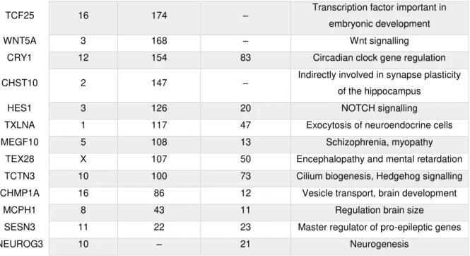
Table 3.01–Gene promoters bound by ZNF675. 26
LIST OF ABBREVIATIONS
XIII
List of Abbreviations
A APS Ammonium persulfate
C cMCPH1 Callithrix jacchus MCPH1
cSESN3 Callithrix jacchus MCPH1
cZNF675 Callithrix jacchus ZNF675
cDNA Complementary DNA
ChIP Chromatin immunoprecipitation
ChIP-seq Chromatin immunoprecipitation sequencing
D DNA Deoxyribonucleic acid
DMEM Dulbecco's modified Eagle medium
E E.coli Escherichia coli
ERV Endogenous retrovirus
eGFP Enhanced green fluorescent protein
EDTA Ethylenediamine tetraacetic acid
G gDNA Genomic DNA
GTEx Genotype-Tissue Expression
GMEM Glasgow's MEM
gRNA Guide RNA
H HES1 Hairy and enhancer of split homolog 1
HiFBS Heat inactivated fetal bovine serum
HP1 Heterochromatin protein 1
HBS HEPES Buffered Saline
H3K9me3 Histone 3 lysine 9-trimethylation
hMCPH1 Homo sapiens MCPH1
hSESN3 Homo sapiens SESN3
hZNF675 Homo sapiens ZNF675
HDR Homology-directed repair
HEK293T Human embryonic kidney 293 T cells
HERV Human endogenous retroviruses
hiPSC Human induced pluripotent stem cells
I IP Immunoprecipitation
K KAP1 KRAB-associated protein 1
KZNF KRAB-ZNF
KRAB-ZNF Krüppel-associated box zinc-fingers
L LCA Last common ancestor
LIF Leukemia inhibitory factor
LINE Long interspersed elements
LTR Long terminal repeats
LIST OF ABBREVIATIONS
XIV
M mZNF675 Macaca fascicularis ZNF675
MCPH1 Microcephalin 1
Mya Millions of years ago
MEM Minimum essential medium
mESC Mouse embryonic stem cells
N NWM New World Monkeys
NHEJ Non-homologous end-joining
NuRD Nucleosome remodelling and deacetylase
O OWM Old World Monkeys
ORF Open reading frame
P P/S Penicillin/streptomycin
PBS Phosphate buffered saline
PAGE Polyacrylamide gel electrophoresis
PCR Polymerase chain reaction
PEI Polyethylenimine
Q qPCR quantitative PCR
R RT-PCR Reverse transcriptase PCR
ROS Reactive oxygen species
RNA Ribonucleic acid
S SESN3 Sestrin 3
SVA SINE-VNTR-Alu element
ssDNA Single-stranded DNA
SINE Short interspersed element
SETDB1 SET domain bifurcated 1
T TEMED Tetramethylethylenediamine
TF Transcription factors
TSS Transcription start site
TE Transposable element
THE1 Transposon-like human element 1
TRIM28 Tripartite motif-containing 28
V VNTR Variable number of tandem repeats
INTRODUCTION
1
1
Introduction
1.1 Evolution of the human brain
The human brain has increased remarkably in size and complexity throughout primate evolution.
The encephalization quotient, a way of measuring brain size relatively to body weight, differs significantly
between human and other primates, which is not observed when comparing nonhuman primates
(Williams, 2002). In fact, the human brain has undergone an overdevelopment, specifically the neocortex
and cerebellum, that has been correlated to an increase in cell populations (Herculano-Houzel, Collins,
Wong, & Kaas, 2007). A lot of effort has been devoted to find the underlying causes of the human brain
singularity, i.e. the genetic and molecular events that led to a bigger-than-expected brain throughout
primate evolution.
In order to search for the genetic basis of brain evolution in primates, differences between
humans and chimpanzees have been investigated. In 1975, Mary-Claire King and Allan Wilson
published a landmark article that shed light on the molecular differences and similarities of humans and
chimpanzees, where they found that both primates share more than 99% of the average polypeptide
coding sequence (King & Wilson, 1975). Even though years later it was described that the difference
between these two species is in fact 4% (Varki & Altheide, 2005), such a small change could not explain
their major anatomical and behavioural differences. However, together with the non-coding part of the
genome, important regulatory regions which control gene expression, and structural genomic
rearrangements might have had an impact on the emergence of the human species.
Brain evolution has been linked to natural selection and genome reshaping, of which
Transposable Elements (TEs) are a major cause (Ayarpadikannan & Kim, 2014; Cordaux & Batzer,
2009; Deininger, Moran, Batzer, & Kazazian, 2003). TEs are derived from ancient viruses that infected
the host millions of years ago (Mya). They were first described by the Nobel Prize Barbara McClintock,
being referred to as “controlling elements” because they are able to regulate gene expression
(McClintock, 1950). They have the unique ability of moving throughout the genome, or even between
genomes (Cordaux & Batzer, 2009), which is also the reason why between half (Lander et al., 2001)
and two-thirds (de Koning, Gu, Castoe, Batzer, & Pollock, 2011) of the human genome is composed of
TEs.
Krüppel-associated box zinc-fingers (KRAB-ZNF) proteins function as transcriptional repressors
of TEs, in a type-specific manner, to prevent transposition and, thereby, maintain genome integrity
(Imbeault, Helleboid, & Trono, 2017; Jacobs et al., 2014; Najafabadi et al., 2015). These proteins
constitute ~50% of human zinc-finger (ZNF) proteins, representing the largest and most rapidly evolving
gene family (Huntley et al., 2006; Najafabadi et al., 2015). They have been described as a driving force
of a new level of gene regulation in primates, in a species-specific manner (Hamilton, Huntley, Gordon,
& Stubbs, 2005; Imbeault et al., 2017; Jacobs et al., 2014; Nowick, Hamilton, Zhang, & Stubbs, 2010;
Shannon, Hamilton, Gordon, Branscomb, & Stubbs, 2003). Interestingly, KRAB-ZNF (KZNF) proteins
INTRODUCTION
2
It was proposed that these regulatory modifications were in part responsible for the humans’ over-sized
and more complex brain (Nowick, Gernat, Almaas, & Stubbs, 2009).
1.2 Transposable elements
TEs are grouped in two major classes according to their transposition mechanism: class 1 and
class 2. Class 2 is composed of transposons, DNA elements that can excise and insert themselves into
another region of the genome, which constitute ~3% of the human genome (Pace, Feschotte, & Ii, 2007).
Class 1 comprises TEs which make use of an RNA intermediate to transpose. For this reason, they are
called retrotransposons. A reverse transcriptase, encoded in the element itself, copies the RNA into
complementary DNA (cDNA) which then gets integrated in another region of the genome. Within this
class, TEs can even be divided in families based on the presence or absence of long terminal repeats
(LTRs): LTR retrotransposons (Figure 1.01), also known as human endogenous retroviruses (HERVs),
are similar to simple retroviruses, lacking only the envelope-coding gene, and constitute ~8% of human
genome (Deininger et al., 2003). In contrast, non-LTR retrotransposons, which do not contain an LTR
region, are further subdivided in long interspersed elements (LINEs) and short interspersed elements
(SINEs), e.g. L1 and Alu elements, respectively. There is also a hominoid-specific element family, the SVA elements, which comprise a SINE region, followed by a variable number of tandem repeats (VNTR)
and an Alu element. The non-LTR elements constitute ~34% of the human genome (Cordaux & Batzer, 2009).
Figure 1.01– Schematic of retrotransposable elements. Retrovirus is composed of three genes: gag
INTRODUCTION
3
Retrotransposons are the major creators of genome instability by inserting themselves within or
close to genes, or by inducing genomic ectopic rearrangements (Ayarpadikannan & Kim, 2014; Cordaux
& Batzer, 2009; Lander et al., 2001). They can cause deleterious mutations when inserted in an exon,
which might change the open reading frame (ORF) giving rise to non-functional proteins. TEs can also
cause abnormal proteins, if they get inserted in an intron, eliminating the canonical splice site or even
creating novel splice sites (Ayarpadikannan & Kim, 2014). On the other hand, TEs can generate ectopic
rearrangement events, such as deletions, duplications and inversions, creating new genes, modifying
pre-existing ones or just changing the GC content (Lander et al., 2001). Moreover, if TEs get inserted in
close proximity with genes, in regulatory regions, they can change their expression levels, thus altering
the network on which that gene acts on (Ayarpadikannan & Kim, 2014). Interestingly, genomic
modifications induced by TEs are proposed to have accelerated evolution of gene regulatory networks
over mammalian evolution (Lowe, Bejerano, & Haussler, 2007).
With the sequencing of the human genome it was possible to estimate TEs’ activity status during
primate evolution, by analysing the sequence divergence between species. It was shown that the activity
of all transposons has declined 35-50 Mya, which means that they were active during early primate
evolution (Lander et al., 2001). This includes some families of LTR (THE1A, THE1B, THE1C, MSTA
and MSTB) and non-LTR retrotransposons (L1 and Alu elements), since they are primate-specific (Cordaux & Batzer, 2009; Pace et al., 2007). On the contrary, SVA elements have been active only
during the hominid evolution. Some families of SVA, L1 and Alu elements are still active in the human genome (Mills, Bennett, Iskow, & Devine, 2007; H. Wang et al., 2005). Interestingly, they have been
associated with a variety of genetic diseases, such as certain types of cancer, haemophilia and X-linked
diseases (Deininger et al., 2003; Kazazian, 1998; Konkel & Batzer, 2010). However, TEs have also
been described to play an important role in early development, being involved in essential processes
such as in placental formation, meiotic recombination during gametogenesis and epigenetic inheritance
(Gifford, Pfaff, & MacFarlan, 2013).
1.3 Krüppel-associated box zinc-finger proteins
KZNF proteins, the TEs’ transcriptional repressors, are composed of C2H2-like ZNFs, which is
the most prevalent DNA-binding motif of transcription factors (TFs) in eukaryotic organisms (Huntley et
al., 2006; Thomas & Schneider, 2011). ZNF proteins often have between 1 and 30 ZNF motifs in tandem
arrays, each one with two cysteines and two histidines interacting with a zinc ion, stabilizing the structure
(Huntley et al., 2006; Krishna, Majumdar, & Grishin, 2003). These ZNF motifs consist of two β-sheets
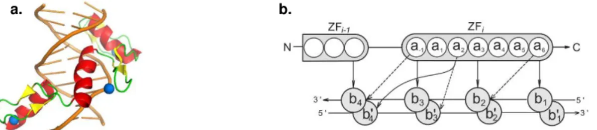
and one α-helix (Figure 1.02a), being the last one the DNA-binding domain by fitting in the major groove of the DNA (Lupo et al., 2013; Persikov & Singh, 2014). It is proposed that the ZNF protein binds to DNA
with high specificity, as each ZNF motif contacts four consecutive base pairs (Figure 1.02b), overlapping
on one, i.e. there is binding of three base pairs per ZNF motif (Persikov & Singh, 2014). There is currently
INTRODUCTION
4
influence of amino acids close to those that bind the DNA, the impact of other ZNF motifs or even the
possibility that not all the motifs bind to DNA (Najafabadi et al., 2015).
a. b.
Figure 1.02 –Representation ofC2H2-like ZNF protein-DNA interaction. (a) Structural model of a protein with
three ZNF motifs interacting with DNA. Each ZNF motif has two β-sheets (yellow), one α-helix (red/orange) and a zinc ion (blue). Adapted from Yang, Wang, & Macfarlan, 2017. (b) Two consecutive ZNF motifs binding to DNA. Amino acids within the i-th ZNF are numbered according to their relative position from the start of the alpha helical domain. DNA bases b1, b2, b3 and b4 are numbered sequentially and the complementary bases are primed. The canonical contacts are shown with solid arrows, and the three additional contacts are shown with dashed arrows.
Adapted from Persikov & Singh, 2014.
KZNF proteins are composed of a 75-amino acid KRAB domain in the N-terminal domain, in
addition to the ZNF domain at the C-terminus. After DNA-binding by the ZNF domain (Figure 1.03),
repression occurs through interaction of the KRAB domain with the KRAB-associated protein 1 (KAP1,
or TRIM28) (Peng et al., 2000). The complex recruits SETDB1, a histone 3 lysine 9-specific
methyltransferase, which in turn enhances heterochromatin protein 1 (HP1) binding (David C Schultz,
Ayyanathan, Negorev, Maul, & Rauscher Iii, 2002). The nucleosome remodelling and deacetylase
(NuRD) complex is also recruited by KZNF/KAP1 (D. C. Schultz, Friedman, & Rauscher, 2001), making
up the repression complex, which induces heterochromatin formation and transcriptional silencing
(Yang, Wang, & Macfarlan, 2017). The induced chromatin remodelling is spread in a long-range
repression through H3K9me3 (histone 3 lysine 9-trimethylation) and HP1, model confirmed in a KZNF
cluster, suggesting an auto-regulatory mechanism (Groner et al., 2010).
Figure 1.03 –Representation of the KRAB-ZNF protein repression complex. Firstly,ZNF domain binds to the
DNA and the KRAB domain interacts with KAP1. The complex recruits other factors such as SETDB1, HP1 and the NuRD complex. Red circles on histone tails are H3K9m3 and green circles are deacetylation of multiple lysines.
INTRODUCTION
5
KZNF proteins are specific to tetrapod vertebrates and are often localized in familial gene
clusters, as they are primarily originated by duplication events. For instance, out of 423 KZNF
protein-coding loci, at least 136 are primate-specific KZNF and have diverged significantly after duplication
(Huntley et al., 2006). Even though these genes coexist in the same loci, each one is under a distinct
selective pressure, giving rise to novel proteins with new recognition sites, as changes occur mostly in
the ZNF domain and the smallest nucleotide variation influence DNA binding (Huntley et al., 2006; Lupo
et al., 2013). Segmental duplications of loci containing KZNF protein-coding genes are also the
underlying cause for the observed diversity among their families, where 24 KZNF protein-coding genes
are only present in great apes and hominids. Interestingly, gene duplications that are human and
chimpanzee-specific have evolved in a higher rate than older genes, which may be evidence for the still
ongoing evolution of regulatory factors (Nowick et al., 2010).
KZNF proteins are involved in essential physiological processes, such as development,
differentiation, metabolism, cell division and cancer (Lupo et al., 2013), in a cell type-specific manner
(Najafabadi et al., 2015). For instance, ZNF809 is a stem cell-specific retroviral repressor (Wolf & Goff,
2009); ZNF382 is a proapoptotic tumour suppressor (Cheng et al., 2010); ZNF268 participates in the
erythroid cells’ differentiation (Zeng et al., 2012); and PRDM9, a well-studied KZNF protein, determines
meiotic recombination hotspots’ location (Altemose et al., 2017). Moreover, KZNF proteins might have a role preventing segmental duplication events by suppressing nonallelic homologous recombination,
stabilizing the genome (Najafabadi et al., 2015).
1.4 Evolutionary arms race between transposable elements and KZNF proteins
In 2009, Emerson and Thomas hypothesized that the evolution of KZNF proteins was being
“driven by an arms race with their viral or transposon targets” (Emerson & Thomas, 2009). The first two main evidences that point in that direction were a strong correlation between the number of ZNF
domains and LTR retrotransposons in the vertebrate genome; and whenever a new endogenous
retrovirus (ERV) family arose in the genome, it was usually followed by a burst of ZNF genes. (Thomas
& Schneider, 2011). This model was later supported by Jacobs and colleagues, who have shown that
retrotransposons would evade repression due to mutations within the binding site of their
primate-specific KZNF (Figure 1.04). In response, KZNF proteins evolved to keep the retrotransposons silenced,
in an evolutionary arms race (Jacobs et al., 2014). Jacobs et al. showed this through luciferase reporter
assays in mouse embryonic stem cells (mESC). An SVA element was cloned upstream of a minimal
SV40 promoter, which controls the expression of the luciferase reporter. The constructs were
co-expressed with different versions of the ZNF91: the human, the version probably present in the last
common ancestor (LCA) of humans and gorillas, in the LCA of humans and orangutans, and the
macaque version. The experiment revealed a difference in luciferase activity, being strongly repressed
with the human ZNF91, and having a decreasing effect in the older species. This implies that the
INTRODUCTION
6
Figure 1.04 –Evolutionary arms race between KZNF proteins and retrotransposons. Retrotransposons have
the ability to transpose to another location in the genome until a KZNF protein emerges and silences them. Later, retrotransposons escape repression due to mutations. KZNF protein mutates too, recovering the ability to silence the retrotransposons. Adapted from Frank Jacobs,frankjacobslab.com (retrieved in: 20/09/2018).
KZNF proteins have been protecting our genome integrity against transposable elements’
emergence since 100 Mya, in the early mammalian radiation (Najafabadi et al., 2015). Throughout
evolution the ZNF domain of KZNF proteins have undergone significant changes, whilst the KRAB
domain stayed conserved, meaning that recently emerged TEs can be silenced by the same
KAP1-mediated repression complex (Huntley et al., 2006; Thomas & Schneider, 2011). These DNA-binding
proteins make up a suitable and smart way of fighting TEs movement, preventing whole-genome
instability.
The arms race can explain in part the rapid expansion of KZNF protein families, but not entirely
since the majority of TEs have lost the transposition potential and the correspondent KZNF protein still
has its full binding capacity (Imbeault et al., 2017). As a collateral consequence of the evolutionary
battle, these family of repressors have acquired novel regulatory functions. They are involved in
regulation of gene expression, acting either directly by binding promoter regions (Lupo et al., 2013), or
indirectly by binding TEs, which they benefit from and use as regulatory platforms nearby genes
(Imbeault et al., 2017; Najafabadi et al., 2015).
1.5 ZNF675 and its target genes
ZNF675 is a KZNF gene which is expressed in the brain according to the Genotype-Tissue
Expression (GTEx) project (Lonsdale et al., 2013). According to DECIPHER (Firth et al., 2009), it is
associated with several neuronal disorders, such as microcephaly, macrocephaly, intellectual disability,
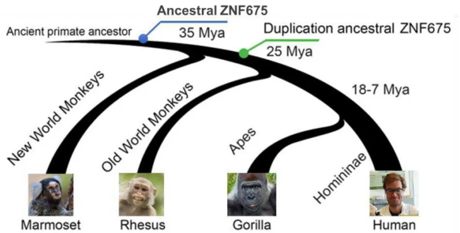
autism and cerebellar atrophy. Like many other KZNF proteins, ZNF675 is a primate-specific protein
INTRODUCTION
7
1.05) (Imbeault et al., 2017). However, a duplication event occurred in the ancestor of humans and
OWMs, giving rise to ZNF681, with a quite different DNA recognizing ZNF domain. Furthermore,
ZNF675 itself underwent significant structural modifications, which changed its DNA-recognizing motifs
(Lodewijk et al., unpublished).
Figure 1.05 –Evolutionary history of ZNF675. ZNF675 first arose in the LCA of NWMs and OWMs. It underwent
a duplication event in the LCA of OWM and apes, giving rise to ZNF681. Adapted from Lodewijk et al., unpublished.
ZNF675 represses LTR retrotransposons, specifically THE1B/C/D/A (transposon-like human
element 1) and MSTA/B/B1 (Najafabadi et al., 2015), acting in a cell-type specific manner (Imbeault et
al., 2017). Interestingly, it appears that a great expansion of THE1 and MST elements happened in the
LCA of NWM and OWM, which was right before the structural changes of ZNF675 (Lodewijk et al.,
unpublished).
Chromatin immunoprecipitation sequencing (ChIP-seq) is a technique widely used to detect the
interactions between DNA and proteins, and allows the identification of the DNA sequence involved, i.e.
the binding site (Schmidt et al., 2009). ChIP-seq data for ZNF675 (GEO accession number:
GSM1407624) (Najafabadi et al., 2015), suggests that besides binding TEs, ZNF675 binds to several
genes related to neurodevelopment or involved in neurological disorders such as HES1 (hairy and
enhancer of split homolog 1), MCPH1 (microcephalin 1) and SESN3 (sestrin 3).
HES1 is a transcription factor involved in the regulation of the central nervous system
development, by maintaining neuronal progenitors’ self-renewal and proliferative capacity (Nakamura et al., 2000). Preserving the neuroprogenitors’ pool is crucial to reach the correct number of neuronal cells,
which is accomplished by HES1 repression of the cyclin-dependent kinase inhibitor p27Kip1 (Murata et
al., 2005).
MCPH1 is considered a pleiotropic factor, since it has a broad range of different functions. It is
implicated in DNA damage repair, genomic stability, chromatin remodelling and cell division, by
regulating the G2/M checkpoint (Liu, Zhou, & Wang, 2016) and the chromosomes biorientation (Arroyo
INTRODUCTION
8
including lung and breast cancer (Mantere et al., 2016; Zhou et al., 2016). MCPH1 plays an important
role in regulating brain size during development, being the first gene to be associated with primary
microcephaly. Microcephaly is a disorder characterized by a small brain, specifically the cerebral cortex,
and a head circumference with more than 3 standard deviations below the age-related mean (Jackson
et al., 2002). Genes involved in brain development are of particular interest since they might have played
a role in humans’ brain enlargement during evolution. In fact, several studies concerning MCPH1 gene
have suggested that it was under positive selection in the lineage from OWMs ancestor to humans and
great apes, especially after the split of lesser apes (Evans, Anderson, Vallender, Choi, & Lahn, 2004;
Y. Q. Wang & Su, 2004).
SESN3 gene is a member of the highly conserved Sestrins family, characterized by having
antioxidant properties. It is regulated by FoxO, subfamily of forkhead transcription factors, in response
to high levels of reactive oxygen species (ROS) induced by Ras proteins. Therefore, SESN3 have a
determinant role against oxidative stress and in preventing apoptosis (Hagenbuchner et al., 2012;
Nogueira et al., 2008). In case of disease, the protective activity of SESN3 is especially important, which
is why this protein has been described to act in certain types of diseases, such as cancer (colorectal
and lung cancer), metabolism disorders, diabetes and cardiovascular disorders (M. Wang et al., 2018).
However, regarding the brain, SESN3 has been associated with epilepsy. In the human epileptic
hippocampus, SESN3 was found to act as a trans-acting genetic control of a proconvulsant gene-regulatory network, which positively regulates proconvulsive cytokines and Toll-like receptor signalling
gene (Johnson et al., 2015).
Genes involved in major cellular processes need to be under a strict chronological and spatial
regulatory mechanism in order to be expressed in a controlled manner. Consequently, changes in the
regulatory machinery have a strong impact on gene expression, thus altering the downstream process
that gene would act on. The LCA of humans and NWMs was probably the first primate suffering from
the regulatory changes caused by the ZNF675 emerging, around 35 Mya.
1.6 Aim of this study
It is hypothesised that ZNF675 arose in the genome to repress THE1 and MST elements, but
the promoters of several genes were caught in the arms race, such as HES1, MCPH1 and SESN3.
Here, ZNF675 repression of MCPH1 and SESN3 is investigated in the context of evolution. By exploring
how ZNF675 structural changes might have affected its DNA binding capacity and how small
modifications in the promoters of these genes over the past millions of years have fine-tune the binding.
ZNF675 structural changes over time are investigated with the Princeton predictive DNA recognition
tool (Persikov & Singh, 2014), where DNA motifs recognized by ZNF675 are unravelled. The human
ZNF675 and marmoset ZNF675 binding affinity to the MCPH1 and SESN3 promoter regions is analysed
INTRODUCTION
9
promoters’ changes during primate evolution are assessed with a multiple sequence alignment of
sequences from NWMs or basal primates to humans. To further investigate these findings, the
CRISPR/Cas9 system is used to induce a targeted deletion of the ZNF675 and ZNF681 genes. This
technique is also used, together with the homology-directed repair (HDR) mechanism, to change the
ZNF675 binding site in human cells by the marmoset. Finally, a chromatin immunoprecipitation (ChIP)
MATERIALS AND METHODS
11
2
Materials and methods
2.1 ZNF675 and ZNF681 structural analysis
The ZNF675 sequences from human (hZNF675), crab-eating macaque (mZNF675) and
marmoset (cZNF675), and the ZNF681 from human (hZNF681) and crab-eating macaque (mZNF681),
were previously obtained through the analysis of sequences from the Whole-Genome Shotgun contigs
on GenBank, NCBI. Using the predictive DNA recognition tool (Persikov & Singh, 2014), ZNF motifs of
ZNFs were characterized as functional, if the ZF score was higher than 25, or degenerated, and on
which DNA motifs they bind to. With that information, the structural changes of all ZNFs were analysed.
2.2 Analysis of ZNF675 binding to THE1B-int elements
Using the RepeatMasker from the UCSC Genome Browser (Kent et al., 2002), all the
THE1B-int sequences, which is the THE1B-internal part of the elements, were obtained from the human reference
genome GRCh37/hg19. Sequences were filtered by sequence similarity score above 8000 and length
greater than 800bp. In order to get the THE1B-int elements that are bound and not bound by ZNF675,
the selected sequences were intersected with Model-based Analysis of ChIP-seq (MACS) peak calling
data from the ChIP-seq data for ZNF675 (GEO accession number: GSM1407624) (Najafabadi et al.,
2015). ZNF675-bound was defined as 50% of the ZNF675 peak was overlapping with a THE1B-int
element, whereas unbound was defined as no overlap at all. From the bound 641 and unbound 1505
THE1B-int elements, 15 sequences with the higher score of each group were aligned using the Multiple
Sequence Alignment MUSCLE in UGENE (Okonechnikov et al., 2012).
2.3 ZNF675 ChIP-seq data for promoter binding analysis
Previously, raw ZNF675 ChIP-seq data fastq files (GSM2466628, GSM1407624) (Imbeault et
al., 2017; Najafabadi et al., 2015) were trimmed with Trimmomatic using the following parameters:
ILLUMINACLIP Truseqv3 single end for Hiseq, SLIDNIGWINDOW: 4:20, MINLEN: 30. Then, reads
were mapped to hg19 or hg38 using Bowtie2, using very sensitive end-to-end settings. The BAM files
were converted to bigwig using DeepTools bamCoverage (bin size = 1). Bigwig files were analysed
using the UCSC genome browser. Using the table browser, bigwig files were filtered for regions with
sequencing depth of >20 reads. Promoter regions were selected from 5kb upstream of genes’
transcription start site (TSS) until 1kb downstream. Both promoter regions and the filtered bigwig files
were intersected with any percentage of overlap. From the output list of intersections, regions with a lot
MATERIALS AND METHODS
12
long non-coding RNA, antisense RNA and small nuclear RNA, if the ZNF675 binding site was in those
elements. The list was further shortened by selecting neuronal related genes, involved in cell signalling
or associated with neuronal diseases.
2.4 Retinoic Acid treatment of a human neuroblastoma cell line
Cells from the human neuroblastoma cell line SK-N-SH (Sigma-Aldrich #86012802) were
seeded at a density of 5 x 10⁴ cells per well in 2x 12-wells plates and grown in minimum essential
medium (MEM, ThermoFisher Scientific #31095029) with 10% heat inactivated fetal bovine serum
(HiFBS, ThermoFisher Scientific #10500-064) and 1x penicillin/streptomycin (P/S, ThermoFisher
Scientific #15140122). After 24 hours (T0) cells were isolated in TRIzol (ThermoFisher Scientific
#15596018) and kept in –80°C. Cells still in culture were treated with 10µM retinoic acid (RA,
Sigma-Aldrich #R2625) or ethanol (Merck Millipore #1.00983.1000) as control. 24 hours later, cells were
isolated in TRIzol and the RNA extracted, including from the T0 cells, following the Direct-zolTM RNA
MicroPrep protocol (Zymo Research). A one-step RT-PCR (QIAGEN) was performed according to the
manufacturer’s protocol, on 20ng of RNA per reaction, with a program of 25 cycles. The following primers were used:
HES1_qFw1: GATGCTCTGAAGAAAGATAGC HES1_qRv1: TTCATGCACTCGCTGAAGC
The amplicons were analysed on a 1% agarose gel electrophoresis and the relative density of
the DNA was analysed using ImageJ (Abràmoff, Magalhães, & Ram, 2004).
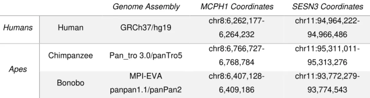
2.5 MCPH1 and SESN3 promoter region in primates
MCPH1 and SESN3 promoter sequences were obtain from the human reference genome
GRCh37/hg19 on the UCSC Genome Browser. The sequences were then converted to other primate’s
genomes, being further described in the following table:
Table 2.01 –Description of each primate species used on the analysis. The columns from the first until the fifth
correspond to: primate’s group, primate’s name, genome assembly used, coordinates of the sequence used of the
MCPH1 and SESN3 genes’ promoters.
Genome Assembly MCPH1 Coordinates SESN3 Coordinates
Humans Human GRCh37/hg19
chr8:6,262,177-6,264,232
chr11:94,964,222-94,966,486
Apes
Chimpanzee Pan_tro 3.0/panTro5 chr8:6,766,727-6,768,784
chr11:95,311,011-95,313,276
Bonobo MPI-EVA panpan1.1/panPan2
chr8:6,407,128-6,409,186
MATERIALS AND METHODS
13
Gorilla gorGor3.1/gorGor3 chr8:6,207,578-6,209,653
chr11:92,578,851-92,581,113
Orangutan WUGSC 2.0.2/ponAbe2
chr8:6,674,658-6,676,746
chr11:91,241,710-91,243,978
Gibbon GGSC Nleu3.0/nomLeu3 chr4:145,850,941-145,853,014 chr15:40,467,613-40,469,875 Old World Monkeys
Green Monkey Chlorocebus_sabeus 1.1/chlSab2 chr8:6,173,985- 6,175,851 + chr8:6,190,182- 6,190,384 chr1:86,486,434-86,488,709 Crab-eating Macaque Macaca_fascicularis_ 5.0/macFas5 chr8:6,619,567-6,621,649 chr14:91,115,965-91,118,237 Rhesus Macaque BCM Mmul_8.0.1/rheMac8 chr8:6,570,411-6,572,491 chr14:87,860,620-87,862,893
Baboon Baylor Panu_2.0/papAnu2 chr8:6,062,211-6,064,321 chr14:84,999,969-85,002,238 Golden
Snub-nosed Monkey Rrox_v1/rhiRox1
KN298329v1:105,736-107,790 KN300223v1:207,295 -209,579 New World Monkeys
Marmoset WUGSC 3.2/calJac3 chr13:5,720,181-5,722,193
chr11:42,482,819-42,485,066 Squirrel
Monkey Broad/saiBol1
JH378324:74,869-76,826 JH378333:178,252-180,348 Basal Primates
Mouse Lemur Mouse
lemur/micMur2 –
KQ057561v1:1,916,7 68-1,919,029
Bushbaby Broad/otoGar3 – GL873520:268,417-270,652
The sequences were aligned using the Multiple Sequence Alignment MUSCLE in UGENE and
their conservation was analysed.
2.6 Luciferase Reporter Assay on human and marmoset MCPH1 and SESN3 promoters
Cloning of sequences into pGL3 vector – A promoter region of 200bp from human and marmoset MCPH1 and SESN3 were obtained on the UCSC Genome Browser, further characterized in
the following table:
Table 2.02 –Description of human and marmoset promoter regions of MCPH1 and SESN3. First column
corresponds to the primate’s name, second is the genome assembly used, third and fourth are the coordinates fo
the sequences used of the MCPH1 and SESN3 genes’ promoters.
Genome Assembly MCPH1 coordinates SESN3 coordinates
Human GRCh37/hg19 chr8:6,263,654-6,263,853 chr11:94,964,744-94,964,943
MATERIALS AND METHODS
14
These sequences were repeated 3 times and the following restriction sites were added: KpnI at
the 5’ end, and XhoI or BglII for the 3’ end. The constructs were synthesized by GeneArt from
ThermoFisher Scientific and cloned into a pGL3-Promoter luciferase vector (Promega #E1761). Ligation
and transformation were done following the Quick ligation kit recommended protocol. pGL3-hMCPH1,
pGL3-cMCPH1, pGL3-hSESN3 and pGL3cSESN3 were Sanger sequenced.
Cell Transfection and Luciferase Assay – Mouse 46c embryonic stem cells (Ying, Stavridis, Griffiths, Li, & Smith, 2003) were seeded from a full 100mm dish in a dilution of ¼ cells per well, in
24-wells plate coated with 0.1% gelatin. The cells were cultured in Glasgow's MEM (GMEM, ThermoFisher
Scientific #11710-035) complete (10% HiFBS, 1x P/S, 5.5nM β-mercaptoethanol (ThermoFisher
Scientific #31350010) 1x Non-essential aminoacids (ThermoFisher Scientific #11140035) and 1x
sodium pyruvate (ThermoFisher Scientific #11360039)) supplemented with 1x leukemia inhibitory factor
(LIF, ESGRO/Merck Millipore #ESG1106). On the following day the medium was changed to GMEM
complete without P/S. Cells were transfected using LipofectamineTM 3000 reagent (ThermoFisher
Scientific), according to the manufacturer’s recommended protocol. The transfection mix per well was
composed of 5ng Red Fluorescent Protein (RFP), 5ng pRenilla (Promega #E2231) and 50ng pGL3
luciferase vector: EV, MSTA (previously cloned), hMCPH1, mMCPH1,
pGL3-hSESN3 or pGL3-mSESN3. Per luciferase vector, it was added 440ng pCAG-EV (with 90ng
pBluescript), pCAG-hZNF675-HA or pCAG-mZNF675-HA. 6 hours after the transfection the medium
was changed again to GMEM complete with 1x LIF. On the next day the cells were washed once with
Phosphate Buffered Saline (PBS, Lonza BioWhittaker™ #BE17-516F/12) and lysed using 100µl Passive Lysis Buffer from Dual-Luciferase® Reporter Assay System (Promega). The cell suspension was
incubated for 15min incubation on an orbital shaker (250rpm) at room temperature and for 15min at
-80°C. The Dual Luciferase Reporter Assay was performed following the manufacturer’s recommended
protocol.
2.7 Cloning of ZNF675-EGFP and ZNF681-EGFP fusion constructs
The enhanced green fluorescent protein (eGFP) was amplified by a PCR on pCAG-eGFP
expression vector (Addgene #11150), with Phusion High-Fidelity DNA Polymerase (ThermoFisher
Scientific). Two sets of primers were used, with different restriction sites, as follows:
pEGFP_ZNF675_PstI_F1:
AGCTCTGCAGAACTGGAACGTGTACCCATACGATGGCGGAGGTGGAGGCGGAGGTGGAGTGAGCAAGGGCGAGGAGCT GTTC
pEGFP_ZNF675_NdeI_F2: AGCTCATATGATGGCGGAGGTGGAGGCGGAGGTGGAGTGAGCAAGGGCGAGGAGCTGTTC
pEGFP_ZNF675_NotI_R1: AGCTGCGGCCGCTCACTTGTACAGCTCGTCCATGCCG
MATERIALS AND METHODS
15
PCR products were analysed on a 1% agarose gel electrophoresis. Amplicons were excised,
and DNA was extracted using QIAEX II Gel Extraction kit (QIAGEN).
The human, crab-eating macaque and marmoset ZNF675 coding sequences were previously
obtained through the analysis of sequences from the Whole-Genome Shotgun contigs on GenBank,
NCBI. An HA-tag sequence was added to the end of each ZNF and the constructs were synthesized by
GeneArt from ThermoFisher Scientific. The vectors and the eGFP fragment were digested with the
restriction enzymes: PstI (ThermoFisher Scientific, #ER0611) for pMA-hZNF675-HA; NdeI
(ThermoFisher Scientific #ER0581) for pMA-mZNF675-HA, pMA-cZNF675-HA and pMA-hZNF681-HA;
and NotI (ThermoFisher Scientific #ER0591) for every construct. The digested fragments were analysed
with 1% agarose gel electrophoresis, the DNA excised and extracted using QIAEX II Gel Extraction kit.
The vectors were treated with Alkaline Phosphatase (New England Biolabs #M0371S) and purified with
DNA Clean & Concentrator-5 Kit (Zymo Research). The linearized vectors were ligated with the eGFP
fragment in a 1:10 molar ratio using the Quick ligation kit (New England Biolabs). Competent E.coli
DH5α was transformed with the construct following the manufacturer’s recommended protocol. Bacteria
was grown overnight in Luria-Bertani (LB)– agar plates (Merck Millipore #1.10285.0500, VWR
Chemicals #84609.05) at 37°C. On the next day colonies were picked and grown in LB medium with
ampicillin (Roche #10835242001) overnight. The plasmids were then isolated from the bacteria using
the Plasmid Miniprep Kit (QIAGEN). The constructs were cloned into the pCAG expression vector
(Addgene #51142) using the restriction enzymes EcoRI (ThermoFisher Scientific #ER0271) and NotI,
for the 5’ and 3’ end, respectively. Finally, the pCAG-hZNF675-GFP, mZNF675-GFP, pCAG-cZNF675-GFP and pCAG-hZNF681-GFP were Sanger sequenced.
2.8 MCPH1 promoter region on different primate species
Genomic DNA from Chimpanzee, Gorilla, Orangutan, Rhesus Macaque, Baboon, Gibbon and
Marmoset were obtained from the Biomedical Primate Research Centre, in Rijswijk. Primers were
designed in the MCPH1 promoter of every primate, which are the following:
MCPH1_Prom_F2_AllPrimates: AAGTACGTGAGTCTTCAATG
MCPH1_Prom_F3_Marmoset-AAGTACTTGGGTCTTCAATG
MCPH1_Prom_F4_Marmoset: TAGGCACCGTTGTCCTCGTTAC
MCPH1_Prom_R2_OWM: TTCCGGGATTTGTCTGTAGC
MCPH1_Prom_R3_Orangutan: TTTCCGGGATTTCTTTGTAGG
MCPH1_Prom_R4_Gibbon: TTTCCGGGATTTCTCTAGGTG
MCPH1_Prom_R5_Marmoset: GCGGGAGGCTGGCGGGAGCT
MCPH1_Prom_R6_Marmoset: CAGGAAGTTCCTCACCTTTC
MATERIALS AND METHODS
16
Genomic regions of 1200bp were amplified with Phusion High-Fidelity DNA Polymerase on
20ng genomic DNA (gDNA) per reaction. PCR products were analysed on a 1% agarose gel
electrophoresis. Amplicons were extracted from gel using the QIAEX II Gel Extraction kit and purified
with DNA Clean & Concentrator-5 Kit. A second PCR was performed, with the same DNA Polymerase,
with primers with restriction sites, as shown below:
MCPH1_Prom-KpnI_F2_AllPrimates: ATGCGGTACCAAGTACGTGAGTCTTCAATG
MCPH1_Prom-NheI_R2_GreatApes (human, chimp, gorilla): ATGCGCTAGCTTTCCGGGATTTCTCTGTAGG
MCPH1_Prom-NheI _R2_Baboon: ATGCGCTAGCTTCCGGGATTTGTCTGTAGC
MCPH1_Prom-NheI _R8_Rhesus: ATGCGCTAGCTTCCGGGATTTATCTGTAGC
MCPH1_Prom-NheI _R3_Orangutan: ATGCGCTAGCTTTCCGGGATTTCTTTGTAGG
MCPH1_Prom-NheI _R4_Gibbon: ATGCGCTAGCTTTCCGGGATTTCTCTAGGTG
The amplicons were analysed on a 1% agarose gel electrophoresis and excised from the gel.
2.9 Replacement of human ZNF675 binding site in the MCPH1 promoter by the marmoset
Cloning of guide RNAs into pX330 vector – The guide RNAs (gRNAs) were designed upstream and downstream of the ZNF675 binding site in the MCPH1 promoter. To clone unidirectionally, on the
5’ end of both sense (s) and antisense (as) oligos an overhang was added that can hybridize with DNA ends created when digesting pX330-U6-Chimeric_BB-CBh-hSpCas9 vector (Addgene #42230) with
BbsI (ThermoFisher Scientific #ER1011). The sequences are the following:
pX330-MCPH1_bs_up_sense: CACCGCCGCACAAAGCCGTCCGCT
pX330-MCPH1_bs_up_antisense: AAACAGCGGACGGCTTTGTGCGGC
pX330-MCPH1_bs_down_sense: CACCGCGCGCCACGTTACTGTTCA
pX330-MCPH1_bs_down_antisense: AAACTGAACAGTAACGTGGCGCGC
10pmol of the gRNAs were mixed in 10x Annealing Buffer (10mM Tris pH8.0, 50mM Sodium
chloride, 1mM ethylenediamine tetraacetic acid (EDTA) (Addgene)) and annealed using the
thermocycler, where the temperature was increased up to 95°C for 10min, and slowly decreased until
25°C, in 30min total. The annealing efficiency was analysed with a 2% agarose gel electrophoresis. The
gRNAs were then cloned into the pX330 with BbsI following the Quick ligation kit recommended protocol,
with the single modification of adding only 0.2pmol of gRNA mix. Constructs were Sanger Sequenced.
Testing of gRNAs – Human embryonic kidney 293 T cells (HEK293T, uncertain origin) were seeded at passage 3+12 from a full 60mm dish in a dilution of ¼ cells per well, in a 12-wells plate and
cultured in Dulbecco's Modified Eagle Medium (DMEM) (1x)+GlutaMaxTM (ThermoFisher Scientific
#61965026) with 10% HiFBS and 1x P/S. On the following day the medium was changed to
MATERIALS AND METHODS
17
250ng of pX330-gRNA or pX330-EV, 50ng of pCAG-GFP and 200ng of pBluescript, one well per gRNA,
according to the manufacturer’s recommended protocol. The medium was changed again to DMEM+GlutaMax with 10% HiFBS and 1x P/S, 6 hours after. 48 hours after transfection, the cells were
lysed and the DNA isolated using Quick-DNA Miniprep Plus Kit (Zymo Research), following
manufacturer’s recommended protocol. A PCR was performed on 25ng of gDNA, using Phusion High-Fidelity DNA Polymerase, with the primers MCPH1_Prom_F2_primates and
MCPH1_Prom_R2_greatapes.
10% of PCR product was analysed on a 1% agarose gel electrophoresis. Amplicon was purified
using DNA clean & Concentrator-5 Kit and annealed, following the same protocol. An 8%
Polyacrylamide gel electrophoresis (PAGE, 8% acrylamide (Bio-Rad #1610146), 1x TAE Buffer (Tris
base (Sigma-Aldrich #10708976001), acetic acid (Merck Millipore #64-19-7), EDTA (Sigma-Aldrich
#E5134)), 0.1% ammonium persulfate (APS, Sigma-Aldrich #A3678), 0.00001%
Tetramethylethylenediamine (TEMED, Sigma-Aldrich #T9281)) was done with 100ng of gDNA. Samples
migrated at 90V until the loading dye left the gel, then the gel was stained with 0.005% ethidium bromide
in 1x TAE for 15min on orbital shaker at 50rpm. The gel was imaged using ChemiDocTM MP Imaging
System (Bio-Rad).
HDR mediated ZNF675 binding sequence replacement – The chosen gRNA was MCPH1_bs_down and a single-stranded DNA (ssDNA) oligo was designed with two homology-arms
with 30bp each. In between the arms the sequence is from the marmoset genome WUGSC 3.2/calJac3.
The oligo has on both ends three phosphorothioate bonds, as shown below:
MCPH1_ssODN_caljac:
G*A*G*CAGGCAGGGCGGTGCGTCTGGCCCTGAGCTGTAACGTGGCGCGCCGGCCCCAGGGGTGTCGGGCTGGAGTGGG GGCGGAAGGCGCCGAAAGTCCGCACAAAGCCGTCCGC*T*G*G
* Indicate phosphothiorate bonds.
HEK293T were seeded at passage 3+22 from a full 100mm dish in a dilution of ¼ cells per well,
in a 12-wells plate and cultured in DMEM+GlutaMax with 10% HiFBS and 1x P/S. Cells were transfected
on the following day using LipofectamineTM 3000 reagent with 250ng of pX330-MCPH1_bs_down or
pX330-EV, 50ng of pCAG-GFP and 200ng of pBluescript. On 2 wells with the pX330- MCPH1_bs_down,
the cells were co-transfected with 10nM and 30nM of MCPH1_ssODN_caljac. 48 hours after
transfection, the cells were lysed and the DNA isolated using Quick-DNA Miniprep Plus Kit. A PCR was
performed on 25ng of gDNA, using Phusion High-Fidelity DNA Polymerase, with following primers:
MCPH1_homJac_Fw1: TGAGGTCTGGAGGTACTCCTG MCPH1_homJac_Rv1: GGACTTTCGGCGCCTTCC
MCPH1_homJac_Rv2: CCACTCCAGCCCGACACC
MATERIALS AND METHODS
18
2.10 ZNF675/ZNF681 KO using CRISPR/Cas9 systemCloning of gRNAs into pX330 vector – The gRNAs were designed upstream (ex1up1 and ex1up2) and downstream (ex1down1, ex1down2, ex1down3 and ex1down4) of the first exon of ZNF681.
The sequences are the following:
pX330-681_ex1up1_sense: CACCGGGCAAATACCCTGTTGGGT
pX330-681_ex1up1_antisense: AAACACCCAACAGGGTATTTGCCC
pX330-681_ex1up2_sense: CACCGACTATGGCTGCCAGAAACC
pX330-681_ex1up2_antisense: AAACGGTTTCTGGCAGCCATAGTC
pX330-681_ex1down1_sense: CACCGCGCCATCTTATGGTCCAAG
pX330-681_ex1down1_antisense: AAACCTTGGACCATAAGATGGCGC
pX330-681_ex1down2_sense: CACCGAAGTGGACTGAGGCCGCCT
pX330-681_ex1down2_antisense: AAACAGGCGGCCTCAGTCCACTTC
pX330-681_ex1down3_sense: CACCGCGTCAGGGGCGAATCCTGA
pX330-681_ex1down3_antisense: AAACTCAGGATTCGCCCCTGACGC
pX330-681_ex1down4_sense: CACCGGCGAATCCTGACGGGTGCC
pX330-681_ex1down4_antisense: AAACGGCACCCGTCAGGATTCGCC
The cloning strategy used was the same as previously described for gRNAs. The gRNAs for
ZNF675 were previously designed upstream and downstream of exon 4 and cloned into a pX330 with
BbsI. The sequences are the following:
pX330-675_ex4up2_sense: CACCGTACACTTAATTATGGCCTG
pX330-675_ex4up2_antisense: AAACCAGGCCATAATTAAGTGTAC
pX330-675_ex4down1_sense: CACCGATATAAGTGACACATGAGG
pX330-675_ex4down1_antisense: AAACCCTCATGTGTCACTTATATC
Testing of gRNAs for ZNF681 – HEK293T were seeded at passage 3+1 at a density of 3.5 x 10⁵ cells per well, in a 12-wells plate. Cells were cultured in DMEM+GlutaMax with 10% HiFBS and 1x
P/S. On the following day the medium was changed to DMEM+GlutaMax without 1x P/S. Using
LipofectamineTM 3000 reagent the cells were transfected with 250ng of pX330-gRNA or pX330-EV, 50ng
of pCAG-GFP and 200ng of pBluescript, one well per gRNA, according to the manufacturer’s
recommended protocol. The medium was changed again to DMEM+GlutaMax with 10% HiFBS and 1x
P/S, 6 hours after. 48 hours after transfection, the cells were lysed and the DNA isolated using
Quick-DNA Miniprep Plus Kit, following manufacturer’s recommended protocol. A PCR was performed on 25ng
of gDNA from the transfected cells, using LongAmp® Taq DNA Polymerase (New England Biolabs),
with the following primers:
MATERIALS AND METHODS
19
10% of PCR product was analysed on a 1% agarose gel electrophoresis. Amplicon was purified
using DNA clean & Concentrator-5 Kit and annealed, as previously described. An 8% PAGE was done
with 100ng of gDNA, following the same protocol, and the gel was imaged using ChemiDoc Imager.
ZNF681 KO and screening – HEK293T cells were seeded at passage 3+14 from a full 60mm dish in a dilution of ¼ cells per well in a 12-wells plate and cultured in DMEM+GlutaMax with 10% HiFBS
and 1x P/S. Cells were transfected, using LipofectamineTM 3000 reagent, with 2x 225ng of pX330-gRNA
or 450ng of pX330-EV, and 50ng of pCAG-GFP. The gRNA pairs are as follows:
pX330-681_ex1up1 + pX330-681_ex1down1 pX330-681_ex1up1 + pX330-681_ex1down2
pX330-681_ex1up1 + pX330-681_ex1down3 pX330-681_ex1up1 + pX330-681_ex1down4
48hours after transfection, the cells were lysed and the DNA isolated using Quick-DNA Miniprep
Plus Kit, following manufacturer’s recommended protocol. A PCR was on 25ng of gDNA, using
LongAmp® Taq DNA Polymerase with the primer pair 681_ex1up_Fw1/Rv4. Amplicons were analysed
on a 1% agarose gel electrophoresis.
ZNF675/ZNF681 KO and screening – HEK293T cells were seeded at passage 9+16 from a full 60mm dish in a dilution of ¼ cells per well, in a 12-wells plate and cultured in DMEM+GlutaMax with
10% HiFBS and 1x P/S. Cells were transfected, using LipofectamineTM 3000 reagent, with 2x 225ng of
pX330-gRNA or 450ng of pX330-EV, and 50ng of pCAG-GFP. The gRNA pairs for ZNF675 and ZNF681
are as follows:
pX330-675_ex4down1 + pX330-681_ex1up1 pX330-675_ex4down1 + pX330-681_ex1down3
pX330-675_ex4down1 + pX330-681_ex1down4 pX330-675_ex4up2 + pX330-681_ex1up1
48 hours after transfection, the cells were lysed and the DNA isolated using Quick-DNA Miniprep
Plus Kit, following manufacturer’s recommended protocol. A PCR was performed on 25ng of gDNA, using LongAmp® Taq DNA Polymerase with the primers 681_ex1up_Fw1 and:
675_ex4up_Rv1: GCATAGTTGGTAAACATTGG 675_ex4down_Rv1: GGGATTACTAGAAATGTTTGTCC
Amplicon was analysed on a 1% agarose gel electrophoresis.
HDR-mediated KO – The gRNA pair 675_ex4down1/681_ex1down3 was chosen and a ssDNA oligo was designed with two homology-arms of 30bp each, with the following sequence:
675-681-dKO-ssDNA:
A*A*T*G*CAGAATATTACCCTGAACACCCACCTCTCGAGGGATTCGCCCCTGACGACCCTCCCGT*G*G*T*C
* Indicate phosphothiorate bonds.
HEK293T cells were seeded at passage 9+24 from a full 60mm dish in a dilution of ¼ cells per
MATERIALS AND METHODS
20
transfected using LipofectamineTM 3000 reagent, with 225ng of 675_ex4down1 and
pX330-681_ex1down3 or 450ng of pX330-EV, and 50ng of pCAG-GFP. On 4 wells, the cells were
co-transfected with 2.5nM, 10nM, 30nM and 100nM of 675-681-dKO-ssDNA. 48 hours after transfection,
the cells were lysed and the DNA isolated using Quick-DNA Miniprep Plus Kit, following manufacturer’s
recommended protocol. A PCR was on 25ng of gDNA, using LongAmp® Taq DNA Polymerase with the
primers 681_ex1up_Fw1 and 675_ex4down_Rv1. Amplicons were analysed on a 1% agarose gel
electrophoresis.
2.11 ChIP of human and marmoset ZNF675
Cell transfection – HEK293T cells were seeded at passage 3+24 from a full 100mm dish in a ¼ dilution of cells per dish, in 8x 100mm dish. Human bone osteosarcoma cells (U2OS, Sigma-Aldrich
#92022711) were seeded at passage 4+5 in 12x 100mm dishes, at a density of 2.5 x 10⁶ cells per dish.
Both types of cells were cultured in DMEM+GlutaMax with 10% HiFBS and 1x P/S. On the following day
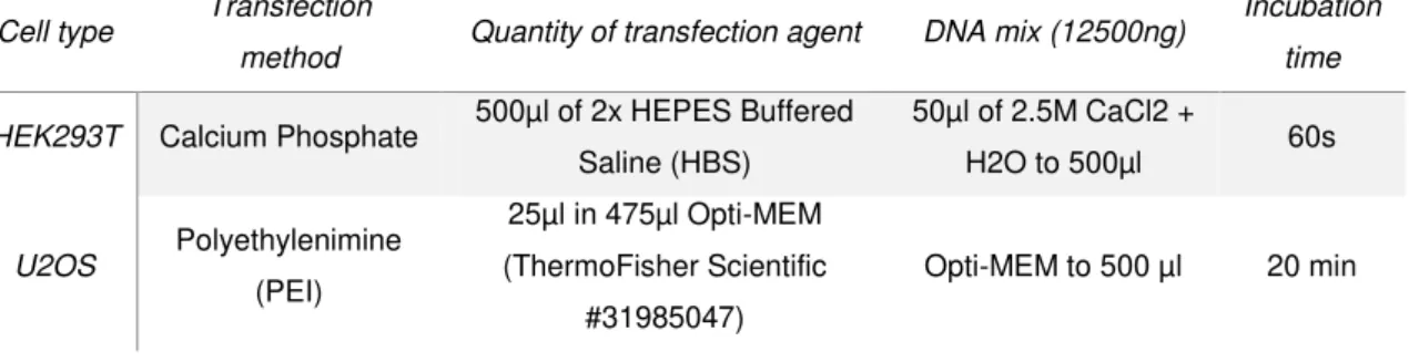
the medium was changed to 9ml DMEM+GlutaMax without 1x P/S. The transfection protocol for one
100mm dish is the following:
Table 2.03 – Description of protocols followed per transfection of one 100mm dish. The first column
corresponds to the cell type transfected, the second is the transfection method used with a detailed description in the third, fourth and fifth columns.
Cell type Transfection
method Quantity of transfection agent DNA mix (12500ng)
Incubation
time
HEK293T Calcium Phosphate 500µl of 2x HEPES Buffered
Saline (HBS)
50µl of 2.5M CaCl2 +
H2O to 500µl 60s
U2OS Polyethylenimine
(PEI)
25µl in 475µl Opti-MEM (ThermoFisher Scientific
#31985047)
Opti-MEM to 500 µl 20 min
The 2x HBS (1.5mM Di-sodium hydrogen phosphate (Merck Millipore #1.06580.1000), 50mM
HEPES-NaOH pH 7.5, 140mM Sodium chloride) was previously prepared.
For the U2OS cells transfection with PEI (Polysciences #23966-1), the DNA mixes per dish
contained 12500ng pCAG-cZNF675-GFP, 11250ng pCAG-hZNF675-GFP and 1250ng pCAG-GFP +
8750ng pCAG-EV (pBluescript was added to make the 12500ng). For the HEK293T cells transfection,
the DNA mixes per dish contained 12500ng of pCAG-cZNF675-GFP and pCAG-hZNF675-GFP. For
both cell types, the medium was changed to DMEM+GlutaMax with 10% HiFBS and 1x P/S, 6 hours