UNIVERSIDADE DE LISBOA
FACULDADE DE CIÊNCIAS
DEPARTAMENTO DE BIOLOGIA VEGETAL
Characterization of Klebsiella pneumoniae bacteriocalins
Duarte João Neves Guerreiro
Mestrado em Microbiologia Aplicada
Dissertação orientada por:
Professor Miguel Valvano (Queen’s University Belfast)
Professora Doutora Mónica Vieira Cunha (FCUL)
Characterization of Klebsiella pneumoniae bacteriocalins
Duarte João Neves Guerreiro
2016
This thesis was fully performed at Wellcome-Wolfson Institute for Experimental Medicine in
Queen’s University Belfast under the direct supervision of Professor Miguel Valvano in the
scope of the Master in Applied Microbiology of the Faculty of Sciences of the University of
Lisbon.
This thesis was also supervised by Professor Mónica Vieira Cunha as the internal
supervisor at FCUL.
I
ACKNOWLEGDMENTS
First and foremost I would like to thank Professor Miguel Valvano for allowing me to develop this thesis under his guidance at his lab and for all the patience and good will that he showed, even when the odds for “good” results were statistically close to zero.
I thank Professor Laura Hobley for she had the patient to teach me about the not-so-cooperative-Klebsiella pneumoniae and for showing many lab methodologies.
Also, I need to thank to my internal supervisor, Professor Monica Cunha, for her help and for the ideia of a B plan that also worked in the end.
To my work group, Valvano’s lab team, for the team work, laughs, awesome environment and share of knowledge during my stay.
To the future Professor Nuno Lopes for he pushed me to become more ambicious and persue even more difficult challenges. Also, for those times that we procrastinated along with other Civilizations.
And a special thanks to Ana Marques for she gave me the strength and support to continue to work on this thesis even when everything seemed about to collapse. For she, even from a distance of 1785 km or less than 3, made me laugh and kept me focus on the objective that made me go away.
II RESUMO
Desde a descoberta dos primeiros antibióticos em meados do seculo XX, que estes têm vindo a ser utilizados, frequentemente, de forma indiscriminada para combater infeções bacterianas, o que tem sido associado a um aumento do número de infeções causadas por bactérias multirresistentes (MDRs). Apesar de várias medidas terem sido já implementadas para evitar a incidência de microrganismos multirresistentes, estas têm-se mostrado ineficazes, ficando a comunidade científica responsável por encontrar novas formas de combater os mecanismos de resistência bacterianos.
Um dos mecanismos recentemente descritos envolve a actuação de proteínas denominadas lipocalinas bacterianas (BCNs, anteriormente conhecidas por YceI). Inicialmente descritas em Burkholderia cenocepacia K56-2, estas proteínas são secretadas pelas bactérias capturando antibióticos hidrofóbicos no meio extracelular, impedindo o antibiótico de atuar sobre a bactéria, aumentando assim a concentração mínima inibitória (CIM) do respetivo antibiótico. Este mecanismo não aparente ser restrito às bactérias produtoras de BCNs, exercendo actividade e proteção sobre a comunidade bacteriana envolvente.
As BCNs consistem em proteínas de baixa massa molecular, altamente conservadas entre as bactérias, apresentando uma conformação característica tridimensional (3D) de barril-β, seguida por uma hélice-α. Geralmente secretadas para o meio periplasmático, livres ou ancoradas à membrana plasmática, estas proteinas podem ainda surgir no meio extracelular ou, ainda, no citosol da bactéria. As BCNs descritas até à data ainda se encontram muito pouco caracterizadas funcionalmente, desconhecendo-se a capacidade de ligação de proteínas ortologas das descritas originalmente em alguns organismos aos antibioticos. Desta forma, esta tese focou-se no estudo do homólogo de BCN em Klebsiella pneumoniae kp52.145 designado BcnK. Esta bactéria Gram-negativa, pertencente à família das Enterobacteriaceae, é um importante agente patogénico responsável por surtos de pneumonia, entres outras infeções, em ambientes hospitalares e na comunidade. Tal como se verifica com outros agentes patogénicos, o número de estirpes de K. pneumoniae multirresistentes tem vindo a aumentar. Os principais mecanismos de resistência presentes nas estirpes clínicas desta espécie consistem em: (i) produção de β-lactamases de largo espectro de atuação, capazes de hidrolisar cefalosporinas e antibióticos do grupo dos monobactâmicos, ou (ii) produção de carbapenemases, que possuem a capacidade de hidrolisar um largo espectro de antibióticos, incluindo carbapenemes. Estes genes possuem a capacidade de se propagar horizontalmente entre estirpes da mesma ou outras espécies, tendo sido detetadas em várias regiões do mundo, constituindo assim uma ameaça para a saúde pública.
Assim, a fim de se caracterizar funcionalmente a proteína BcnK, o gene correspondente (bcnK) foi expresso por clonagem em Escherichia coli no vetor pDA-CTHis, contendo uma cauda de seis histidinas na extremidade C-terminal, dando origem a pDG1. A proteína recombinante expressa foi purificada com sucesso por cromatografia de afinidade. A verificação da expressão desta proteína foi efectuada através de SDS-PAGE e Western-blot. Apesar de se ter verificado que a expressão de BcnK conferiu um aumento da CMI, traduzido por uma diferença de
III crescimento de cerca de 65.5% OD600 a uma concentração de 1.0 µg/mL de polimixina B, em
testes de proteção com Pseudomonas aeruginosa PAO1, não foi possível repetir este ensaio devido à agregação da proteína observada durante a diálise em tampão PBS, o que impossibilitou a quantificação da proteína e a sua utilização em ensaios subsequentes. Especula-se que a agregação obEspecula-servada Especula-se deveu, possivelmente, à componente lipídica N-acil-S-sn-1,2-diacilcerilcisteína presente na exterminade N-terminal de BcnK. Desta forma, procedeu-se a nova tentativa de clonagem de bcnK no mesmo vetor, removendo-se a sequência do péptido sinal. Contudo, não se obtiveram quantidades suficientes da proteína produzida para se proceder à sua purificação. Assim, alternativamente, clonou-se bcnK no vetor induzível por IPTG, pET-28a (+), contendo caudas de histidina nas extremidades N- e C- terminal (pDG7) e, em paralelo, clonou-se no mesmo vetor, usando apenas uma cauda de histidinas na extremidade N-terminal (pDG8). A expressão do gene bcnK nestes vetores levou à produção de quantidades suficientes de proteína para purificação. No entanto, a expressão da construção genética em pDG7 conduziu a elevados níveis de agregação da proteína durante a diálise, possivelmente devido à presença das caudas de histidina. Contrariamente ao esperado, a expressão de BcnK em pDG8 não conduziu ao aumento da CMI nos ensaios de proteção. Desta forma, admitiu-se a hipótese de que a proteína recombinante produzida sem péptido sinal teria uma conformação incorreta para o exercício da sua função biológica ou de que a cauda de histidinas presente na extremidade N-terminal pudesse gerar interferência com a atividade da proteína. Um novo plasmídeo foi construído utilizando o vetor pUC19 contendo uma caude de histidinas na extremidade C-terminal. Contudo, a expressão neste plasmídeo também não produziu quantidades suficientes de proteína para purificação. Uma nova abordagem será realizada ao clonar bcnK num vector contendo um péptido sinal, secretando BcnK para o espaço perisplásmico sem a componente lipidicaN-acil-S-sn-1,2-diacilcerilcisteína, permitindo a solubilização da proteína.
De forma a investigar o papel de BcnK na resistência a antibióticos exibida por K. pneumoniae, tentou-se inativar bcnK no genoma bacteriano por duas metodologias, através de mutagénese dirigida não marcada, o que permitiria a obtanção de um mutante de eliminação isogénico, e através de mutação por inserção, por recurso aos plasmídeos construídos neste trabalho, pDG2 e pDG9, respetivamente. No entanto, não foram obtidos mutantes por quaiquer dos métodos, tendo-se obtido mutantes merodiplóides apenas na estratégia de inativação por mutagénese dirigida não marcada. Assim, em alternativa, procurou-se testar a essencialidade de BcnK por expressão de bcnK sob o controlo de um promotor induzível por ramnose. Contudo, foi necessário testar a funcionalidade deste promotor em K. pneumoniae. Para o efeito, utilizou-se o vetor pSCrhaB2-e-GFP, que tem um promotor induzível por ramnoutilizou-se fundido transcricionalmente com o gene GFP (green fluorescent protein), o qual permite a deteção da sua expressão por fluorescência. Conjugou-se em K. pneumoniae, a qual foi crescida em 0.2% e 0.5% de ramnose e 0.5% glucose, respetivamente, tendo-se registado fluorescência na presença das diferentes concentrações de ramnose e, por outro lado, a ausência de fluorescência na presença de glucose, o que sugere o correto funcionamento do promotor no hospedeiro K. pneumoniae. Um fragmento de bcnK foi então clonado no vetor suicida contendo
IV um promotor induzível por ramnose pSC200, dando origem a pDG10, sendo este posteriormente conjugado em K. pneumoniae. Os transconjugantes obtidos foram crescidos em meio M9 contendo 0.5% de ramnose (condições permissivas) ou glucose (condições não permissivas). Em ambos os meios, observou-se crescimento, sugerindo que bcnK não é um gene essencial à viabilidade de K. pneumoniae, pensando-se ainda que a expressão de bcnK na presença de glucose, ainda que em níveis basais, poderá ser suficiente para suportar a viabilidade de K. pneumoniae. Informação recolhida durante a pesquisa bibliografica sugere que BcnK intervem ao nivel da cadeia de transporte eletrónico, sendo a expressão de BcnK suprimida em anaerobiose. Desta forma, numa tentativa de se desligar a cadeia de transporte eletrónico, tentou-se mutagenizar bcnK em condições de anaerobiose.
Alternativamente, testou-se o papel de BcnK na resposta da célula ao stress oxidativo, usando para o efeito um plasmídeo com um gene repórter que codifica para uma proteína luminescente, a luciferase. Os resultados preliminares mostram o aumento da expressão de PbcnK::luxCDABE e PoxyR::luxCDABE (controlo positivo) nas condições testadas, sem grandes
diferenças aparentes. No entanto, pretende-se utilizar no futuro a construção PwaaE::luxCDABE
como controlo negativo, pois é previsto que a expressão de waaE não seja induzida por stress oxidativo.
O mutante de B. cenocepacia ΔBcnAΔBcnB foi complementado com o plasmídeo pDG1 expressando BcnK para verificação da possibilidade de recuperação do fenótipo em relação à estirpe selvagem. Verificou-se que a complementação com BcnK conduz a níveis de CMI similares aos verificados com a estirpe selvagem, no entanto o mesmo resultado foi obtido quando se introduziu apenas o vetor (controlo), sugerindo que um efeito inespecífico. No entanto, bcnK será clonado no vetor pSCrhaB2, vetor este utilizado originalmente nos estudos de complementação em B. cenocepacia.
O alinhamento das sequências aminoacídicas de BcnK e BcnA por recurso à ferramenta Clustal Omega demonstrou que os resíduos Val107 e Glu118 de BcnK parecem corresponder aos resíduos Asp82 e Asp93 de BcnA, respetivamente, que foram demonstrados como essenciais para a ligação de BcnA a antibióticos. Apesar destas evidências in silico, a intervenção dos resíduos correspondentes em BcnK ao nível da ligação com antibióticos permanece por demonstrar experimentalmente.
A análise de polimorfismos de BcnK por pesquisa de homologia nas bases de dados internacionais, restringindo a busca ao género Klebsiella, seguida pelo alinhamento das respetivas sequências aminoacídicas, demonstrou que BcnK é altamente conservada neste género (99 a 81% de homologia), contudo desconhece-se ainda a influência das diferenças registadas na função biológica exercida pela proteína a nível celular.
O estudo da conservação de genes vizinhos de bcnK por análise de sintenia foi realizado a partir da ferramenta SyntTax. Os resultados obtidos permitiram observar a conservação do locus genético deste gene entre as várias espécies testadas. Em algumas espécies, o gene de BCNs encontra-se associado ao gene que codifica para o citocromo b561 (CybB). Contudo, não foi encontrada sintenia para Enterococcus faecium, Streptococcus pyogenes e Acetobacterium
V woodii, nos quais se registou a ausência de BCNs e CybB. Estas espécies bacterianas são conhecidas por não possuírem cadeia de transporte eletrónico funcional e por serem anaeróbios restritos. Assim, considerando a globalidade dos resultados obtidos, propõe-se um modelo da funcionalidade das BCNs, sugerindo-se que estas proteínas participam no transporte ou sequestro de compostos hidrofóbicos, tais como quinonas isoprenóides ou vitamina E, especificamente no meio extracelular, por ação de BcnA, ou no meio periplásmico, por ação de BcnB ou BcnK. Estes compostos hidrofóbicos são transportados até CybB, onde são reduzidos, e posteriormente transportados para o meio extracelular e/ou periplásmico, atuando como agentes antioxidantes.
Fica, no entanto, por demonstrar experimentalmente este papel.
VI ABSTRACT
Antibiotic resistant bacteria have become one of the greatest threats to modern society, especially those bacteria that resist multiple antibiotics (referred to as multidrug resistant; MDRs). Although most well known resistance mechanisms operate within bacterial cells, recent evidence suggests extracellular mechanisms. One of such mechanisms involves bacterial lipocalins (BCNs), which are secreted proteins that capture hydrophobic antibiotics in the extracellular space. BCNs are widely distributed in bacteria. Klebsiella pneumoniae, a Gram-negative enteric bacterium, possesses a BCN ortholog. Klebsiella species cause hospital and community-acquired infections and antibiotic resistance is in part due to the spread of β-lactamases. In this thesis, I cloned, expressed and purified a K. pneumoniae kp52.145 BCN (BcnK). Recombinant BcnK proteins were employed in antibiotic protection assays using Pseudomonas aeruginosa PAO1 against polymyxin B (PmB) as a model system. Full-length recombinant BcnK was unstable and formed aggregates that complicated its quantification. However, this protein caused an increase of 65.5% in the OD600 of P. aeruginosa in the presence of sublethal amount of PmB. Other bcnK
constructs were made, but either lacked activity or could not be purified. A bcnK chromosomal deletion was attempted using protocol to proceed unmarked deletion and another one to mutate by inserting a polar gene cassette. No mutants were obtained in both cases. K. pneumoniae kp52.145 bcnK gene expression was placed under control of a rhamnose-inducible promoter, but the resulting constructs did exhibit the expected growth defect, showing the same growth phenotype irrespective of the presence of rhamnose (permissive condition) or glucose (non-permissive condition), suggesting that bcnK is not essential for K. pneumoniae viability. I also investigated the regulation of bcnK gene expression. Preliminary results suggest that bcnK expression is upregulated under different concentrations of paraquat, a compound that stimulates the production of oxygen radicals. Recombinant bcnK was used to complement a ΔbcnAΔbcnB Burkholderia cenocepacia mutant by assessing the restoration of rifampicin resistance to parental levels. However, increased resistance could only be attributed to the plasmid vector control but not to the plasmid expressing BcnK. Alignments done using amino acid sequence for BcnK and BcnA from B. cenocepacia J2315 showed two residues, Val107 and Glu118 of BcnK to correspond to Asp82 and Asp93 of BcnA, respectively. Previous reports have shown that these residues, in BcnA, are these residues were shown to be crucial for antibiotic binding. BCN genomic studies showed a highly conserved protein (99 to 81% homology) among Klebsiella species. Synteny and BLASTp results showed that in some species BCNs are associated with a cytochrome b561 (cybB) gene. However, both BCNs and cybB genes are absent in strict anaerobes. I suggest a model of BCNs cellular function that involves the hijacking of hydrophobic compounds, such as isoprenoid quinones, and their transport to the membrane where these compounds are reduced and further transported in the extracellular and/or periplasmic space acting as antioxidants.
VII LIST OF CONTENTS
ACKNOWLEGDMENTS ... I RESUMO ... II ABSTRACT ... VI LIST OF CONTENTS ... VII LIST OF TABLES ... IX LIST OF FIGURE ... X LIST OF ABBREVIATIONS ... XII
CHAPTER I – INTRODUCTION ... 1
1.1. The Antibiotic Crisis ... 1
1.2. A new resistance mechanism ... 3
1.2.1. Extracellular antibiotic hijacking ... 3
1.2.2. Bacteriocalins (BCNs) ... 4
1.3. A rising multidrug resistance bacterium ... 6
1.3.1. The Klebsiella genus ... 6
1.3.2. Klebsiella pneumoniae ... 7
1.3.2.1. Epidemiology ... 7
1.3.2.2. K. pneumoniae MDRs overview ... 8
1.4. The present work objectives ... 9
CHAPTER II – MATERIALS AND METHODS ... 9
2.1. General protocols ... 9
2.1.1. Bacterial strains and growth conditions ... 9
2.1.2. General molecular techniques ... 10
2.1.3. Biparental conjugation ... 10
2.2. K. pneumoniae BCN studies ... 11
2.2.1. Cloning, expression and purification of K. pneumoniae kp52.145 BCN ... 11
2.2.2. SGS-PAGE & Western-blot ... 12
2.2.3. P. aeruginosa polymyxin B (PmB) protection assays ... 12
2.3. K. pneumoniae mutagenesis ... 12
VIII
2.3.2. Rhamnose conditional promoter and essentiality assessment ... 13
2.4. Oxidative stress studies ... 13
2.4.1. Transcriptional fusions to luxCDABE and luminescence assays ... 13
2.5. B. cenocepacia BCN complementation and MIC assays ... 14
2.6. BcnK genomic studies ... 14
2.7. Computational methods ... 14
CHAPTER III – RESULTS AND DISCUSSION ... 15
3.1. Activity of purified BcnK in an antibiotic protection assay ... 15
3.2. The K. pneumoniae bcnK gene seems to be essential for bacterial viability ... 17
3.3. bcnK regulation under various stress conditions ... 19
3.4. B. cenocepacia BCN complementation ... 20
3.5. BCN in silico structural and genomic characterization of BCNs ... 20
3.5.1. BcnA vs BcnK secondary structure comparison ... 20
3.5.2. Klebsiella spp. BcnK genomic comparison ... 22
3.5.3. bcnK neighborhood studies ... 22
3.8. A general hypothesis for BCNs cellular function ... 22
4. Conclusions and perspectives ... 25
CHAPTER IV - BIBLIOGRAFY ... 26 CHAPTER V – APPENDIXES ... XV 5.1 – SUPPLEMENTARY TABLES ... XV 5.2 – SUPPLEMENTARY FIGURES ... XX
IX LIST OF TABLES
Table 3.1 – Rifampicin MIC determination of complemented B. cenocepacia (WT); non-complemented B. cenocepacia ΔbcnAΔbcnB (OME4); B. cenocepacia ΔbcnAΔbcnB complementation with bcnK (pDG1) and B. cenocepacia ΔbcnAΔbcnB complemented with pDG1 backbone (pDA-CTHis). Results expressed in growth (+) and no growth (-) (n=9, 3 independent experiments)………. 20 Table 3.2 – Presence/absence of BCN and CybB in the genome of the indicated species based on BLASTp results. ETC presence/absence based on bibliography. cybB association with BCN gene obtained from synteny results. Among Enterobacter species only Enterobacter sp. and
Enterobacter cloacae have cybB associated to BCN. NA = Not Applicable……… 24
SUPPLEMENTARY TABLES
Table S1– Strains, mutants and vectors used or created in this study. The following abbreviations stands for the respective antibiotic resistance: TpR: trimethoprim; TetR: tetracycline; AmpR:
ampicillin; KmR: kanamycin; EmR: erythromycin……….... XV
Table S2 – Primes generated for this study and respective restriction enzymes. Underlined bases represent the restriction site. Bases in bold represents the homology region with pGSVTp-lux vector………... XVI Table S3 – List of genomic and protein sequences obtained from NCBI used in this thesis with information referring species, strain, type of sequence (nucleotide or protein), accession number, description of the sequence and entry date……… XVI Table S4 – Composition of the solution used for protein purification protocols. (A) Lysis buffer; (B) Equilibration buffer; (C) Washing and elution buffers used for BcnK purification………….. XVII Table S5 – List of bacterial strains possessing a BCNs homologous obtained from BLASTp results, deploying BcnK as query and used to construct the cladogram from Figure S3. The species code, strains, accession number and entry date are associated with the attributed cluster (1, 2 or 3). Similar protein sequences are grouped in MULTISPECIES (more than one species) or MULTISTRAINS (more than one strain). Klebsiella pneumoniae (Kp), Klebsiella oxytoca (Ko),
X LIST OF FIGURE
Figure 1.1 – Timelines of antibiotic introduction (above) and bacterial antibiotic resistance (below). *Not in clinical use; Ampicillin hydrolyzing (AmpC); Cefotaximase (CTX-M); Imipenemase (IMP); Klebsiella pneumoniae carbapenemase (KPC); Methicillin resistant
Staphylococcus aureus (MRSA); New Delhi metallo-β-lactamase (NDM); Penicillin resistant S.
aureus (PRSA); β-lactam hydrolyzing enzymes (TEM, SHV, OXA); Vancomycin resistant
enterococci (VRE); Vancomycin resistant S. aureus (VRSA); Verona integron encoded metallo
β-lactamase (VIM)………. 2 Figure 1.2 – BCNs antibiotic resistance action model. Depending on the homologue, BCN is shown in different cellular localizations, bound to antibiotics………. 3 Figure 1.3 – BCNs subcellular localization in bacteria, showing BCNs in cytosol, anchored on inner and outer membrane, in periplasmic space. Its localization depends on the presence or absence and type of leader peptide present on BCN coding region (Image adapted from Bishop, 2000)……….... 4 Figure 1.4 – Polyisopreniod-binding protein TT1927b (protein data bank code 1wub), an example of YceI-like crystal structure with a polyisoprenoid within the β-barrel structure, obtained from Thermus thermophiles HB8. (http://www.ebi.ac.uk/pdbe/entry/pdb/1wub)... 5 Figure 1.5 - Klebsiella pneumoniae (dark violet) surrounded by its capsule (white) grown in skim milk broth and stained with Anthony’s capsule stain. (Roxana B. Hughes, University of North Texas, Denton, TX)……….... 6 Figure 1.6 - Epidemiological incidence of several types of KPCs producer types by country of origin (Munoz-Price et al. 2013)……… 8 Figure 3.1 – Verification of BcnK presence expressed from pDG1 during several steps of -Ni2+
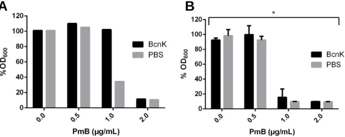
chromatography purification procedure (A) SDS-PAGE of the soluble and membrane protein fractions; purification column flow through; washes with imidazole and elution of BcnK bound to the column, performed with the respective and concentrations of imidazole and the respective dialysis in PBS; (B) Western-Blot performed on crucial steps of BcnK purification…………... 15 Figure 3.2 – In vitro protection assay of P. aeruginosa against PmB with 1.5 µM of BcnK. (A) BcnK expressed from pDG1 (n=3; from an individual assay); (B) BcnK expressed from pDG8 (n=9; from 3 independent assays). Results correspond to the end points of 24 hours incubation shown in % OD600 relative to untreated control. Significant differences (* P<0.0001) were tested
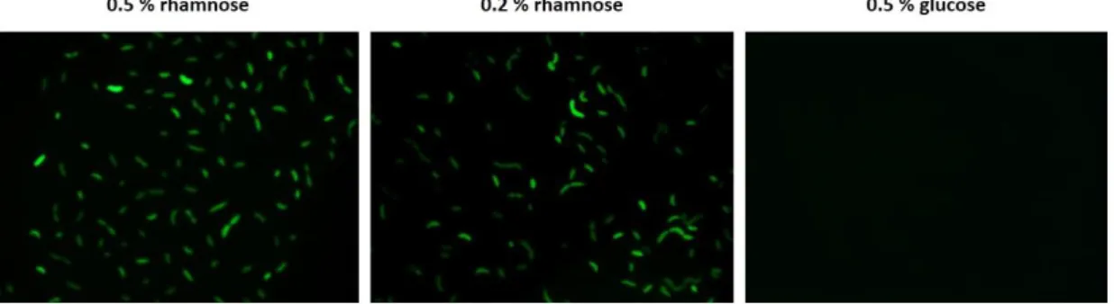
using 2way ANOVA……….. 15 Figure 3.3 – cPCR performed of K. pneumoniae trimethoprim sensitive colonies obtained after the second conjugation of bcnK of unmarked deletion using pDG2. K. pneumoniae merodiploid colonies 1 and 2 were compared against K. pneumoniae WT purified chromosome and the purified knockout plasmid pDG2……… 17 Figure 3.4 – Microscopy of K. pneumoniae exconjugants expressing eGFP under the rhamnose inducible promoter. Bacteria were grown at the indicated concentrations of rhamnose and glucose……….. 18
XI Figure 3.5 – K. pneumoniae bcnK essentiality assessment using the obtained exconjugants containing rhamnose inducible promoters (DNG9-11). K. pneumoniae wild type was complemented with pSCrhaB2 (kp52.145/pSCrhaB2) using rhamnose inducible promoter as control. XOA12 and XOA10 were used as rhamnose-dependent and rhamnose-independent control mutant (Ortega et al., 2007). Bacteria were grown on the indicated rhamnose and glucose concentrations……….. 19 Figure 3.6 – 3D prediction of BcnK (left) and BcnA (right) visualized in Jmol. In red, are shown the corresponding residues for BcnK’s Val107 and BcnA’s Asp82 and in blue the corresponding BcnK’s Glu118 and BcnA’s Asp93………. 21 Figure 3.7 – BcnK and BcnA amino acid sequence alignment obtained from Clustal Omega. In blue is BcnK’s residue, Val107, corresponding to BcnA’s Asp82 and in red is BcnK’s residue, Glu118, corresponding to BcnA’s Asp93. The following symbols stand for (*) identical residues;
(:) identical charge; (.) identical property (hydrophobic or hydrophilic)……… 21
Figure 3.8 – Example of electron transport chain during aerobic respiration from Paracoccus
denitrificans, a model organism for respiration studies, (Mandigan et al., 2010)……….. 23
Figure 3.9 – Schematic model of BCN cellular function. 1 – Extracellular BcnA or BcnK/BcnB on the periplasmic space can bind hydrophobic compounds such as quinones or vitamin E. 2 – BCNs deliver these compounds to cytochrome b561 (CybB). 3 – ETC substrates are oxidized on the cytosol by CybB that transports electron across the membrane reducing the compound present on BCNs. 4 – the reduced compound is transported in the periplasmic space acting as an antioxidant or 5 – Reduced isoprenoid quinones are transported into the inner membrane where they enter in the ETC or act as antioxidants………. 25 SUPPLEMENTARY FIGURES
Figure S1 – BcnK amino acid sequence displaying its putative signal peptide (green), predicted to be cleaved between Alanine45 and Alanine46 (SQA-AA)……… XX Figure S2 – Constructed vector pDG11 showing lacZα gene fragment and N-region of bcnK coding region with the respective primers for point mutation assays. The primers display the adenine to be added aiming to fuse both genes………. XX Figure S3 – Cladogram of Klebsiella genus amino acidic comparison. K. pneumoniae kp52.145 BcnK is shown in red letters on Cluster 1. BCNs are separated in three main cluster 1 (Blue), 2 (Red) and 3 (Green). Cladogram constructed using Clustal Omega v1.2.2 and FigTree v1.4.2. The code of each sequence in displayed on Table S4……….. XXI Figure S4 – SyntTax report obtain for BcnK. At the center, in blue and bold, stands bcnK, each arrow represents conserved a gene with an associated color. Genes with no synteny are presented in white genes………...….. XXII
XII LIST OF ABBREVIATIONS
MDRs – Multidrug resistant
MIC - Minimum inhibitory concentration AmpC – Ampicillin hydrolysing
CTX-M – Cefotaximase IMP – Imipenemase
MRSA – Methicillin resistant Staphylococcus aureus NDM – New Delhi metallo-β-lactamase
PRSA – Penicillin resistant Staphylococcus aureus VRE – Vancomycin resistant Enterococci
VRSA – Vancomycin resistant Staphylococcus aureus VIM – Verona integron encoded metallo β-lactamase BCNs – Bacteriocalins
CF – Cystic fibrosis
Bcc – Burkholderia cepacia complex PmB - Polymyxin B
RpoS – S sigma factor
PAMPs – Pathogen-associated molecular patterns ICUs – Intensive care units
ESKAPE – Enterococcus faecium, Staphylococcus aureus, Klebsiella pneumoniae, Acinetobacter baumannii, Pseudominas aeruginosa and Enterobacter species
rRNA – Ribosomal RNA LPS – Lipopolysaccharides HV – Hypervirulent
ESBLs – Extended-spectrum β-lactamases KPCs – K. pneumoniae carbapenemases MBL – Metalle-β-lactamases
CRE – Carbapenem-resistant Enterobacteriaceae MLST – Multilocus sequence typing
LB – Lysogeny broth
PCR – Polymerase chain reaction cPCR – Colony-PCR
NCBI – National Center for Biotechnology Information BLASTp – Basic Local Alignment Search Tool for proteins His-tag – Hexahistidine tag
IPTG – Isopropyl thiol-β-D-galactoside PBS – Phosphate-buffered saline TBS – Tris-buffered saline WT – Wild type
XIII CDD – Conserved Domain Database
STRING – Search Tool for the Retrieval of Interacting Genes/Proteins 3D – Tridimensional
Phyre2 – Protein Homology/analogy Recognition Engine SD – Shine-Dalgarno
IBs – Inclusion bodies
GFP – Green fluorescent protein IQ – Isoprenoid quinone
ETC – Electron-transport chain ATP – Adenosine triphosphate ROS – Reactive oxygen species FNR – Fumarate and Nitrate reductase sRNA – Small RNAs
Na2HPO4 – Disodium phosphate
NaCl – Sodium chloride NH4Cl – Ammonium chloride
CaCl2 – Calcium chloride
MgSO4 – Magnesium sulfate
Mg2+ - Magnesium
Ca2+ - Calcium
DAP – Diaminopimelic acid Ni2+ - Nickel CO2 – Carbon dioxide H2 - Hydrogen N2 – Nitrogen mM – Millimolar µM – Micromolar
µg/mL – Micrograms per milliliter mg/L – Milligrams per liter w/v – weigh per volume ˚C – Celsius s – Seconds min – Minutes h – Hour mL – Milliliter µL – Microliter
rpm – Rotations per minute g – Gravity
ksi – Kilopound per square inch GPa - GigaPascal
XIV µm - Micrometer V - Volt mV - Millivolt A – Ampere bp – Base pairs OD600 – Optical density at 600 nm nm – Nanometer %OD600 – Percentage of OD600
RLU/OD600 – Relative luminescence unites per OD600
kDa - Kilodalton % - Percent
1 CHAPTER I – INTRODUCTION
1.1. The Antibiotic Crisis
Since the discovery of penicillin, the first known antibiotic, by Alexander Fleming in 1928, mankind has relied on this and other antibacterial molecules for the treatment of bacterial infections. The antibiotics are molecules used in the treatment and prevention of infections caused by bacteria. However, the “antibiotic era” might come to an end as the majority of the clinically available antibiotics are becoming useless to treat bacterial infections (Figure 1.1) caused by multidrug resistant (MDRs) Gram-positive and Gram-negative strains (Llor & Cots, 2009). This scenario, named the “post-antibiotic era” is considered one of the greatest threats for mankind (WHO, 2014; WEF, 2015). Despite the investment and incentives to research to identify new antimicrobial molecules in the last years (Piddock, 2012), there are considerable challenges to bring them to the market resulting on a long and fastidious process (Nathan, 2004; Wright, 2015). Aggravating this situation, the interest of pharmaceutical companies to search for new molecules has decreased in the last years, resulting in only a few newly approved and reliable drugs (Spellberg, 2011).
Antibiotic molecules inhibit the growth or kill bacteria with minimum impact on the human body. They have different mechanisms of action for which they are categorized in different classes. For instance, (i) β-lactams interfere with cell wall synthesis, (ii) macrolides inhibit protein synthesis, (iii) fluoroquinolones interfere with nucleic acids synthesis, (iv) trimethoprim inhibits metabolic pathways and (v) polymyxins disrupt bacterial membrane. In some cases, bacteria are capable of overcoming this toxicity through several mechanisms which give rise to resistant strains. The antibiotic therapy represents a stressful environment for a sensitive bacterial community, and resistant subpopulations are selected and ultimately able to prevail and proliferate in the community giving rise to one or more resistant strains. In clinical microbiology, a strain is defined as resistant, susceptible or intermediate by comparing the minimum inhibitory concentration (MIC). This is the minimal antibiotic concentration at which bacterial growth is inhibited under standardized conditions in vitro (Turnidge & Paterson, 2007), with the predefined susceptibility “breakpoint” of the tested species.
In the past, the bulk of research efforts in antibiotic resistance focused on bacterial cellular functions associated with decreased susceptibility. These included (i) modification of the antibiotics target due to chromosomal mutations, making it unrecognizable to the antibiotic, (ii) production of enzymes that breakdown or modify antibiotic molecules inactivating them, (iii) extrachromosomal elements from other bacteria, such as plasmids, transposons and integrons, which can be accumulated on a single or several strains expressing proteins that inactivate the antibiotic affect, (iv) efflux pumps responsible for expelling several types of antibiotics from inside the cell (v) and decreasing membrane permeability to antibiotics reducing the access to their targets (Levy & Marshall 2004; Tenover, 2006; Alekshun et al., 2007). DNA analysis of human bacterial microbiota revealed identical genes harbored by major bacterial pathogens (Sommer et al., 2009) and similar genes responsible for the present modern antibiotic resistance were found in the environment and in samples dating back millions of years (Forsberg et al., 2012; Bhullar et
2 al., 2012) suggesting a great adaptation capacity to resist the action of antibiotics whose existence predates their use in clinical therapies (Iredell et al., 2016) (Figure 1.1).
Compounding the above-described mechanisms of resistance, other factors not related to bacteria adaptation may influence the rise of resistance. Indeed, the necessary MIC to be achieved on an infected patient, regardless of the drug dosing (pharmacokinetics), may not always be attained in the patient (Andersson & Hughes, 2014),especially at the site of infection, since antibiotics do not readily diffuse inside inflamed tissues. Also, the MIC depends on the population density at the site of infection (inoculum effect), as bacteria produce antibiotic resistance proteins that concentrate locally, destroying more antibiotic than an individual cell (Martínez et al., 2015). Thus, bacteria may be exposed to sub-lethal antibiotic concentrations, promoting the rise and selection of resistance.
To reduce the incidence of MDRs, new strategies of drug administration are being implemented in hospital facilities (Baquero et al., 2011; Spellberg et al., 2013). However other measures must also be applied to increase the efficiency of old and currently used drugs. So it falls on scientific community to research for new antimicrobial drugs or augment currently licensed antibacterial drugs (Piddock, 2012) and generate a deep understanding of biological and molecular mechanisms of antibiotic action and resistance (Wright, 2015).
Figure 1.1 – Timelines of antibiotic introduction (above) and bacterial antibiotic resistance (below). *Not in clinical use; Ampicillin hydrolyzing (AmpC); Cefotaximase (CTX-M); Imipenemase (IMP); Klebsiella pneumoniae carbapenemase (KPC); Methicillin resistant
Staphylococcus aureus (MRSA); New Delhi metallo-β-lactamase (NDM); Penicillin resistant S. aureus (PRSA); β-lactam hydrolyzing enzymes (TEM, SHV, OXA); Vancomycin resistant Enterococci (VRE); Vancomycin resistant S. aureus (VRSA); Verona integron encoded metallo β-lactamase (VIM) (Iredell et al., 2016).
3 1.2. A new resistance mechanism
1.2.1. Extracellular antibiotic hijacking
Recent work in Valvano’s laboratory has identified a group of previous described proteins known as bacterial lipocalins, herein bacteriocalins (BCNs; formerly known as YceI), which possess the ability to bind and hijack antibiotics in the extracellular space rendering them ineffective, thus augmenting the MIC value. The initial study was carried out using Burkholderia cenocepacia K56-2, an opportunistic pathogen responsible for causing chronic infection on immunocompromised patients (O’Neil et al., 1986; Poe et al., 1977), especially in those with cystic fibrosis (CF) (Isles et al., 1984). B. cenocepacia K56-2 belongs to B. cepacia complex (Bcc), a group of 20 closely related species, phenotypically similar but genetically discrete (De Smet et
al., 2015) of motile, aerobic, rod-shaped, non-spore forming Gram-negative β-Proteobacteria. In
that study, data based on the phenotypes of B. cenocepaciawild-type and two mutants defective in the production of BCN paralogues, BcnA (BCAL3311) and BcnB (BCAL3310) (El-Halfawy & Valvano, unpublished data) indicated that these proteins, and in particular BcnA, not only hijack antibiotics but their function could be inhibited by vitamin E.
BCNs are proteins synthesized and secreted into bacterial milieu leading to increased MIC but also virulence augmentation. The described mechanism of action consists on binding to antibiotics rendering them ineffective. This extracellular scavenging of antibiotics by BCNs represents a novel mechanism (Figure 1.2) of intrinsic bacterial resistance. Moreover, as some of these proteins homologous are naturally secreted into the extracellular milieu, its effect is not restricted to BCNs producing bacteria. Other more susceptible bacteria present on the milieu, of the same or different species, can benefit from this protection and thus, this mechanism of
Figure 1.2 – BCNs antibiotic resistance action model. Depending on the homologue, BCN is shown in different cellular localizations, bound to antibiotics.
4 resistance can act at the community level (El-Halfawy & Valvano, 2012; 2013). The same authors also demonstrated that the addition of purified B. cenocepacia BCNs is also capable to increasing the virulence of other bacteria species in in vivo assays with Galleria mellonella. Further, BCNs were also capable to protect Pseudomonas aeruginosa, when injected in mice, against Polymyxin B (PmB) action. It was also observed that BCNs expression is upregulated in response to antibiotic oxidative stress conditions (El-Halawy & Valvano, 2013).
1.2.2. Bacteriocalins (BCNs)
BCNs consist on a large family of low-molecular weight proteins with more than 5400 homologous distributed in both Gram-negative and positive bacteria (Bishop, 2000; Smart, 2016). Most members of this family are annotated as “conserved hypothetical proteins” referred to as YceI. Apart from a few examples of cytoplasmic BCNs from Campylobacter jejuni and Chlorobium tepidum, most of BCN coding regions possess a type-1 or type-2 signal peptide, suggesting these proteins are secreted into the extracellular space in Gram-positives and into the periplasmic space in Gram-negative bacteria,or they are covalently modified on the N-terminal with a N-acyl-S-sn-1,2-diacylglycerylcysteine moiety, enabling the protein to anchor on the inner leaflet of the outer membrane as lipoproteins (Bishop, 2000)(Figure 1.3).
The BCN’s expression in Escherichia coli was shown to depend on S sigma factor (RpoS), which activates gene transcription under several environmental stressful conditions, such as starvation, osmotic stress (Bishop, 2000) and alkaline pH (Stancik et al., 2002). Other example of induced expression occurs in the homologous Helicobacter pylori BCN, which is overexpressed under low pH contributing for bacterial survival during a stomachic infection (Sisinni et al., 2010).
Figure 1.3 – BCNs subcellular localization in bacteria, showing BCNs in cytosol, anchored on inner and outer
membrane, in periplasmic space. Its localization depends on the presence or absence and type of leader peptide present on BCN coding region (Image adapted from Bishop, 2000).
5 There are also indications that BCNs homologs from different bacteria possess different functionalities based on the capacity to bind to different molecules, such as fatty acids and amines (Sisinni et al., 2010), isoprenoid lipids (Handa et al., 2005; Vincent et al., 2010), chlorophenoxy herbicides (Benndorf et al., 2004), lipophilic antibiotics (El-Halfawy & Valvano, 2013), and fat-soluble vitamins such as α-tocopherol (vitamin E) and menaquinone (vitamin K2) (El-Halfawy & Valvano, unpublished).
The BCNs three-dimensional fold generally consists of an extended, eight to nine stranded antiparallel β-sheet, folding back on itself forming a β-barrel with one closed end, establishing a pocket inside and followed by a C-terminal α-helix (Figure 1.4). The lipophilic ligands are hosted inside the pocket of the β-barrel structure (Bishop, 2000; Handa et al., 2005). However, BCNs molecular modeling suggests two binding ways: (i) one by antibiotic polar interactions, with several amino acids residues, at the rim of the BCNs pocket, and another (ii) for more lipophilic interactions deeper into its pocket. Aromatic moieties may also play a role in molecular recognition of these proteins
(El-Halfawy & Valvano, unpublished).
Like many other components present on the bacterial envelope (BCNs are uniquely synthesized by bacteria and are cell surface-exposed), BCNs are recognized as pathogen-associated molecular patterns
(PAMPs). The
acyl-S-sn-1,2-diacylcerylcysteine modification on BCNs N-terminus stimulates an immune response through CD14 receptors recognition in macrophages plasmatic membrane surface
(Medzhitov & Janeway, 1997; Hoffmann et al., 1999), resulting in activation of immune cells leading to inflammatory responses (Brightbill et al., 1999; Aliprantis et al., 1999). BCNs stimulate innate and adaptive immune systems, as also indicated by the presence of BCNs antibodies in patients' sera (Scott et al., 2013; Yoder-Himes et al., 2010; Upritchard et al., 2008).
BCNs cellular function is still unclear. Even with all the available information, is not possible to predict the binding preference of the BCN orthologs. In this study, we focused on the Klebsiella pneumoniae BCN homolog. K. pneumoniae is an opportunistic pathogen responsible for more than 15% of Gram-negative infections in hospital intensive care units (ICUs) in the United States (Lockhart et al., 2007), and the main cause of nosocomial infections caused by the Enterobacteriaceae in hospitals (Chien Ko et al., 2002; Sanchez et al., 2013) and community centers (Carpenter, 1990). Rice (2008) referred K. pneumoniae as one of the ESKAPE bugs, along with Enterococcus faecium, Staphylococcus aureus, Acinetobacter baumannii, Pseudomonas aeruginosa and Enterobacter species. These bacteria are responsible for the largest share of nosocomial infections as well representing paradigms of pathogenesis, transmission and, most importantly, antibiotic resistance (Rice, 2008).
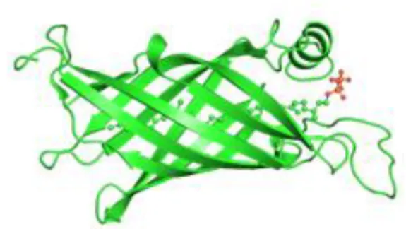
Figure 1.4 – Polyisopreniod-binding protein TT1927b (protein data bank code 1wub), an example of YceI-like crystal structure with a polyisoprenoid within the β-barrel structure, obtained from Thermus thermophiles HB8. (http://www.ebi.ac.uk/pdbe/entry/pdb/1wub).
6 1.3. A rising multidrug resistance bacterium
1.3.1. The Klebsiella genus
The Klebsiella genus includes nonmotile, capsule forming, rod shaped Gram-negative bacteria of the Enterobacteriaceae family and belongs to the γ-Proteobacteria. They were named after the German microbiologist Edwin Klebs. This bacterium is human commensal normally found among the skin and gastrointestinal tract microflora (Bagley, 1985). It can also be found in sewage, polluted waters, soil and plants. Some strains associated with plant roots have shown to fix nitrogen, converting into ammonia (Postgate, 1998) through the expression of the nitrogenase enzyme complex, encoded by the nif gene cluster (Ausubel et al., 1983; Wang et al., 2013). Consequently, those strains have the potential to be used for agriculture (Riggs et al., 2001; Temme et al., 2012). Through genetic comparison of 16S rRNA and rpoB gene sequences, Klebsiella genus can be organized in three distinct clusters. Cluster I: K. pneumoniae containing three sub-species; K. pneumoniae subsp. pneumoniae; K. pneumoniae subsp rhinoscleromatis and K. pneumoniae subsp. ozaenea.
Cluster II containing K. ornithinolytica, K. planticola, K. trevisanii and K. terrigena. And cluster III containing K. oxytoca (Drancourt et al., 2001). Klebsiella species are characteristic for producing a thick polysaccharide capsule (Figure 1.5), giving their colonies a mucoid appearance. The capsule synthesis represents a key element to Klebsiella’s virulence, as capsulated strains exhibits higher virulence than capsule defective mutants (Simoons-Smit et al., 1986). The capsules are generally composed by polysaccharides in repeating units of three to six sugars and
uronic acids giving rise to 77 varieties (Li et al., 2014; Follador et al., 2016), known as K-antigens, enabling to distinguish Klebsiella strains into serotypes (Podschun & Ullmann, 1998). Within these K-antigens varieties, strains harboring K1 and K2 capsules types exhibit hypermucoviscous phenotypes due to the elevated production of capsule and are associated with hypervirulence of Klebsiella pneumoniae strains (Follador et al., 2016), although not all of K1 and K2 strains are virulent (Kauffmann, 1949; Mizuta et al., 1983). Klebsiella polysaccharide capsule prevents bacteria from being recognized by innate immune defenses, avoiding phagocytosis and inhibiting the complement activation (Domenico et al., 1994). The polysaccharides also inhibit differentiation and functional capacity of macrophages in in vitro assays (Yokochi et al., 1979). The O-antigen, the outermost part of lipopolysaccharides (LPS), represent another virulence factor of the Klebsiella genus, as it activates the complement system, causing C3b molecule deposition far from the bacterial cell membrane, thus preventing the insertion of the complement’s
Figure 1.5 - Klebsiella pneumoniae (dark violet) surrounded by
its capsule (white) grown in skim milk broth and stained with Anthony’s capsule stain. (Roxana B. Hughes, University of North Texas, Denton, TX)
7 membrane attack complex into the bacteria cell envelope (Podshun & Ullmann, 1998). Additional virulence factors in Klebsiella include: (i) adhesins (pili, fimbriae) allowing the bacteria to bind to the host cell and catheters (Schroll et al., 2010), (ii) siderophores, which are secreted low-molecular-weight, high-affinity iron chelators scavenging iron bound to host proteins (Podshun & Ullmann, 1998), (iii) biofilm formation, which provides increased antibiotic resistance, and (iv) urease production, which, by hydrolyzing urea increases the pH in the infection locale, leading to precipitation and encrustation of organic salts and facilitating the formation of biofilms (Clegg & Murphy, 2016). However, there is a significant heterogeneity in Klebsiella strains and not every one of these factors plays the same critical role in virulent strains (Paczosa & Mecsas, 2016). The virulence factors used by Klebsiella are not focus in this work, so the reader is referred to recent reviews for additional information (Li et al., 2014; Paczosa & Mecsas, 2016).
1.3.2. Klebsiella pneumoniae 1.3.2.1. Epidemiology
K. pneumoniae is the most prevalent species of nosocomial agents that cause pneumonia, followed by Klebsiella oxitoca(Jarvis et al., 1985). Klebsiella can rapidly spread in healthcare facilities by direct contact with infected personal or other patients and cause hospital outbreaks (ECDC, 2014) and community-acquired pneumonias (Paczosa & Mecsas, 2016). Apart from pneumonia, K. pneumoniae is also frequently responsible for urinary tract infections, being the second most frequent cause after E. coli (Czaja et al., 2007; Lorente et al., 2005). K. pneumoniae primary infections on lungs and bladder can spread into the blood causing bacteremia, resulting in high rates of morbidity and mortality (Clegg & Murphy, 2016). Individuals suffering from diabetes mellitus, neuropathic bladders, chronic renal insufficiency, chronic obstructive pulmonary diseases, immunocompromised patients and alcoholics are generally considered risk groups for K. pneumoniae infections (Montgomerie, 1979; Ko et al., 2002; Clegg & Murphy, 2016), although K. pneumoniae hypervirulent (HV) strains were previously reported to cause life-threatening infections on healthy individuals (Shon & Russo, 2012; Shon et al., 2013). Liver infections are likely initiated from a breach in host defenses in the gastrointestinal tract (Paczosa & Mecsas, 2016). These type of strains are primarily responsible of causing liver abscess and are able to spread through metastasis causing further complications such as meningitis, endophthalmitis, necrotizing fasciitis and abscesses in other tissues (Siu et al., 2012) and its prevalence seems to be restricted mainly in Taiwan and Southeast Asia (Chung et al., 2007; Siu et al., 2012). Infections caused by hypervirulent strains are treatable through very aggressive therapies, although these may not prevent catastrophic disabilities on infected patients (Fang et al., 2000).
8 1.3.2.2. K. pneumoniae MDRs overview
Much like other important pathogens, K. pneumoniae MDRs strains have emerged due to several mechanisms such as efflux pumps (Ogawa et al., 2005), chromosomal mutations and plasmid-borne resistance (George et al., 1995; Hudson et al., 2014). Two major types of resistance have been commonly observed in K. pneumoniae. One is the expression of extended-spectrum β-lactamases (ESBLs) capable to hydrolyze cephalosporins and monobactams. The other mechanism which is considered more concerning is the expression of carbapenemases. These are β-lactamases with the ability to hydrolyze carbapenems. First discovered in 1996 (Yigit et al., 2001), the K. pneumoniae carbapenemases-producing strains (KPCs) render useless a broad spectrum of antibiotics like penicillins, all cephalosporins, monobactams, β-lactamases inhibitors and last-resort carbapenems (Papp-Wallace et al., 2010). The functional Ambler classification divides β-lactamases in four major classes (class A to D) based upon their amino acid sequence homology (Hall & Barlow, 2005). β-lactamases from classes A, C and D have serine in their active site, while class B possesses zinc (Hall & Barlow, 2005; Paterson, 2006). In this classification, carbapenemases are included on classes A, B and D, while ESBLs are strict to class A (Queenan & Bush, 2007). All KPCs fall into class A and are divided 16 different variants (KPC-2 to KPC-17), being KPC-2 and KPC-3 the most prevalent (Hirsch & Tam, 2010; Wang et al., 2014). Additional types of carbapenemase were also found in K. pneumoniae strains such as metallo-β-lactamases (MBL), which fall into Class B and OXA-β-lactamases, which fall into class D (Pitout et al., 2015). Irrespective of the type of carbapenemases they encode carbapenemase-producing isolates are usually termed carbapenem-resistant Enterobacteriaceae (CRE) (Paczosa & Mecsas, 2016). Only few antibiotics, such as tigecycline and polymyxins, can be used to treat
Figure 1.6 - Epidemiological incidence of several types of KPCs producer types by country of origin (Munoz-Price et al.
9 infections by CRE bacteria, but with variable degree of success (Urban et al., 2008). Therefore, these bacteria cause a high mortality rate among patients with bloodstream infections (Munoz-Price, 2013; Clegg & Murphy, 2016).
KPC genes possess a great potential to spread horizontally as some of them are encoded on transposons and often found present on several types of plasmids (Queenan & Bush, 2007). Horizontal transfer is not restricted to Klebsiella as KPC genes have been reported on other Enterobacteriaceae, including E. coli, Enterobacter species, Salmonella enterica, Proteus mirabilis, Citrobabacter freundii (Queenan & Bush, 2007; Bush et al., 1995; Villegas et al., 2005) and Pseudomonas species (Munoz-Price et al., 2013). K. pneumoniae KPC ST258, defined by multilocus sequence typing (MLST) of 7 loci, played a major role in disseminating its associated KPC enzymes worldwide (Munoz-Price et al., 2013). More detailed information on ST258 and genes concerned in MRDs dissemination can be found in recent reviews (Iredell et al., 2016; Paczosa & Mecsas., 2016).
KPC strains incidence has been steadily increasing worldwide (Munoz-Price et al., 2013; Iredell et al., 2016) (Figure 1.6), as well as ESBLs producing K. pneumoniae (Boucher et al., 2009), making urgent to find new ways to successfully treat infections caused by these MDRs bacteria.
1.4. The present work objectives
In this dissertation, I will explore the role of BCNs ortholog of K. pneumoniae kp52.145 virulent strain, to confer an antibiotic resistance mechanism as described for B. cenocepacia (El-Halfawy & Valvano, 2013). In doing so, I aim to demonstrate that BCNs provide general resistance mechanism that can be exploited by important antibiotic-resistant pathogens.Initially I will assess the protection capacity of a recombinant K. pneumoniae BCNs protein. Simultaneously, I will attempt to delete BCNs gene from K. pneumoniae chromosome. Also, the regulation properties of the same gene under oxidative stress will be evaluated. Finally, I will perform in silico studies of K. pneumoniae BCNs by comparing with other BCNs from different bacteria. Additionally, synteny studies will be performed.
Here, I will describe all the developed work done until the submission of this dissertation. Additional experimental work is still underway and new data generated will be presented during the public defense.
CHAPTER II – MATERIALS AND METHODS 2.1. General protocols
2.1.1. Bacterial strains and growth conditions
Strains and plasmids used for this thesis are listed onTable S1(see Supplementary data on CHAPTER V). Bacteria were grown at 37˚C, 180 rpm, in Difco™ LB broth. Rhamnose
conditional mutants were grown on M9 minimal medium (42 mM Na2HPO4, 8 mM NaCl, 10 mM
NH4Cl supplemented with Casamino Acids (80 µg/mL), vitamin B1 (10 µg/mL), tryptophan (40
10 required. Mueller-Hinton media cation adjusted with 10 mg/L Mg2+ and 20 mg/L Ca2+, final
concentration, was used for MIC determinations. Each medium were added with antibiotics trimethoprim (50 µg/mL for E. coli strains; 100 µg/mL for K. pneumoniae), ampicillin (100 µg/mL), tetracycline (100 µg/mL for B. cenocepacia; 30 µg/mL for E. coli; 12.5 µg/mL for K. pneumoniae), kanamycin (40 µg/mL) final concentrations when required.
2.1.2. General molecular techniques
K. pneumoniae kp52.145 genomic DNA extraction was carried out using Genomic DNA Mini kit (Invitrogen), chromosomal amplicons were generated by PCR using HotStar Hifidelity polymerase Kit (Qiagen), using 20% Q-solution final concentration and thermocycled at the following temperatures: 5 min at 95˚C; 35 cycles of 30 s at 95˚C, 30 s at 55˚C and 1 min 72˚C and final extension 10 min at 72˚C and purified by QIAquick PCR purification kit (Qiagen). Plasmid extractions were carried out using QIAprep Spin Miniprep Kit (Qiagen). DNA restriction endonuclease digestions, plasmid dephosphorylation, ligations reactions and agarose gel electrophoresis were performed according to standard techniques (Sambrook et al., 1990). Restriction enzymes and Antarctic phosphatase were purchased from New England BioLabs, T4 DNA ligase from Roche Diagnostics. DNA transformation with E. coli strains was carried out by calcium chloride method (Cohen et al., 1972). Colony-PCR (cPCR) were carried out with Taq
polymerase kit (Qiagen) with the following parameters: 3 min at 95˚C; 45 cycles of 15 s at 95˚C,
30 s at 56˚C and 1 min 72˚C; and a final extension at 72˚C for 10 min. PCR products were screened on 0.7% (w/v) agarose gels. Gene sequence of positive transformants was verified by sequencing. All designed primers and respective restriction enzymes are listed on Table S2.
2.1.3. Biparental conjugation
The various constructed vectors from all other procedures, which were transformed in E.
coli DH-5α and E. coli GT115, were extracted and transformed into diaminopimelic acid (DAP)
dependent E. coli β2163, capable to mobilize the vectors into the recipient strains, such as K. pneumoniae or B. cenocepacia, by biparental conjugation. The biparental conjugations were carried out using 2,6-Diaminopimelic acid bought from Sigma-Aldrich®.
Mobilization of the vectors was performed by growing overnight of recipient strain, with 180 rpm orbital shaking and the donor strain without shaking. Next day, both strains were pelleted by centrifugation at 4000 rpm for 20 min, washed in 5 mL of 10 mM MgSO4, pelleted again and
resuspended in 500 µL 10mM MgSO4. A mixture of 100 µL of each strain patched in LB agar
supplemented with 0.3 mM of DAP final concentration, incubated overnight at 37˚C. Next day, serial dilutions were made, until 10-4, from the recovered patched biomass and plated on LB
without the addition of DAP and with the appropriate antibiotic. The grown colonies were screened by cPCR and/or luminescence on UVP (BioSpectrum® AC Imaging System).
11 2.2. K. pneumoniae BCN studies
2.2.1. Cloning, expression and purification of K. pneumoniae kp52.145 BCN
K. pneumoniae kp52.145 yceI nucleotide sequence (GenBank ID: FO834906.1) and YceI amino acid sequence (GenBank ID: CDO15049.1) was retrieved from National Center for Biotechnology Information (NCBI) using Basic Local Alignment Search Tool for proteins (BLASTp) algorithm, deploying as query sequence the protein sequence of the putative exported protein from B. cenocepacia J2315 (GenBank: CAR53634.1) (Table S3).
To facilitate the differentiation from other BCN orthologues, such as B. cenocepacia’s BcnA and BcnB, the gene encoding K. pneumoniae’s BCN (yceI) will be referred to as bcnK and its respective encoded protein will be mentioned as BcnK.
bcnK was amplified by PCR with (primers 775 and 776) and without (873 and Q-776) its signal peptides, cloned into pDA-CTHis, which contains an N-terminal hexahistidine tag (His-tag), originating pDG1 and pDG5, respectively. To clone into pET28a (+) isopropyl thiol-β-D-galactoside (IPTG) inducible vector,bcnK was amplified without its signal peptide and encoding an N- and C-terminal His-tag (Q-873 and Q-880) giving rise to pDG7. Also, bcnK was amplified without signal peptide and coding for a N-terminal His-tag (Q-873 and Q-895), and cloned into pET28a (+). Primers (Q-907 and Q-908) for bcnK cloning into pUC19 inducible vector were design without bcnK’s signal peptide and to contain a His-tag followed by a STOP codon on bcnK’s C-terminal giving rise to pDG11. This last vector was point mutated by amplifying (961 and Q-962) to add an adenine base upstream of bcnK start codon and digested with DpnI overnight at 37˚C afterwards, transformed into E. coli and the resulting colonies were selected in ampicillin. The generated amplicons and respective vectors were digested with restriction enzymes, listed on Table S2, ligated and transformed into E. coli DH-5α. IPTG inducible vectors were transformed into E. coli BL2. Overnight cultures induction were carried out using 0.05 mM IPTG, final concentration and further incubated for 3 h at 25˚C, centrifuged at 10,000 x g for 15 min at 4˚C, washed with Tris-buffer 50 mM, pH 7.4 and pelleted again, resuspended in Lysis buffer (Table S4 A) and passed through One Shot (E1061, Constant System) at 18 ksi (124.1 GPa). The resulting lysate was centrifuged at 15,000 x g for 20 min at 4˚C for cell debris removal and to obtain the total protein fraction. Soluble and membrane protein fractions were obtained by centrifuging the total protein fraction at 30,000 x g for 45 min at 4˚C. BcnK purification was carried out by mixing the soluble fraction with coated Ni2+ Chelating Sepharose™ Fast Flow (GE Healthcare) beads
overnight at 4˚C previously treated with equilibration buffer (Table S4 B). Next day, the supernatants were collected, labelled as Flow through, and the beads were washed with increasing concentration of imidazole (50 mM and 75 mM), eluted in 400 mM (Table S4 C) and dialyzed in 4 L of phosphate-buffered saline (PBS) overnight at 4˚C. In each step an aliquot was collected for further analysis. The dialyzed protein was filter sterilized with 0.45 µm Whatman™ (SPARTAN Syringe Filter) and conserved at -80ºC until used. The presence of the protein was confirmed by SDS-PAGE and Western-Blot.
12 2.2.2. SGS-PAGE & Western-blot
The various protein fractions obtained were boiled at 100˚C for 10 min, loaded in a 16% SDS-PAGE gel, run for 75 V for 35 min and 130 V for 2 h. For SDS-PAGE staining the gel were dyed with PAGE-Blue™ (Thermo Scientific) for 2 h and distained overnight.
For Western-blot assays, the protein transfer was carried out using Biorad Trans-Blot®
Turbo™ Kit into a nitrocellulose membrane for 20 min with 1.3 A and 25 V, blocked overnight at
4˚C with Blocker™ Casein in TBS (Thermo Scientific). Next day, the membrane was washed with Tris-buffered saline (TBS), the primary antibody Anti-His Antibody (GE Healthcare Life Sciences) was added diluted 1:3000 and incubated at 4˚C for 2 h, washed three times with TBS, added the secondary antibody AlexaFluor® 680 anti-mouse IgG (Life Technologies) diluted 1:20000, incubated for 45 min, washed three times and checked at Li-cor (Odyssey®) at the wavelength of 700 nm.
2.2.3. P. aeruginosa polymyxin B (PmB) protection assays
The purified and dialyzed BcnK obtained from the expression of the various constructed vectors, were concentrated if required using Vivaspin 500 (3000 MWCO PES, Sartorius Stedim Biotech), quantified by NanoVue Plus™Spectrophotometer. Overnight cultures of P. aeruginosa PAO1 were subcultured for 2 hours, OD600 adjusted to 0.04, loaded in a 100 well honeycomb
plate along with 2; 1; 0.5; 0 µg/mL final concentration of PmB and 1.5 µM, final concentration, of purified BcnK. Controls were performed with the same antibiotic concentrations using PBS instead of purified BcnK. Each antibiotic concentration, with and without BcnK, was tested within triplicates. The OD600 was read each hour at 37ºC for 24 h on Bioscreen C
(
Oy Growth CurvesAb Ltd.).
2.3. K. pneumoniae mutagenesis
2.3.1. bcnK deletion in K. pneumoniae kp52.145
Unmarked deletion method was performed as previously described (Flannagan et al., 2008). To delete bcnK, PCR amplifications of ≈ 300 bp flanking regions bcnK were performed (Q-786 and Q-787; Q-788 and Q-789). Amplicons were digested with XbaI-XhoI and XhoI-EcoRI respectively and cloned into pGPI-SceI-2 digested with XbaI-EcoRI giving rise to pDG4. Simultaneously, it was also created a vector containing ≈ 1000 bp flanking regions bcnK (Q-810 and Q-811; Q-812 and Q-813), the amplicons were joined together using Ex Taq® DNA polymerase (TaKaRa) and ligated into pGEM®-TEasy resulting on the vector ΔyceI-pGEMT. The vector was digest with EcoRI, gel purified and ligated with pGPI-SecI-2 giving rise to pDG2. Each plasmid was introduced to wild type (WT) strain of K. pneumoniae kp52.145 by conjugation, separately, and selected with trimethoprim. The resulting conjugants were subjected to a new conjugation with pGPI-SceI-SacB and selected through tetracycline resistance.
Insertional inactivation was performed by cloning an bcnK internal fragment with 297 bp (Q-902 and Q-903) into suicide vector pGPΩTp (Flannagan et al., 2007) which contains dhfr
13 flanked with Ω-fragments, which when conjugated into K. pneumoniae, creates a polar mutation stopping bcnK transcription. The constructed vector was named pDG9.
The procedures described above were carried out in both aerobic and anaerobic conditions. For anaerobic conditions, samples were manipulated and incubated in Whitley A35
Anaerobic Workstation under an atmosphere composed by 10% CO2, 10% H2 and 80% N2.
2.3.2. Rhamnose conditional promoter and essentiality assessment
The vector pSCrhaB2-e-GFP (Cardona et al., 2006) was conjugated into K. pneumoniae kp52.145 WT. The exconjugants were selected in LB supplemented with trimethoprim. Ten colonies were picked and each grown overnight at 37ºC in M9 medium supplemented with 0.5%, 0.2% of rhamnose or 0.5% of glucose. Next day, the bacterial suspensions were deposited in slides covered with thin layer of 0.8% (w/v) agarose and observed by light and fluorescent microscopy. The images were acquired using Axioscope 10 (Carl Zeiss) microscope coupled to a camera AxioCam MRm (Zeiss) and an endow GFP bandpass emission filter set with the 470 ± 25 nm emission range, 525 ± 25 nm excitation. Images were digitally processed using ZEN 2012 (Blue Edition) Service Pack 1 image software.
The rhamnose conditional promoter construct was performed as previously described (Ortega et al., 2007) by cloning a 306 bp fragment (Q-775 and Q-904) spanning from the 5’ region of bcnK into pSC200, giving rise to pDG10. The resulting vector possesses a rhamnose-inducible PrhaB upstream of the multiple cloning site, enabling to drive the expression of bcnK in the
presence of rhamnose and repressing it in glucose. The vector’s conjugation was performed in LB supplemented with 0.5% rhamnose and selected in LB with 0.5% rhamnose and trimethoprim. The conditional mutants were grown overnight at 37ºC in M9 medium with 0.5% rhamnose, spun down and washed three times with PBS, resuspended in PBS and adjusted to an OD600 of 1 (Neat
solution). Drops (10 µL) of Neat, 10-1, 10-2, 10-3 and 10-4 dilutions were plated in M9 agar square
plates supplement with 0.5% (w/v) rhamnose or 0.5% (w/v) glucose and incubated at 37ºC. The strains XOA10 and XOA12 were used as a negative and positive control, respectively.
2.4. Oxidative stress studies
2.4.1. Transcriptional fusions to luxCDABE and luminescence assays
Promoter region of bcnK, was amplified by PCR (Q-909 and Q-910). The amplicon with ≈900 bp was digested and cloned into the digested and dephosphorylated pGSVTp-lux suicide vector and transformed into E. coli GT115. The primers for oxyR and waaE (Q-841 and Q-842; Q-852 and Q-853, respectively) were designed after ELIC method (Koskela & Frey, 2015) containing ≈25 bp homologous with the vector’s region adjacent to region where it will be cloned in pGSVTp-lux. The amplicons and digested vector were quantified in NanoVue Plus™ Spectrophotometer, mixed in a ratio of 3:1, respectively, in a final volume of 10 µL of Mili-Q water and incubated for 1 h at room temperature and transformed into E. coli GT115. The transformants colonies were selected in LB with trimethoprim, grown overnight and checked for luminescence on POLARstar® Omega for 12 h at 37ºC treated with and without paraquat 1.5 µM final