UNIVERSIDADE TÉCNICA DE LISBOA
Faculdade de Medicina Veterinária
GENETIC ANALYSIS OF THEILERIA ORIENTALIS POPULATION IN CATTLE FOLLOWING A THEILERIOSIS OUTBREAK IN VICTORIA, AUSTRALIA
NÁDIA SORAIA SEGREDO SPIRO CUFOS
DISSERTAÇÃO DE MESTRADO EM MEDICINA VETERINÁRIA
CONSTITUIÇÃO DO JÚRI ORIENTADOR
Doutora Isabel Maria Soares Pereira da Fonseca Doutor Robin Beat Gasser
de Sampaio
Doutor Robin Beat Gasser
Doutor Luís Manuel Madeira de Carvalho CO-ORIENTADOR Doutor Vítor Manuel Diogo de Oliveira Alves Doutor Luís Manuel Madeira de Carvalho
2012 LISBOA
2
UNIVERSIDADE TÉCNICA DE LISBOA
Faculdade de Medicina Veterinária
GENETIC ANALYSIS OF THEILERIA ORIENTALIS POPULATION IN CATTLE FOLLOWING A THEILERIOSIS OUTBREAK IN VICTORIA, AUSTRALIA
NÁDIA SORAIA SEGREDO SPIRO CUFOS
DISSERTAÇÃO DE MESTRADO EM MEDICINA VETERINÁRIA
CONSTITUIÇÃO DO JÚRI ORIENTADOR
Doutora Isabel Maria Soares Pereira da Fonseca Doutor Robin Beat Gasser
de Sampaio
Doutor Robin Beat Gasser
Doutor Luís Manuel Madeira de Carvalho CO-ORIENTADOR Doutor Vítor Manuel Diogo de Oliveira Alves Doutor Luís Manuel Madeira de Carvalho
ii
iii
Acknowledgments
This project took place in the Department of Parasitology, Faculty of Veterinary Science, University of Melbourne, with the aim of completing the Integrated Master's degree in Veterinary Medicine, and therefore I could not fail to thank all those, whose presence was essential for this dream to come true.
In first place I would like to thank Professor Robin B. Gasser for accepting me as a student and for giving me the opportunity to work in Australia and in such renowned laboratory. Also I would like to thank for all the knowledge passed on to me, help and concerning. To Doctor Abdul Jabbar, for the friendship, teaching, for the support while I was in Australia and after flying back to Portugal, and above all things for being there whenever needed. It was a great experience that could not be possible without their help.
I would also like to thank Professor Luis Madeira de Carvalho for sending me to Australia, for being an excellent co-supervisor, and for always being there, whenever needed, even though he as so many “children” to look after.
To John Dalziel and Andrew Hogan, Seymour Veterinary Surgery, Victoria, for all the support in this project.
To doctor Lídia Gomes, my baby sitter during my training time in the parasitology lab in the Veterinary Medicine Faculty in Lisbon.
To all my colleagues in Gasser’s lab, especially to Harshanie Abeywardena and Namitha Mohandas, for the friendship, encouragement, laughs, crazy conversations and for making my lunch and tea breaks so funny.
To my house mates, Ebrahim Bani and Sandeep Purba, for the companionship, for helping me settle down in Melbourne, and most importantly for letting me be the “alpha dog” of the house.
To my friends and colleagues who accompanied me this far and enjoyed my “good temper”, especially to FLA’s boys, to Ana Catarina and to Patricia Fernandes, who made this journey incredibly rich, adventurous and funny.
To Mafalda Ferreira, whose friendship multiplies with distance, for being my diary and my shoulder, especially when I was in Australia. For being the wonderful person and for making me better, simply by being my friend.
iv
To my family, especially to my cousin Raquel Gaspar by the constant presence from the first minute of this journey, but mostly for taking care of my mother during the time that I was off to do my internship. To all, my sincere thanks.
To my grandfather, Jaime Pires Lopes, for always expecting the best of me and for encouraging me to wonder more and more.
And lastly, I would like to thank the three women in my life. To my grandmother Efigenia Silva Rodrigues for raising me as a daughter and for loving me above all things. To my sister Carolina Cufos for making my day to day a constant challenge, and to my mother, Zilda Segredo, this great woman with the power to transform the difficult in easy, the impossible into possible, to make (my) dreams come true, for I love more than life itself and especially for letting me be part of hers. To my mother the most sincere thanks, for without her I am nothing.
v Financial support
Financial support of supervisors for the present study was provided from bodies including the Australian Research Council (ARC) the National Health and Medical Research Council (NH&MRC) and Melbourne Water Corporation [to Robin B. Gasser] and by Early Career Researcher (ECR) grant from The University of Melbourne [to Abdul Jabbar].
vi
Publications resulting from this research
Cufos, N., Jabbar, A., Carvalho, L.M., Gasser, R.B. (2012). Mutation scanning-based analysis of Theileria orientalis populations in cattle following an outbreak. Electrophoresis 33, 2036– 2040
This research was also presented in the 2012 ASP Annual Conference (July, 2012) as an oral communication entitled: Abdul Jabbar, Nadia Cufos, Robin Gasser (2012) Bovine theileriosis - an emerging problem in south-eastern states of Australia? Australian Society for Parasitology Inc. Annual Conference, 2-5 July, Country Club Tasmania, Launceston,
vii
GENETIC ANALYSIS OF THEILERIA ORIENTALIS POPULATIONS IN CATTLE FOLLOWING A THEILERIOSIS OUTBREAK IN VICTORIA, AUSTRALIA
Abstract
Bovine theileriosis is a tick-borne disease caused by one or more haemoprotozoan parasites of the genus Theileria. In the past, Theileria infection in cattle in Australia was largely asymptomatic and recognized to be associated with Theileria buffeli. However, in the recent years, outbreaks of theileriosis have occurred in beef and dairy cattle in subtropical climatic regions (New South Wales) of Australia. There is also one published report of a recent theileriosis outbreak on a beef farm near Seymour in the south-eastern state of Victoria. In order to gain an improved insight into the genetic composition of Theileria populations following this outbreak, we undertook herein an integrated PCR-coupled mutation scanning-sequencing-phylogenetic analysis of sequence variation in part of the major piroplasm surface protein (MPSP) gene within and among samples from cattle involved in the outbreak. Theileria DNA was detected in 89.4% of 94 cattle on the Seymour farm; the genetic analysis showed that the ikeda and chitose genotypes representing the Theileria orientalis complex were detected in 75% and 4.8% of 84 infected cattle, respectively, and that mixed populations of these two genotypes were found in 20.2% of infected cattle. Given unpublished reports of a significant increase in the number of outbreaks in Victoria, future investigations should focus sharply on elucidating the epidemiology of Theileria to subvert the economic impact on the cattle industry in this state. Although used here to explore genetic variation within the T. orientalis complex in Australia, a mutation scanning-based approach has broad applicability to other species of Theileria in other countries.
Keywords: Theileria orientalis / Cattle / Major piroplasm surface protein (MPSP) gene / Mutation scanning-based analysis / Phylogeny
viii
ANÁLISE GENÉTICA DE POPULAÇÕES DE THEILERIA ORIENTALIS, EM BOVINOS, APÓS UM SURTO DE THEILERIOSE EM VITORIA, AUSTRÁLIA
Resumo
A teileriose é uma doença transmitida por carraças e causada por hemoprotozoários pertencentes a uma ou mais espécies do género Theileria.
Historicamente, a infecção de gado na Austrália, com este parasita, é considerada assintomática e associada especificamente à espécie Theileria buffeli. Contudo, nos últimos anos, surtos de teileriose têm ocorrido tanto em explorações de carne como de leite em
regiões de clima subtropical da Austrália (Nova Gales do Sul).
Recentemente foi publicado um relatório, correspondente a um surto de teileriose perto de Seymour, Victoria, um estado a sudeste do país.
A fim de obter uma melhor compreensão sobre a composição genética das populações de Theileria envolvidas neste surto, foi levado a cabo um sistema de análise integrada de PCR - análise de mutações – sequenciação– filogenia, das variações existentes na sequência de parte do gene codificador da principal proteína de superfície do piroplasma (major piroplasm surface protein – MPSP), dentro e entre diferentes amostras provenientes de animais residentes na exploração envolvida no surto.
O ADN do parasita foi detectado em 89,4% de 94 bovinos testados, na exploração de Seymour e a subsequente análise genética mostrou que os genótipos Ikeda e Chitose, representativos do complexo formado por diferentes estirpes pertencentes à espécie Theileria orientalis, foram detectados em 75% e 4,8% de 84 animais infectados, respectivamente, e que populações mistas compostas por ambos os genótipos foram detectadas em 20,2% desses mesmos animais.
Dado que, relatórios não publicados apontam para um aumento significativo do número de surtos de teileriose em Victoria, futuras investigações deverão centrar-se fortemente na elucidação da epidemiologia deste parasita, a fim de avaliar o impacto económico que este poderá ter sobre a indústria bovina neste Estado.
Ademais, apesar de usados neste estudo para explorar a variação genética das populações de T. orientalis na Austrália, uma abordagem baseada na análise de mutações tem ampla aplicabilidade para outras espécies de Theileria presentes em outros países.
Palavras-chave: Theileria orientalis / bovinos / Major Piroplasm Surface Protein (MPSP) gene / mutation scanning-based analysis / filogenia
ix
TABLE
OF
CONTENTS
CHAPTER1–UNDERTAKINGATRAINEESHIPINMELBOURNEUNIVERSITY ... 1
CHAPTER2–LITERATUREREVIEW ... 7
2.1 Introduction ... 7
2.2 Literature Review aims ... 8
2.3 Beef production in Australia ... 8
2.3.1 Geography and climate in Australia ... 8
2.3.2 Beef production system ... 10
2.3.3 Beef industry ... 12
2.4 Classification and background ... 13
2.5 Life cycle and pathogenicity ... 19
2.5.1 The tick vector ... 19
2.5.2 Life cycle in the tick vector ... 20
2.5.3 Life cycle in the mammalian host ... 20
2.5.4 Pathogenicity ... 21
2.6 Epidemiology ... 22
2.7 Treatment and control ... 24
2.7.1 Chemotherapy ... 24
2.7.2 Tick control ... 25
2.7.3 Immunization ... 25
2.8 Detection of T. orientalis parasites ... 26
2.8.1 Parasitological methods ... 26
2.8.2 Serological methods... 27
2.8.3 Polymerase Chain reaction (PCR) – based methods ... 28
2.8.3.1 Target region ... 30
2.8.3.2 Restriction Fragment Length Polymorphism (RFLP) ... 31
2.8.3.3 Reverse Line Blot hybridization (RLB) ... 31
2.8.3.4 Single-Strand Conformation Polymorphism (SSCP) ... 31
2.8.3.5 DNA Sequencing ... 32
2.9 Final remarks ... 32
CHAPTER3–GENETICANALYSISOFTHEILERIAORIENTALISPOPULATIONSIN CATTLEFOLLOWINGATHEILERIOSISOUTBREAKINVICTORIA,AUSTRALIA .. 34
3.1 Research aims ... 34
3.2 Characterization of the “problem” farm ... 34
x
3.4 Collection of blood samples ... 38
3.5 Enzymatic amplification ... 38
3.6 Single-strand conformation polymorphism analysis ... 41
3.7 Sequencing ... 42
3.8 Phylogenetic analysis... 43
CHAPTER4–RESULTSANDGENERALDISCUSSION ... 44
4.1 Amplification and mutation scanning of blood samples ... 44
4.2 Phylogenetic analysis... 47
4.3 General discussion ... 49
CHPATER5-CONCLUSION ... 52
xi
FIGURES
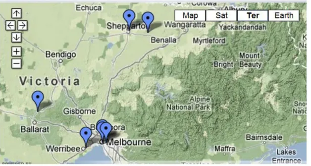
Figure 1 – Map with different locations of different campuses of university of Melbourne ... 2
Figure 2 – A view from University of Melbourne, Parkville campus ... 2
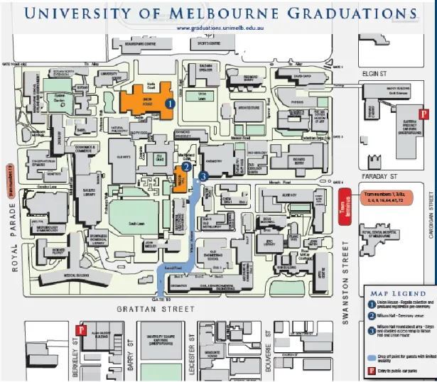
Figure 3 – Melbourne University Parkville campus map ... 3
Figure 4 – Veterinary Science Faculty, Parkville campus building ... 4
Figure 5 – Gasser’s Laboratory structures ... 5
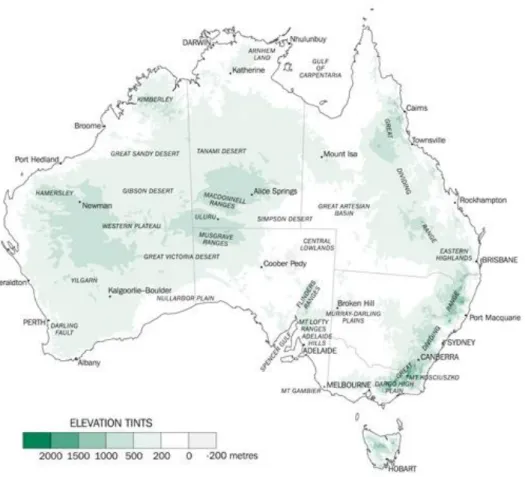
Figure 6 – Australian map, showing the different elevation landscape... 9
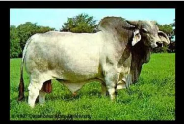
Figure 7 – Brahman breed ... 10
Figure 8 – Temperate breeds ... 11
Figure 9 – Beef cattle distribution in Australian territory in 2001 ... 12
Figure 10 – Theileria genus classification ... 13
Figure 11 – Most important Theileria species distribution ... 14
Figure 12 – Life cycle of Theileria species ... 21
Figure 13 – Australian map showing T. orientalis record in different states. ... 23
Figure 14 – Piroplasms in the red blood cells from cattle ... 27
Figure 15 – Map of the Victoria state showing the localization of the farm suffering with the initial outbreak of theileriosis ... 35
Figure 16 – Geldoc system ... 41
Figure 17 – SSCP rig apparatus ... 42
Figure 18 – Representative agarose gels of the MPSP amplicons ... 44
Figure 19 – Representative SSCP gel displaying profiles P1, P2 and P3 ... 46
Figure 20 – Relationships of partial MPSP nucleotide sequences from Theileria from cattle in Victoria ... 48
TABLES
Table 1 – Most important Theileria species affecting cattle, their vector, distribution and pathogenicity ………...14Table 2 – Chronology of different studies which attempted to classify members of the Theileria sergenti/buffeli/orientalis complex ……… 17
Table 3 – Detailed chronology of the Theileriosis outbreak ………...…... 36
Table 4 – Representative table of blood samples collection from different farms... 38
xii
A
BBREVIATIONSbp – base pair
BVD – Bovine Viral Disease C. – Clostridium
DNA – deoxyribonucleic acid ECF – East Coast Fever
ELISA – Enzyme Linked Immunosorbent Assay H. - Haemaphysalis
IFAt – Indirect Fluorescent Antibody test ITS – Internal Transcribed Spacers mpsp – major piroplasm surface protein NSW – New South Wales
PCR – polymerase chain reaction QLD – Queensland
RBC’s – red blood cells
RFLP – Restriction Fragment Length Polymorphism RLB – Reverse Line Blot
RNA – ribonucleic acid spp. – species
SSCP – Single-Strand Conformation Polymorphism T. – Theileria
1
CHAPTER 1 – UNDERTAKING A TRAINEESHIP IN MELBOURNE UNIVERSITY
The present Thesis is based on the traineeship, implemented in the curriculum of the
Integrated Master in Veterinary Medicine of the Faculty of Veterinary Medicine, Technical University of Lisbon, (FVM - UTL), which aims the practical application of the acquired knowledge, during the first 5 years of study, in order to prepare the future veterinarian for the labor market, as well as, enable the same with some orientation to the area that most appeals to him/her, since this is a profession with many different applications in many different fields. For this purpose, there was a chance of a nine months traineeship, from 15th July 2011 to 15th April 2012, at the University of Melbourne in the state of Victoria, Australia.
The University of Melbourne was founded in 1854, the second in the country, by four talented professors from four different schools: WP Wilson (Mathematics), Henry E Rowe (Classics and Ancient History), Frederick McCoy (Natural Sciences) and WE Hearn (Modern History, Literature and Political Economy) (The University of Melbourne, 2012). Currently comprises about 16 schools distributed in seven different campuses (Figure 1), being the main one located in Parkville (Figure 2, 3). It is a prestigious institution, being for a few years in the top 20 of world's best universities, and recognized as a pioneer in research, integrating about 40,000 students from over 120 countries (The University of Melbourne, 2012).
The Faculty of Veterinary Science, it was the first Veterinary school in the country and with more than 100 years of history, has as its mission the training of world-class professionals in veterinary field, and is divided into two campuses: Parkville, in the northern-center of the Melbourne city (Figure 4) and Werribee, in the suburbs (The University of Melbourne, Faculty of Veterinary Science 2012).
2
Figure 1 – Map with different locations of different campuses of university of Melbourne (adapted from The University of Melbourne, http://brand.unimelb.edu.au/global/contact-maps.html)
3
Figure 3 – Melbourne University Parkville campus map (adapted from The University of Melbourne,
http://graduation.unimelb.edu.au/__data/assets/pdf_file/0020/530606/Online_map_-_reduced.pdf
4
Figure 4 – Veterinary Science Faculty, Parkville campus building
Beyond a focus on training new veterinarian practioners, it also has a large responsibility in different research areas, such as animal production, performance and welfare, infectious diseases, public health, biosecurity, cell biology and morphology, animal biotechnology and small animal medicine.
I had the pleasure to join the Parasite Genetics and Genomics laboratory (Figure 5), directed by the world renowned Professor Robin Beat Gasser, which main goals are “the study of parasites with socio-economic impact in order to reveal their molecular biology to enable further development of new and improved diagnostic techniques, control methods and treatment” (The University of Melbourne, Faculty of Veterinary Science, 2012). The same is composed of a diverse team which includes lecturers, post-doctoral scientists, research assistants and post graduate students, all working together to obtain better results. From the different projects undertaken by this laboratory, stand out transcription projects of different Platyhelminthes, such as Fasciola hepatica, and Haemonchus contortus; Nematode diagnosis in small ruminants; Theileria diagnosis and support on control; and projects in Cryptosporidium spp. and Giardia spp. in conjunction with Melbourne Water Corporation.
5 Figure 5 – Gasser’s Laboratory structures (original)
As part of the internship, based on the investigation of a recent outbreak of theileriosis in the state of Victoria, we were able to learn new techniques, engage in the daily routine of laboratory work, as well as develop and refine a critical spirit, which opened doors to research world, an unknown field until now, and from which much knowledge and understanding was gathered.
Obviously, at the arrival to the laboratory we were introduced to the entire team to understand the work in which each member was involved. After being explained the specific project for our master’s, three weeks were given to get acquainted with the theme of the project and to remember everything that had been learned and that somehow it would be useful in life in the laboratory and to understand thoroughly all techniques (why, how and when to use/apply them).
The second stage of my internship consisted in the laboratory work, during which I was allowed to learn and apply the following techniques:
1. Extraction and purification of DNA for further analysis - learn different techniques for the extraction of genomic material from tissue (ticks and round worms) and blood (cattle), with different kits and protocols, as well as optimization of the method in different situations;
6
2. Preparation, development and identification of PCR products - learning how to prepare samples and how to assemble the PCR tubes, as well as, handling the different machinery for this purpose and optimization of the reaction to assemble different situations (optimal concentration of reagents, and various stages of the reaction). At this stage I got my first “contact” with bioinformatic tools for primer design;
3. Scanning and analysis of mutations - learning techniques that allow the analysis of mutations within and among different samples, such as SSCP and RFLP;
4. Cloning techniques – preparation of PCR products for further cloning techniques with pGEM T easy vector® (with or without the white/blue selection method) and further analysis of the results (extraction of DNA from clones for posterior sequencing analysis);
5. Sequencing of samples - learning sample preparation for automated sequencing on an independent laboratory, and analysis of results with different bioinformatics tools; 6. Phylogenetic analysis - using different bioinformatic tools that allow an accurate
analysis of the results and drawing of phylogenetic relationships among different organisms / populations (Mega5®, Mrbayes®, clustal X®);
Besides all the work done in the laboratory, I was also able to attend different conferences and be aware of other surveys conducted in different laboratories and departments of the college, as well as a short guide tour to the farm affected with the Theileriosis outbreak, in order to collect some information from the responsible veterinarian.
It was surely a unique experience that could not otherwise be possible if not within a traineeship.
7 CHAPTER 2 – LITERATURE REVIEW
2.1 Introduction
Infectious diseases are of major importance as they can cause substantial damage to humans, animals and plants. Infectious agents include different organisms such as prions, viruses, bacteria, fungi and also protozoan and metazoan parasites.
The metazoan parasites are divided into arthropods (tick, mites, flies and fleas – characterized by their exoskeleton, joined limbs and segmented body) and worms. The latter group includes round worms (Nematoda) and flat worms (Platyhelminthes: Trematoda and Cestoda) (Taylor, Coop & Wall, 2007)
Protozoan parasites are unicellular organisms and comprise 4 main groups, amoeba, flagellates, ciliates and apicomplexans. The Apicomplexa are characterized by the presence of an apical complex, as well by schizogony and merogony stages in their life cycle and include coccidia, haemosporidia and piroplasmidia such as Theileria (T.) species (Taylor et al., 2007) Theileria parasites are responsible for causing clinical and subclinical infection, known as theileriosis, an important disease of (mainly) livestock throughout the world and characterized by lymphocytic proliferation and anaemia that can lead to death or, in less severe cases, to a decreased productivity. The most important species are T. parva, the causative agent of African East Coast Fever (ECF), and T. annulata responsible for tropical theileriosis affecting animals in the Mediterranean basin, middle East and Indo-china (Radostits, Done & Blood, 2007).
In Australia, theileriosis is caused by the T. orientalis/buffeli/sergenti group whose classification is still controversial. They are sometimes thought to be benign (in terms of disease), but in Japan, Korea and some parts of China members of this group have a major economic impact in cattle (Kakuda et al., 1998). Parasites belonging to this group are now having an increased impact in Australia. In 2010, Izzo, Poe, Horadagoda, De Vos & House reported a small number of haemolytic disease outbreaks in New South Wales (NSW) associated with T. orientalis/buffeli/sergenti group infection, mainly in peri parturient cattle moved from coastal areas to inland farms. In early 2011, there was a report of a similar outbreak in Victoria (VIC) (Islam, Jabbar, Campbell, Cantacessi & Gasser, 2011) a state in which theileriosis disease was never recorded before. Following this report, there has been a series of new outbreaks in this particular state of Australia (Dr. Michael Jeffers, Department of Primary Industries [DPI], Victoria, personal communication; January 2012).
8
2.2 Literature Review aims
The present thesis investigates, the genetic composition of Theileria populations infecting cattle on one particular farm suffering from the initial outbreak in Victoria. In order to provide a foundation for the study it was essential to first review and critically appraise the literature, establish the current state of knowledge and then to identify all the gaps in the knowledge and understanding of this disease globally, in Australia and in Victoria. Therefore, the aims of this chapter were to explore some areas, including the Australian beef production system, taxonomy and classification of the parasite, as well as, life cycle, pathogenesis, epidemiology and aspects of treatment, control and diagnosis and then formulate the specific aims of the thesis.
2.3 Beef production in Australia
Australia is one of the largest exporters of beef in the world (Australian Red Meat, 2012), and exports more than 60% of its entire herd, mainly for the United States and Japan (The Australian beef industry – the basics at www.pwc.com.au).
To this fact contributes not only its extensive territory, as well as the fact that most of the restrictive diseases to cattle production are absent in Australian farms, which puts their meat on top of the safest, and therefore most sought after (Australian Red Meat, 2012).
Listed below are some factors that contribute to this success.
2.3.1 Geography and climate in Australia
The Australian territory has approximately 7.7 million km2 and is divided into seven states: Northern Territory, Queensland, New South Wales, Victoria, Western Australia, South Australia and Tasmania to which are added up to 12,000 islands. Its location in coordinates is: latitude, between 10°41´ south (Queensland) and 43°38´ south (Tasmania) and longitude between 113°09´ east (Western Australia) and 153°38´ east (New South Wales).
It is the second flattest and driest country-continent, right after Antarctica, and many of its landscape are dated to millions of years. It is possible to observe their different elevations in the map in Figure 6 and one should draw attention that mountainous areas, i.e. landscape above 2000m, are concentrated in a small region of NSW (The Australian Bureau of Statistics, 2012).
9
Figure 6 – Australian map, showing the different elevation landscape (adapted from The Australian Bureau of Statistics, http://www.abs.gov.au)
It is without doubt one of the countries with the greatest variety of climates, that creak from the tropical north and temperate south, passing through the more arid climates in the most interior areas of continental plate. For the same reason fluctuations in temperature and rainfall may be large. By way of example, at north, temperatures are more or less constant throughout the year, having rainfall season from November to April and a drier season during the remaining six months of the year. Unlike in most southern states, where the variations are more pronounced and therefore the four seasons of the year are well defined (The Australian Bureau of Statistics, 2012).
10
2.3.2 Beef production system
The cattle settling began on the arrival of the first Europeans in 1788 that brought with them six head of cattle purchased in South Africa. The herds have grown ever since, and in 2005 Australia had 2% of world’s bovine population and ranked in 10th place of the largest meat exporters (The Australian Bureau of Statistics, 2012).
Nowadays, the herd consists mainly of two types of breeds:
- Bos indicus or tropical breeds (Brahman) (Figure 7), imported from India in 1930s (The Australian Bureau of Statistics, 2012) that comprises mostly cattle from the north as they are adapted to extreme temperature conditions in that region (Australian Red Meat, 2012);
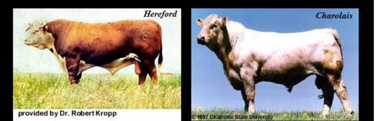
- Bos taurus or temperate breeds originating from Great Britain (Hereford and Shorthorn) and the rest of Europe (Limousine, Charolais etc.) (Figure 8). These make up the herds from further southern areas, in rainfall regions. The crossbreeds from these animals are used mainly for later finishing as they give larger animals for target markets (Australian red meat, 2012).
11
Figure 8 – Temperate breeds (adapted from http://www.ansi.okstate.edu/breeds/cattle)
As breeds adapt to different Australian climates, also production systems vary, depending on the region where we are.
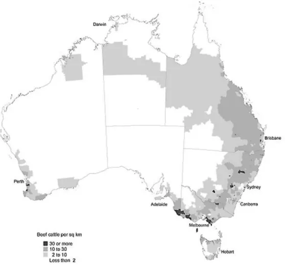
In northern states such as Northern Territory, northern Queensland, and Western Australia, there is a more extensive system of production, with grazing cattle, and low population densities. Whereas in the southern states the holdings are maintained in an intensive system of small farms where animals are fed with introduced pastures and fodder crops (Figure 9) (Australian Red Meat, 2012).
12
Figure 9 – Beef cattle distribution in Australian territory in 2001 (adapted from The Australian Bureau of Statistics, http://www.abs.gov.au)
2.3.3 Beef industry
The Australian beef industry constitutes the majority of agricultural industry. In 2005 from 133,000 farming establishments, 33250 were destined to growing cattle for beef industry, which represents almost 27% of all agricultural house holdings (The Australian Bureau of Statistics, 2012).
Most of the beef produced in Northern regions, extensively, is sold to United States, as livestock for other countries, or goes to feed-lot properties, while the cattle produced in south is destined to Japanese and Australian market.
The feedlot system was introduced in Australia in late 1950’s, but only during the 1980’s it started developing and gain more supporters (Australian Red Meat, 2012). Usually it receives grazing cattle for late finishing on highly energy grain diets for 30 to 300 days depending on the target market (fewer days for national consumption and more days for Japanese and American market).
13
2.4 Classification and background
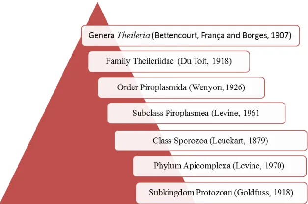
Theileriosis is an important tick-borne disease caused by heteroxen haemoparasites of the family Theileriidae (order Piroplasmidia, class Sporozoa and phylum Apicomplexa) (Figure 10).
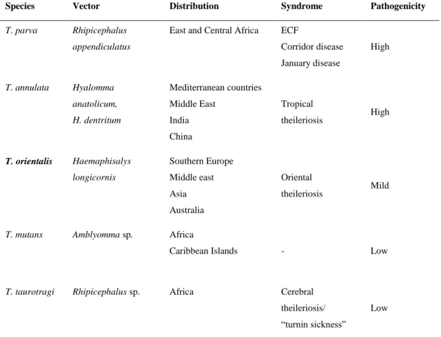
Nowadays, five species are known to infect cattle worldwide, causing different syndromes, which are characterized from severe clinical disease to mild or non-pathogenic infection (Figure 11 and Table 1) (Radostits et al., 2007).
Figure 10 - Theileria genus classification (adapted from Navarrete et al., in Parasitologia Veterinaria, 2002)
14
Figure 11 – Most important Theileria species distribution (adapted from Welcome Trust Project for Tropical theileriosis, 2007, at http://www.theileria.org)
Table 1 – Most important Theileria species affecting cattle, their vector, distribution and pathogenicity (based on Radostits et al. 2007)
Species Vector Distribution Syndrome Pathogenicity
T. parva Rhipicephalus appendiculatus
East and Central Africa ECF
Corridor disease January disease High T. annulata Hyalomma anatolicum, H. dentritum Mediterranean countries Middle East India China Tropical theileriosis High T. orientalis Haemaphisalys longicornis Southern Europe Middle east Asia Australia Oriental theileriosis Mild
T. mutans Amblyomma sp. Africa
Caribbean Islands - Low
T. taurotragi Rhipicephalus sp. Africa Cerebral theileriosis/ “turnin sickness”
15
Theileria organisms parasite mononuclear phagocyte system and the species considered with most impact on livestock production, also known as lymphoproliferative species (Dobbelaere & McKeever, 2002), are T. parva, responsible for different syndromes in East and central Africa, and T. annulata which infects cattle, in the Mediterranean basin, Middle East and Indo-China. These parasites are highly pathogenic due to their schizonts pathogenicity, which induce, along with their own multiplication, uncontrolled proliferation of their host cells. In other words, each infected lymphoid cell that divides originate two daughter-cells, both infected, resulting in the exponential multiplication of the parasite in a short period of time. Although these species have this point in common, T. parva and T. annulata differ in some aspects.
T. parva affects cattle from countries in Africa and is transmitted by Rhipicephalus spp. Its main reservoir is the African buffalo, and the parasite is responsible for three different syndromes: East coast fever, transmitted by ticks feeding on infected cattle and transferring it to susceptible animals. In enzootic areas the mortality rate may exceed 90% (Mehlhorn, 2001), and recovered animals may have their productivity affected by several months. It was eradicated from southern Africa by restrictive control and slaughter measures; Corridor disease, transmitted from infected buffalo to cattle, is similar to the former syndrome, however the course of the disease is faster, and death occurs within days. Recovered animals stay infected for life; January disease occurs in Zimbabwe and appeared after eradication of ECF. It is closely related to activity peak of the tick vector and therefore its name. As for the first syndrome its transmission is from cattle to cattle, and symptomatology and epidemiology are the same.
T. annulata is the causative agent of tropical theileriosis, has a wider distribution than T. parva and is transmitted by ticks of the genus Hyalomma. Navarrete, Serrano & Reina et al. (2002), estimates that more than 250 million animals are infected by this agent, which parasites both, cattle and buffalo. Although the mortality rate is slightly lower than in ECF (around 70% in enzootic areas), it represents a huge problem, mainly for small producers, since its control is costly.
Despite of the fact that initial symptoms of T. annulata infection resemble the ones caused by T. parva, such as general lymphadenopathy and fever, in more advanced stages, animals show up with symptoms of pronounced haemolytic anaemia and jaundice, most probably caused by their intraerythrocitic stage.
More benign species, the non-lymphoproliferative group is composed by T. orientalis, T. mutans and T. taurotragi, whose impact is not that important due to their low pathogenicity. The mortality rate is estimated to be up to 30% in introduced and exotic cattle (Irwin, 2012).
16
Moreover, Theileria spp. can produce nervous symptomatology recognized as turning sickness disease, when schizonts build up in capillaries of the brain. The cattle present circling movements, incoordination of the hind legs, loss of orientation, ataxia, opisthotonus and paralysis (Capucchio et al., 2011).
In Australia theileriosis is thought to have entered the country by introduced goods from Japan infested with the tick vector (Riek, 1982). It is caused by members of the Theileria orientalis/buffeli/sergenti group, which presently comprises eight different genotypes based on the major piroplasm surface protein (MPSP) gene: 1- chitose, 2- ikeda, 3- buffeli and genotypes 4 to 8, (Ota et al., 2009; Kamau et al., 2011). The classification of Theileria spp. is based mainly on morphology, presence of schizonts, geographical origin, pathogenicity, mammalian and tick preferences, and/or serology. As these criteria have usually proved unreliable, in recent years, molecular markers have been used to provide a better insight into their classification (Gubbels et al., 2000). Despite this fact the nomenclature/classification of the T. orientalis/buffeli/sergenti group is not yet clear, with different studies adopting different names: T. sergenti in Japan, T. buffeli in Australia and T. orientalis elsewhere (Fujisaki, 1992).
Some researchers believe that all genotypes of the group belong to a single species, while others believe that the group comprises different, but closely related species (Kawazu, Sugimoto, Kamio & Fujisaki, 1992; Fujisaki, Kawazu & Kamio, 1994; Stewart, Uilenberg & deVos, 1996). For a better understanding of the evolution of T. orientalis/buffeli/sergenti classification, a summary is given below (Table 2).
17
Table 2 – Chronology of different studies which attempted to classify members of the Theileria sergenti/buffeli/orientalis complex
Year Researcher Achievement
1906 Theiler Described T. mutans for the first time;
1908 Schein Isolated a theileria from the water buffalo, in Vietnam, and called it
T.buffeli;
1910 Seddon Recorded a Theileria parasite in Australian cattle and name it T. mutans; 1912 Neveu-Lemaire Isolated a Theileria parasite from Asian water buffalo in Southeast Asia
and named it T. buffeli;
1923 Schein Failed transmission of the parasite from cattle to buffalo and vice versa;
1926 Wenyon Named sheep’s Theileria as T. sergenti;
1930 Yakinnoff Dekhtereff
Described a parasite causing clinical theileriosis in Eastern Siberia and named it T. sergenti;
1931 Yakinnoff Soudatschenkoff
Described a similar parasite as the previous in the same area and named it
Theileria orientalis;
1966 Rogers Reported 3 fatal cases of infection with Theileria mutans in Queensland; 1976 Callow Showed that T. buffeli can be transmitted from buffalo to cattle;
1977 Uilenberg Stated that Australian T. mutans is in fact T. sergenti Yakimoff and Dekhtereff 1930;
1981 Morel Uilenberg
Suggested that Australian Theileria is either T. orientalis (Yakimoff and Dekhtereff 1930) or T. buffeli (Neveu-Lemaire 1912);
1984 Callow Based on Theileria transmission from buffalo to cattle state that it should be named T. buffeli;
1985 Uilenberg Concluded, based on serological and morphological characteristics, that strains from Japan, Australia, Iran, Britain and USA were identical, with a more pathogenic strain from Korea, and named them all T. orientalis; 1985 Shastri Failed transmission of T. buffeli from buffalo to cattle;
1987 Stewart Designated Australian strain as T. bufelli (Callow 1984) after demonstrating the non-transmissibility with Haemophysalis longicornis ticks and suggested H. brancofti and H. humerosa as the main vectors; 1991 Kawazu After transmission studies and protein analysis of piroplasm suggested two
groups to classify the benign Theileria from Japan (T. sergenti), Australia and Britain (T. bufelli/orientalis);
1991 Sugimoto Analysed proteins from different isolates in a 2D gel and concluded that T.
buffeli and T. orientalis were identical, with T. sergenti showing a
completely different spot-pattern suggesting that this parasite may belong to a different species;
1992 Fujisaki After a review of transmission, phenotypic and genomic experiments, suggested that Japanese T. sergenti might be a different species and the Australian, T. buffeli, and British, T. orientalis, might belong to one and the same species;
18
Table 2 (cont.) – Chronology of different studies which attempted to classify members of the Theileria sergenti/buffeli/orientalis complex.
1992 Kawazu They compared the nucleotide sequence of T. sergenti and T. buffeli for the 33/34kDa proteins and showed high similarity, but when compared by RFLP they showed distinct patterns; Built cDNA libraries for T. buffeli and
T. sergenti and screened them with rabbit anti sera. The proteins 33/34kDa
showed species-common and species –specific epitopes;
1994 Fujisaki Considered T. bufelli and T. orientalis as the same species and T. sergenti as a separated one;
1994 Kubota Found two major allelic forms for T. Sergenti: ikeda and chitose types; 1995 Kubota They found a new buffeli allele in Japanese isolate and named it B2, being
the B1 the buffeli type from Australian isolates;
1996 Kubota After compared chitose types sequences from Australian and Japanese isolates concluded that they were 98.5% similar;
1996 Stewart After a review of different characteristics concluded that there was no valid reason for distinguish more than one species and that they all should be name T. buffeli;
1998 Kim Found parasites with three different MPSP sequences. Demonstrated that
among the same type the differences were very small regardless the geographic origin, but different types are very different even when they come from the same isolate;
1999 Kawasu Based on phylogenetic analysis and vector tick subgenus experiments, proposed the name T. buffeli to characterize the parasites of Asian buffalo,
T. orientalis to benign theilerias of cattle. Divided the latter into two
subspecies: T. orientalis orientalis and T. orientalis sergenti;
1998 Chae After analysis of the variable region V4 of SSU rRNA, they described 7 genotypes A, B, C, D, E, H and Warwick for cattle;
2000 Gubbels After a phylogenetic analysis of SSU rRNA and MPSP genes concluded that all known T. bufelli isolates must been originated from the buffalo stock and suggested that all should be in the same species, T. buffeli, until further studies suggest otherwise subspecies;
2004 Kim Found a seventh type and divided the species in two sister groups: group 1(type 2 + type 7) and group 2 (type 3 + type 5);
2006 Kim Re-grouped the species in (type 2+ type 7) (type 3 + type 4);
2009 Jeong Classified the parasites into 8 different types and the Brisbane isolate (unclassified) divided into 2 sister groups: (type 2 + type 7) (type 3 + Brisbane);
2010 Liu Reclassified T. orientalis MPSP type 6 as T. sinensis;
2010 Khukhuu Found a ninth genotype: N3, and divided into two groups: Ikeda (type 2 + type 7) and Chitose ( type 1, 3 ,4 ,5 8 N3)
19
As shown in the table above, there is still much to be done to reach a consensus regarding the name and number of species that constitute this group, as well as in the knowledge of the disease.
Therefore, in order to simplify the reading, from this point on, the group will be referred to as T. orientalis, as the name T. sergenti was already used for a sheep’s parasite and it is not yet proved that buffalo can be infected with this Theileria spp., therefore the name T. buffeli is still controversial (Fujisaki, 1992).
2.5 Life cycle and pathogenicity
The life cycle of T. orientalis is similar to those of other Theileria spp. (Figure 12). The parasite undergoes gametogony (sexual reproduction) and sporogony (asexual reproduction) in the tick vector and schizogony and merogny (asexual reproduction) in the mammalian host (Navarrete et al., 2002).
2.5.1 The tick vector
Theileria parasites are transmitted by arachnids belonging to the SubOrder Ixodides, commonly known as ticks. It is divided into two distinct families, the Argasidae family and the Ixodidae family also called as hard ticks.
The latter is extremely important, both in veterinary practice as in public health not only due to the direct damage caused by the tick, through the ingestion of blood, secretion of toxins (as in the tick paralysis disease) and induction of an exacerbated inflammatory reaction, but also due to the fact that different species act as vectors of different diseases caused by viruses, bacteria, and parasites (piroplasms and ricketsias).
They owe their designation of hard ticks, due to the presence of a dorsal shield, complete in the male and partial in the engorged female, and among other characteristics of the family we also highlight the terminal position of the capitulum and pronounced sexual dimorphism of the adult forms.
Cosmopolitan parasites, mostly ubiquitous and heteroxens, comprising numerous genera capable of transmitting different Theileria spp., depending in the region of the planet: Ixodes, Amblyoma, Hyalomma, Boophilus, Rhipicephalus, Dermacentor and Haemaphysalis.
In Australia, currently it is accepted that the species responsible for T. orientalis transmission is Haemaphysalis longicornis, also known as bush tick, parasitizing, preferably, cattle but also
20
found in other mammals and sometimes birds. These ticks have a small capitulum and no eyes, being the place of excellence for attachment the ear, neck, shoulders and perineum. They are 3 host ticks, which means that every different stage will feed in a different host, falling to the ground after the blood meal for moulting.
2.5.2 Life cycle in the tick vector
The ixodid tick becomes infected through the ingestion of intraerythrocytic piroplasms when it feeds on an infected animal. In the gut, macro and microgametes merge to give rise to the zygote that develops to an ookinete in order to penetrate the intestinal wall. This mobile body travels via haemolymph, to specialised cells of the salivary glands (Navarrete et al., 2002), where it undergoes sporulation, which culminate in the formation of sporoblasts. When the tick feeds in the next animal a new multiplication cycle begins to form sporozoites (infective form to the mammalian host), inoculated along with the saliva of the feeding tick.
2.5.3 Life cycle in the mammalian host
Once injected into the definitive host, the sporozoites infect reticuloendothelial cells and approximately 10 days after inoculation, macro- and microschizonts are found in lymph nodes, spleen and liver. Schizogony is transient for this species, and the parasite is not able to induce uncontrolled multiplication in the host cell: therefore fatal lymphoproliferation, so characteristic of highly pathogenic species, is not observed (Sugimoto & Fujisaki, 2002). The schizonts, or Koch’s blue bodies, differentiate into merozoites which, after rupturing the host cell, invade red blood cells (RBCs) giving raise to piroplasms that will be ingested by the next tick vector, after which, a new cycle begins.
21
Figure 12 – Life cycle of Theileria species (generalised) (adapted from Welcome Trust Project for Tropical theileriosis, 2007, http://www.theileria.org)
2.5.4 Pathogenicity
It is believed that T. orientalis’ piroplasms are responsible for causing disease, since, at the time they appear in blood stream, anaemia, transient fever and a reduction in white blood cells counts can be observed (Kawazu et al., 1991; Sugimoto & Fujisaki, 2002). Although the mechanism responsible for the onset of haemolytic anaemia is still unclear, some published works showed that haemolytic and oxidative mechanisms may be involved in RBCs damage (Hagiwara et al., 1995; Shiono et al., 2001). This may lead to an increase on the osmotic fragility of the erythrocyte as well as in the appearance of abnormal cells (Yagi, Furuuchi, Takahashi & Koyama, 1989) that will accelerate the removal of parasite as well as non-parasite cells by activated T cells, NK cells and monocytes (Ishii et al., 1992). Although anaemia induced by T. orientalis is not fatal, in the presence of mixed infections with another haemoparasites, it can lead to death in the absence of treatment.
22
2.6 Epidemiology
T. orientalis is transmitted by ticks of the genus Haemaphysalis, but it is possible that other congeners are implied in its transmission, which may explain its wide distribution (Gubbels et al., 2000). As the tick vector is essential in the parasite life cycle, the distribution of both, Theileria and tick, are linked, with seasonal peaks of the tick activity corresponding to an increased infection rate with the parasite (Ota et al., 2009).
Transmission is transtadial, i.e., stage to stage in the tick vector (Riek, 1982), iatrogenic, by parasitized blood transfusion or inoculation (Uilenberg, Perie, Spanjer & Franssen, 1985) and Baek et al. (2003) demonstrated transplacental transmission, by showing the presence of piroplasms in blood samples of “pre-colostral” calves, and schizonts in spleen smears of aborted foetuses. There are no records of transovarial transmission in the three-host tick till date.
Several studies were carried out and suggested a worldwide distribution of T. orientalis from temperate to subtropical zones (Sugimoto & Fujisaki, 2002). The parasite is prevalent in southern Europe countries (Papadopoulos, Brossard & Perie, 1996; Ceci et al., 1997; Brigido et al., 2004; Garcia-Sanmartin, Nagore, Garcia-Perez, Juste & Hurtado, 2006), Middle East (Uilenberg & Hashemi-Fesharki, 1984; Cicek, Eser & Tandogan, 2009) and Indo-Pacific region (James et al., 1984; Luo & Lu, 1997). Although there is little information about the prevalence of T. orientalis in America, a fatal case was reported in Missouri (Stockham et al. 2000), and the parasite is known in other states such as Texas, North Carolina and Michigan (Cossio-Bayugar, Pillars, Schlater &Holman, 2002).
In Australia, this taxon was first recorded in 1910 and it is thought to have entered the country via goods infested with Haemaphysalis ticks from Japan (Seddon, 1966 cited by Riek, 1982). Subsequently the parasite has spread throughout the country, with prevalence recorded from all states except South Australia and Tasmania (Figure 13) (Stewart et al., 1996). Highest prevalence is recorded in the eastern parts of the country, namely in New South Wales (NSW) and Queensland (QLD) with animal prevalences of 60% and 41% respectively (Sedon, 1966 cited by Stewart, Standfast, Baldock, Reid & de Vos, 1992).
23
Figure 13 – Australian map showing T. orientalis record in different states. States in green represent those whose presence of the parasite has never been registered. States in yellow represent the states without official/studied prevalence and, finally, the orange states represent those with the highest prevalence recorded to date (original).
Oriental theileriosis has been reported mainly in grazing cattle, which had contact with the parasite early in their life, becoming carriers, after recovery, and a source of infection for ticks, maintaining an endemic status in the herd (Stewart, de Vos, McHardy & Standfast, 1990). Although, in general, it causes subclinical disease, leading to a decrease in daily gain/growth of calves and low productivity in adults (Jeong, Kweon, Kang & Paik, 2003), is known that, under stress conditions such as mixed infections with other pathogens, transportation or environmental alterations, animals may succumb to disease, and death may even occur in more severe cases (Tanaka et al., 1993; Izzo et al., 2010). In some Asian countries like Japan, Korea and some areas of China it causes clinical disease associated with haemolytic anaemia, causing major economic losses. Recently in Australia there have been reports of outbreaks in NSW and VIC (Izzo et al. 2010; Islam et al. 2011), and a fatal case was reported from a single cow in Michigan, USA (Cossio-Bayugar et al. 2002).
Onuma, Kakuda & Sugimoto (1998) demonstrated that most of these field isolates were associated to the presence of Ikeda type, the most pathogenic strain of T. orientalis.
24
It must be noted that outbreaks occur especially when naive animals are exposed to the parasite, either by cattle movement to endemic areas, or by the introduction of infected ticks into clean areas.
Most of field isolate observed are composed by mixed infection of two or more different types of T. orientalis, with rare records of single infection. Thus Matsuba et al. (1993) Kubota, Sugimoto & Onuma (1996) and Onuma et al. (1998), showed changes in the dominant population during chronic infection or during transmission from cattle to vector or vector to cattle. Both events may show a way of the parasite to evade and disrupt the immune system of the host.
It is however interesting that, a significant level of breed resistance to infection with this parasite has been observed. In Japan, Japanese Black cattle (Terada, Ishida & Yamanaka, 1995) appear to be more resistant to infection than Holstein Frisian breed, and in other studies local breeds showed less or no symptoms of disease when compared with exotic breeds (Liu et al., 2010; Aparna et al., 2011; Yokoyama et al., 2011).
Notwithstanding this can allow the animal to build an immune response against more pathogenic strains, and Gale, Leatch, Dimmock & Gartside (1997) showed that natural infected animals when challenge with Anaplasma spp. develop low parasitemia levels compared with free- Theileria animals.
2.7 Treatment and control
An early diagnosis, a proper choice of chemotherapy against piroplasms and the adoption of the best prevention method for each particular geographical region are the best way to fight theileriosis.
2.7.1 Chemotherapy
As an intraerythrocytic parasite, Theileria is challenging to treat, and researchers have not yet found, a drug, thus far, capable of eliminating infection. Thus, recovered animal usually become carriers for life. Nevertheless, if chemotherapy is given on time, at an early stage, clinical signs can be reduced and the animal can be saved (Stewart et al., 1996).
In Australia, only tetracyclines are approved for use against theileriosis. Therapy usually involves the administration of oxytetracycline (10-20 mg/kg, intramuscular, daily), intravascular fluid therapy and blood transfusion (Izzo et al., 2010).
25
However other drugs are known to decrease clinical signs. Aminoquilonine compounds such as primaquine were showed to act against piroplasm stage (Neitz, 1950 and Callow, 1984 cited by Stewart et al. 1996). Stewart and de Vos (1990) published two works in which demonstrated that primaquine alone does not work, but in conjunction with buparvaquone or halofuginone, showed distinguished efficacy in removing T. orientalis infection. In India, Aparna et al. (2011) showed decrease in the prevalence of new infections after administration of buparvaquone.
2.7.2 Tick control
Once treated, animals become carriers and reservoir of the disease, thus a source of infection for new ticks that will feed on them. The main objective in the fight against theileriosis should be to prevent animals to become infected in the first place, through tick control (Office International des Épizooties [OIE], 2009). The most used tick control is the use of acaricides. Dipping or pour on throughout the year, mainly with flumetrin has shown to decrease the incidence of new infections in different countries (Shimizu, Nojiri, Matsunaga, Yamane & Minami, 2000; Yokoyama et al., 2011).
However this method requires a proper monitoring, and raises several issues such as the high price of acaricides, the increased tick resistance to these drugs, the environmental damage and the fact that, due to climate change, some species are appearing in areas where they had never been recorded before (OIE, 2009). Therefore, other methods for the decline of tick burden, such as rotational grazing, proper fencing of the herd, strict cattle movement laws or even the development of a vaccine against exposed tick antigen, are gaining ground (Sugimoto & Fujisaki, 2002).
2.7.3 Immunization
With respect to vaccination much remains to be done since there is no approved vaccine against T. orientalis. A live vaccine has been tested in Japan, but its use was prohibited, as it was proposed to transmit other blood-borne pathogens (Sugimoto & Fujisaki, 2002). The mix nature of the infection and the genetic diversity of the parasite make the development of an effective vaccine challenging (Jeong et al., 2009). Nowadays researchers are still trying to develop vaccines. Some studies are exploring a sporozoite surface antigen similar to P67 of T. parva or Su6-1 of T. annulata (Sugimoto & Fujisaki, 2002), since these two species have effective vaccination.
26
Also the major piroplasm surface protein (MPSP) has been studied for the development of a vaccine, given that the passive transfer of anti-MPSP mononuclear antibodies inhibits the progression of parasitemia (Boulter & Hall, 1999).
2.8 Detection of T. orientalis parasites
T. orientalis causes subclinical disease characterized by low growth rates and feed inefficiency (Jeong, Kweon, Kang & Paik, 2003). However acute syndromes may occur, where the animal shows pale or icteric mucous membranes, apathy, inappetence, with reports of abortion and still births in preparturient cattle (Izzo et al., 2010; Islam et al., 2011). After eliminating other plausible causes of haemolytic anaemia such as babesiosis, anaplasmosis, trypanosomosis, leptospirosis, Bovine Viral Disease (BVD), copper poisoning or preparturient hypophosphataemia (Aparna et al., 2011) a presumptive diagnosis of theileriosis is made. However, as this syndrome shows no pathognomonic signs, the confirmation of the parasite in the laboratory is a required demand for a final diagnosis.
2.8.1 Parasitological methods
Conventional diagnosis of theileriosis in Australia is by microscopic observation of the parasite on Giemsa-stained blood smears, where it appears as round, oval, pyriform, comma or rod-shaped basophilic inclusions in erythrocytes (Taylor et al. 2007) (Figure 14).
However, this approach requires expertise, does not always allow the diagnosis of carrier animals or those with low parasitemia and, in the presence of other parasites, such as Babesia or Anaplasma species, a misdiagnosis is readily possible (Kajiwara, Kirisawa, Onuma & Kawakami, 1990; Stewart et al., 1996; Liu et al., 2010).
27
Figure 14 –Theileria spp. in the red blood cells from cattle (original)
2.8.2 Serological methods
Another approach for diagnosis of T. orientalis infections is through the use of serological tests. These tests allow the identification of the parasite, by showing the presence of antigen-antibody reaction, and although largely used for epidemiological studies, they fail in the early detection of the parasite (Kajiwara et al., 1990).
The indirect fluorescent antibody test (IFAt) detects the antigen-antibody reaction through immunofluorescent microscopic examination using ultraviolet light. The most common dyes/fluorochromes are fluorescein isothiocynate and rhodamine as they can be attached to the Fc region of the antibody without jeopardizing their specificity (Kuby, 1997). It is the most widely used serological test for East Coast Fever (T. parva) and tropical theileriosis (T. annulata) (OIE, 2009). Regarding T. orientalis it was used in the past for epidemiological surveys (Stewart, Standfast, Baldock, Reid & de Vos, 1992; Papadopoulos, Brossard & Perie, 1996) and it was considered to be highly specific under Australian conditions in the detection of T. orientalis parasites.
The World Organisation for Animal Health (OIE) (2009) considers the Enzyme-Linked Immunosorbent Assay (ELISA) test easy to interpret, more robust for field and more specific than IFAT. ELISA is based on the principle that an enzyme can be used to detect an antibody-antigen reaction, using an enzymatic colour reaction (Kuby, 1997).
28
Indirect ELISA were developed for T. parva and T. mutans, based on recombinant antigens, and showed higher sensitivity, detecting antibodies for a longer period of time when compared to IFAT (OIE 2009). Kawazu et al. (1992) used the ELISA test to compare different stocks of T. sergenti, T. orientalis and T. buffeli, and recently Wang et al. (2010) developed an indirect method, employing recombinant piroplasm protein.
Although these tests are fairly specific and easy to perform, their sensitivity depends on the course of infection, since the antibodies titres of carrier animals decrease as the disease progresses, which means that after some time, these animals can show a negative result and, in regions where different Theileria spp. co-occur it is difficult to achieve a reliable diagnosis, as cross reactivity commonly occurs.
2.8.3 Polymerase Chain reaction (PCR) – based methods
As serological and immunological (phenetic) methods are often not sufficiently specific and sensitive, particularly for animals with low parasitaemia, and/or in geographical regions where more than one strain/species of parasite is present, molecular tools have better characteristics for the diagnosis of infection/s. In addition some of these tools are also useful for epidemiological and population genetic studies, which is an advantage when studying a species whose classification is still controversial at this stage. PCR-based methods are widely used, and they have the potential to detect the presence of a single organism by specific amplification of its DNA.
The polymerase chain reaction (PCR) can be described as the “art” of replicate DNA in vitro. This method enables the amplification of a target zone in the gene in study by hybridization of a pair of oligonucleotides probes which serve as primers for the DNA synthesis. PCR occurs in a number of denaturation, annealing and extension cycles, resulting in millions of molecules which represent the final amplicons (Brown, 2010).
In theory, any sequence from any DNA can be amplified by PCR as long as specific primers are designed.
The amount of DNA to be amplified can be greatly reduced since just one double-strand is needed, in the first cycle, to hybridize with the primers, for the process to begin. After each cycle, the started amount is amplified by a factor of 2 (Nelson, Lehninger & Cox, 2008). It is important to note that this process is very sensitive, so any sequence of DNA that pair with the primers will potentially be amplified. That said the contamination leads to erroneous result. So it is important to work carefully and to ensure that reagents are free of nucleic acids, DNA-ases and RNA-ases. Critical is the use of a positive control (a sample already known to
29
amplify) and a negative control (without template). If the positive control reveals a negative, then PCR steps should be optimized and the concentration of the reagents carefully studied. If, on the other hand, the negative control is positive, then the PCR was contaminated with erroneous DNA, and this problem should be addressed.
The main constituents/parameters needed for a PCR reaction are:
1. Primers: the key to the specificity of the PCR, since they determine the region to be amplified. They should be complementary to the region to be analysed and the 3' end should point towards to each other and must not hybridize between them. As to its length, must be neither too short so they won’t hybridize with non-specific regions, nor too long, as the hybridization goes at slower rates, which culminates in the production of small quantities of product.
2. DNA polymerase: there are multiple choices of DNA polymerases but for PCR usually
Taq polymerase is used as it is thermo stable, and therefore it resists the denaturation temperatures. This enzyme is isolated from Thermus aquaticus, a bacterium that lives in hot springs. It should be used in a concentration of 0.5 – 1.25U/ 50µl of PCR reaction, as low amounts may lead to less amount of product, and excessive amounts may lead to misincorporation of nucleotides, when associated to long extension timings ;
3. Deoxyribonucleotide triphosphate (dNTP): equal amounts of deoxyadenine
triphosphate adenine (dATP), deoxythymidine triphosphate (dTTP), deoxycitosine triphosphate (dCTP) and deoxyguanidine triphosphate (dGTP), should be used in the reaction as they will be incorporated in the new strand. If one or more of the dNTPs is in higher concentrations than the remaining, misincorporations can occur, which will decrease the fidelity of the method; its stated that 200µl of each dNTP is sufficient in a 50µl PCR reaction; 4. Magnesium chloride: it is present in the reaction for delivery of magnesium, a co-factor of Taq polymerase;
5. Buffer: Tris-HCl provides an optimal pH, 8.3–8.8 at 20 ºC, for the PCR reaction; 6. Temperatures: denaturation – usually temperatures of 94-95 ºC are used in this step, which allows the breakage on hydrogen bonds of the double stranded DNA, and doesn’t affect the polymerase activity. It also depends on the sequence content of C and G, since the higher their concentration is more hydrogen bonds are present, higher temperatures are needed to break them; annealing – the most important step as it can affect the specificity of the method. Usually temperatures ranging 50 to 72 ºC are used. The important is that the annealing temperature is not that high that will not allow the hybridization to occur, and not too low that will give raise to mis-hybridization and misincorporation. Normally this temperature is set in
30
agreement with the melting temperature (Tm) of primer-template (1-2 degrees below); extension – the optimal temperature for Taq activity is 72 ºC.
Using optimal primers, PCR has proved to be highly sensitive and specific for the detection of T. orientalis infections (Tanaka et al., 1993; d'Oliveira, van der Weide,Habela, Jacquiet & Jongejan, 1995; Kawazu, Kamio,Sekizaki & Fujisaki, 1995).
Following PCR-amplification, products are analysed using the agarose gel electrophoresis. This technique is performed by running a portion of the PCR product in an agarose gel, dipped in buffer that will allow the electrical flow to be carried out from the negative to the positive pole. This way the amplicons will migrate through the gel and therefore they will be separated on basis on their molecular weight. A DNA leather of known size is used to help the characterization of the PCR products. After electrophoresis the agarose gel is stained and photographed.
2.8.3.1 Target region
Multiple DNA markers have been used for the detection and epidemiological surveys of T. orientalis populations.
The major piroplasm surface protein (MPSP) gene is the most studied genetic marker for T. orientalis (Gubbels et al., 2000). It is a single copy gene, conserved among all Theileria spp. and encodes an immunodominant protein in the piroplasm surface expressed during its intraerithrocytic stage (Kubota et al., 1996). It has a molecular weight that ranges from 32 to 34 kDa (Matsuba et al., 1995) and it is believed to have an important role in the pathogenesis of the parasite by modulating the host’s immune response (Jeong, 2010).
The analysis of the small subunit ribosomal RNA (SSU rRNA) gene have proven useful for the classification of different Theileria spp. (Allsopp, 1993). This multiple copy gene has been used in many studies and typing of the V4 variable loop region revealed 7 different genotypes for the organisms in the T. orientalis group: types A to E, H and Warwick (Chae et al., 1998). A study by Gubbels et al. (2000) showed good correlation between MPSP and SSU classification within the same isolate.
Recently studies of the internal transcribed spacers (ITS 1, ITS 2 and the 5.8S rRNA gene) were carried out in order to discriminate between different species of Theileria (Aktas et al., 2007; Bendele, 2004).