Exome sequencing identifies a novel mutation of the
GDI1
gene in a Chinese
non-syndromic X-linked intellectual disability family
Yongheng Duan
1, Sheng Lin
1, Lichun Xie
1, Kaifeng Zheng
1, Shiguo Chen
1,Hui Song
1, Xuchun Zeng
1,
Xueying Gu
1, Heyun Wang
1, Linghua Zhang
1, Hao Shao
1, Wenxu Hong
1, Lijie Zhang
2and Shan Duan
1 1Laboratory of Medical Genetics, Center for Birth Defect Research and Prevention, Shenzhen Research
Institute of Population and Family Planning, Shenzhen City, People’s Republic of China.
2
College of Pharmacy, Nankai University, Tianjin City, People’s Republic of China.
Abstract
X-linked intellectual disability (XLID) has been associated with various genes. Diagnosis of XLID, especially for non-syndromic ones (NS-XLID), is often hampered by the heterogeneity of this disease. Here we report the case of a Chinese family in which three males suffer from intellectual disability (ID). The three patients shared the same pheno-type: no typical clinical manifestation other than IQ score£70. For a genetic diagnosis for this family we carried out whole exome sequencing on the proband, and validated 16 variants of interest in the genomic DNA of all the family members. A missense mutation (c.710G > T), which mapped to exon 6 of the Rab GDP-Dissociation Inhibitor 1 (GDI1) gene, was found segregating with the ID phenotype, and this mutation changes the 237th position in the guanosine diphosphate dissociation inhibitor (GDI) protein from glycine to valine (p.Gly237Val). Through molecular dynamics simulations we found that this substitution results in a conformational change of GDI, possibly affecting the Rab-binding capacity of this protein. In conclusion, our study identified a novel GDI1 mutation that is possibly NS-XLID causative, and showed that whole exome sequencing provides advantages for detecting novel ID-associated variants and can greatly facilitate the genetic diagnosis of the disease.
Keywords: Intellectual disability,GDI1gene, guanosine diphosphate dissociation inhibitor, whole exome sequencing.
Received: September 18, 2016; Accepted: March 18, 2017.
Introduction
Intellectual disability (ID) is a neurodevelopmental disorder that appears before the age of 18. Its main clinical manifestations are intellectual deficits and social adjust-ment problems (Raymond, 2006). The average prevalence of ID in the last two decades was 13.0 per 1,000 in eight-year-old children in Atlanta, USA (Van Naarden Braunet al., 2015), and 7.5 per 1,000 in the general population in China during the same period (Wuet al., 2010). Impaired cognitive ability and social adaptation difficulties make it difficult for ID patients to live independently. Thus, the pa-tients usually need lifelong care at home or in welfare cen-ters, which pose enormous socioeconomic burdens for their family and the society (van Schrojenstein Lantman-de Valk and Walsh, 2008).
ID may arise from environmental factors, genetic pre-disposition, or a combination of both. In addition, the clini-cal manifestations of ID are highly heterogenic, which
makes it difficult to confirm the etiology of most ID pa-tients by traditional clinical diagnostic processes (Steven-sonet al., 2003). As to the cases in which genetic factors play a role, a great variety of chromosomal abnormalities and gene mutations are involved (Inlow and Restifo, 2004). Hitherto, there are approximately 820 genes considered re-sponsible for ID (Kochinkeet al., 2016). Examining all of those genes in each ID case by Sanger sequencing is im-practical. Therefore, there are increasing needs for new technical improvements to make precise molecular diagno-sis for inherited ID patients. Nowadays, the next-gene-ration sequencing (NGS) technology provides advantages for the genetic diagnosis of ID. The application of exome sequencing, a variant of NGS that focuses on the coding re-gions of the genome, improves the efficiency of molecular diagnosis and helps to uncover novel mutations present in either sporadic or familial cases with non-specific pheno-types (Topperet al., 2011; de Ligtet al., 2012; Rauchet al., 2012).
Here, we present the genetic diagnosis of a Chinese family of Han origin with three males suffering from non-syndromic X-linked intellectual disability (NS-XLID) car-ried out by whole exome sequencing (WES). To our knowl-edge, this family might be the fifth XLID case caused by
DOI: http://dx.doi.org/10.1590/1678-4685-GMB-2016-0249
Send correspondence to Shan Duan. Laboratory of Medical Genet-ics, Center for Birth Defect Research and Prevention, Shenzhen Research Institute of Population and Family Planning, 4009# Xinzhou Road, Futian District, 518040 Shenzhen City, People’s Republic of China.E-mail: [email protected].
mutations located in the Rab GDP-Dissociation Inhibitor 1 (GDI1) gene (OMIM*300104) ever reported (Strobl-Wildemannet al., 2011).
Materials and Methods
Ethical statement
This study was approved by the Ethics Committee of Shenzhen Research Institute of Population and Family Planning (SZIPP) in accordance with the Declaration of Helsinki (review list No. 20150411001). Written informed consents for publication of clinical information and genetic investigation were obtained from each participant or their legal guardians.
Clinical information of patients and family ascertainment
This family, coded SZMRX, is of Chinese Han ori-gin, with three generations including six members and one fetus (Figure 1A). There are three affected males belonging to two generations. The proband (III:1) is a 5-year old male, who was initially noted to have language developmental delay at 3 years old. His spoken language was limited to single words, like “father” and “mother”, and his emotions were expressed with purposeful body movements in most times. He was born after normal pregnancy and spontane-ous delivery course, and his growth parameters were within the normal ranges of the Chinese reference for children’s growth. He started to walk at the age of 18 months and his muscle tonus was within normal. Clinical, physical and mental examinations showed only a moderate intellectual disability, with verbal intelligence quotient (VIQ) = 44, performance IQ (PIQ) = 55, and full scale IQ = 45, evalu-ated by the Wechsler Preschool and Primary Scale of Intel-ligence III (WPPSI-III). The two maternal uncles of the proband (II:1 and II:3) presented the same phenotype: retardations were apparent in the first three years of life and the development of intelligence was non-progressive; there was no typical clinical manifestation other than the limita-tions in intellectual function and adaptive behaviors. The cranial magnetic resonance imaging (MRI) examination was performed in the 3 patients of this family and no abnor-mal anatomical feature was detected. Chromosoabnor-mal aberra-tions and fragile X-syndrome were ruled out by G-banding karyotype examination and FMR1 mutation analysis (data not shown).
All obligate and possible carriers were of normal in-telligence, and the pregnancy and delivery courses were uneventful in all female members. The mother of the proband got pregnant two years after his birth, but she ter-minated her pregnancy at the 12thweek for personal rea-sons. She was pregnant recently and the amniocentesis was executed at the 18th week of this pregnancy. The pathological phenotype is considered inherited in X-linked recessive mode for several reasons. First, multiple
affected members exist in continuous two generations re-veal that the ID phenotype of the proband is not from ade novo mutation. Second, the normal phenotype of the proband’s parents demonstrates that the ID phenotype is not inherited in an autosomal-dominant or X-linked domi-nant mode. Third, in consideration of the relatively rare allele frequency of ID-causing mutations, autosomal-re-cessive causes would be rather unlikely, as several indi-viduals from two generations of this family with no consanguineous marriage were affected.
DNA isolation
Genomic deoxyribonucleic acid (gDNA) was iso-lated from peripheral blood of the proband and all family members, and from the amniotic fluid of the fetus using the QIAamp DNA blood mini kit (Qiagen, Hilden, Germany).
Whole exome sequencing
The proband underwent WES investigation, which was carried out on an Ion Torrent PGM platform (Life Technologies, Carlsbad, CA, USA). In brief, gDNA of the proband was fragmented by Ion Shear Plus Reagents to generate the fragment library. Exome capture was con-ducted by hybridizing the fragment library with biotin-labeled blocker at 47 ºC for 72 h, followed by extraction us-ing streptavidin-coated magnetic beads. Then the exome-enriched library was amplified by Ion TargetSeq Amplifi-cation Primer and purified using Agencourt AMPure XP Reagent (reagents mentioned above were all from Ion Plus Fragment Library Kit, Life Technologies). Thereafter, the template was produced by emulsion PCR (Ion PGM Tem-plate OT2 200 Kit) using the Ion OneTouch System (Life Technologies), and the sequencing program was executed on Ion 318 Chip V2 using the Ion PGM Sequencer (Life Technologies).
Data analysis and validation
The whole exome sequencing data obtained from the Ion Torrent PGM platform were qualified as 98.76% of the exonic bases covered by at least 1 read, and 87.45% of those covered by 10 reads or more. The sequenced reads were aligned using the human reference genome (GRCh37/ hg19) as the reference sequence, then the aligned reads were applied to call variants by TVC4.2 and annotated by Ion Reporter 4.4 (Life Technologies). The alternative allele frequency (AAF) of all the variants was further adjusted by self-written script to browse the 1000 Genomes Project da-tabase, and common variants were excluded by filtering out those AAF > 0.01, in other words, only rare mutations were left for further investigation. Next, a script was executed to reserve only dangerous mutations, which were defined as variants that probably disrupt protein functions, such as frame shift indels, splice site variations, stop gain or loss, and deleterious missense(SIFT score < 0.05 or PolyPhen score > 0.5) (De Rubeiset al., 2014). Finally, all the in silico predicted dangerous mutations were validated by Sanger sequencing in the proband, all family members and in the fetus as follows. The locations of interest in genomic DNA were amplified by PCR using primers designed by Primer Premier 5.0 (PREMIER Biosoft), shown in Supple-mentary Table S1.Sanger sequencing was carried out using the ABI Prism Big Dye Terminator Cycle Sequencing v3.1 Kit (Applied Biosystems, Foster City, CA, USA) on an ABI-3130xl genetic analyzer (Applied Biosystems). Co-segregation analysis was performed and mutations of X-linked recessive mode inheritance were considered because
of the multiple affected members and the normal phenotype of the proband’s parents.
In silicoanalysis of the impact of amino acid
conversion on the structure and molecular dynamics of GDI protein
The amino acid homology sequence alignment was performed using ClustalX to verify the evolutionary con-servation of amino acid sequences we concerned between several vertebrates and human (Larkinet al., 2007). The structure modeling work of both wild type and mutated pro-tein was done by Modeller 9.11 on the basis of bovine GDP-Dissociation Inhibitor a-isoform (PDB ID: 1D5T),
and assessed by SAVES. The structural effects of interested amino acid conversion were predicted by HOPE (Have (y)Our Protein Explained) website (Venselaaret al., 2010). Molecular dynamics simulations of both wild type and mu-tated protein were conducted using a version of the PMEMD module from AMBER 12 (Caseet al., 2005).
Results
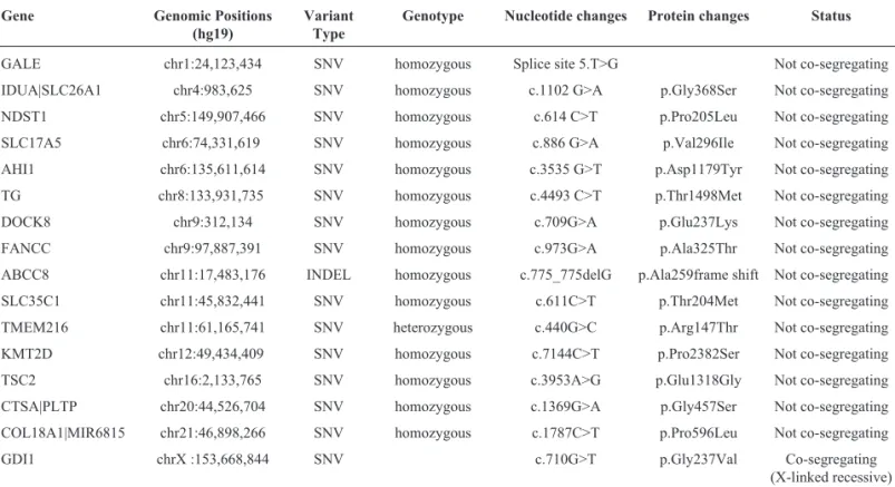
Sixteen candidate variants (15 homozygous and 1 heterozygous, located in 12 genes) were identified from the proband by analysis of the WES data (Table 1). Subsequent Sanger validation followed by co-segregation analysis de-tected a c.710G > T (Chr.X: 153, 668, 844G > T) missense mutation, which mapped to exon 6 of theGDI1gene in all male patients (II: 1, II: 3 and III: 1), segregating with the phenotype of NS-XLID (Figure 1B, C). Meanwhile, the proband’s mother and grandmother, who have a normal in-tellectual phenotype and social adjustment ability, were proved to be obligate carriers of this mutation (Figure 1C). Additionally, we also checked this variant by comparing to ExAC and 5000 Exomes (for URLs see the Internet Re-sources), and insured that they were not present in either of these two large scale exome consortia.
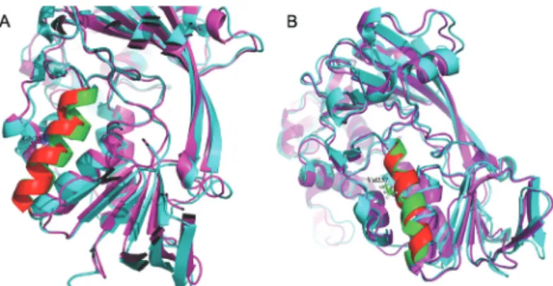
This missense mutation changes the 237thposition amino acid in the alpha-isoform GDP-Dissociation Inhibi-tor protein from glycine to valine (p.Gly237Val). Amino acid sequence alignment by ClustalX showed that the re-gion, which p.Gly237Val mutant residue was situated in, was conserved across various vertebrates. Namely, neither this mutant residue (Val) nor any other residue with similar properties was observed at this position in other homolo-gous sequences of vertebrates (Figure 2A). Several patho-genicity prediction score programs gave deleterious predic-tions for this p.Gly237Val mutation (Table S2).The computer built model of the GDI protein revealed that the location of residue 237 belongs to a hydrophobic domain composed by four helices, and substitution of glycine with valine introduces a larger side-chain into this four-helix hy-drophobic pocket (Figure 2B). Molecular dynamics simu-lations lasting 50 nanoseconds (ns) also revealed that the residue 237 located in the helix of wild typeaGDI
minimi-zation, heating and equilibration procedures (Figure 3A), while the p.Gly237Val mutant showed a relatively less ob-vious conformational change (Figure 3B).
Discussion
Here we report a family (SZMRX) with three patients suffering from intellectual disability. Ultimately, the inher-itance pattern of ID in this family was identified to be an X-linked recessive type by the WES and subsequent Sanger validation followed by co-segregation analysis, as the two unaffected females (mother-daughter relationship) share one rare heterozygous mutation related to XLID, and three consanguineous male patients are homozygous of the same loci.
The unique phenotype presented in the three patients is a moderate ID without any other recognizable clinical signs, and therefore the potential cause of this phenotype cannot be determined by G-banding karyotype examination and FMR1 mutation analysis. Whole exome sequencing was performed for the proband, and the probable patho-genic variants were validated by Sanger sequencing for all the family members and the fetus. Analysis of the WES data and subsequent Sanger validation followed by co-segregation analysis showed a missense mutation that seg-regated with the ID phenotype. This variant is a G to T transversion at position 710 of the coding sequence (c.710G > T), which is mapped to exon 6 of theGDI1gene, and it results in a p.Gly237Val substitution in the encoded protein. TheGDI1gene contains 11 exons, spans 6.29 kb, and is located at chr. Xq28. This gene encodes the protein GDI, which belongs to the TCD/MRS6 family of GDP
dis-Table 1- Candidate mutations of the SZMRX family identified by whole exome sequencing#.
Gene Genomic Positions
(hg19)
Variant Type
Genotype Nucleotide changes Protein changes Status
GALE chr1:24,123,434 SNV homozygous Splice site 5.T>G Not co-segregating
IDUA|SLC26A1 chr4:983,625 SNV homozygous c.1102 G>A p.Gly368Ser Not co-segregating
NDST1 chr5:149,907,466 SNV homozygous c.614 C>T p.Pro205Leu Not co-segregating
SLC17A5 chr6:74,331,619 SNV homozygous c.886 G>A p.Val296Ile Not co-segregating
AHI1 chr6:135,611,614 SNV homozygous c.3535 G>T p.Asp1179Tyr Not co-segregating
TG chr8:133,931,735 SNV homozygous c.4493 C>T p.Thr1498Met Not co-segregating
DOCK8 chr9:312,134 SNV homozygous c.709G>A p.Glu237Lys Not co-segregating
FANCC chr9:97,887,391 SNV homozygous c.973G>A p.Ala325Thr Not co-segregating
ABCC8 chr11:17,483,176 INDEL homozygous c.775_775delG p.Ala259frame shift Not co-segregating
SLC35C1 chr11:45,832,441 SNV homozygous c.611C>T p.Thr204Met Not co-segregating
TMEM216 chr11:61,165,741 SNV heterozygous c.440G>C p.Arg147Thr Not co-segregating
KMT2D chr12:49,434,409 SNV homozygous c.7144C>T p.Pro2382Ser Not co-segregating
TSC2 chr16:2,133,765 SNV homozygous c.3953A>G p.Glu1318Gly Not co-segregating
CTSA|PLTP chr20:44,526,704 SNV homozygous c.1369G>A p.Gly457Ser Not co-segregating
COL18A1|MIR6815 chr21:46,898,266 SNV homozygous c.1787C>T p.Pro596Leu Not co-segregating
GDI1 chrX :153,668,844 SNV c.710G>T p.Gly237Val Co-segregating (X-linked recessive)
#: Sixteen candidate mutations (located in 12 genes) were identified from the proband by analysis of the whole exome sequencing data, and finally a c.710 G > T (chrX: 153,668,844G > T) missense mutation, which mapped to exon 6 of the GDI1 gene (OMIM* 300104), was confirmed to be a pathogenic vari-ant bySanger sequencing and co-segregation analysis in all male patients (II:1, II:3 and III:1).
sociation inhibitors, and serves as a regulator in the mem-brane-traffic process of vesicles by retrieving Rab (a protein family of Ras-like GTPases) from target mem-branes after a vesicular transport event (Pereira-Leal and Seabra, 2001). In brief, GDI has the capacity to bind the guanosine diphosphate (GDP)-bound form of Rab and to facilitate its release from the target membrane through forming cytosolic complexes. GDI subsequently delivers Rab to the cytoplasmic donor membrane, and after the dis-assembly of Rab-GDI complexes, GDI returns to the cyto-sol and becomes available for a new cycle (Pfeffer, 2013). In eukaryotes, exocytic and/or endocytic processes medi-ated by vesicles are major transport pathways between indi-vidual cells (Aridor and Balch, 1996). Rab proteins function as both directors, which point the specific transit way, and motors for vesicles to move between the mem-branes of cytoplasm and subcelluar organelles (Pfeffer, 2001; Goud, 2002). Meanwhile, the recycling of Rabs be-tween the donor membrane and the target membrane needs cytosolic elements that act as guides for this traffic. Among Rab-interacting proteins, GDI not only serves as a retriever of Rabs, but also helps to maintain a pool of GDP-Rab in the cytoplasm (D’Adamoet al., 2014). Thus, GDI has been considered a significant regulator for vesicular trafficking. The phenotypes of ID caused by mutations ofGDI1
gene were first located in two ID families in 1998 (D’Adamo et al., 1998). A Pro to Leu substitution was identified at the 92ndposition of the GDI protein’s amino acid sequence in the MRX 41 family (OMIM#300849). This mutation affects a conserved residue in thea-helix
be-neath the Rab-binding platform of GDI protein (Schalket al., 1996) and subsequently decreases GDI affinity for Rab3A (a locally abundant Rab in brain) remarkably (Alo-ry and Balch, 2001). A sequence-conserved region (SCR) named SCR3B (residues 232 to 259) was described as a vi-tal part of the Rab-binding platform by studies on the X-ray structure of bovine brain GDIa-isoform (Schalk et al.,
1996). In that research, Glu233 and Arg240, which are near the 237th position mutant residue, were found to be critical foraGDI’s binding capacity to Rab. Our genetic screening
based on WES technique and subsequentin silicoanalysis located a pathogenic transition p.Gly237Val on ana-helix
of humanaGDI, which is buried in a hydrophobic pocket
of the protein, just within this major functional SCR (resi-dues 232 to 259) of GDI. Wildtype of the 237 residue is a glycine, which is the most flexible one among all amino ac-ids. Meanwhile, the substitution of glycine with valine, which introduces a side-chain into this packed hydrophobic pocket between helices, results in numerous clashes with surrounding side-chains. Therefore, we speculate that the flexibility provided by glycine might be necessary for the protein’s function, and the mutant residue (bigger and more hydrophobic than the wildtype) might disturb the binding capacity of GDI with Rabs. To verify our hypotheses, we carried out molecular dynamics simulations to exhibit the effect of the 237 residue variants on the function ofaGDI.
Our findings that the 237 position mutantaGDI exhibited a
relatively less obvious conformational change than the wild type, confirm the loss of flexibility resulting from the sub-stitution of glycine with valine. Hence, the Rab-binding ca-pacity of GDI would probably be reduced by this restric-tion. Recently, new experimental evidence achieved by studies focusing on theaGDI mutated astrocytes revealed
that impaired aGDI function could also affect the
endo-lysosomal trafficking process in astrocytes. The mobility of vesicles in wild-type mouse astrocytes, which were trans-fected with two reported XLID-related aGDI mutations,
was reduced (Potokar,et al., 2016).Functional researches focusing on the detailed functional mechanism of this p.Gly237Val mutation are needed in future works.
In summary, our genetic analysis based on whole exome sequencing followed by Sanger validation revealed a novel missense mutation located in theGDI1gene in a Chinese family with three males suffering from XLID. Our findings extended the spectrum of GDI1 mutations that cause X-linked non-specific intellectual disability (NS-XLID). Moreover, identification of the genetic cause for this NS-XLID family allowed us to assess the risk of the proband’s mother for having an impaired child by prenatal testing during her pregnancy and to arrange appropriate management. Although the detailed functional mechanism and the frequency of this novelGDI1mutation await future investigations, we believe that the progressive identifica-tion of genetic defects associated with ID will eventually shed light on the underlying pathological mechanisms and help develop more effective treatment strategies for ID pa-tients in the future.
Acknowledgments
We thank the patients and their family members for their generous support. This work was supported by the
Figure 3- The 237 mutation. (A) The 237 residue located helix of the wild typeaGDI exhibited a distinct conformational change after a 50 ns
Key Project of the Population and Family planning Com-mittee of Guangdong Province (grant number: 2008004) and the Funds for Science and Technology Research from Shenzhen municipality (grant number: JCYJ20150401095437130).
References
Alory C and Balch WE (2001) Organization of the Rab-GDI/CHM superfamily: The functional basis for choroide-remia disease. Traffic 2:532-543.
Aridor M and Balch WE (1996) Principles of selective transport: Coat complexes hold the key. Trends Cell Biol 6:315-320. Case DA, Cheatham 3rd TE, Darden T, Gohlke H, Luo R, Merz Jr
KM, Onufriev A, Simmerling C, Wang B and Woods RJ (2005) The Amber biomolecular simulation programs. J Comput Chem 26:1668-1688.
D’Adamo P, Menegon A, Lo Nigro C, Grasso M, Gulisano M, Tamanini F, Bienvenu T, Gedeon AK, Oostra B, Wu SK,et al.(1998) Mutations in GDI1 are responsible for X-linked non-specific mental retardation. Nat Genet 19:134-139. D’Adamo P, Masetti M, Bianchi V, Morè L, Mignogna ML,
Giannandrea M and Gatti S (2014) RAB GTPases and RAB-interacting proteins and their role in the control of cog-nitive functions. Neurosci Biobehav Rev 46:302-314. de Ligt J, Willemsen MH, van Bon BW, Kleefstra T, Yntema HG,
Kroes T, Vulto-van Silfhout AT, Koolen DA, de Vries P, Gilissen C,et al.(2012) Diagnostic exome sequencing in persons with severe intellectual disability. N Engl J Med 367:1921-1929.
De Rubeis S, He X, Goldberg AP, Poultney CS, Samocha K, Cicek AE, Kou Y, Liu L, Fromer M, Walker S,et al.(2014) Synaptic, transcriptional and chromatin genes disrupted in autism. Nature 515:209-215.
Goud B (2002) How Rab proteins link motors to membranes. Nat Cell Biol 4:E77-E78.
Inlow JK and Restifo LL (2004) Molecular and comparative ge-netics of mental retardation. Gege-netics 166:835-881. Kochinke K, Zweier C, Nijhof B, Fenckova M, Cizek P, Honti F,
Keerthikumar S, Oortveld MA, Kleefstra T, Kramer JM,et al.(2016) Systematic phenomics analysis deconvolutes ge-nes mutated in intellectual disability into biologically coher-ent modules. Am J Hum Genet 98:149-164.
Larkin MA, Blackshields G, Brown NP, Chenna R, McGettigan PA, McWilliam H, Valentin F, Wallace IM, Wilm A, Lopez R,et al.(2007) Clustal W and Clustal X version 2.0. Bio-informatics23:2947-2948.
Pereira-Leal JB and Seabra MC (2001) Evolution of the Rab fam-ily of small GTP-binding proteins. J Mol Biol313:889-901. Pfeffer SR (2001) Rab GTPases: Specifying and deciphering organelle identity and function. Trends Cell Biol 11:487-491.
Pfeffer SR (2013) Rab GTPase regulation of membrane identity. Curr Opin Cell Biol 25:414-419.
Potokar M, Jorgacevski J, Lacovich V, Kreft M, Vardjan N, Bianchi V, D’Adamo P and Zorec R (2016) ImpairedaGDI function in the X-linked intellectual disability: The impact on astroglia vesicle dynamics. Mol Neurobiol 54:2458-2468.
Rauch A, Wieczorek D, Graf E, Wieland T, Endele S, Schwarz-mayr T, Albrecht B, Bartholdi D, Beygo J, Di Donato N,et
al.(2012) Range of genetic mutations associated with severe non-syndromic sporadic intellectual disability: An exome sequencing study. Lancet 380:1674-1682.
Raymond FL (2006) X linked mental retardation: A clinical guide. J Med Genet43:193-200.
Schalk I, Zeng K, Wu SK, Stura EA, Matteson J, Huang M, Tandon A, Wilson IA and Balch WE (1996) Structure and mutational analysis of Rab GDP-dissociation inhibitor. Na-ture 381:42-48.
Stevenson RE, Procopio-Allen AM, Schroer RJ and Collins JS (2003) Genetic syndromes among individuals with mental retardation. Am J Med Genet A 123A:29-32.
Strobl-Wildemann G, Kalscheuer VM, Hu H, Wrogemann K, Ropers HH and Tzschach A (2011) Novel GDI1 mutation in a large family with nonsyndromic X-linked intellectual dis-ability. Am J Med Genet A 155A:3067-3070.
Topper S, Ober C and Das S (2011) Exome sequencing and the ge-netics of intellectual disability. Clin Genet 80:117-126. Van Naarden Braun K, Christensen D, Doernberg N, Schieve L,
Rice C, Wiggins L, Schendel D and Yeargin-Allsopp M (2015) Trends in the prevalence of autism spectrum disor-der, cerebral palsy, hearing loss, intellectual disability, and vision impairment, Metropolitan Atlanta, 1991-2010. PLoS One 10:e0124120.
van Schrojenstein Lantman-de Valk HM and Walsh PN (2008) Managing health problems in people with intellectual dis-ability. BMJ 337:a2507.
Venselaar H, Te Beek TA, Kuipers RK, Hekkelman ML and Vriend G (2010) Protein structure analysis of mutations causing inheritable diseases. An e-Science approach with life scientist friendly interfaces. BMC Bioinformatics 11:548.
Wu L, Qiu Z, Wong D, Hernandez LW and Zhao Q (2010) The re-search on the status, rehabilitation, education, vocational de-velopment, social integration and support services related to intellectual disability in China. Res Dev Disabil 31:1216-1222.
Internet Resources
5000 Exomes, http://evs.gs.washington.edu/EVS/ (December 15, 2015).
ExAC, http://exac.broadinstitute.org/ (December 15, 2015). HOPE, http://www.cmbi.ru.nl/hope/ (January 11, 2016). Online Mendelian Inheritance in Man (OMIM),
https://www.ncbi.nlm.nih.gov/omim/ (March 6, 2015). SAVES, http://services.mbi.ucla.edu/SAVES/ (January 11,
2015).
Supplementary material
The following online material is available for this article: Table S1 - Characteristics of primers for amplification of candidate pathogenic regions on the genomic DNA Table S2 - Possible consequence of GDI1 p.Gly237Val mutation predicted by various algorithms
Associate Editor: Mara H. Hutz