Pedro Miguel de Sousa Vieira
Hypericum perforatum phenolic compounds:
Protective role in the toxicity induced by
heterologous expression of
-synuclein and
huntingtin in yeast cells
α
Mestrado em Biotecnologia e Bioempreendedorismo de
Plantas Aromáticas e Medicinais
Trabalho realizado sobre a orientação de
Alberto Dias
e co-orientação de
Paula Ludovico
Outubro de 2011
Este trabalho foi realizado nas instalações do Centro de Investigação e de Tecnologias Agro-Ambientais e Biológicas (CITAB), Departamento de Biologia da Escola de Ciências da Universidade do Minho
e nas instalações do laboratório de Microbiologia e Infeção, do Instituto de Ciências da Vida e da Saúde, na Escola de Ciências da Saúde da Universidade do Minho.
Nome: Pedro Miguel de Sousa Vieira
Endereço electrónico: [email protected] Telefone: 917466260 Número do Bilhete de Identidade: 13456782
Título da tese:
Hypericum perforatum phenolic compounds: Protective role in the toxicity induced by heterologous expression of α-synuclein and huntingtin in yeast cells
Orientador: Alberto Dias Co-orientador: Paula Ludovico Ano de conclusão: 2011
Tese de Mestrado em:
Biotecnologia e Bioempreendedorismo de Plantas Aromáticas e Medicinais
É AUTORIZADA A REPRODUÇÃO INTEGRAL DESTA TESE/TRABALHO APENAS PARA EFEITOS DE INVESTIGAÇÃO, MEDIANTE DECLARAÇÃO ESCRITA DO INTERESSADO, QUE A TAL SE COMPROMETE.
Universidade do Minho, 31/10/2011
ii
Chegou o fim de uma caminhada importante na minha vida e foram bastantes as pessoas que contribuíram, de uma forma ou de outra, para a escrita de cada uma destas páginas. Desta forma, não poderia deixar de agradecer o apoio, o carinho e a amizade que me encorajaram a caminhar sempre no bom sentido.
Em primeiro lugar queria agradecer ao meu orientador, Professor Doutor Alberto Dias, pela sua orientação que tornou possível a elaboração deste trabalho. Não menos importante foi o apoio da minha co-orientadora, Professora Doutora Paula Ludovico, que me aceitou no seu grupo de trabalho e me orientou de forma a possibilitar o trabalho elaborado.
Quero fazer um agradecimento especial, à Belém, que me acompanhou e ajudou nas tarefas laboratoriais e ainda na escrita da tese, de uma forma amigável e disponível.
Agradeço a todos os meus colegas de mestrado e também aos colegas de laboratório pela amizade e ajuda dispensada durante este tempo. Obrigado Rose, Alberta, Xana, Jéssica, Maria João, Conceição, Márcia, Susana e Ana Oliveira, pelas horas de desabafos e aconchego.
Não menos importante foram os reencontros com pessoas que apesar de longe, estiveram sempre disponíveis e acreditaram em mim, Andreia, Didi, Diana e Ita.
Indispensável, é o agradecimento às pessoas que mais acreditaram e me apoiaram nesta luta, os meus pais e o meu irmão que nunca se recusaram a ajuda-me e apadrinhar as decisões e as ações. Claro que foi também importante o apoio da restante família, em especial a minha avó e prima Patrícia. Agradeço também a todos os meus amigos de Penafiel e aos que se encontram espalhadas pelo restante país.
Agradeço igualmente à Associação Cultural e Recreativa de Croca (ACRO) e a todos os membros, em especial a Cátia Quintas, Jorge, Nuno, Sónia, Ricardo, Cátia, João Paulo, João Cunha e Rosa. Durante este tempo por Braga, integrei-me nos Bomboémia, perdura aqui um agradecimento especial pelos momentos de descontração e carinho, em especial à Gina, ao Leo, à Tânia e à Lu.
iii
A realização deste trabalho na cidade de Braga, proporcionou momentos de todas as formas e feitios. O melhor momento foi ter encontrado uma namorada fantástica, que sem dúvida contribuiu de uma forma única para a concretização da minha tese. Obrigado Raquel.
iv
Parkinson’s (PD) and Huntington’s (HD) diseases are neurodegenerative disorders with high prevalence. Several studies implicate abnormal protein accumulation, protein phosphorylation, mitochondrial dysfunction and oxidative stress as common pathways implicated in PD and HD pathogenesis. Polyphenolic compounds are commonly found in both edible and medicinal plants, and they have been reported to have multiple biological effects, including antioxidant activity. The budding yeast Saccharomyces cerevisiae has been used as a model to study the toxicity and the biological functions of α-synuclein in PD and huntingtin in HD. The heterologous expression of wild-type and A53T mutant form of α-synuclein and huntingtin 103Q mutant form in yeast model to PD and HD, respectively, causes toxicity and death of yeast cells. Therefore, the aim of this study was to evaluate the possible protective effect of Hypericum perforatum phenolic compounds (quercetin, kaempferol and biapigenin) in the toxicity induced by the heterologous expression of α-synuclein and huntingtin, in S. cerevisiae. Preliminary results indicate that the presence of these phenolic compounds decreased the α-synuclein toxicity. Our results showed a possible synergistic effect between the phenolic compounds, and biapigenin was the compound with higher protective effects in the α-synuclein-induced toxicity. Mixtures of these compounds inhibited the foci formation, and kaempferol increased the aggresome formation. Relatively to the expression of huntingtin 103Q mutant form, our results showed that the presence of phenolic compounds did not interfere in the huntingtin-induced toxicity. We concluded that these phenolic compounds apparently show beneficial biological properties that consequently could have a potential use in preventing Parkinson’s disease. Key words: Parkinson’s disease, α-synuclein, Huntington’s disease, huntingtin, quercetin, kaempferol, biapigenin, ROS, foci and yeast.
v
Resumo
A doença de Parkinson (DP) e a doença de Huntington (DH) são desordens neurodegenerativas com bastante prevalência. Diversos estudos associam a acumulação de proteínas disfuncionais, fosforilação de proteínas, disfunção mitocondrial e stress oxidativo à patogénese da DP e da DH. Os compostos fenólicos são normalmente encontrados em plantas comestíveis e em plantas medicinais e têm sido estudados devido aos seus múltiplos efeitos biológicos, incluindo a actividade antioxidante. A levedura Saccharomyces cerevisiae tem sido usada como modelo de estudo de toxicidade e das funções biológicas das proteínas α-sinucleína e huntingtina inplicadas na DP e na DH, respectivamente. A expressão heteróloga das formas tóxicas da proteína α-sinucleína (normal e da forma mutante A53T) e da forma patogénica (103Q) da huntingtina, em leveduras, causa toxicidade e morte celular. Assim, o objectivo deste trabalho foi a avaliação de possíveis efeitos protetores de compostos fenólicos (quercetina, kaempferol e biapigenina), presentes na planta Hypericum perforatum, na toxicidade induzida pela expressão heteróloga das proteínas, α-sinucleína e huntingtina, usando para tal a levedura S. cerevisiae como modelo. Resultados preliminares demonstraram que a presença dos compostos fenólicos diminuiu a toxicidade da α-sinucleína. Os resultados mostraram ainda a possibilidade de efeitos sinergéticos entre os compostos fenólicos e que a biapigenina foi o composto com maior efeito protetor na toxicidade induzida pela α-sinucleína. Misturas desses compostos inibiram a formação de “foci” e o kaempferol aumentou a formação de agressomas. Os resultados mostraram ainda que a presença dos compostos fenólicos não interferiu na toxicidade induzida pela expressão da forma patogénica da huntingtina. Conclui-se assim que os compostos fenólicos utilizados aparentam ter propriedades biológicas benéficas e consequentemente poderão ter um potencial uso na prevenção da DP.
Palavras-chave: Doença de Parkinson, α-sinucleína, Doença de Huntington, huntingtina, quercetina, kaempferol, biapigenina, espécies reactivas de oxigénio, “foci” e levedura.
vi Declaração ... i Agradecimentos ... ii Abstract………iv Resumo………...v Abreviations ... viii I. Introduction... 1 1. Neurodegenerative disorders ... 1
Parkinson disease and α- synuclein ... 2
Huntington’s disease ... 4
2. Oxidative stress, mitochondrial dysfunction and ubiquitin proteasomal system in Parkinson’s and Huntington’s diseases ... 6
3. Yeast model ... 9
Yeast model for α-synuclein aggregation and toxicity ... 10
Yeast model of Huntington’s disease... 12
4. Hypericum perforatum and phenolic compounds ... 13
Quercetin ... 15
Kaempferol ... 16
Biapigenin ... 16
5. Goals ... 17
II. Material and Methods ... 18
1. Phenolic compounds ... 18
2. Strains and growth conditions ... 18
vii
4. Measurements of cellular reactive oxygen species levels... 21
5. Statistical analysis ... 21
III. Results and Discussion ... 22
1. Effect of phenolic compounds in toxicity induced by heterologous expression of α-synuclein…….. ... 22
2. Effect of the mix compounds in toxicity induced by heterologous expression of α-synuclein 26 3. Effect of phenolic compounds in huntingtin expression ... 29
4. Effect of the mix compounds in huntingtin expression ... 32
5. Epifluorescent microscopy ... 34
6. Measurements of cellular ROS levels ... 41
IV. Conclusion ... 44
Conclusion and future perspectives ... 44
viii Aβ AD ALS ATP ATP7B CFUs CLN3 DHE DMSO DNA GFP GSH GSSH HD HSP Htt mPTP NFT OS Amyloid β-protein Alzheimer’s disease
Amyotrophic Lateral Sclerosis Adenosine triphosphate Wilson’s disease protein Conting colony-forming units Ceroid lipofuscinosis neuronal 3 Dihydroethidium
Dimethyl sulfoxide Deoxyribonucleic acid Green fluorescent protein Glutathione
Oxidized glutathione Huntington’s disease
Hereditary Spastic Paraplegia Huntingtin
Mitochondrial permeability transition pore Neurofibrillary tangles
ix PD PolyQ RNA RNS ROS SNcp UPS WT YEPD Parkinson´s disease Polyglutamine Ribonucleic acid
Reactive nitrogen species Reactive oxygen species
Substantia nigra pars compacta Ubiquitin proteasome system Wild-type
1
I.
Introduction
1. Neurodegenerative disorders
Neurodegenerative diseases are progressive disorders of the central nervous system. They have clinical heterogeneity and are characterized by motor, cognitive, and/or behavioural dysfunction (Woulfe, 2008).
Age is directly linked with these diseases and not only makes patients more bent to neurodegenerative diseases, but also impairs their abilities of self-repairing. During ageing, DNA, RNA and proteins become more susceptible to suffer damage, such as oxidation, and ubiquitin proteasome system (UPS) activity decreases (Hung et al., 2010; Woulfe, 2008). Another factor associated with many neurodegenerative diseases (table 1) is the accumulation of disease-specific misfolded proteins in the central nervous system – the conversion of a protein from normal, functional, and benign conformations to aberrant and toxic conformations (Shastry, 2003). The proteins α-synuclein in Parkinson’s disease (PD), amyloid β-protein (Aβ) and tau protein in Alzheimer’s disease (AD) and polyglutamine (polyQ) – expansion in huntingtin (htt) protein in Huntington’s disease (HD) are good examples of misfolded proteins in neurodegenerative disorders (Wood-Kaczmar et al., 2006; Shastry, 2003). The aggregation of misfolded proteins leads to a selective loss of neurons and affect a diverse neuronal population but the exact causes of misfolding and accumulation of proteins are still not totally understood (Hung et al., 2010; Wood-Kaczmar et al., 2006 and Shastry, 2003).
Table 1 – Three neurodegenerative diseases, their inclusions and protein (Shastry, 2003)
Disease Inclusions Protein
Alzheimer’s Plaques and neurofibrillary tangles (NFT)
Presenilins 1 and 2; Apolipoprotein E and Amyloid precursor protein
Parkinson’s Lewy bodies α-Synuclein; Parkin and Ubiquitin carboxy-terminal hydrolase L1
2
Parkinson’s disease and α- synuclein
Parkinson's disease is one of the most common neurodegenerative diseases and was first described by James Parkinson in 1817. Most patients are over 55 years old and men seem to be more affected than women (Wood-Kaczmar et al., 2006). PD is a chronic, progressive neurodegenerative disorder and its typical clinical symptoms are resting tremor, slowness of movement, cogwheel rigidity, bradykinesia, and postural instability (Ziemssen & Reichmann, 2007). Patients with PD also exhibit nom-motor symptoms that consist of disturbances of sleep, cognition, autonomy and olfaction (Hou & Lai, 2007).
A study on the prevalence of PD in Portugal shows that age-standardized rates were 1.4/1000 for males and 1.3/1000 for females and sex-specific rates increase gradually with age for males and females, covering a level of 9/1000 for ages over 75 years (Dias et al., 1994). It is believed that most cases are sporadic. However in some cases, PD is thought to have a genetic component in recessive and dominant modes of inheritance (Wood-Kaczmar et al., 2006). Some of the key factors associated with this disease process, both familiar and sporadic forms, are protein phosphorylation, oxidative stress, mitochondrial dysfunction, protein misfolding and impairment of the UPS (Dauer & Przedborski, 2003).
From the pathological point of view, PD is characterized principally by the loss of dopaminergic neurons in the substantia nigra resulting in a deficiency of dopamine, an important neurotransmitter for the basal ganglia circuit and the presence of cytoplasmic inclusions known as Lewy bodies (Fig.1) (Hou & Lai, 2007 and Ruipérez, Darios, & Davletov, 2010)
α- Synuclein is an abundant presynaptic protein and is expressed in a number of neuronal and non-neuronal cell types. Aggregations of this protein have been implicated in the pathogenesis of PD (Outeiro et al., 2006; Brighina et al., 2009 and Dev et al., 2003). The α-synuclein protein has a highly conserved N-terminal domain that consists of two α-helical regions interrupted by a short break and a C-terminal region. The protein is small and the normal function is not well understood. However, α-synuclein is known to interact with membranes and to bind to fatty acids. It is also implicated in the regulation of dopamine neurotransmission and certain enzymes and involved in neuronal survival (Wood-Kaczmar et al., 2006 and Dev et al., 2003).
3
Studies show that α-synuclein is the main component of Lewy bodies, having an important role in PD (Dauer & Przedborski, 2003). Three mutations of this protein, A30P, A53T and E46K, lead to the development of familial PD. Many studies show that triplications and duplications of the α-synuclein gene cause an early onset of PD. Mutations in this protein have not been found in sporadic PD (Dauer & Przedborski, 2003; Ruipérez et al., 2010).
Overexpression of α-synuclein in several cellular models (animal, yeast and mammalian cells) leads to cytotoxicity and inclusion body formation, facilitates cell death, promotes mitochondrial deficit, improves vulnerability to oxidative stress, and can impair cellular organelles including lysosomes. The pathological role of inclusion bodies is still a debated topic, but the fibrillation and aggregation of α-synuclein have been implicated in the pathogenicity of PD (Outeiro et al., 2006 and Brighina et al., 2009). Aggregation of α-synuclein in disease (Fig.2), is caused by increased oxidative stress and decreased protein degradation of abnormal proteins by UPS (Raichur et al., 2006 and Branco et al., 2010).
Several studies show differences in fibrils formation between α-synuclein mutant forms and wildtype (WT) α-synuclein. The A53T mutant fibrilizes more rapidly than WT α-synuclein while the A30P mutant fibrilizes more slowly (Dauer & Przedborski, 2003). Apart from their divergent rates in fibril formation, the A30P mutant contrasting the A53T mutant is also defective in binding to vesicles and membranes. The mutant forms, seem to sensitize cells to proteasome inhibition and thus reduce the
4
neuroprotective effects as compared to WT α-synuclein (Dev et al., 2003; Dauer & Przedborski, 2003).
Huntington’s disease
HD belongs to a family of dominantly inherited neurodegenerative disorders characterized by degeneration of neurons in the striatum and, to a lesser extent, in the cerebral cortex (Sorolla et al., 2011). This disease begins in middle age and is caused by an abnormal expansion of a CAG repeat, located in the exon 1 of the IT15 gene that encodes a stretch of polyglutamines (polyQ) at the N-terminal of the htt protein (Damiano et al., 2010). Htt is a 350-kDa multidomain protein that contains a polymorphic glutamine/proline-rich domain at its amino-terminus (Sorolla et al., 2008; Qin & Gu, 2004). The normal function of this protein has been complicated to establish because it encloses very little sequence homology to other known proteins, is ubiquitously expressed and is localized in many subcellular compartments. Htt is present in the nucleus, cell body, dendrites and nerve terminals of neurons, and is also associated with a number of organelles including the
5
endoplasmic reticulum, mitochondria and Golgi apparatus (Cattaneo et al., 2005; Landles & Bates, 2004).
Clinical symptoms include psychiatric and cognitive abnormalities, chorea, and dementia. Degenerative events begin years before the occurrence of clinical symptoms. With the progression of the disease, motor rigidity and dementia dominate and death normally occurs after 15–20 years of progression (Damiano et al., 2010).
Age of onset and severity of this disease, correlates with the length of CAG expansion and HD appears when the number of glutamines expands beyond 40. A polyQ extension in proteins is implicated in some autosomal dominant neurological diseases and HD is the most prevalent of these diseases, affecting 1 in 105 persons (Sorolla et al., 2011).
The major hallmark of cytopathology of HD and other polyglutamine disorders is the accumulation of misfolded polyglutamine proteins as aggregates or neuronal intranuclear inclusions (Duennwald et al., 2006). In fact, mutant proteins with extended polyQ tracts gain an unusual conformation that simplifies their oligomerization and aggregation, generating cell toxicity. In addition to the toxicity induced by aggregates, polyQ proteins can also be toxic in the absence of a detectable formation of aggregates and misfolded htt monomers and oligomers are believed to be toxic before development of macromolecular inclusions (Sorolla et al., 2011; Damiano et al., 2010).
Several pathways are affected by the existence of pathological polyQ such as: vesicular and organelle trafficking; UPS; kinurenine pathway; transcription and mitochondrial function (Sorolla et al., 2011). In HD, the combination of function failure of the wild type protein with a gain-of-function mutation, probably leads to neuronal degeneration. The mutant htt polypeptides get an uncommon conformation that produced cell toxicity, and facilitates the aggregation the aggregation into intracellular inclusion bodies (Landles & Bates, 2004).
Different models have been used to study this disease, and most of them express exon 1 or it’s fragment, which promotes protein misfolding and aggregation, and causes toxicity (Sorolla et al., 2011; Damiano et al., 2010). Examples of these models are mouse, worm, fly, cell cultures and yeast (Sorolla et al., 2011).
6
2. Oxidative stress, mitochondrial dysfunction and ubiquitin
proteasomal system in Parkinson’s and Huntington’s diseases
Oxidative stress (OS) happens when oxygen free radicals are formed in excess through the reduction of oxygen. Many of these radicals are generated during normal physiological conditions at low levels and are scavenged by endogenous antioxidant systems (Gilgun-sherki et al., 2001). The production of reactive oxygen species (ROS) leads to oxidative stress when endogenous antioxidant system is not capable to remove the high amount of ROS. This process has been suggested to concur in the onset of several neurodegenerative diseases, including PD and HD (Gilgun-sherki et al., 2001; Floyd & Hensley, 2002).
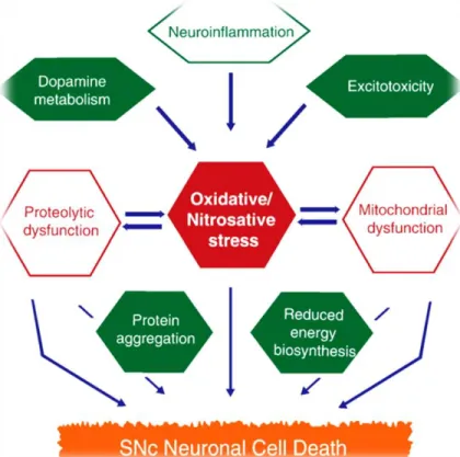
In fact, the brain is inclined to ROS production and oxidative stress due to its elevated metabolic rate, combined with the content of oxidizable molecules, such as dopamine and neuromelanin, whose metabolism generates ROS. In addition, the elevated concentration of transition metals and the low content of antioxidants, can contribute to exacerbate oxidative challenge (Raichur et al., 2006; Branco et al., 2010; Tsang & Chung, 2009). Indeed, many studies have showed a reduction in glutathione (GSH) and an increase in oxidized glutathione (GSSH) levels (Gilgun-sherki et al., 2001). The oxidative stress in dopaminergic neurons of the substantia nigra pars compacta (SNpc) is connected with increased ROS levels, decreased levels of cellular antioxidants and increased levels of iron. Oxidative stress may contribute to SNpc neuronal cell death (Fig. 3) and plays an important role in neuronal degeneration of pigmented dopaminergic neurons in the SNpc (Raichur et al., 2006; Branco et al., 2010 and Tsang & Chung, 2009).
Various studies in HD brains have demonstrated the existence of oxidative damage, and that antioxidants slow the disease development. Therefore, the role of oxidative stress in the pathogenesis of HD has been growing attention (Sorolla et al., 2011).
Oxidative damage in mitochondria has also been implicated in the pathogenesis of PD and HD. Mitochondria are organelles with a vital role in the cells such as fatty acid oxidation, steroid metabolism, amino acid biosynthesis and principally, mitochondria maintain the cellular energy reserves with ATP production (Chong et al., 2005 and Hald & Lotharius, 2005).
Post-mortem studies show a complex I deficiency in the substantia nigra of patients with PD (Hald & Lotharius, 2005). In fact the inhibition of complex I activity leads to: an energy crisis; a discrepancy
7
of mitochondrial membrane potential; an increase on ROS levels; and, eventually, to neuronal death (Chong et al., 2005 and Hald & Lotharius, 2005). The active generation of ROS can lead to a subsequent irreversible inhibition of complex I in mitochondria and enhance ROS production (Tsang & Chung, 2009).
HD is characterized by degeneration of neurons in the striatum and it is known that this brain region is very sensitive to deficiency in energy metabolism (Cattaneo et al., 2005). In fact, some studies suggested a association between mitochondrial defects and striatum susceptibility to ROS, which presents the initial and most striking neuropathological changes in patients (Damiano et al., 2010). Mutant htt possibly may destabilize mitochondrial membrane, and increase the sensitivity of the mitochondrial permeability transition pore (mPTP) to calcium or other apoptotic stimuli through interactions with the external mitochondrial membrane (Damiano et al., 2010).
Post mortem studies shows that defects in respiratory chain, diminution in the activity of complex II/III and milder reduction of complex IV, were observed in HD patients (Damiano et al., 2010).
Figure 3 – Pathways that contribute to the SNcp neuronal cell death (Tsang & Chung, 2009)
8
The regulation of protein levels involved in fundamental cellular processes is made through mechanisms of protein degradation (Betarbet et al., 2005). The UPS is one of the pathways involved in targeting and detoxification of short-lived damaged proteins for degradation and provides the specificity and control of several regulatory proteins (Nedelsky et al., 2008). The UPS also contribute to maintain amino acid pools in acute starvation and contribute considerably to the degradation of defective proteins (Salomons et al, 2010).
The UPS involves the coordinated actions of a large number of proteins that consecutively recognize the substrates, tags substrates with ubiquitin chains, and promote the destruction of poly-ubiquitylated substrates (Nedelsky et al., 2008). The recognition and tagging are mediated by a large group of ubiquitylation enzymes that conjugate poly-ubiquitin chains to substrates, while the final destruction is executed by the proteasome,a large multi-subunit proteolytic complex (Salomons et al., 2010 and Esser et al., 2004).
The accumulation of toxic proteins can arise due to a directly or indirectly impairment of UPS, under conditions of stress or other insults and lead to neuronal dysfunction or death (Betarbet et al., 2005). Anomalous accumulation of protein and aggregation affect the normal function of degradative pathways to degrade mutated or oxidized proteins (Nedelsky et al., 2008 and Ross & Pickart, 2004). The aggregation of mutated α-synuclein can inhibit the UPS and it is thought that an impairment of the UPS and protein aggregation may affect each other (Betarbet et al., 2005). In fact, besides α-synuclein, proteasomal subunits are found in Lewy bodies suggesting the involvement of UPS in PD pathogenesis (Ross & Pickart, 2004).
Post mortem studies, show a deficit of proteasomal activity in nigral tissue from sporadic PD patients, suggesting that a dysfunctional UPS may cause susceptibility and deterioration of nigral dopaminergic neurons in PD (Betarbet et al., 2005). Other studies show the possibility of α-synuclein to block the polybiquitin recognition by binding to a subunit of 19S complex – UPS (Ross & Pickart, 2004).
In what concerns HD, the formation of both intranuclear and cytoplasmic ubiquitinated aggregates result from de existence of pathological polyQ and these aggregates containing the protease-resistant mutated N-terminal htt fragment in neurons of affected areas. This proposes that abnormal htt is targeted for proteolysis but is resistant to its removal (Bennett et al., 2007 and Sorolla et al., 2008).
9
3. Yeast model
Yeast models have been developed to study several neurodegenerative disorders (Scherzer & Feany, 2004; Summers & Cyr, 2011 and Winderickx et al., 2008). Saccharomyces cerevisiae, also known as budding yeast, is extensively used for studying the molecular events and cell composition relevant for human diseases and is one of the most recognised model organisms (Summers & Cyr, 2011 and Zabrocki et al., 2008). S. cerevisiae was the first eukaryote organism to be fully sequenced and there is a well-functioning research infrastructure, such as the Saccharomyces Genome Database (Zabrocki et al., 2008 and Petranovic & Nielsen, 2008).
S. cerevisiae is a unicellular microorganism and several cellular processes such, cell division, protein turnover, DNA replication and recombination, are well conserved between yeast and higher eukaryotes, including humans. In fact around 31% of yeast genes have a mammalian homolog and an additional 30% of yeast genes have domain similarity with mammalian (Scherzer & Feany, 2004 and Winderickx et al., 2008). There are some factors that make yeast an exceptional model: easy to cultivate in large populations, quickly and inexpensive; exists in haploid and diploid forms that makes possible sexual and budding reproduction, allowing genetic manipulations and screenings; can express heterologous genes, being rather simple to mutate, insert or delete any genomic sequence due to the presence of homologous recombination pathway (Winderickx et al., 2008; Zabrocki et al., 2008 and Miller-Fleming et al., 2008).
S. cerevisiae offers an experimental model system to explore the cell biological effects of several proteins involved in neurodegenerative diseases (table 2) (Scherzer & Feany, 2004). Although it is impossible to study the neuronal effects of this proteins in yeast, some mechanisms and pathways correlated to neurodegenerative disorders such as proteasomal dysfunction and mitochondrial dysfunction, are well conserved between yeast and humans, enabling the modelling of neurodegenerative diseases in these cells (Winderickx et al., 2008 and Miller-Fleming et al., 2008).
10
Table 2 – Proteins involved in neurodegenerative diseases modeled in yeast
Protein involved Disease
Superoxide dismutase 1 Amyotrophic Lateral Sclerosis (ALS) (Scherzer & Feany, 2004)
Frataxin Friedreich’s Ataxia (Scherzer & Feany, 2004)
Huntingtin HD and PoyQ disorders (Winderickx et al., 2008)
α-synuclein PD and synucleinopathies (Winderickx et al., 2008)
Tau and Amyloid precursor
protein AD and tauopathies (Winderickx et al., 2008)
ATP- dependent proteases Hereditary Spastic Paraplegia (HSP) (Winderickx et al., 2008)
Wilson’s disease protein
(ATP7B) Wilson’s disease (Scherzer & Feany, 2004) CLN3 Ceroid lipofuscinosis (Scherzer & Feany, 2004)
Yeast model for α-synuclein aggregation and toxicity
The yeast model has been used in the study of the pathogenic basis of PD (Fig.4), especially the biochemistry and toxicity of α-synuclein and its mutant forms (Scherzer & Feany, 2004). Through this model it is possible to comprehend the molecular pathways underlying the cytotoxic effects of α-synuclein malfunction, the cell response to an accumulation of toxic aggregates and the intracellular location of α-synuclein aggregates (Petranovic & Nielsen, 2008; Braun et al., 2010 and Outeiro & Giorgini, 2006).
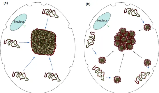
When α-synuclein is expressed in yeast, the formation of inclusions (Fig.5) seems to be a process that involves the aggregation of monomers at the plasma membrane, which are then delivered and continue to grow in the cytoplasm (Braun et al., 2010 and Wang et al., 2009).
11
Figure 4 – Yeast model for α-synucleinopathies (Braun et al., 2010)
The heterologous expression of WT α-synuclein and A53T mutant form inhibited the yeast growth, which is not the case of A30P mutant that merely produced partial growth inhibition when expressed from high-copy plasmid (Winderickx et al., 2008).
Unlike the WT α-synuclein and the A53T, the A30P mutant does not form cytoplasmatic inclusions, due to its low membrane-binding capability. The WT α-synuclein and the A53T inclusions are involved in impaired proteasomal degradation, inhibition of phospholipase D, blockage of endoplasmic reticulum to Golgi transport and delayed endocytosis (Zabrocki et al., 2008 and Winderickx et al., 2008).
Several studies show that the heterologous expression of WT α-synuclein or mutant forms, leave the yeast cell more vulnerable to oxidative stress induced by peroxide, and this is because the expression of the protein induced the ROS production and release of cytochrome c from mitochondria, proposing that α-synuclein activates the apoptotic cell death program (Braun et al., 2010 and Winderickx et al., 2008).
12
Yeast model of Huntington’s disease
Studies have been developed in order to understand the folding and behaviour of mutant htt, as well as to determine the basic conserved mechanism of mutant htt-dependent toxicity and yeast models of HD have been developed as valuable tools in these studies (Miller-Fleming et al., 2008).
Yeast models express a fragment of the human polyQ protein huntingtin (htt), htt exon I. The first studies using yeast as a model demonstrated that heterologous expression of htt results in aggregation dependent of polyQ length (Duennwald et al., 2006). One of these models shows toxicity dependent of polyQ length, such that expression of a htt fragment with higher polyQ length (htt103Q) produces cellular toxicity, whereas the expression of a non-expanded polyQ tract (htt25Q) shows no effects upon growth (Duennwald, 2011).
These models offer a useful tool to discover modulators of polyQ toxicity in a defined and consistent cellular environment and provide a powerful instrument to explore the intramolecular and intermolecular factors that are connected with polyQ toxicity (Duennwald, 2011 and Duennwald et al., 2006).
Figure 5 – Two models to cytoplasmic inclusion formation. (a) Monomers directly diffuse to the deposition site. (b) Monomers aggregate in the cell periphery and are transported to the inclusion body (Kopito, 2000).
13
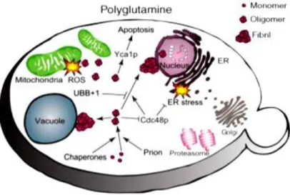
Figure 6 - Yeast model for polyglutamine disorders (Braun et al., 2010).
Mitochondrial dysfunction, increased levels of ROS, defects in endocytosis, apoptotic-like events and perturbations in the kynurenine pathway are examples of factors that many studies related to toxicity in yeast model of HD (Solans et al., 2006 and Miller-Fleming et al., 2008).
4.
Hypericum perforatum
and phenolic compounds
Medicinal plants have been used for long time, due to their significant biological activities, as well as many natural compounds from these plants (Maciel et al., 2002).
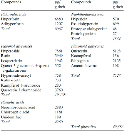
Hypericum, a genus of the family Clusiaceae, has been commonly used in traditional medicine for a long time. H. perforatum is common in Europe, North Africa, Asia and North America (Langosch et al., 2002). The plant spreads quickly and can emerge in different locations such as plains and sides of roads. Its popular name St John’s wort raises to its flowering period around St John’s day, the 24th of June (Langosch et al., 2002 and Saddiqe et al., 2010). The medicinal plant H. perforatum has been used as an antidepressant with scarce side effects, and it is very interesting to observe that several Hypericum species showed good antioxidant activity (Saddiqe et al., 2010). Evidences from the literature show that the quercetin, kaempferol and biapigenin are present in H. perforatum extracts (Table 3), in particular biapigenin, which is only produced by this plant (Silva et al., 2008 and Schulte-Löbbert et al., 2003).
14
Table 3 – Phenolic composition of Hypericum perforatum total ethanolic extract (Silva et al., 2008)
Phenolic compounds are abundant in plant foods, and have been associated with the sensory and nutritional value of fresh and processed plant foods (Gonzalez et al., 1999). They are secondary metabolites that are derivatives of the pentose phosphate, shikimate, and phenylpropanoid pathways in plants (Balasundram et al., 2006). These compounds, ubiquitous in plants, have physiological and morphological relevance in these organisms. Phenolic compounds show an important role in growth and reproduction, providing defense against pathogens and predators, also contributing to the color and sensory characteristics of fruits and vegetables (Balasundram et al., 2006 and Martins et al., 2011). These compounds are considered relatively non-toxic bioactive substances and have various biological activities, such as antioxidants, chelation of metals, scavengers of active oxygen species, blockers of nitration, allergenic, artherogenic, inflammatory, microbial, anti-thrombotic, cardioprotective and vasodilatory effects. (Martins et al., 2011; Cai et al., 2004;
15
Balasundram et al., 2006 and Saddiqe et al., 2010) They can suffer autoxidation to produce hydrogen peroxide in the presence of metals and are capable of modulating certain cellular enzyme activities (Saddiqe et al., 2010). Flavonoids have a great antioxidant potential due to structural characteristics such as the arrangement of hydroxyl groups on the benzene ring (García-Mediavilla et al., 2007). These compounds have gained much attention, since their consumption has been associated with a decrease in age-related cognitive declines and risk of developing neurodegeneration (Filomeni et al., 2010). In fact, studies with rat brain have demonstrated that biapigenin, kaempferol and quercetin, may play a neuroprotective role against neuronal excitotoxicity and mitochondrial dysfunction (Silva et al., 2008).
Quercetin
Quercetin is a major dietary flavonoid (Fig. 7) present in fruits and vegetables and several others foods, and has gained attention as a good antioxidant (Cornish et al., 2002).
Within the flavonoids, quercetin is the most active scavenger of ROS and reactive nitrogen species (RNS). This can be explained by the structure of the molecule that has the optimum configuration for free radical scavenging (García-Mediavilla et al., 2007). Several studies using human and rat cells, show that quercetin exhibits anti-inflammatory effects, inhibiting tumor necrosis factor receptor family signaling in bone cells and can display a neuroprotective role (García-Mediavilla et al., 2007; Pang et al., 2006 and Arredondo et al., 2010).
Figure 7 – Structural formula of quercetin (Silva et al., 2008)
16
Kaempferol
Kaempferol is a natural flavonoid (Fig. 8) which has been isolated from tea, broccoli, witch-hazel, propolis, grapefruit, and other plant sources (Yoshida et al., 2008). Some studies with neuronal cells have shown that this compound, even at the dose of 30 µM, may provide neuroprotection by autophagy and protect against mitochondrial dysfunction (Filomeni et al., 2010). This compound such as quercetin, have been reported as antioxidant, as illustrated by a study with cerebellar granule cells that shows that kaempferol decreased oxidative stress (Samhan-Arias et al., 2004).
Biapigenin
Biapigenin is the most abundant biflavone (Fig. 9) in Hypericum perforatum and some studies suggest that biapigenin may contribute to some of the pharmacological actions of this herb (Colovic & Caccia, 2008). It is mainly localized in the buds and flowers of this plant, reaching its maximum amount just before blooming of the buds (Schulte-Löbbert et al., 2003).
This compound has a variety of biological effects. Biapigenin was remarkably active in an established screening model for antidepressants (Colovic & Caccia, 2008). A study using macrophage cells show that biapigenin may inhibit the activation of nuclear factor-κB and thus contribute to blocking effects in inducible nitric oxide synthase and ciclooxygenase-2 (Woo et al., 2006). Another study with rat hippocampal neurons shows that this compound confers neuroprotection against excitotoxic insults by protection from calcium deregulation (Silva et al., 2008).
Figure 8 – Structural formula of Kaempferol (Silva et al., 2008).
17
Figure 9 - Structural formula of Biapigenin (Silva et al., 2008).
5. Goals
The aims this study were to evaluate the possible effects of Hypericum perforatum phenolic compounds (quercetin, kaempferol and biapigenine) in the toxicity induced by the heterologous expression of α-synuclein and huntingtin. For these studies the yeast Saccharomyces cerevisiae was used as a model.
18
II.
Material and Methods
1. Phenolic compounds
Quercetin (≥ 98%), kaempferol (≥ 98%) and biapigenin (≥ 98%) were purchased from Sigma. All compounds were dissolved in dimethyl sulfoxide (DMSO) (Sigma).
The compounds were used at concentration 15 µM and were made two different mixtures of the 3 compounds (Mix a: 10µM quercetin, 10µM kaempferol and 10µM biapigenin; Mix b: 15µM quecetin, 15µM kaempferol and 15µM biapigenin).
1. Strains and growth conditions
The yeast strains and plasmids used in this study are listed in Table 3. Table 3 – Yeast strains and plasmids used in this study
Strain Genotype Plasmids Source
W303-1A MATα ade2-1 can1-100 his3-11,14 leu2-3 112 trp1-1 ura3-1 WT pRS304Gal-αSynWT-GFP pRS306Gal-αSynWT-GFP Outeiro, T.F. A53T pRS304Gal-αSynA53T-GFP pRS306Gal-αSynA53T-GFP A30P pRS304Gal-αSynA30P-GFP pRS306Gal-αSynA30P-GFP W303-1A trp1Δ 2 leu2-3,112 MATa ura3-52
his3-11 ade2-1
25Q pRS303Gal-htt25Q-GFP Outeiro,
T.F.
103Q pRS303Gal-htt103Q-GFP
To perform the assays (Fig. 10) with the yeast Parkinson’s disease (PD) models, the cells were inoculated in YEPD medium with 0.5 % (w/v) yeast extract (Difco Laboratories), 1% (w/v) peptone (Difco Laboratories) and 2% (w/v) glucose at 26ºC and 150 rpm, until the stationary phase. In the assays (Fig. 11) with the yeast Huntington’s disease (HD) models, the cells were inoculated in
19
selective YNB medium, containing 0.67 % (w/v) Yeast Nitrogen Base (Difco Laboratories), 2% (w/v) glucose as carbon source, supplemented with the appropriate amino acids: 100 mg/L uracil, 300 mg/L leucine, 100 mg/L tryptophan and 100 mg/L adenine. Growth was monitored (each 2 hours) by measuring the turbidity of the culture at 640 nm in a Spectronic Genesys 20 spectrophotometer (Thermo Spectronic).
To study the effects of the phenolic compounds, cells were collected at exponential phase (OD 640 nm ≈ 0.4), washed twice with distilled water and inoculated in YEPDGal medium with 0.5 % (w/v) yeast extract (Difco Laboratories), 1% (w/v) peptone (Difco Laboratories) and 2% (w/v) galactose or YNBGal medium with 0.5 % (w/v) yeast extract (Difco Laboratories), 1% (w/v) peptone (Difco Laboratories) and 2% (w/v) galactose at optical density ≈ 0.24 at 640 nm. Then, the yeast cells were transferred to 12-well culture plates and Falcon tubes to PD and HD yeast model, respectively. Next the phenolic compounds were added.
For determination of yeast cell survival, cellular samples were serially diluted and platted on YEPD agar plates, consisting in 0.5 % (w/v) yeast extract (Difco Laboratories), 1% (w/v) peptone (Difco Laboratories), 2% (w/v) glucose and 2% (w/v) agar. The viability was determined by counting colony-forming units (CFUs) after 2 days of incubation at 30°C. The CFUs on time 0 (when the phenolic compounds were added to the culture medium) were considered as 100% survival.
20
Figure 10 -Schematic representation of the methods used to study the toxicity induced by heterologous expression of α- synuclein.
21
2. Epifluorescence microscopy
All the samples were collected at specific time points, 20h and 40h, and then they were washed and suspended in PBS and Vectashield® (Vector Laboratories, Burlingame, Canada).
Images were acquired with an Olympus BX61 (Olympus, United States) microscope with filter wheels, to control excitation and emission wavelengths, equipped with a high-resolution DP70 digital camera and using an Olympus UPlanSApo 100X/oil objective, with a numerical aperture of 1.40.
3. Measurements of cellular reactive oxygen species levels
Cellular reactive oxigen species (ROS) production was kinetically monitored by flow cytometry with dihydroethidium (DHE) staining. This assessment was obtained by measuring the ROS production of cell culture samples harvested at different time points of α-syn expression. For DHE staining, cells were harvested by centrifugation, resuspended in PBS and incubated with 10 µg/ml DHE in the dark for 10 minutes at 30°C. Afterwards, the fluorescence was detected by flow cytometry. Cells presenting high red fluorescence were considered to have high intracellular ROS levels.
Flow cytometry assays were performed on an EPICS XL-MCL (Beckman-Coulter Corporation, Hialeah, FL) flow cytometer, equipped with an argon-ion laser emitting a 488-nm beam at 15mW. DHE was collected through a 488-nm blocking filter, a 590 nm long-pass dichroic and a 620-nm bandpass. Twenty thousand cells per sample were analyzed. The data were evaluated with the MULTIGRAPH software included in the system II acquisition software for the EPICS XL/XL-MCL version 1.0.
4. Statistical analysis
The results shown are mean values and standard deviations of at least three independent assays. Statistical analyses were determined using t -test. A p -value less than 0.05 was assumed to denote a significant difference.
22
III. Results and Discussion
1. Effect of phenolic compounds in toxicity induced by heterologous
expression of α-synuclein
The protein α-synuclein is the main component of Lewy bodies, one of the hallmarks of Parkinson’s disease (PD) (Corti et al., 2005). Several studies in different cellular models showed that overexpression of the α-synuclein resulted in cell toxicity and culminates in cell death (Raichur et al., 2006). Saccharomyces cerevisiae, genome does not encode an orthologue of α-synuclein and thus could be used to study the effects of heterologous expression of α-synuclein. In fact, yeast cells have already proved to be an excellent model to study the cellular processes linked to PD (Outeiro & Lindquist, 2003). To study the cellular toxic effects of α-synuclein a model in which the yeast cells carrying human wild-type (WT) or mutant α-synuclein fused with GFP (green fluorescent protein) was developed. The α-synuclein-GFP fusion construct was integrated into the yeast genome under the control of a galactose-inducible promoter (Outeiro & Lindquist, 2003), meaning that the presence of galactose in the yeast culture medium allows the α-synuclein-GFP heterologous expression.
In yeast, the toxicity of neurotoxic proteins is tested by clonogenic assays on agar plates or in liquid cultures, and cell survival is calculated by the number of colony-forming units (CFUs) (Braun et al., 2010). Therefore, clonogenic assays were performed with the addition of phenolic compounds (quercetin, kaempferol and biapigenin) in the medium.
The phenolic compounds have various biological activities (Silva et al., 2008), including quercetin and kaempferol which have been used in several studies due to their antioxidant properties (Pang et al., 2006). The use of polyphenols has been associated with a decrease in age-related cognitive declines and risk of developing neurodegeneration (Filomeni et al., 2010). In addition, studies in isolated rat brain mitochondria have been developed and showed that biapigenin confers protection against excitotoxic insults (Silva et al., 2010).
To gain further insight into the bioactivities of these compounds, yeast clonogenic assays were performed on cells expressing an vector control (Empty), WT α-synuclein and mutant forms, A30P
23
and A53T in the presence of quercetin, kaempferol and biapigenin at concentration of 15µM each, to verify the occurrence of possible effects of the phenolic compounds on the toxicity caused by heterologous expression of the proteins. The data showed that biapigenin seems to be more effective in reducing the toxicity of heterologous expression of WT α-synuclein (Fig. 12B) when compared to cells without the addition of compounds, or even when compared with the quercetin and kaempferol.
Empty 0 20 40 0 500 1000 1500 2000 2500 Control DMSO Quercetin Kaempferol Biapigenin Time (h) % s u rv iv a l WT 0 20 40 0 50 100 150 Control DMSO Quercetin Kaempferol Biapigenin Time (hours) % s u rv iv a l A B * * * ** ** **
24 A30P 0 20 40 0 1000 2000 3000 Control DMSO Quercetin Kaempferol Biapigenin Time (h) % s u rv iv a l A53T 0 20 40 0 50 100 150 Control DMSO Quercetin Kaempferol Biapigenin Time (h) % s u rv iv a l
Figure 12 - Survival determined by colony-forming units of cell expressing WT (B) and point mutants A53T (D) and A30P (C) forms of α-synuclein or harboring the empty vector (A) after induction of expression by changing to 2% galactose medium in presence of quercetin, kaempferol and biapigenin. Data represent mean ± S.E. of 3 independent experiments. Statistical significance (*p <0.05, **p <0.01) were determined by t -test.
As expected there was a continued growth in the case of the cells harboring the empty vector (Fig. 12A) functioning as a control. Also in the case of the A30P mutant form (Fig. 12C) there was a
C
D
** **
25
continuous growth, similarly to the control conditions (empty). The non-toxic phenotype of α-synuclein A30P mutant form was also expected, since the A30P mutation reduces the affinity of the protein to interact with phospholipids, a pre-requisite to aggregation (Auluck et al., 2010). The A30P mutant form only produced partial growth inhibition when expressed in a high-copy plasmid (Scherzer & Feany, 2004). In the case of WT α-synuclein (Fig. 12B), the heterologous expression of the protein reduced the cell survival to values of 22.57% and 4.82% after 20h and 40h of induction of protein expression, respectively. While the A53T mutant form (Fig. 12D) the heterologous expression reduces the cell survival to values of 35.24% and 7.1% to 20h and 40h, respectively. The treatment with biapigenin appears to reduce the toxicity induced by heterologous expression of α-synuclein, since the values of cell survival in the presence of this compound were about 41.95% and 10.82% after 20h and 40h of induction of WT α-synuclein expression, respectively (Fig. 12B). In the case of A53T mutant form the values of cell survival (Fig. 12D) in the presence of biapigenin were 55.27% and 16.35% to 20h and 40h, respectively. The research about biapigenin and its actions is scarce, but some studies using rat brain mitochondria, suggest that biapigenin contributes for neuroprotection against excitotoxicity, since this compound can modulate mitochondrial permeability transition pore opening (mPTP), probably through modulating adenosine nucleotide translocator function, which contributes to improve the mitochondrial calcium efflux, reducing in this manner the calcium burden (Silva et al., 2008 and Silva et al, 2010).
Additionally, the presence of quercetin, also demonstrated a reduction of the toxicity induced by heterologous expression of α-synuclein, since the values of cell survival were about 40.75% and 11.28% to 20h and 40h respectively (Fig. 12D). Nevertheless, relatively to the cells expressing the A53T mutant form in the presence of quercetin, the toxicity reduction was only observed at 20h. The effects observed with quercetin may be due to its role as an antioxidant, catalysing electron transport, chelating transition metal and scavenging free radicals, since several studies using human and rat cells show that quercetin decreases oxidative stress (Dias et al., 2005 and Jamshidzadeh & Mehrabadi, 2010).
26
2. Effect of the mix compounds in toxicity induced by heterologous
expression of α-synuclein
An effect in reducing the toxicity induced by heterologous expression of α-synuclein under phenolic compounds treatment was observed, therefore, we wanted to understand if the presence of the three compounds together could induce a synergistic effect. Hence, to perform this assay two mixtures with different concentrations (Mix a: 10µM quercetin, 10µM kaempferol and 10µM biapigenin and Mix b: 15µM quecetin, 15µM kaempferol and 15µM biapigenin) were used. We used two different compounds concentrations, for as little or nothing is known about the use of the compounds together, we decided to try the two concentrations mentioned above. The data (Fig. 13) showed that the mix a, has significant effects in protecting against the toxicity of the protein expression in the case of α-synuclein A53T mutant form. Although the mix b had no significant effects, however there was observed a strong tendency to reduce the toxicity induced by heterologous expression of α-synuclein.
27 0 20 40 0 500 1000 1500 Control DMSO Mix a Mix b Empty Time (hours) S u rv iv a l (% ) 0 20 40 0 50 100 150 Control DMSO Mix a Mix b WT Time (hours) S u rv iv a l (% ) A B
28 0 20 40 0 500 1000 1500 Control DMSO Mix a Mix b A30P Time (hours) S u rv iv a l (% ) 0 20 40 0 50 100 150 Control DMSO Mix a Mix b
A53T
Time (hours) S u rv iv a l ( % )Figure 13 - Survival determined by colony-forming units of cell expressing WT (B) and point mutants A53T (D) and A30P (C) forms of α-synuclein or harboring the empty vector (A) after induction of expression by changing to 2% galactose medium in presence of compounds mix. Data represent mean ± S.E. of 3 independent experiments. Statistical significance (*p <0.05, **p <0.01) were determined by t -test.
Once again, as expected, the cells harboring the empty vector and the A30P mutant form (Fig. 13A and 13C) showed a non-toxic phenotype, being possible to observe cell growth. In the case of expression of -synuclein A53T mutant form (Fig. 13D), the values of cell survival at time 20h and
C
D
** **
29
40h without compounds were about 22.13% and 13.84%, respectively. In the presence of mix a, we could observe an higher percentage of cell survival, 33.51% and 42.53% after 20h and 40h of induction of protein expression, respectively. Relatively to the mix b, that has the higher concentration of compounds, no significant effect has been detected, however there was a strong tendency of this mixture to reduce the -synuclein-induced toxicity, also in the case of α-synuclein A53T mutant form (Fig. 13D). Relatively to the expression of WT α-synuclein, again, no significant effects were shown (Fig. 13B), but there was a strong tendency of the both mixtures to reduce the toxicity induced by heterologous expression of WT α-synuclein. Therefore, it is necessary to repeat the experiments with mix b, to validate the observations and to confirm their statistical significance.
The data showed that, contrary to previous results, the presence of DMSO reduces the cell survival, since the amount of DMSO used (1.3%) is toxic to yeast cells. Thus, we can assume that, in this case, the effects of the compounds are more significant than those observed.
Although there are some studies about quercetin, kaempferol and biapigenin, little or nothing is known about their use as a mixture. The data showed possible synergistic effects of these compounds, that may have been originated by the combined action of quercetin and kaempferol, due to their antioxidant properties (Dias et al., 2005 and Samhan-Arias et al., 2004) and/or ability of biapigenin to regulate the calcium homeostasis (Silva et al., 2010). A study with neuronal cells showed that kaempferol can interfere with mitochondrial homeostasis (Filomeni et al., 2010), a target of the α-synuclein induced toxicity. Therefore, we can assume that there may be a combined effect of the actions of kaempferol and biapigenin by impinging on cellular energetics.
3. Effect of phenolic compounds in huntingtin expression
The formation of aggregates and misfolded huntingtin (htt) monomers and oligomers are the major hallmark of cytopathology of Huntington’s disease (HD) and several pathways are affected by their presence (Qin & Gu, 2004). The disease is caused by an abnormal expansion of a CAG repeat located in the exon 1 of the IT15 gene of htt protein. The longer polyQ domain seems to induce conformational changes in the protein, which causes it to form intracellular aggregates that, in most cases, manifest as nuclear inclusions (Sorolla et al., 2011 and Damiano et al., 2010). The increase
30
in the length of polyglutamine tract alters biochemical and biophysical properties of proteins, and consequently, these proteins accumulate and forms aggregates (Dehay & Bertolotti, 2006).
The yeast genome does not have a homologous htt gene, however it is possible to induce its heterologous expression. The yeast model of HD used, employs the W303-1a strain harboring a plasmid containing the first exon of htt gene, with a polyQ tract of 25 glutamines (25Q) or 103 glutamines (103Q), under the control of a galactose-inducible promoter (Solans et al., 2006 and Hughes et al., 2001), furthermore, the exon 1 of htt gene is fused with the GFP, allowing its visualization.
Like what was developed with the PD yeast model, with the HD model, we studied the effect of phenolic compounds on the survival of yeast cells expressing the normal or extended htt polyQ tract (Fig. 14), to access the possible effects of the phenolic compounds on the toxicity levels caused by an abnormal expansion of a CAG repeat.
31 Empty 0 5 10 15 0 200 400 600 800 Time (days) % s u rv iv a l 103Q 0 5 10 15 0 50 100 150 Time (days) % s u rv iv a l 25Q 0 5 10 15 0 200 400 600 Time (days) % s u rv iv a l
Figure 14 - Survival determined by colony-forming units of cell expressing point mutants 25Q (C) and 103Q (B) forms of htt or harboring the empty vector (A) after induction of expression by changing to 2% galactose medium in presence of quercetin, kaempferol and biapigenin. Data represent mean ± S.E. of 3 independent experiments.
A
B
32
The yeast model used to study the effect of phenolic compounds in htt expression has lower toxicity than the yeast model used to α-synuclein expression. Thus, for this study the cell survival was monitored for 15 days.
In the case of cells harboring the empty vector (Fig. 14A), it was possible to observe a continued growth, as expected, since protein expression does not occur. The same happened in case of normal htt form 25Q (Fig. 14C) since the severity of the disease is correlated with the length of CAG expansion and the toxicity appears when the number of glutamines expands beyond 40 (Sorolla et al., 2011). The data showed a reduction in cell survival in the case of htt 103Q mutant form (Fig. 14B). These results are not surprising because the length of CAG expansion is higher and consequently the toxicity occurs (Dehay & Bertolotti, 2006). In fact, other studies using yeast and other cell models showed that the expression of htt 103Q form has a greater toxic effect when compared to normal htt 25Q form (Truant, 2003). Several investigations using various models show that unusually long polyQ tracts induce disease because they interfere with the normal function of cellular proteins (Sorolla et al., 2008 and Sorolla et al., 2011).
The presence of quercetin, kaempferol or biapigenin seems to have no effect in the toxicity induced by expression of toxic protein, htt 103Q (Fig. 14B), since the values of cell survival were 70.2% and 35.8% after 10 and 15 days of induction of protein expression, respectively, while in the presence of the phenolic compounds, the values of cell survival ranged between 65.3% and 71.3% after 10 days of induction of htt expression and 35.2% and 42.9% after 15 days of induction of htt expression. Probably, the concentrations of compounds used were not high enough to cause effects in htt expression, therefore it is necessary to repeat the experiments with other concentrations.
4. Effect of the mix compounds in huntingtin expression
Although there have been no effects of phenolic compounds in cells expressing htt toxic form, it was performed an assay in which two different mixtures of the phenolic compounds, quercetin, kaempferol and biapigenin, were used (Fig. 15) to understand if there was the possible synergistic effects occurring between the compounds, that consequently could rescue the cells expressing the toxic form of htt.
33 Empty 0 5 10 15 0 200 400 600 800 Time (days) % s u rv iv a l 103Q 0 5 10 15 0 50 100 150 Time (days) % s u rv iv a l 25Q 0 5 10 15 0 200 400 600 Time (days) % s u rv iv a l
Figure 15 - Survival determined by colony-forming units of cell expressing point mutants 25Q (C) and 103Q (B) forms of htt or harboring the empty vector (A) after induction of expression by changing to 2% galactose medium in presence of compounds mix. Data represent mean ± S.E. of 3 independent experiments.
A
B
34
Once again, and as expected, the cells expressing the empty vector and the normal htt 25Q form (Fig. 15A and 15C), revealed a continuous growth and presented a non-toxic phenotype, since it was not observed cellular death during the time that the assay was performed. On other hand, the cells expressing the 103Q mutant form (Fig. 15B), presented a reduction in cell survival, of about 30% and 60% at time 10 days and 15 days, respectively, as expected, and the use of mixtures apparently, did not demonstrate any interference with htt induced toxicity, since it was not observed any difference between the survival of untreated and treated cells with the phenolic compounds.
Previous results seemed to demonstrate that the effects of the phenolic compounds were essentially on the α-synuclein-induced toxicity. Therefore, other studies about α-synuclein aggregation and ROS levels were performed.
5. Epifluorescent microscopy
The heterologous expression of α-synuclein can induce the foci formation (Griffioen et al., 2006). The protein aggregation results from a tendency of unfolded polypeptides to associate with each other (Kopito, 2000). In fact, in certain aggregation pathways, small foci may converge, by microtubule-based transport, forming a large inclusion of protein foci, called an aggresome (Wang et al., 2009). In order to understand whether phenolic compounds interfere with the aggregation of α-synuclein and since that in the models used in this work the α-synuclein protein is fused with GFP, it was performed the microscopic visualization of the cells in the presence of quercetin, kaempferol and biapigenin.
Examination of cells, using epifluorescent microscopy analysis in the studied conditions, allowed the visualization of GFP fused with the α-synuclein forms. The cells expressing the WT α-synuclein and the A53T mutant form (Fig. 16) revealed the presence of several foci, being more pronounced after 20h of expression induction, while in the case of cells expressing the α-synuclein A30P mutant form, there are no cytoplasmatic inclusions, due to its low membrane-binding capacity (Dauer & Przedborski, 2003).
The analyses of the images of epifluorescent microscopy led to the counts observed in results described below. The images (Fig. 16), made possible the distinction between small foci and foci with large size (aggresomes).
35
Figure 16 - Foci visualization of the cells expressing WT and point mutant A53T form of α-synuclein, after induction of expression by changing to 2% galactose medium in presence of phenolic compounds and compounds mix. 20 µm.
20 40 WT 20 40 A53T Time (hours) Control DMSO Mix a Mix b Quercetin Kaempferol Biapigenin
Aggresome
Small foci
36
It was performed a count of 300 cells in triplicate, and it was estimated the amount of the cells containing foci, to study the possibility of the compounds interfering in the aggregation of α-synuclein. Thus if during the treatment of cells with the phenolic compounds occurs a decrease in the cell number with foci, it is possible to admit that the compounds have effects in the protein aggregation. WT 20 0 10 20 30 40 50 Time (hours) %c e ll s w it h f o c i WT 40 0 10 20 30 40 Time (hours) %c e ll s w it h f o c i A53T 20 0 10 20 30 40 50 Time (hours) %c e ll s w it h f o c i A53T 40 0 10 20 30 40 Time (hours) %c e ll s w it h f o c i
Figure 17 -% of the cells expressing WT (A and B) and point mutant A53T form of α-synuclein (C and D), after induction of expression by changing to 2% galactose medium in presence of phenolic compounds. Data represent mean ± S.E. of 3 independent experiments. Statistical significance (*p <0.05, **p <0.01 and ***p <0.001) were determined by t -test. The data showed that the compounds mix have effects (Fig. 17) on protein aggregation, since the number of cells with foci is significantly reduced, during the time considered. For WT α-synuclein, mix a reduced the cell number with foci approximately 13.67 % (Fig. 17A) when compared with control, after 20h of protein induction. After 40h of α-synuclein induction, the mix a and mix b reduced the
A B D C * * * *** ***
37
percentage of cells with foci around 8.33% and 9.1% respectively. In the case of α-synuclein A53T mutant form, significant effects of mix a and b, after 40h of protein induction, were observed that reduced the cell number with foci approximately 7.56% and 9.56% respectively.
Once more, the results showed the synergistic effects between different compounds. These suggest that compounds mixtures may have a potential use for preventing foci formation. In fact, some natural compounds can depolymerize microtubules (Wilson & Jordan, 1995) and thus inhibit the aggregation. The aggregation inhibitors that act early on the aggregation pathway can prevent toxic species from forming, and allow cells to clear the mutant protein before it can do much damage (Bodner et al., 2006).
Next and using a similar approach, we determined the number of foci per cells. For this count were admitted only small foci.
38 WT 20 0 50 100 150 200 250 Time (hours) N ºf o c i WT 40 0 100 200 300 Time (hours) N ºf o c i A53T 20 0 100 200 300 400 Time (hours) N ºf o c i A53T 40 0 100 200 300 Time (hours) N ºf o c i
Figure 18 – Number of foci present in the cells expressing WT (A and B) and point mutant A53T form of α-synuclein (C and D), after induction of expression by changing to 2% galactose medium in presence of phenolic compounds. Data represent mean ± S.E. of 3 independent experiments. Statistical significance (*p <0.05, **p <0.01 and ***p <0.001) were determined by t -test.
The number of foci in the presence of the phenolic compounds and compounds mixture was analyzed. The data (Fig. 18) showed that mix a and mix b, have significant effects in reducing the number of foci mainly for WT synuclein (Fig. 18A and 18B) After 20h of induction of WT α-synuclein expression (Fig. 18A), the number of foci was reduced from 217 to 123 and 162 in the case of mix a and b, respectively. Also after 40h both mix a and b, reduced the foci number from 217 to 178 and 168, in that order (Fig. 18B). In the case of α-synuclein A53T mutant form expression, the foci number was reduced significantly only at time 20 and in the presence of the mix a, from 243 to 202 (Fig. 18C). Thus, these results accentuate once again the idea of synergistic effects between different compounds.
A B D C * * * * *
39
Next we determined the number of aggresomes per cells, since several investigations into the mechanisms of intracellular protein aggregation show that the aggresomes facilitate the degradation by autophagy (Wang et al., 2009) and we also observe the presence of higher foci. This suggests that aggresome formation represents a protective response of cells to an accumulation of foci under conditions in which chaperones and ubiquitin proteasomal system (UPS) machineries fail (Wang et al., 2009).
Investigations with mammalian cells showed that therapeutic intervention for PD and HD may involve the discover of compounds that decrease the size or number of inclusions and showed that the aggresome formation can be blocked by drugs that depolymerize microtubules (García-Mata et al., 1999). On the other hand, studies showed that there are compounds that enhance the formation of aggresome. These compounds can contribute for the treatment of neurodegenerative disorders (Olzmann et al., 2008).