Review article:
ANAPLASTIC THYROID CANCER: HOW FAR CAN WE GO?
Mariana Amaral1, Ricardo A. Afonso2,3,4, M. Manuela Gaspar1, Catarina Pinto Reis1,5,*1 Research Institute for Medicines (iMed.ULisboa), Faculdade de Farmácia, Universidade
de Lisboa, Lisboa, Portugal
2 CEDOC, NOVA Medical School, Faculdade de Ciências Médicas (NMS/FCM),
Universidade Nova de Lisboa, Lisboa, Portugal
3 Área de Ensino e Investigação em Ciências Funcionais e Alvos Terapêuticos, NOVA
Medical School, Faculdade de Ciências Médicas (NMS|FCM), Universidade Nova de Lisboa, Lisboa, Portugal
4 Departamento de Física, Faculdade de Ciências e Tecnologia, Universidade Nova de
Lisboa, Lisboa, Portugal
5 IBEB, Institute of Biophysics and Biomedical Engineering, Faculdade de Ciências,
Universidade de Lisboa, Portugal
* Corresponding author: Catarina Pinto Reis, Research Institute for Medicines
(iMed.ULisboa), Faculdade de Farmácia, Universidade de Lisboa, Lisboa, Portugal, E-mail: [email protected]
http://dx.doi.org/10.17179/excli2020-2257
This is an Open Access article distributed under the terms of the Creative Commons Attribution License (http://creativecommons.org/licenses/by/4.0/).
ABSTRACT
Globally, thyroid cancer accounts for 2 % of all cancer diagnoses, and can be classified as well-differentiated or undifferentiated. Currently, differentiated thyroid carcinomas have good prognoses, and can be treated with a combination of therapies, including surgical thyroidectomy, radioactive iodine therapy and hormone-based ther-apy. On the other hand, anaplastic thyroid carcinoma, a subtype of undifferentiated thyroid carcinoma character-ized by the loss of thyroid-like phenotype and function, does not respond to either radioactive iodine or hormone therapies. In most cases, anaplastic thyroid carcinomas are diagnosed in later stages of the disease, deeming them inoperable, and showing poor response rates to systemic chemotherapy. Recently, treatment courses using multi-ple-target agents are being explored and clinical trials have shown very promising results, such as overall survival rates, progression-free survival and tumor shrinkage. This review is focused on thyroid carcinomas, with particular focus on anaplastic thyroid carcinoma, exploring its undifferentiated nature. Special interest will be given to the treatment approaches currently available and respective obstacles or drawbacks. Our purpose is to contribute to understand why this malignancy presents low responsiveness to current treatments, while overviewing novel ther-apies and clinical trials.
Keywords: Anaplastic thyroid cancer, treatments and obstacles, challenges for innovative therapies
INTRODUCTION
The thyroid is a butterfly-shaped gland composed by two lobes, located medially in front of the neck, below the larynx and poste-riorly to the tracheal thyroid cartilage
(Stathatos, 2006). This gland has a functional unit, the thyroid follicle, a cystic structure composed of a single layer of follicular cells (Stathatos, 2012). These units store thyro-globulin, involved in the synthesis of thyroid
hormones (i.e., tri-iodothyronine, T3; and
tetraiodothyronine, T4) (Rousset et al., 2000;
Stathatos, 2012). Thyroid hormone produc-tion and secreproduc-tion are controlled by the hypo-thalamus-pituitary axis, comprising the re-lease of thyrotropin-releasing hormone (TRH) from the hypothalamus, and thyroid-stimulating hormone (TSH), from the pitui-tary (Opitz et al., 2009; Stathatos, 2012). Upon release by the pituitary, TSH binds to the TSH receptor (TSHR) of the follicular cells’ membrane, stimulating the synthesis and release of the thyroid hormones (Opitz et al., 2009; Stathatos, 2012). Regulation of thy-roid function is achieved almost entirely by a negative feedback mechanism carried out by T3 and T4 on hypothalamus and pituitary.
T3 and T4 are known to play important
roles in the human body, such as promoting the growth and differentiation of many tis-sues, as well as energy and metabolic homeo-stasis, due to their involvement in different metabolic pathways (Jugan et al., 2010). Moreover, the thyroid gland also comprises neural-crest derived parafollicular C-cells, lo-cated in-between thyroid follicles and respon-sible for calcitonin secretion (Khan and Farhana, 2020). This hormone promotes cal-cium and phosphate deposition in different tissues (Khan and Farhana, 2020).
Although thyroid cancers account for only 2.1 % of all cancers diagnosed worldwide, it is one of the most frequent endocrine malig-nancies (Gimm, 2001; Kitahara and Sosa, 2016; Nikiforova and Nikiforov, 2008). As with other malignancies, thyroid cancers dif-fer in their morphology, invasiveness and mo-lecular profile (Lin, 2011; Nikiforova and Nikiforov, 2008).
Taking into account their histopathology, thyroid carcinomas can be classified as well-differentiated (medullary, papillary and fol-licular thyroid carcinoma) or undifferentiated (anaplastic thyroid carcinoma) (Kondo et al., 2006; Lin, 2011; Soares et al., 2011), as sum-marized in Table 1.
Table 1: Classification of thyroid carcinomas
Cell type Subtype
Unknown Anaplastic TC Undifferen-tiated Follicular Papillary TC Well-differ-entiated Follicular TC Parafollicular C-Cells Medullary TC TC – Thyroid Carcinoma
EPIDEMIOLOGY AND PROGNOSIS
Papillary thyroid carcinoma accounts for the largest portion of thyroid carcinomas (70-80 %), being the least aggressive due to slowly-forming metastasis and low invasive-ness (Nguyen et al., 2015). Follicular thyroid carcinoma is a more aggressive subtype of well-differentiated thyroid carcinoma, due to its usual later diagnosis, and accounts for 10 % of thyroid malignancies (D'Avanzo et al., 2004). Medullary thyroid carcinoma ac-counts for 5-10 % of thyroid malignancies, originates from parafollicular C-cells, is asso-ciated with a mutation of the RET proto-onco-gene and can be sporadic or familial (25 % of medullary thyroid carcinomas) (Leboulleux et al., 2004). Another form of hereditary roid carcinoma is familial non-medullary roid carcinoma, englobing all hereditary thy-roid carcinomas originating from thythy-roid fol-licular cells (Nosé, 2008; Robenshtok et al., 2011). Although familial non-medullary thy-roid carcinoma is inherited through an auto-somal dominant pattern, the associated mu-tated genes are not yet identified (Robenshtok et al., 2011).
Anaplastic thyroid carcinoma is a form of undifferentiated thyroid carcinoma, which although rare (<2 %), is one of the most lethal malignancies, being characterized by high ag-gressiveness, due to both fast growth and strong invasiveness, as well as low respon-siveness to most therapies currently available (Kebebew et al., 2005; Wiseman et al., 2003). Moreover, although anaplastic thyroid carci-noma arises from thyroid follicular cells, these cells loose its thyroid-like features, leading to very poor prognosis (Lang and Lo,
2007). The overall 5-year survival rate upon anaplastic thyroid carcinoma diagnosis is lower than 10 %, and most patients do not live longer than a few months after diagnosis (Liu et al., 2016).
These two groups of malignancies have different aggressiveness. Indeed, well-differ-entiated thyroid carcinomas are known to be more manageable, with higher survival rates, whereas undifferentiated thyroid carcinomas are known to be more aggressive, with higher invasiveness and poorer prognosis, normally non-operable and having poor treatment re-sponse rates (DeLellis, 2006). Well-differen-tiated thyroid carcinomas include malignan-cies derived from the thyroids’ follicular cells, such as papillary and follicular thyroid carcinomas (Soares et al., 2004). Anaplastic thyroid carcinoma is an undifferentiated sub-type with very poor survival prognosis, esti-mated to be 3 to 5 months after diagnosis, and survival rates of 10-20 % and less than 5 % after 1 and 10 years, respectively (Nagaiah et al., 2011). Although this rare tumor has an in-cidence of only 1-2 persons per million per year, it is responsible for 40 % of all thyroid cancer deaths (Green et al., 2006; Tiedje et al., 2018). The very poor prognosis is associated to anaplastic thyroid carcinoma only being detectable by the current diagnostic tools at advanced stages and, furthermore, being un-responsive to the current treatments available (Lin, 2011).
In similarity to what is seen for malignan-cies of other tissues and/or organs, there are risk factors associated with the increased chances of developing thyroid carcinomas. Such risk factors include radiation exposure to the chest or neck area, abnormal iodine in-take leading to iodine deficits, previously-ex-isting thyroid pathologies (i.e., goiter and Hashimoto’s Thyroiditis) and metabolic dis-orders (i.e., diabetes and obesity) (Liu et al., 2017). There are some etiologic factors spe-cifically associated with the development of anaplastic thyroid carcinoma, both as primary disease or by dedifferentiation of other thy-roid malignancies. Such etiological factors in-clude irradiation and abnormal TSH levels
(Khairy, 2009). The biggest risk factor for de-veloping this rare undifferentiated carcinoma seems to be prior history of goiter, both of self and familial (Nagaiah et al., 2011). Further-more, risk factors generally include previous history of other thyroid malignancies, as these can give rise to anaplastic thyroid carcinoma through dedifferentiation (Dackiw, 2010; Khairy, 2009). Anaplastic thyroid carcinoma seems to occur most frequently in the elderly, being diagnosed at around 65-72 years old (Zivaljevic et al., 2014). Generally, both well-differentiated and anaplastic thyroid carcino-mas affect women 2 to 3 times more than men (Tuttle et al., 2010).
PATHOPHYSIOLOGY AND HISTOLOGY
As previously mentioned, based on their histology and behavior, thyroid carcinomas can be subcategorized in well-differentiated and undifferentiated (anaplastic) thyroid car-cinomas. Regarding histology, well-differen-tiated thyroid carcinoma arises from the thy-roids’ follicular cells and can be classified as papillary, if a papillary pattern is seen, or fol-licular, if a follicular pattern is found (Shah, 2015). Although both patterns may be pre-sent, classification is based on the most prev-alent pattern observed (Shah, 2015). Further-more, well-differentiated thyroid carcinoma aggressiveness is determined by assessing the presence of capsular and/or blood vessels in-vasion (Filetti et al., 2019). Usually, papillary thyroid carcinomas present as an encapsu-lated mass, not invasive, whereas the follicu-lar subtype presents high invasiveness, of both capsule and blood vessels. Papillary thy-roid carcinomas can be further distinguished into two classes according to molecular pro-filing: BRAF-predominant; and RAS-predom-inant, the last associated with increased ag-gressiveness (Filetti et al., 2019).
Usually, thyroid carcinoma staging is de-termined by age, histology, size, extra-glan-dular invasion and presence of distance me-tastasis (Cady, 1998). Regardless of the
pa-tient’s and tumor status based on the men-tioned characteristics, anaplastic thyroid car-cinomas are always classified as stage IV (Kebebew et al., 2005; Nguyen et al., 2015; Tahara et al., 2017). Then, by assessing dif-ferent parameters, it can be sub-classified as: stage IVA, if it is confined to the thyroid; stage IVB, when there is extra thyroidal dis-ease; or stage IVC, once distant metastasis are present (Ranganath et al., 2015).
Anaplastic thyroid carcinoma is often clinically characterized as a large palpable rapidly growing mass, causing symptoms such as hoarseness, dysphagia, dyspnea, and in advanced cases, superior vena cava syn-drome and Horner’s synsyn-drome (Cabanillas et al., 2016; Wein and Weber, 2011). Histologi-cally, the characteristic cells of this tumor are known to have undergone epithelial-mesen-chymal cell phenotype transition (Lin, 2011). Furthermore, histological findings may fol-low one of three patterns according to the main cellular population present being giant, spindle or squamous cells (Cabanillas et al., 2016; Wein and Weber, 2011). This leads to uncertainty of the organ of origin, culminat-ing in delays in diagnostic and in initiation of treatment (Cabanillas et al., 2016; Wein and Weber, 2011). Although these histological differences may be present, they do not sig-nificantly influence prognosis (Are and Shaha, 2006). Macroscopically, regardless of its cellular heterogeneity, anaplastic thyroid carcinoma presents characteristically as large light tan color tumors, with marked invasive-ness and mitotic activity, high proliferation, presence of hemorrhage and large areas of ne-crotic tissue, but decreased apoptosis (Are and Shaha, 2006).
Although this undifferentiated malig-nancy can arise primarily, there is clinical, pathologic and epidemiologic evidences sup-porting that it can originate from the dediffer-entiation of previously-existing well-differen-tiated thyroid carcinomas (Neff et al., 2008; Nikiforov, 2004). Such evidence includes the fact that these tumors can coexist and that some treated well-differentiated thyroid
car-cinomas relapse as anaplastic thyroid carcino-mas (Santarpia et al., 2008). Furthermore, the genetic modifications and oncogenes that give rise to follicular and/or papillary thyroid carcinomas are also observed in anaplastic thyroid carcinomas (Neff et al., 2008; Nikiforov, 2004; Wang et al., 2007). Moreo-ver, anaplastic thyroid carcinoma harbors some characteristic genetic features (Ragazzi et al., 2014). For example, gain of function mutations of the PIK3CA gene are frequently seen in anaplastic thyroid carcinoma, but not in well-differentiated thyroid carcinomas (Ragazzi et al., 2014). Mutations in the gene encoding ߚ-Catenin, CTNNB1, are commonly associated with epithelial-mesenchymal tran-sition, which has been speculated as being one of the main processes behind this malignancy pathogenesis (DeLellis, 2006; Ragazzi et al., 2014). The previously mentioned mutations are gain of function of important oncogenes, but the loss of function and inactivation of tu-mor suppressor genes are also present in ana-plastic thyroid carcinoma (Ragazzi et al., 2014; Salvatore et al., 2007). Such genes in-clude p53 and PTEN, both negative regulators of proliferation and inducers of apoptosis (DeLellis, 2006; Quiros et al., 2005; Salvatore et al., 2007). Thus, the inactivity of these genes lead to increased aggressiveness, and are present in this non-differentiated tumor (Ragazzi et al., 2014). Anaplastic thyroid car-cinoma shares mutations with follicular and/ or papillary thyroid carcinomas, such as point mutations in BRAF and RAS, but these muta-tions are more common in the differentiated subtypes (Antonelli et al., 2008; Ragazzi et al., 2014; Salvatore et al., 2007). Furthermore, the overexpression of receptors, such as epi-dermal growth factor receptor (EGFR), are not only characteristic of anaplastic thyroid carcinoma but also of thyroid and primary thyroid carcinomas dedifferentiation or ana-plastic carcinoma transformation (Fisher et al., 2013; Landriscina et al., 2011).
DIAGNOSIS
Well-differentiated thyroid carcinomas, such as follicular or papillary thyroid carcino-mas, are usually asymptomatic and are diag-nosed upon physical and/or ultrasonography examination (Paschke et al., 2015). In rare oc-casions, well-differentiated thyroid carcino-mas may present symptoms such as a palpable and growing neck mass, hoarseness, dyspha-gia and/or with cervical lymph-node metasta-ses (Paschke et al., 2015).
Anaplastic thyroid carcinoma generally presents more serious symptoms, including hoarseness, airway distress, dyspnea and dys-phagia, caused by a fast growing neck mass (Akaishi et al., 2011; Shaha, 2008).
In order to classify a thyroid nodule as malignant or benign, TSH serum levels are evaluated, and a combination of histologic, cytologic and imaging techniques are used. TSH serum levels allow to differentiate be-tween hyperfunctioning and non-functioning nodules (Nguyen et al., 2015). Thyroid carci-nomas often present non-functioning thyroid nodules, and thus, other tests are generally re-quired (Nguyen et al., 2015).
Definitive diagnostic is usually achieved by fine-needle aspiration biopsy and/or high-resolution ultrasonography (Huang et al., 2015; Lewinski et al., 2000; Nguyen et al., 2015). Papillary, medullary and anaplastic thyroid carcinomas are diagnosed according to the results of these examinations, but addi-tional histological tests can be necessary to differentiate between follicular thyroid carci-noma and benign follicular thyroid adecarci-nomas (Sherman, 2003).
CURRENT TREATMENT APPROACHES
Currently, thyroid cancer is treated by us-ing a combination of radioactive iodine ther-apy, thyroid hormone suppression therapy and surgery. Nevertheless, the chosen treat-ment is defined according to different factors, such as the subtype of cancer and stage of dis-ease (Nguyen et al., 2015).
Total or partial surgical resection of the thyroid gland remains one of the first options for both well-differentiated and undifferenti-ated thyroid carcinomas, although in the latter total thyroidectomy is unusual due to inva-siveness of the disease (Cabanillas et al., 2016; Giuffrida and Gharib, 2000).
Therapy with radioactive iodine has been used to treat thyroid cancer since 1946 (Lee, 2010). For radioactive iodine therapy to be ef-fective, a high level of thyroid-stimulating hormone (TSH or thyrotropin) must be pre-sent in the blood to promote its uptake/or ab-sorption. To synthetize thyroid hormones, the thyroid follicular cells need to uptake iodine, a substrate required for the synthesis of these hormones. Iodine is internalized by the follic-ular cells through iodine symporter channels. Inside the follicular cells, the iodine is oxi-dized and bound to tyrosyl residues of thy-roglobulin, giving rise to tri-iodothyronine (T3) and tetraiodothyronine (T4), with three
and four atoms of iodine, respectively (Biondi et al., 2005; Ross, 2011). When in the pres-ence of Radioactive Iodine (131I) instead of normal iodine, the 131I undergoes the
previ-ously described processes in the follicular cells, leading to tissue necrosis mediated by its beta emissions. As a result, this necrosis will lead to the ablation of the functional tis-sues of the thyroid gland (Ross, 2011; Schlumberger et al., 2014). Conventionally, this therapy is used after surgery, either as an adjuvant therapy or to treat any tumoral resid-ual tissue (Jonklaas et al., 2006; Sawka et al., 2004). Undifferentiated and medullary thy-roid carcinomas do not respond to this therapy as they are characterized by a lack of expres-sion of thyroid cell markers and behavior, be-ing unable to uptake iodine and consequently produce T3 and T4 (Cabanillas et al., 2016;
Sherman, 2003).
Usually, life-long thyroid hormone ther-apy (THST) is used to treat well-differenti-ated and medullary thyroid carcinomas after thyroidectomy and radioactive iodine therapy to prevent thyroid-stimulating hormone (TSH)-dependent proliferation of any resid-ual well-differentiated thyroid cancer cells
(Brabant, 2008; McGriff et al., 2002; Sherman, 2003). Physiologically, TSH re-lease by the pituitary is inhibited by high se-rum levels of T3 and T4, i.e., through a
nega-tive feedback mechanism (Biondi et al., 2005). Thus, this therapy involves the admin-istration of T3 and T4, increasing serum levels
of these two hormones and inhibiting pituitary TSH release (Biondi et al., 2005). Moreover, besides inhibiting TSH-dependent proliferat-ion of cancer cells, THST also corrects the surgically-induced hypothyroidism of pa-tients who undergo total or partial thyroid re-section (Biondi et al., 2005).
Medullary thyroid carcinomas and undif-ferentiated thyroid carcinomas are usually un-responsive to the conventional course of ther-apy used for well-differentiated thyroid carci-nomas: radioactive iodine therapy, THST and surgery (Sherman, 2010). For this reason, sys-temic chemotherapy is used for the treatment of the mentioned malignancies, and also for the treatment of non-resectable, radioactive-iodine-non-responsive, recurrent or meta-static, well-differentiated thyroid carcinomas (Busaidy and Cabanillas, 2012; Sherman, 2010).
Treatment of well-differentiated thyroid carcinomas
Currently, well-differentiated (papillary and follicular) thyroid carcinomas are treated by using a combination of surgery to remove the thyroid and, if necessary, radioactive io-dine therapy and THST. It has been reported that patients with stage II or well-differenti-ated high-risk thyroid carcinoma benefit from radioactive iodine therapy, improving their overall survival (Jonklaas et al., 2006). This is not the case for patients with stage I well-dif-ferentiated thyroid carcinomas, whose overall survival worsens when 131I is used (Jonklaas
et al., 2006). Furthermore, radioactive iodine therapy can also be useful for patients with metastatic disease, in which the distant neo-plastic foci have thyroid-like features, re-sponding to treatment and 131I uptake (Cabanillas et al., 2016). Thus, although radi-oactive iodine therapy is efficient in many
cases, it has severe side effects associated to it such as off-target organ damage (i.e., sali-vary glands and bone marrow) and increasing the patients risk of developing hematologic malignancies (Kloos, 2009).
Furthermore, THST has been shown to be an efficient therapy for the treatment of well-differentiated thyroid carcinomas, increasing the patients’ overall survival, reducing dis-ease recurrence and cancer-related mortality (Cabanillas et al., 2016; Jonklaas et al., 2006). Moreover, high-risk well-differentiated thy-roid carcinoma patients strongly benefit from this therapy, whose survival can be increased 2 to 3 fold-factor (Sherman, 2003).
In the case of metastatic and/or advanced well-differentiated thyroid carcinomas, sys-temic chemotherapies are used. But, unfortu-nately, these malignancies are associated with poor response rates and short time of response when systemic therapies are used (Pacini et al., 2010). Some of the cytotoxic drugs used for the treatment of advanced thyroid carci-noma include doxorubicin, paclitaxel, tamox-ifen, bleomycin, epirubicin, cisplatin and oc-treotide (Harada et al., 1971; Liebner et al., 2016; Pacini et al., 2010; Sherman, 2003, 2010). The choice of systemic therapies is somewhat inconclusive, as reports with dif-ferent response rates to the same protocols are observed. For example, doxorubicin, the most studied and used chemotherapy agent for these malignancies, seems to be one of the cy-totoxic drugs with the highest response rate, ~40 %; but this response is temporary and most responses to this drug are incomplete (Sherman, 2003). Other studies show that some patients only have a response rate of ~17 %, for the single use of doxorubicin, dis-playing however, an increase in response rates when combined with cisplatin or even achieving complete responses (Lim, 2012; Shimaoka et al., 1985). Altogether, doxorubi-cin seems to be the most effective in treating metastatic disease, that are non-responsive to other treatments, as well as for medullary thy-roid carcinoma, either as a single-agent chem-otherapy or in combination with other agents
(Liebner et al., 2016). Such combined regi-mens include: doxorubicin, bleomycin and vincristine; or bleomycin, doxorubicin and cisplatin (Liebner et al., 2016).
When administered at low doses, cyto-toxic agents can be used as radiosensitizers for external radiation therapy to increase its efficacy (Lim, 2012).
Treatment of anaplastic thyroid carcinoma According to the most recent American Thyroid Association (ATA) guidelines, to manage and control local and metastatic ana-plastic thyroid carcinoma, a combination of surgery, chemotherapy and radiotherapy should be used (Smallridge et al., 2012). Sur-gery for local disease management should be applied when total or major part of the mass is resectable, without damaging the surround-ing vital structures (i.e., trachea, larynx, vocal cords, esophagus, major vessels and nerves, etc.). ATA also describes that only a small percentage of patients fit these criteria. In pal-liative care, surgery can improve quality of life, for example by decompressing the pa-tient’s airway. Regardless of potential-cura-tive or palliapotential-cura-tive intent of treatment, trache-ostomy is recommended by ATA for airway management (Ranganath et al., 2015). As ad-juvant or primary course of treatment, radio- and chemotherapy are also used. Different cy-totoxic drugs are used, such as regimens com-bining paclitaxel and carboplatin, or docet-axel and doxorubicin, but multimodal chemo-therapy regimens composed of only one cyto-static drug, such as cisplatin, paclitaxel and doxorubicin, are also part of the therapeutic options. In the case of advanced metastatic disease, surgery and radiotherapy are not rec-ommended, being the systemic therapies the most appropriate.
For this, anthracyclines and platins are used as first-line cytotoxic chemotherapy reg-imens. Overall survival seems to be maxim-ized when the three presented therapeutic op-tions are combined (Molinaro et al., 2017).
Although the above-described therapeutic options are available, both individually and combined, its efficacy in treating and curing
patients with anaplastic thyroid carcinoma is very low (Molinaro et al., 2017). For exam-ple, although it has been described that total resection of tumor increases 1 year survival to 92 %, the mean survival of patients that un-dergo surgery is 3.5 months (Smallridge and Copland, 2010). Radiotherapy alone does not seem to improve survival (2.3 months), but in combination with surgery and chemotherapy, survival rates of 2 years after diagnosis, for some patients, are reported (Nagaiah et al., 2011; Smallridge and Copland, 2010).
Radiotherapy has no effect in disease re-occurrence, with half of the patients relapsing when treated with radiotherapy alone (Small-ridge and Copland, 2010). Due to the recur-rence rate, cytotoxic agents such as doxorubi-cin, can also be used as radiosensitizers, im-proving local long-term control rate (68 %). Although this cytotoxic agent is the most used in chemotherapy regimens for anaplastic thy-roid carcinoma, its response rate is around 22 % (Molinaro et al., 2017; Nagaiah et al., 2011; Smallridge and Copland, 2010). Paclitaxel and docetaxel, also used in chemo-therapy regimens, show a response rate of around 53 and 14 %, respectively (Molinaro et al., 2017; Nagaiah et al., 2011). Regimens composed of only cisplatin, bleomycin and methotrexate also have poor response rates (Nagaiah et al., 2011). The poor response rates presented might be due to anaplastic thy-roid carcinoma expressing multidrug re-sistance-associated proteins and due to the presence of cancer stem cells (Haghpanah et al., 2016; Nagaiah et al., 2011). Although sys-temic treatment with chemotherapy has achieved promising results, combining doxo-rubicin and cisplatin seems to be more effec-tive in reducing mass size, being, for this rea-son, the standard systemic therapeutic regi-men (Molinaro et al., 2017; Smallridge and Copland, 2010).
Novel therapies
To overcome the lack of both efficacy and tumor selectivity, nanosystems-based thera-peutical approaches have been developed with promising results. One example is the
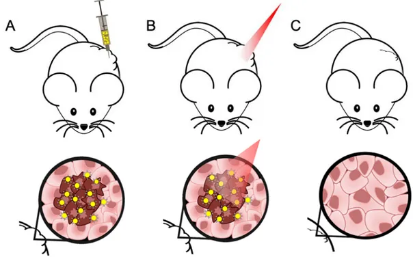
copper-based nanosystems for radiotherapy and Photothermal Therapy (PTT), used to treat thyroid carcinomas (Zhou et al., 2015). PTT is a non-pharmacologic and less invasive approach to target and reduce tumors by ther-mal ablation of cancer cells, with minither-mal side effects when compared to other therapeu-tic options (Silva et al., 2016b), as represented in Figure 1. A gold-based nano hybrid ap-proach has been developed for the treatment of melanoma, also showing very promising results for the treatment of other tumors, such as anaplastic thyroid carcinoma (Silva et al., 2016a, 2016b; Amaral, 2020).
Moreover, an incoming therapeutic ap-proach for the treatment of anaplastic thyroid carcinoma is using targeted inhibitors for hy-peractive and/or mutant components of sig-naling pathways (Saini et al., 2018). As previ-ously mentioned, members of the RAF/ MAPK and MEK pathway are mutated in this malignancy (i.e., EGFR, BRAF, Ras) (Saini et al., 2018; Santarpia et al., 2008; Smallridge
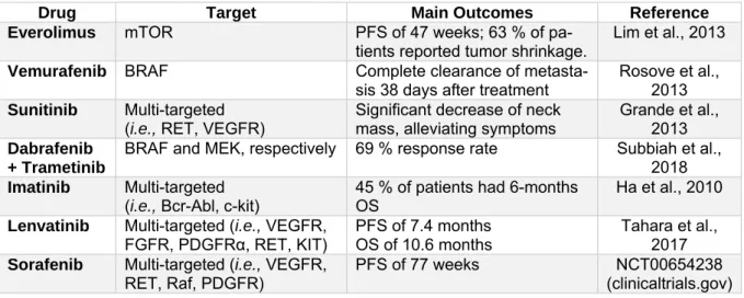
et al., 2009). Recent studies reported promis-ing results, either of decrease in tumor mass or good responses, using targeted/multi-tar-geted therapies, both individually or com-bined, enlightening the benefits of using se-quencing tools to identify possible targets for each patient (Tiedje et al., 2018). Further-more, small-molecule tyrosine kinase inhibi-tors, such as sorafenib (Nagaiah et al., 2011), vemurafenib (Rosove et al., 2013), dabrafenib (Subbiah et al., 2018), and trametinib (Subbiah et al., 2018), have been shown to be promising for the treatment of anaplastic thy-roid carcinoma, being currently in clinical tri-als, alone or in combination (i.e., dabrafenib + trametinib) (Nagaiah et al., 2011; Subbiah et al., 2018; Tiedje et al., 2018). However, when a small-molecule tyrosine kinase inhibitor was used alone, disease reoccurrence was re-ported by most patients (Saini et al., 2018). Table 2 summarizes the promising results of completed clinical trials using small-mole-cule tyrosine kinase inhibitors for the treat-ment of anaplastic thyroid carcinoma.
Figure 1: Different steps of PTT using a targeted hybrid gold-based nanosystem developed and a near-infrared (NIR) laser, in a xenograft mice model. (A) In situ administration of hybrid nanosystem at the tumor site, specific for tumor cells (represented in a darker color); (B) Irradiation of the injected site with a NIR laser to activate the formulation; (C) Reduction of tumor mass through thermal ablation.
Some of these therapeutic results dis-played much higher response rates in compar-ison to the current therapies used, being 10 months the longest overall survival rate re-ported. The 2-3-fold increase in overall sur-vival corresponds to less than a year.
Table 3 resents ongoing clinical trials (Phase I and II) registered on the ClinicalTri-als.gov database, and its respective database
ID, being conducted worldwide, using differ-ent treatmdiffer-ent approaches and protocols (i.e., radiotherapy, chemotherapy, immunotherapy, small-molecule tyrosine kinase inhibitors, and a combination of these therapies) for the treatment of anaplastic thyroid carcinoma.
Table 2: Results of completed clinical trials using targeted or multi-target therapies for the treatment of anaplastic thyroid carcinoma
Drug Target Main Outcomes Reference
Everolimus mTOR PFS of 47 weeks; 63 % of
pa-tients reported tumor shrinkage. Lim et al., 2013 Vemurafenib BRAF Complete clearance of
metasta-sis 38 days after treatment Rosove et al., 2013 Sunitinib Multi-targeted
(i.e., RET, VEGFR)
Significant decrease of neck mass, alleviating symptoms
Grande et al., 2013 Dabrafenib
+ Trametinib
BRAF and MEK, respectively 69 % response rate Subbiah et al., 2018 Imatinib Multi-targeted
(i.e., Bcr-Abl, c-kit) 45 % of patients had 6-months OS Ha et al., 2010 Lenvatinib Multi-targeted (i.e., VEGFR,
FGFR, PDGFRα, RET, KIT) PFS of 7.4 months OS of 10.6 months Tahara et al., 2017 Sorafenib Multi-targeted (i.e., VEGFR,
RET, Raf, PDGFR)
PFS of 77 weeks NCT00654238 (clinicaltrials.gov) PFS – Progression-Free Survival
OS – Overall Survival
Table 3: Ongoing clinical trials using targeted or multi-targeted therapies, radiotherapy, immunotherapy and chemotherapy for the treatment of anaplastic thyroid carcinoma
Drug Target Clinical.gov ID Phase
Lenvatinib Multi-Targeted (i.e., VEGFR,
FGFR, PDGFRα, RET, KIT) NCT02726503 II
Trametinib + Paclitaxel MEK, BRAF NCT03085056 Early I
Durvalumab + Tremelimumab + Stereotactic Body Radiotherapy
PD-1 and CTL-4, respectively NCT03122496 I
MLN0128 mTOR NCT02244463 II
Pembrolizumab PD-1 NCT02688608 II
Nexavar Multi-Targeted (i.e., RAF, MEK,
ERK, VEGFR, PDGFR) NCT03565536 II
Efatutazone + Paclitaxel PPAR γ NCT02152137 II
Intensity-Modulated Radiation Therapy + Paclitaxel + Pazopanib Hydrochloride
Multi-Targeted (i.e., VEGFR,
PDGFR, c-kit) NCT01236547 II Atezolizumab + Bevacizumab +
Cobimetinib + Paclitaxel + Vemurafenib
PD-1, VEGFR, MEK and BRAF, respectively
CONCLUSION
Although some advances have been done regarding the treatment of anaplastic thyroid carcinoma, it still presents low survival rates. There is an urgent need to improve the treat-ment of this rare malignancy, in order to sig-nificantly increase patients’ survival and their life quality improvement. Altogether, the best approach seems to be a more personalized multimodal course of treatment, since there is considerable variability of response to treat-ments using targeted/multi-targeted therapies between individuals with tumor molecular profile.
Funding
This research was funded by Fundação para a Ciência e a Tecnologia (FCT) through the Project Reference UID/DTP/04138/2019. Conflict of interest
The authors declare that they have no con-flict of interest.
REFERENCES
Akaishi J, Sugino K, Kitagawa W, Nagahama M, Kameyama K, Shimizu K, et al. Prognostic factors and treatment outcomes of 100 cases of anaplastic thyroid carcinoma. Thyroid. 2011;21:1183-9.
Amaral M. Non-invasive elimination of superficial tumours: a step forward [master's thesis]. Lisboa: Universidade NOVA de Lisboa, 2020.
Antonelli A, Fallahi P, Ferrari SM, Carpi A, Berti P, Materazzi G, et al. Dedifferentiated thyroid cancer: A therapeutic challenge. Biomed Pharmacother. 2008;62: 559-63.
Are C, Shaha AR. Anaplastic thyroid carcinoma: biology, pathogenesis, prognostic factors, and treat-ment approaches. Ann Surg Oncol. 2006;13:453-64. Biondi B, Filetti S, Schlumberger M. Thyroid-hormone therapy and thyroid cancer: a reassessment. Nat Clin Pract Endocrinol Metab. 2005;1:32-40.
Brabant G. Thyrotropin suppressive therapy in thyroid carcinoma: what are the targets? J Clin Endocrinol Metab. 2008;93:1167-9.
Busaidy NL, Cabanillas ME. Differentiated thyroid cancer: Management of patients with radioiodine nonresponsive disease. J Thyroid Res. 2012;2012:1-12.
Cabanillas ME, McFadden DG, Durante C. Thyroid cancer. Lancet. 2016;388:2783-95.
Cady B. Staging in thyroid carcinoma. Cancer. 1998; 83:844-7.
D'Avanzo A, Treseler P, Ituarte PHG, Wong M, Streja L, Greenspan FS, et al. Follicular thyroid carcinoma: Histology and prognosis. Cancer. 2004;100:1123-9. Dackiw APB. Anaplastic thyroid cancer. In: Sturgeon C (d): Endocrine neoplasia (pp 75-84). Boston, MA: Springer, 2010. (Cancer Treatment and Research, Vol. 153).
DeLellis RA. Pathology and genetics of thyroid carcinoma. J Surg Oncol. 2006;94:662-9.
Filetti S, Durante C, Hartl D, Leboulleux S, Locati LD, Newbold K, et al. Thyroid cancer: ESMO clinical practice guidelines for diagnosis, treatment and follow-up. Ann Oncol. 2019;30:1856-83.
Fisher KE, Jani JC, Fisher SB, Foulks C, Hill CE, Weber CJ, et al. Epidermal growth factor receptor overexpression is a marker for adverse pathologic features in papillary thyroid carcinoma. J Surg Res. 2013;185:217-24.
Gimm O. Thyroid cancer. Cancer Lett. 2001;163:143-56.
Giuffrida D, Gharib H. Anaplastic thyroid carcinoma: Current diagnosis and treatment. Ann Oncol. 2000;11: 1083-9.
Grande E, Capdevila J, Díez JJ, Longo F, Carrato A. A significant response to sunitinib in a patient with anaplastic thyroid carcinoma. J Res Med Sci. 2013;18: 623-5.
Green LD, Mack L, Pasieka JL. Anaplastic thyroid cancer and primary thyroid lymphoma: A review of these rare thyroid malignancies. J Surg Oncol. 2006; 94:725-36.
Ha HT, Lee JS, Urba S, Koenig RJ, Sisson J, Giordano T, et al. A phase II study of imatinib in patients with advanced anaplastic thyroid cancer. Thyroid. 2010;20: 975-80.
Haghpanah V, Fallah P, Naderi M, Tavakoli R, Soleimani M, Larijani B. Cancer stem-like cell behavior in anaplastic thyroid cancer: A challenging dilemma. Life Sci. 2016;146:34-9.
Harada T, Nishikawa Y, Suzuki T, Ito K, Baba S. Bleomycin treatment for cancer of the thyroid. Am J Surg. 1971;122:53-7.
Huang L-Y, Lee Y-L, Chou P, Chiu W-Y, Chu D. Thyroid fine-needle aspiration biopsy and thyroid cancer diagnosis: a nationwide population-based study. PLoS One. 2015;10:e0127354-e.
Jonklaas J, Sarlis NJ, Litofsky D, Ain KB, Bigos ST, Brierley JD, et al. Outcomes of patients with differentiated thyroid carcinoma following initial therapy. Thyroid. 2006;16:1229-42.
Jugan M-L, Levi Y, Blondeau J-P. Endocrine disruptors and thyroid hormone physiology. Biochem Pharmacol. 2010;79:939-47.
Kebebew E, Greenspan FS, Clark OH, Woeber KA, McMillan A. Anaplastic thyroid carcinoma. Cancer. 2005;103:1330-5.
Khairy G. Anaplastic transformation of differentiated thyroid carcinoma. Int J Health Sci. 2009;3:93-6. Khan YS, Farhana A. Histology, thyroid gland. Treasure Island, FL: StatPearls Publ., 2020.
Kitahara CM, Sosa JA. The changing incidence of thyroid cancer. Nat Rev Endocrinol. 2016;12:646-53. Kloos RT. Protecting thyroid cancer patients from untoward effects of radioactive iodine treatment. Thyroid. 2009;19:925-8.
Kondo T, Ezzat S, Asa SL. Pathogenetic mechanisms in thyroid follicular-cell neoplasia. Nat Rev Cancer. 2006;6:292-306.
Landriscina M, Pannone G, Piscazzi A, Toti P, Fabiano A, Tortorella S, et al. Epidermal growth factor receptor 1 expression is upregulated in undifferentiated thyroid carcinomas in humans. Thyroid. 2011;21:1227-34. Lang BH-H, Lo C-Y. Surgical options in undifferen-tiated thyroid carcinoma. World J Surg. 2007;31:969-77.
Leboulleux S, Baudin E, Travagli J-P, Schlumberger M. Medullary thyroid carcinoma. Clin Endocrinol. 2004;61:299-310.
Lee SL. Complications of radioactive iodine treatment of thyroid carcinoma. J Natl Compr Canc Netw. 2010; 8:1277-86; quiz 1287.
Lewinski A, Ferenc T, Sporny S, Jarzab B. Thyroid carcinoma: diagnostic and therapeutic approach; genetic background (review). Endocr Regul. 2000;34: 99-113.
Liebner DA, Haraldsdottir S, Shah MH. Chemotherapy of thyroid cancer: General principles. In: Wartofsky L, Van Nostrand D (eds): Thyroid cancer: A compre-hensive guide to clinical management (pp 717-21). New York, NY: Springer, 2016.
Lim SM, Chang H, Yoon MJ, Hong YK, Kim H, Chung WY, et al. A multicenter, phase II trial of everolimus in locally advanced or metastatic thyroid cancer of all histologic subtypes. Ann Oncol. 2013;24: 3089-94.
Lim SW. Targeted therapy of thyroid cancer. In: Braunstein G (ed): Thyroid cancer (pp 301-15). Boston, MA: Springer, 2012 (Endocrine Updates, Vol. 32).
Lin R-Y. Thyroid cancer stem cells. Nat Rev Endo-crinol. 2011;7:609-16.
Liu T-R, Xiao Z-W, Xu H-N, Long Z, Wei F-Q, Zhuang S-M, et al. Treatment and prognosis of anaplastic thyroid carcinoma: A clinical study of 50 cases. PLoS One. 2016;11:e0164840.
Liu Y, Su L, Xiao H. Review of factors related to the thyroid cancer epidemic. Int J Endocrinol. 2017;2017: 1-9.
McGriff NJ, Csako G, Gourgiotis L, Guthrie LC, Pucino F, Sarlis NJ. Effects of thyroid hormone suppression therapy on adverse clinical outcomes in thyroid cancer. Ann Med. 2002;34:554-64.
Molinaro E, Romei C, Biagini A, Sabini E, Agate L, Mazzeo S, et al. Anaplastic thyroid carcinoma: from clinicopathology to genetics and advanced therapies. Nat Rev Endocrinol. 2017;13:644-60.
Nagaiah G, Hossain A, Mooney CJ, Parmentier J, Remick SC. Anaplastic thyroid cancer: a review of epidemiology, pathogenesis, and treatment. J Oncol. 2011;2011:1-13.
Neff RL, Farrar WB, Kloos RT, Burman KD. Anaplastic thyroid cancer. Endocrinol Metab Clin North Am. 2008;37:525-38.
Nguyen QT, Lee EJ, Huang MG, Park YI, Khullar A, Plodkowski RA. Diagnosis and treatment of patients with thyroid cancer. Am Health Drug Benefits. 2015; 8:30-40.
Nikiforov YE. Genetic alterations involved in the transition from well-differentiated to poorly differentiated and anaplastic thyroid carcinomas. Endocr Pathol. 2004;15:319-28.
Nikiforova MN, Nikiforov YE. Molecular genetics of thyroid cancer: implications for diagnosis, treatment and prognosis. Exp Rev Mol Diagn. 2008;8:83-95.
Nosé V. Familial non-medullary thyroid carcinoma: An update. Endocr Pathol: 2008;19:226-40.
Opitz R, Schmidt F, Braunbeck T, Wuertz S, Kloas W. Perchlorate and ethylenethiourea induce different histological and molecular alterations in a non-mammalian vertebrate model of thyroid goitrogenesis. Mol Cell Endocrinol. 2009;298:101-14.
Pacini F, Castagna MG, Cipri C, Schlumberger M. Medullary thyroid carcinoma. Clin Oncol 2010;22: 475-85.
Paschke R, Lincke T, Müller SP, Kreissl MC, Dralle H, Fassnacht M. The treatment of well-differentiated thyroid carcinoma. Dtsch Arztebl Int. 2015;112:452-8. Quiros RM, Ding HG, Gattuso P, Prinz RA, Xu X. Evidence that one subset of anaplastic thyroid carcinomas are derived from papillary carcinomas due toBRAF andp53 mutations. Cancer. 2005;103:2261-8. Ragazzi M, Ciarrocchi A, Sancisi V, Gandolfi G, Bisagni A, Piana S. Update on anaplastic thyroid carcinoma: Morphological, molecular, and genetic features of the most aggressive thyroid cancer. Int J Endocrinol. 2014;2014:1-13.
Ranganath R, Shah MA, Shah AR. Anaplastic thyroid cancer. Curr Opin Endocrinol Diabet Obes. 2015;22: 387-91.
Robenshtok E, Tzvetov G, Grozinsky-Glasberg S, Shraga-Slutzky I, Weinstein R, Lazar L, et al. Clinical characteristics and outcome of familial nonmedullary thyroid cancer: A retrospective controlled study. Thyroid. 2011;21:43-8.
Rosove MH, Peddi PF, Glaspy JA. BRAF V600E inhibition in anaplastic thyroid cancer. N Engl J Med. 2013;368:684-5.
Ross DS. Radioiodine therapy for hyperthyroidism. N Engl J Med. 2011;364:542-50.
Rousset B, Dupuy C, Miot F, Dumont J. Chapter 2 Thyroid hormone synthesis and secretion. In: Feingold KR, Anawalt B, Boyce A, et al. (eds): Endotext. South Dartmouth, MA: MDText.com, Inc., 2000.
Saini S, Tulla K, Maker AV, Burman KD, Prabhakar BS. Therapeutic advances in anaplastic thyroid cancer: A current perspective. Mol Cancer. 2018;17(1):154. Salvatore G, Nappi TC, Salerno P, Jiang Y, Garbi C, Ugolini C, et al. A cell proliferation and chromosomal instability signature in anaplastic thyroid carcinoma. Cancer Res. 2007;67:10148-58.
Santarpia L, El-Naggar AK, Cote GJ, Myers JN, Sherman SI. Phosphatidylinositol 3-kinase/Akt and Ras/Raf-mitogen-activated protein kinase pathway mutations in anaplastic thyroid cancer. J Clin Endo-crinol Metabol. 2008;93:278-84.
Sawka AM, Thephamongkhol K, Brouwers M, Thabane L, Browman G, Gerstein HC. A systematic review and metaanalysis of the effectiveness of radioactive iodine remnant ablation for well-differentiated thyroid cancer. J Clin Endocrinol Metab. 2004;89:3668-76.
Schlumberger M, Brose M, Elisei R, Leboulleux S, Luster M, Pitoia F, et al. Definition and management of radioactive iodine-refractory differentiated thyroid cancer. Lancet Diabet Endocrinol. 2014;2:356-8. Shah JP. Thyroid carcinoma: epidemiology, histology, and diagnosis. Clin Adv Hematol Oncol. 2015;13:3-6. Shaha AR. Airway management in anaplastic thyroid carcinoma. Laryngoscope. 2008;118:1195-8.
Sherman SI. Thyroid carcinoma. Lancet. 2003;361: 501-11.
Sherman SI. Cytotoxic chemotherapy for differentiated thyroid carcinoma. Clin Oncol. 2010;22:464-8. Shimaoka K, Schoenfeld DA, Dewys WD, Creech RH, Deconti R. A randomized trial of doxorubicin versus doxorubicin plus cisplatin in patients with advanced thyroid carcinoma. Cancer. 1985;56:2155-60.
Silva CO, Petersen SB, Reis CP, Rijo P, Molpeceres J, Fernandes AS, et al. EGF functionalized polymer-coated gold nanoparticles promote egf photostability and EGFR internalization for photothermal therapy. PLoS One. 2016a;11:e0165419-e.
Silva CO, Rijo P, Molpeceres J, Ascensão L, Roberto A, Fernandes AS, et al. Bioproduction of gold nanoparticles for photothermal therapy. Ther Deliv. 2016b;7:287-304.
Smallridge RC, Copland JA. Anaplastic thyroid carcinoma: Pathogenesis and emerging therapies. Clin Oncol. 2010;22:486-97.
Smallridge RC, Marlow LA, Copland JA. Anaplastic thyroid cancer: Molecular pathogenesis and emerging therapies. Endocr Relat Cancer. 2009;16:17-44. Smallridge RC, Ain KB, Asa SL, Bible KC, Brierley JD, Burman KD, et al. American Thyroid Association guidelines for management of patients with anaplastic thyroid cancer. Thyroid. 2012;22:1104-39.
Soares P, Trovisco V, Rocha AS, Feijão T, Rebocho AP, Fonseca E, et al. BRAF mutations typical of papillary thyroid carcinoma are more frequently detected in undifferentiated than in insular and insular-like poorly differentiated carcinomas. Virchows Arch. 2004;444:572-6.
Soares P, Lima J, Preto A, Castro P, Vinagre J, Celestino R, et al. Genetic alterations in poorly differentiated and undifferentiated thyroid carcinomas. Curr Genom. 2011;12:609-17.
Stathatos N. Anatomy and physiology of the thyroid gland. In: Wartofsky L, Van Nostrand D (eds): Thyroid cancer (pp 3-7). Totowa, NJ: Humana Press, 2006. Stathatos N. Thyroid physiology. Med Clin North Am. 2012;96:165-73.
Subbiah V, Kreitman RJ, Wainberg ZA, Cho JY, Schellens JHM, Soria JC, et al. Dabrafenib and trametinib treatment in patients with locally advanced or metastatic BRAF V600–mutant anaplastic thyroid cancer. J Clin Oncol. 2018;36:7-13.
Tahara M, Kiyota N, Yamazaki T, Chayahara N, Nakano K, Inagaki L, et al. Lenvatinib for anaplastic thyroid cancer. Front Oncol. 2017;7:25.
Tiedje V, Stuschke M, Weber F, Dralle H, Moss L, Führer D. Anaplastic thyroid carcinoma: review of treatment protocols. Endocr Relat Cancer. 2018;25: R153-R61.
Tuttle RM, Ball DW, Byrd D, Dilawari RA, Doherty GM, Duh Q-Y, et al. Thyroid carcinoma. J Natl Compr Canc Netw. 2010;8:1228-74.
Wang H-M, Huang Y-W, Huang J-S, Wang C-H, Kok VC, Hung C-M, et al. Anaplastic carcinoma of the thyroid arising more often from follicular carcinoma than papillary carcinoma. Ann Surg Oncol. 2007;14: 3011-8.
Wein RO, Weber RS. Anaplastic thyroid carcinoma: palliation or treatment? Curr Opin Otolaryngol Head Neck Surg. 2011;19:113-8.
Wiseman SM, Loree TR, Rigual NR, Hicks WL, Douglas WG, Anderson GR, et al. Anaplastic trans-formation of thyroid cancer: Review of clinical, pathologic, and molecular evidence provides new insights into disease biology and future therapy. Head Neck. 2003;25:662-70.
Zhou M, Chen Y, Adachi M, Wen X, Erwin B, Mawlawi O, et al. Single agent nanoparticle for radio-therapy and radio-photothermal radio-therapy in anaplastic thyroid cancer. Biomaterials. 2015;57:41-9.
Zivaljevic V, Tausanovic K, Paunovic I, Diklic A, Kalezic N, Zoric G, et al. Age as a prognostic factor in anaplastic thyroid cancer. Int J Endocrinol. 2014;2014: 1-5.