HAL Id: hal-01583804
https://hal.archives-ouvertes.fr/hal-01583804
Submitted on 7 Sep 2017
HAL is a multi-disciplinary open access archive for the deposit and dissemination of sci- entific research documents, whether they are pub- lished or not. The documents may come from teaching and research institutions in France or abroad, or from public or private research centers.
L’archive ouverte pluridisciplinaire HAL, est destinée au dépôt et à la diffusion de documents scientifiques de niveau recherche, publiés ou non, émanant des établissements d’enseignement et de recherche français ou étrangers, des laboratoires publics ou privés.
Distributed under a Creative CommonsAttribution - ShareAlike| 4.0 International
Health
Fillipe Luiz Rosa Do Carmo, Houem Rabah, Bárbara Fernandes Cordeiro, Sara Heloisa da Silva, Gwénaël Jan, Vasco A Azevedo, Rodrigo Dias de
Oliveira Carvalho
To cite this version:
Fillipe Luiz Rosa Do Carmo, Houem Rabah, Bárbara Fernandes Cordeiro, Sara Heloisa da Silva, Gwénaël Jan, et al.. Applications of Probiotic Bacteria and Dairy Foods in Health. Current Research in Microbiology, Open Access eBooks 919 North Market Street Suite 425 Wilmington, DE 19801, 2017.
�hal-01583804�
Applications of Probiotic Bacteria and Dairy Foods in Health
Fillipe Luiz Rosa do Carmo
1,2; Houem Rabah
2,3; Bárbara Fernandes Cordeiro
1; Sara Heloisa Da Silva
1; Gwénaël Jan
2; Vasco Azevedo
1*; Rodrigo Dias de Oliveira Carvalho
11
UFMG, Federal University of Minas Gerais, 31270-901, Belo Horizonte, Minas Gerais, Brazil
2
STLO, INRA, Agrocampus Ouest, 35000, Rennes, France
3
Pôle Agronomique Ouest, F-35042, Rennes, France
*Correspondence to: Vasco Azevedo, UFMG - Federal University of Minas Gerais, Av. Antônio Carlos, 6627, Belo Horizonte - Minas Gerais, Brazil - 31.270-901
Tel.: +55 31 34092610; Fax: +55 31 34092614; Email: vascoariston@gmail.com
Chapter 1
Current Research in Microbiology
Abstract
The intestinal microbiota composition has a great impact on physiology and health, since commensal bacteria are crucial to maintain homeostasis and immune regulation of the gut.
Consequently, disturbances of this microbiota, a process known as dysbiosis, have severe im- plications for the host health such as the rise of many gastrointestinal (GI) problems; including inflammatory disorders like the Inflammatory Bowel Diseases (IBD), mucositis, as well as colorectal cancer (CRC). The consumption of probiotics with beneficial effects is a promis- ing tool to help treating such disorders. Indeed, they modulate diverse biological mechanisms involved in GI homeostasis and have been commonly used to reduce such disorders. In this chapter, we present the molecular mechanisms triggered by probiotic bacteria to modulate the gut physiology during gastrointestinal disorder and the importance of the gastrointestinal stresses tolerance as a limiting factors for probiotic application. Moreover, we focus on the emergence of functional probiotic foods, which can act as excellent vehicles, by enhancing stress tolerance and providing a protective matrix towards digestive stresses.
1. Introduction
Bacteria-host cross talk within the gut is a growing field of interest. While significant-
knowledge has been achieved by studies of interactions between pathogenic bacteria and the
host, much research is required for understanding the impact of commensal bacteria that reside
www.openaccessebooks.com
Azevedo V
within the human gastrointestinal tract (GIT) [1]. An increasing number of studies indicate that the intestinal microbiota is essential for host functions, especially immune responses that contributes to gut homeostasis [2-5]. More recently, several studies show correlations between disturbed microbiota composition (dysbiosis) and diseases which involve gastrointestinal in- flammation [6-9]. Individuals presenting these inflammatory conditions are colonized by an abnormal microbiota and it has been revealed that the lack of bacteria involved in regulation of the gut immune system might be a key factor in the chronicity of mucosal inflammation [10,11]. Therefore, a novel rationale aiming at the restoration of a healthy microbiota has been glimpsed by researchers to prevent and/or help in treating gastrointestinal diseases. In this context, there has been much encouragement for the use of probiotics and functional foods as therapies for such disorders. In this chapter, we describe the most recent advances of dairy foods and probiotic strains protective effects in animal models of intestinal inflammation and in human clinical trials. Furthermore, the challenges and limitations in regard of stability and safety of these approaches are discussed.
2. Gastrointestinal Tract 2.1. Microbiota
The GIT of mammals is a complex biological system whose main function is the diges- tion of food. As the GIT is an environment that is very rich in nutrients, particularly the ileum and colon parts, there is a dynamic community of microorganisms, known as intestinal micro- biota, which plays a role in the intestinal physiology and immune regulation [12-14].
The community of bacteria found in the GIT contains both indigenous and transient members. The first ones are well adapted to the intestinal environment and thus colonize the lumen. In turn, the transient microorganisms are not able to survive more than a few days.
Some transient species are frequently ingested in substantial amounts as they are present in fermented dairy foods such as yogurts, cheeses and fermented milk. However, transiting bac- teria also include several of the enteric food-borne pathogens [15,16]. The survival of alloch- thonous bacteria in the GIT depends on several factors including the ability to tolerate gastric acid, bile salts and pancreatic juice [17].
Many academic and industrial consortiums, such as the MetaHIT (Metagenomics of
the Human Intestinal Tract), have attempted to characterize the microbiota associated with
the human GIT through genomic sequencing, thus giving a more detailed description of the
human intestinal microbiota composition and of its function [18,19]. It is estimated that the
intestinal microbiota comprises 500 to 1,000 species of bacteria, exceeding 10 times or more
the total number of host cells [7,20,21]. Nowadays, it is known that most species found in
mammalian GIT can be classified into four phyla: Bacteroidetes, Firmicutes, Actinobacteria
and Proteobacteria [21-23]. These phyla keep symbiotic relationships with the host, making
fundamental contributions to the host metabolism while occupying a protected environment rich in nutrients [24,25]. In this context, the intestinal microbiota plays major roles such as nu- tritional functions, prevention of pathogen colonization, trophic functions on the proliferation and differentiation of the intestinal epithelium, and development and modulation of the host immune system [21,26-9].
Although the microbiome composition varies greatly among individuals, its composi- tion is relatively simple during the first years of life. It has been reported that Escherichia coli and streptococci are the most common organisms isolated from the upper GIT shortly after birth [21,30]. These species are responsible for creating a favorable environment, promoting in turn the colonization by anaerobic bacteria, among which Bifidobacterium and Bacteroidetes are most prevalent. The Bifidobacterium genus plays an important role in the intestines, as these bacteria can down-regulate the expression of key proinflammatory mediators in the gut and inhibit pathogens. Bacteroidetes species have great capability to digest complex sugars, and thus maintain stable symbiotic relationship with the host and central position in the gut microbiome [31-34]. Afterwards, in the course of life, there can be an increase of Firmicutes, especially of the Lactobacillales and Clostridiales Orders. Among these, several members, including remarkably Faecalibacterium prausnitzii, are Short Chain Fatty Acid-producers, SCFAs playing an important role on the maturation of regulatory T cells [35]. Although micro- biota is stable in older persons, it can be altered in short term duration by dietary intervention [36]. Dietary intake shifts, mainly of dairy foods containing Lactoccocus sp or Propionibacte- rium sp and non digestible carbohydrates, may change the composition of the gut microbiota by increasing the number of Bifidobacterium sp and F. prausnitzii, although these bacteria do not respond in the same way in all human subjects [27,37,38].
In adulthood, the diversity and abundance of bacterial populations vary along the differ- ent parts of GIT [13,21,39,40]. In the stomach and duodenum, a small number of microorgan- isms can be found, while up to 10
3bacterial cells are present per gram of duodenal content.
These bacteria are adhered to the mucosal surface or in transit through the GIT [26,41,42].
Streptococci and lactobacilli are among the most common groups of bacteria found in this part of the intestine [39,43-45]. The bacterial population increases gradually along the jejunum and the ileum, reaching numbers around 10
4-10
7per gram of small intestinal content. However, it is in the lower GIT (colon) that the highest density of bacteria population is encountered, reaching a number of 10
11-10
12per gram, making this area one of the most complex microbial ecosystems known to date on Earth [13,45].
2.2. Immune system regulation
Commensal species from the intestinal microbiota are not ignored by the immune sys-
tem but on the contrary need to be recognized by mammalian cells to deliver tolerogenic sig-
nals and promote intestinal immune homeostasis with an impact on the immune system of the body. Actually, commensal bacteria and their host have co-evolved diverse biological mecha- nisms making this cross talk possible [46]. For example, pattern recognition receptors (PRRs), especially Toll-like receptors (TLRs), expressed by Intestinal Epithelial Cells (IECs), are able to recognize microbe-associated molecular patterns (MAMP) of the commensal microbiota. In fact, the expression of PRR and their interaction with the microbiota is very important for the healthy development of the host immune system [47-49].
These MAMP are microbial components, such as lipoproteins, nucleic acids (RNA and unmethylated CpG dinucleotides), lipopotheic acids, lipopolysaccharide (LPS), surface pro- teins such as flagellin and peptidoglycan [7,50-52]. The recognition of a MAMP transduces signals that subsequently activates innate immune responses [50,52].
Species from Lactobacillales order and the Actinobacteria phylum (Bifidobacterium sp.
and Propionibacterium sp.) [30,53] are capable of stimulating luminal secretion of antimicro- bial peptides by Paneth cells, mucins by goblet cells and fortifying tight junctions of IECs [54, 55]. Furthermore, commensals are reported to induce signals of immunological tolerance, such as secretion of the TGF-β cytokine that inhibits the NF-κB signaling pathway inside epithelial cells. It has been shown that activation of TLRs by commensal bacteria also promotes the de- velopment of CD103 dendritic cells, which are responsible for driving the activation of Treg cells [56-58]. It is known that Treg cells suppress effector T cell responses mainly through the production of IL-10 and TGF-β. The stimulation of these cytokines prevents the recruitment of granulocytes, suppressing the activation of macrophages, neutrophils and endothelial cells.
In addition, Treg cells expressing TGF-β and IL-10 drive B cells to undergo antibody class switching to produce IgA antibody, the major humoral defense of mucosal surfaces. Secretory IgA (sIgA) contributes to mucosal homeostasis through a process known as immune exclu- sion. sIgA is able to bind to opportunistic pathogens avoiding its dissemination throughout the body [59-61].
Although commensal microorganisms show beneficial effects on the host, some mi- crobes of the GIT might present potential risk if case of outgrowth. In this context, potentially pathogenic species, known as pathobionts, composed mainly of Proteobacteria members, such as Escherichia coli, and species from the phylum Firmicutes, as Clostridium difficile and En- terococcus faecalis can elicit a pro-inflammatory immune response after binding to TLR [47, 53,62].
When pathobionts translocate to intestinal epithelium, the host immunity is activated
and is usually enough to eliminate the intruder. Nonetheless, the overproduction of pro-inflam-
matory cytokines, which may occur during dysbiosis, represents a risk once inflammation may
also be problematic causing cell disruption and infection to the host. Therefore, to reach intes-
tinal homeostasis, the gut immune system must be able to recognize and eliminate specifically these pathobionts from the GIT [54,63]. Intestinal barrier dysfunction generates an imbalance between immune responses observed for protective and harmful intestinal bacteria and thus contributes to the onset of several inflammatory conditions of the GIT [7,64].
2.3. Inflammatory disorders
The GIT is permanently challenged by antigens from the intestinal microbiota. Under normal conditions, the intestinal mucosa maintains tolerance to commensals, mainly through the action of Treg cells. When the dynamic balance between Treg and activated effector cells is broken, the homeostasis is compromised and this may lead to the development of mucosal inflammation [2]. Besides dysbiosis, multiple factors can influence the proper functioning of the GIT immune system, including individual genetic background, diet, use of drugs and envi- ronmental stress. The intersection of these factors generates an exaggerated pro-inflammatory reaction against commensal antigens leading to Inflammatory Bowel Diseases (IBD), a group of chronic inflammatory conditions of the GIT, which primarily includes ulcerative colitis (UC), and Crohn’s disease (CD) [21,30]. Clinical symptoms of both diseases are similarly found in patients, such as abdominal pain, diarrhea, rectal bleeding and weight loss [65,66].
Relapse symptoms can last days, weeks, or even months [67]. CD is characteristically dis- continuous, with inflamed areas that can be found in all the layers of the intestinal wall, while UC is characterized as a continuous and superficial inflammation limited to the colon [68,69].
The incidence of these diseases varies widely across countries, however, in recent years, it has increased considerably worldwide, being considered a global public health problem. This increase has been associated with the modern lifestyle that includes the ingestion of processed foods usually high in fats and sugar and low in fiber [70-72].
Caesarean delivery and the inappropriate use of antibiotics, especially during childhood, when the microbiota has not yet been established, are factors that can contribute to the devel- opment of intestinal inflammation [30,72–74]. Both CD and UC have different immunological aspects when it comes to innate and adaptive immunity [75,76]. Pro-inflammatory cytokines are over expressed in IBD patients; however, the predominant set of cytokines observed in CD patients is the one secreted by Th1 and Th17 cells (IL-12, IL-23, IL-27, IFN-γ) whereas in UC patients a Th2 immune response, characterized by the production of IL-4 and IL-13, appears to be predominant [3,4,74].
Chronic inflammation also plays a role in the pathogenesis in several cancers and it has been shown that there is a direct link between IBD and colorectal cancer (CRC) [77]. Indi- viduals suffering from long-term ulcerative colitis or Crohn’s disease have increased risk of developing CRC [78].
Reactive Oxygen Species (ROS), stimulated by proinflammatory response in the intes-
tinal mucosa, play an important role in the development of CRC, as their excessive levels can result in oxidative stress and significant damage to cell structures and macromolecular constit- uents, such as DNA, RNA, proteins and lipids [79,80]. Large amounts of hydrogen peroxide (H
2O
2) are produced and excreted by human tumor cells, and might participate in tumor inva- sion and proliferationas well [78,81]. Furthermore, current studies are investigating the role of effector immune responses against intestinal microbiota in modulating the gut microbiota into a carcinogenic profile composition. In fact, a correlation between diet-driven sulfidogenic bacteria and CRC in African Americans has been demonstrated [82,83].
Other factors such as the use of some medicines can also contribute to the breakdown of this immunological tolerance against commensals commonly observed under normal condi- tions. It has been described that chemotherapeutic agents, as 5-Fluoracil (5-FU), doxorubicin and irinotecan (CPT-11), widely used in the treatment of advanced solid tumors, may also lead to the development of another inflammatory condition of the GIT, known as mucositis. This painful inflammation of the mucosa can affect all portions of the human GIT and has great medical importance as it arises as an adverse effect of chemotherapy [84,85]. These medicines are effective in cancer treatment because they inhibit cell proliferation. 5-FU, for example, causes cytotoxic effect by inhibiting DNA replication in cells with a high mitotic index such as malignant cells. Moreover, this drug can also be incorporated into RNA molecules interfer- ing with their processing and function. However, as an adverse consequence, the drug also shows an effect in normal cells that presents a higher turnover rate, such as GIT enterocytes [86]. Gastrointestinal mucositis is being regarded as a major risk, occurring in 80% of patients receiving 5-FU [87,88].
Patients with mucositis develop symptoms like odynophagia (pain in swallowing), vomiting, abdominal pain and diarrhea, which make eating difficult. Therefore, weight loss and malnutrition are also reported and quality of patient`s life is Gastrointestinal mucositis is characterized by morphological alterations in the mucosal architecture, as villous atrophy, increased cryptsapoptosis, that expose the mucosa to intestinal pathogens, which are able to translocate across intestinal epithelial cells leading to inflammatory responses [89].
The pathophysiology process of mucositis is very complex and involves the release
of endogenous damage-associated molecular pattern (DAMP) molecules and activation ofthe
NF-kB pathway, which induces in turn the expression of several genes, including pro-apop-
totic enzymes such as caspases, tumor necrosis factor-alpha (TNF-α), IL1-β and IL-6 cytok-
ines, and chemokines involved in the recruitment of neutrophils and eosinophils. Commensal
bacteria might have a very important role in promoting many clinical aspects involved in the
pathogenesis of mucositis [90-94], as it was demonstrated that germ-free mice are more resis-
tant to mucositis induction [85,95]. Moreover, opportunistic species belonging to the gut mi-
crobiota, such as Entercocccus faecalis, Escherichia sp and Clostridium sp can alter intestinal
permeability during anticancer treatments and promote disruption of the epithelial layer. The translocation of commensals across IEC exacerbates inflammatory responses and amplifies the damage to the intestinal mucosa [92,95].
As alterations of the intestinal microbiota have been implicated in all of these patholo- gies, the scientific community has been investigating the use of probiotics in order to restore the original gut microbiota, which is responsible for regulating the mucosal immune system.
3. Probiotics
The administration of probiotics for treating gastrointestinal inflammatory disorders have been proposed by many research groups, as they are able to occupy niches that compete with pathogens in the GIT [96-101]. Probiotics are defined as “live microorganisms, which, when administered in adequate amounts, confer a health benefit on the host” [102]. These mi- croorganisms must be safe and present beneficial effects during their transit through the gut.
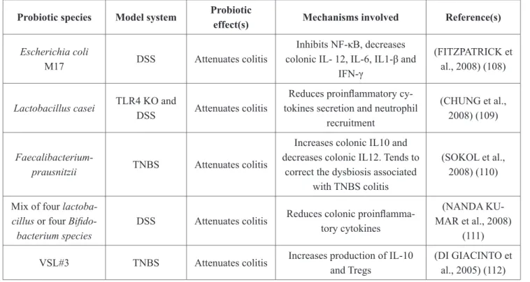
Hence, their ability to resist the stomach and intestine environments are crucial alongwith the capacity of adhesion to intestinal cells, inhibition of pathogens and immunomodulatory effects [103]. Currently, several species of probiotic bacteria are used to prevent or treat a diversity of diseases, including gastrointestinal inflammatory disorders (Table 1). Lactobacilli and bi- fidobacteria, for a long time, were at the front of the stage in the field of this probiotic action.
However, outsider bacterial species such as Lactococcus lactis, Streptococcus thermophilus, Escherichia coli and Propionibacterium freudenreichii recently revealed promising potential for the treatment of intestinal inflammation as well [104-107]. This is summarized in section 4.
Table 1: Immunomodulatory effects of probiotics in experimental animal model Probiotic species Model system Probiotic
effect(s) Mechanisms involved Reference(s) Escherichia coli
M17 DSS Attenuates colitis
Inhibits NF-κB, decreases colonic IL- 12, IL-6, IL1-β and
IFN-γ
(FITZPATRICK et al., 2008) (108)
Lactobacillus casei TLR4 KO and
DSS Attenuates colitis
Reduces proinflammatory cy- tokines secretion and neutrophil
recruitment
(CHUNG et al., 2008) (109)
Faecalibacterium-
prausnitzii TNBS Attenuates colitis
Increases colonic IL10 and decreases colonic IL12. Tends to
correct the dysbiosis associated with TNBS colitis
(SOKOL et al., 2008) (110)
Mix of four lactoba- cillus or four Bifido- bacterium species
DSS Attenuates colitis Reduces colonic proinflamma- tory cytokines
(NANDA KU- MAR et al., 2008)
(111) VSL#3 TNBS Attenuates colitis Increases production of IL-10
and Tregs
(DI GIACINTO et al., 2005) (112)
Lactobacillussali-
varius Ls33 TNBS Attenuates colitis Increases IL-10 production and Tregs
(MACHO FER- NANDEZ et al.,
2011) (113) Lactobacillus
plantarum DSM 15313, Lactobacillus
fermentum 35D
DSS Attenuates colitis Reduces bacterial translocation (OSMAN et al., 2008) (114)
Bacteroides fragilis TNBS Attenuates colitis Increases production of IL-10 and Tregs
(ROUND and MAZMANIAN,
2010) (115) Lactobacillus
salivarius 433118, Bifidobacterium
infantis
IL-10 KO Attenuates colitis Reduces inflammatory cytokines (MCCARTHY et al., 2003) (116)
Lactobacillus casei
Shirota DSS Attenuates colitis
Reduces IL-6 production by lamina propria mononuclear
cells
(MATSUMOTO et al., 2005)(117)
Enterococcus fae- cium CRL 183
1,2 dimethylhy- drazine (DMH)
Reduces ACF and adenocarcinomas
incidence
Improved the immune response by increasing IL-4, IFN-γ, and
TNF-α production
(SIVIEIRI et al., 2008)(118)
Saccharomyces boulardii
C57BL/6J Min/+ (Apc- Min) mice (7
wk old).
Reduces number and diameter of the tumors, the
score for low- grade dysplasia,
numbers of polyps, and cell
proliferation
Inactivation of the EGFR-Mek- Erk pathway signaling. Increase
apoptosis
(CHEN et al., 2009)(119)
Lactobacillus acido-
philus NCFM CT-26 cells
Reduces tumor size and the extraintestinal metastatic tissue
Increase apoptosis through in- crease caspase-9 and caspase-3
and reduces Bcl-2 expression
(CHEN et al., 2012) (88)
Lactobacillus plantarum AdF10 and Lactobacillus rhamnosus GG
1,2 dimethylhy- drazine (DMH)
Reduces tumor incidence, multi-
plicity, and size
Reduces COX-2 protein expres- sion
(WALIA et al., 2015) (120)
Lactobacillus sali- varius Ren
1,2 dimethylhy- drazine (DMH)
Reduces tumor incidence
Reduces Intestinal population of Ruminococcussp and Clostridi- ales bacteria ↑ Intestinal popula-
tion of Prevotellasp
(ZHANG et al., 2015) (121)
Dead nanosized Lac- tobacillus plantarum
Azoxymethane/
Dextran Sulfate Sodium-In-
duced
Reduces tumor incidence; areas
of dysplasia, adenocarcinoma,
and structural disruption
Reduces Over expression of proinflammatory cytokines and
inflammatory genes Increase Apoptosis and cell cycle arrest
(LEE et al., 2015) (122)
Lactobacillus rham- nosus and Lactoba- cillus acidophilus
1,2 dimethylhy- drazine (DMH)
Reduces tumor incidence, burden
and multiplicity;
lipid peroxidation
Reduces GSH, SOD, and GPx activity
(VERMA and SHUKLA 2014)
(123)
Lactobacillus casei BL23
1,2 dimethylhy- drazine (DMH)
Reduces colorec- tal cancer
Regulates Treg and Th17 T-cell populations
(LENOIR et al., 2016) (124) Pediococcus pen-
tosaceus GS4 Azoxymethane Attenuates colon cancer
Triggered apoptosis in colono- cytes
(DUBEY et al., 2016) (125)
3.1. Bacteria for the treatment of gastrointestinal disorders
Many strains of bacteria are known to exert anti-inflammatory effects through the mod- ulation of factors that are involved in maintaining intestinal homeostasis in humans and other animals [97,101]. In this context, bacterial effectors of distinct nature have been implicated in probiotic effects. These include metabolites, peptidoglycan, surface proteins, lipoproteins and lipoteichoic acids, lipopolysaccharides, flagelin and CpG motifs in DNA. Some of these molecules, such as anti-microbial peptides and prebiotic metabolites may interact directly with other species of bacteria that colonize the gut, modulating their growth. Others bacterial fac- tors, called MAMP, bind to PRRs of eukaryotic cells and stimulate different patterns of gene expression in the host involved in innate immunity activation and differentiation of antigen- specific immunity [53,126]. The mechanism of action of these bacteria can be classified in three main categories: alteration of gut microbiome composition, stimulation of epithelial bar- rier function; and induction of the immune responses [127].
Recent advances in genomic sequencing technologies have provided the scientific com- munity with tools to explore the human microbiome and how different treatments affect its global composition and function. Several studies have shown that probiotics can increase or decrease the abundance and diversity in gut microbial species composition. The secretion of antimicrobial compounds acts by directly inhibiting the growth of pathogens. In addition, pro- biotic strains may also reduce the impact of pathogens through a mechanism known as com- petitive exclusion, in which they occupy binding sites at the mucosal surface [97,128,129].
Epithelial barrier function enhancement is a well-established mechanism of probiotic bacteria in the protection of the host against invasive harmful bacteria. Numerous studies have shown that probiotics have the potential to modulate many of the processes involved in mucosal barrier formation and are able to upregulate expression of defensins, mucins or proteins associated with tight junctions such as claudins and occludins [130-132]. This effect is therefore considered as one of most important for the prevention and treatment of IBD and mucositis, as it might avoid translocation of opportunistic pathogens to systemic circulation [84,101].
Probiotics can affect the host heath by modulating inflammatory signaling pathways.
Several probiotics are reported to inhibit the NF-kB activation and thus to influence down-
stream cytokine secretion [133]. Recent studies demonstrated that the anti-inflammatory effects
of some bacteria involve inhibition of IkB degradation by targeting the different steps involved
in this process which are phosphorylation, ubiquitination or proteasome degradation[134].
Some Lactobacilli have shown inhibitory activity of TNF-alpha induced secretion of IL-8 [135]. Other well established immunological mechanism of probiotics is the stimulation of immunological tolerance to GIT microbiota through the increase in IL-10 secretion. For in- stance, Santos and collaborators (2014) showed that the probiotic effect of L. delbrueckii strain CNRZ327 was related to an expansion of Treg cells and an increase of total IgA in Dextran sulfate sodium (DSS)-induced colitis in mice. Recently, it was reported that a Lactococcus- lactiss sp. lactis NCDO2118 strain prevented DSS-induced colitis in mice and the protective effect was related to increased IL-10 levels in the colon and to the induction of Treg cells in the mesenteric lymph nodes [99].
Most studies focused on the beneficial effects autochthonous Lactobacilli (Table 1).
However, recent studies have demonstrated that some allochthonous strains have anti-inflam- matory properties. Ballal and colleagues (2015) found that L. lactis I-1631 prevents colitis in T-bet−/− Rag2−/− mice. Two additional studies have shown that, among the L. lactis species, NCDO2118 subsp. lactis or FC subsp. cremoris are anti-inflammatory when inoculated in in- flamed mice receiving the chemical agent DSS [99,136]. Moreover, L. lactis NZ9000 by itself was able to prevent histological damage and reduce neutrophil and eosinophil infiltration in mice injected with 5-FU. Another allochthonous species with anti-inflammatory effects in IBD models is Propionibacterium freudenreichii, used extensively as a ripening starter of Emmen- tal cheese [104,137,138].
3.2. Challenges and limitations to select probiotic bacteria
For probiotic bacteria selection, the robustness of a bacterium against different abiotic and biotic stresses is crucial, and may constitute a limiting factor for its application as probi- otic. Firstly, to prepare probiotic ingredients, a plethora of stresses are applied, thus the bacte- rial tolerance is a prerequisite for reaching a high survival rate in the product. In the traditional cheese products, the manipulation of bacterial population could be limited by other factors.
However, for the probiotic powders, great efforts were made to maintain a high viable bacte- rial population during freeze-drying or spray drying, such as usage of encapsulation methods.
Gastrointestinal stresses also constitute the main bottleneck of probiotic efficacy.
A probiotic microorganism must be able to tolerate digestive stresses and to adhere to intestinal epithelium, for a long persistence in the host and for an enhanced beneficial effect.
Gastric acid and bile salts are defense mechanisms encountered during intestinal transit where- as pancreatic secretions can also exert some antimicrobial activity via the digestive enzymes.
The existing microbiota may also interfere with the probiotic effect by competition for adhe-
sion or nutrients. The investigation of molecular basis of the adaptive response to stresses and
identification of the pivotal genes involved provided pertinent tools for probiotic screening.
3.2.1. Acid stress
The probiotic resistance to acid stress is a desired characteristic of selected strains, as low pH is widely encountered both during technological processing and during gastric diges- tion. The bacterial adaptive responses to acid challenge have been investigated and some of the molecular mechanisms involved were elucidated, such as induction of proton ATP-dependent pumps F
1F
0-ATPase. The function of this transmembrane protein complexis the extrusion of protons from the cell cytoplasm, resulting in a Proton Motive Force (PMF), and avoiding acid- stress induced drop in intracellular pH [139]. Mutations leading to a reduction of membrane- bound ATPase activity were observed in some strains of Lactococcus lactis subsp. lactis and Lactobacillus helveticus, where they cause growth inhibition under acid conditions [140,141].
Gram-positive bacteria, such as Lactococcus lactis [142] and Lactobacillus brevis [143], pos- sess a second mechanism for an adaptive response to acid stress, involving the enzyme glu- tamate decarboxylase (GAD). The GAD system imports glutamate into the cell prior to its decarboxylation, which consumes protons, participating in intracellular pH homeostasis, fol- lowed by the efflux of the resulting γ-aminobutyrate (GABA), thanks to a GAD/GABA anti- porter. Another mechanism involved in pH homeostasis is the proton-consuming malolactic fermentation (MLF). This metabolic pathway leads to the conversion of the dicarboxylic malic acid to the monocarboxylic lactic acid. The latter is excreted via a lactate-malate antiporter, resulting in intracellular alkanization. Such mechanism was observed within several bacteria like Lactobacillus sakei [144], Lactobacillus plantarum [145], and Lactococcus lactis [146].
Finally, other acid-adaptive mechanisms induced in lactic acid bacteria includethe citrate-lac- tate antiporter (CitP), the arginine deiminase (ADI) system and some heat shock [147-149].
3.2.2. Bile salts
Conjugated bile salts are synthesized by the liver with the amino acids glycine or tau-
rine, those amphipathic molecules act like biological detergents with strong antimicrobial ac-
tivity cause can emulsify biological membrane lipids [150,151]. These compounds may enter
into the bacterial cytoplasm by flip-flop mechanism and cause oxidative stress which leads to
DNA damage [152-154]. In fact, there are different remarkable mechanisms leading to bile
salts tolerance; those molecular actors can also provide bacteria a cross protection towards
other stress types. Some probiotic bacteria hold the ability to hydrolyze bile salts by bile
salt hydrolases (BSHs) which enhances their survival in the digestive tract [155]. Alternative
mechanisms exist such as bile-efflux systems, which are multidrug transporters that mediate
the active extrusion of bile salts from the bacterial cytoplasm [156]. Regarding Lactobacillus
acidophilus particularly, an eight-gene operon encoding for, a two-component regulatory sys-
tem, a transporter belonging to the major facilitator super family, an oxido reductase, and four
hypothetical proteins, has been implicated in bile salts removal [157].
3.2.3. Heat stress
Heat stress is another type of ordeal that is commonly suffered during technological processes, either during food fermentation (cheese cooking-step) or during drying, which may impose high temperatures (>60°C) or low temperatures, depending on the chosen technology.
The response to heat stress involves a set of proteins called Heat Shock Proteins (HSP), which include chaperones and proteases. They are essential for overcoming protein denaturation, maintaining cell homeostasis in response to variations of temperature, which can affect mem- brane fluidity and compromise cellular integrity and basic cell processes [158,159]. Among those crucial proteins, DnaK and GroEL, are two HPSs that have a critical role in cellular processes by maintaining DNA replication process, preventing mutagenesis and preventing protein denaturation [158,160]. Otherwise, low temperatures are frequently used to prevent spoilage during frozen and freeze-dried storing process. Such a cold stress leads to induction of specific proteins called cold shock proteins (CSPs). Their role consists in maintaining tran- scription and translation processes under cold stress adaptation [159].
3.3. Protective matrix and vectorization
Although the adaptive response to various stresses is a quite important feature to screen tolerant or sensitive probiotic strains, vehicle matrix can confer a protection for an efficient delivery of probiotic bacteria to the GIT. Probiotics are commonly consumed under the form of dried powder, in capsules or tablets. Recently, various studies focused on “2-in-1”starter bacteria: microorganisms widely used in food fermentation and which exert beneficial effects.
There is a huge variety of fermentative microorganisms known for their probiotic properties like S. thermophilus, L. delbrueckii ssp. bulgaricus, L. lactis, and other strains used for specific fermented foods [161]. The growth in such stressful medium as dairy fermented foods selects bacteria with a high robustness to GIT stresses there by promoting long-term survival during storage in industrial process.
3.3.1. Encapsulation by biopolymers
Encapsulation is a standard process used to produce protected dried probiotic ingre-
dients. Encapsulation may confer a protection against industrial stresses during the drying
process, and allow a controlled release in the GIT [162,163]. Depending on the type of drying
technology (capsule spray-drying, emulsification, extrusion, co-extrusion, or spray-coating),
different particles sizes may be obtained and interfere in the encapsulation yield. Moreover,
semi-permeable and biocompatible matrices including food-grade biopolymers like alginate,
pectin and cellulose acetate phthalate are used for preventing oxidative reaction, masking fla-
vor and odor changes. The encapsulation essentially provides a protection for bacteria and a
specific addressing of active probiotic compounds to specifics sites [164,165]. Among poly-
mers used for encapsulation; alginate, a polysaccharide composed of β-D-mannuronic and
αL-guluronic acids, is widely used, because of its simplicity, biocompatibility, low cost, and non-toxicity. Recently, the encapsulation by alginate was shown to confer enhanced viability upon storage and simulated gastrointestinal digestion for Lactococcus lactis subsp. cremoris LM0230, Lactobacillus casei NCDC 298, Bifidobacterium longum and other probiotics [166- 168].
3.3.2. Encapsulation by milk proteins
The utilization of milk proteins for probiotic encapsulation is a high quality choice due to their biocompatibility, structural and physico-chemical properties [169]. Milk proteins are categorized in two types: caseins and whey proteins. Caseins are a complex aggregate of phos- phoproteins and are extremely heat-stable proteins, present in colloidal form known as caseins micelles in fresh milk [170]. Whey proteins are a group of globular proteins, α-lactalbumin, ß-lactooglobulin, immunoglobulins, and serum albumin and also various other minor proteins [171]. Milk proteins clotting followed by spray-drying appears as new innovative method- ology to encapsulate probiotic bacteria to enhance survival in GIT [172]. Heidebach et al.
in 2009 demonstrated new methodologies based on a transglutaminase-catalyzed gelation of casein suspensions and spray drying to encapsulate Lactobacillus paracei ssp. paracasei F19 and Bifidobacterium lactis Bb12. It was shown to enhance robustness instressing conditions [173,174].
3.3.3. Dairy Fermented foods
The emergence of functional foods concept such as fermented products is a promising research area. Indeed, the dairy fermented foods constitute an important part of our daily diet [175], as well as our main microbial daily intake. The dairy product matrix may increase toler- ance of bacteria towards digestive stresses and adhesion to cells, depending on its biochemical composition, its physical microstructure and the existing microbial ecosystem, which affects directly the viable bacterial amount reaching the gut. Beyond the protection effect of the ve- hicle matrix, fermentation allows improvement of food nutritional value through the microbial release of a high amount of essential nutrient for consumers [176], including vitamins.
Probiotic bacteria convert different molecules, producing valuable nutrients like con-
jugated fatty acid, B-galactosidase enzyme, beneficial dairy peptide, which can enhance their
probiotic functionality [24,25]. Indeed, L. casei BL23 incubated in milk reduced significantly
the symptoms of a dextran sulfate sodium (DSS) - induced colitis in a murine model, com-
pared to the same strain provided in phosphate buffered saline [178,179]. Yogurt enhances the
therapeutic value of some probiotic bacteria, however the low pH of yogurt decreases viable
population [177,180,181]. To contend this problem, a combination of encapsulated probiotic
bacteria was used to increase survival in yogurt. For example, L. paracasei subsp. paracasei
E6 were encapsulated using whey proteins and gum arabic, before being added to the yogurt
matrix after fermentation. This bacterium exhibited greater viability, compared to cells with- out encapsulation, upon exposure of the probiotic yogurt to simulated gastric juice [182,183].
Cheese matrix favors probiotic beneficial effects by providing a favorable environment with relatively high fat content, enhancing probiotic survival in transit through the GIT, espe- cially towards lethal conditions of the stomach [184]. Özer and colleagues showed increased viability of Bifidobacterium bifidum BB-12 and of Lactobacillus acidophilus LA-5, when these bacteria were microencapsulated in white-brined cheese, compared to same strains without encapsulation protection [185]. Other strains have been used to produce experimental probi- otic cheese, including Lactobacillus casei and Lactobacillus acidophilus in Crescenza cheese.
They exhibited improved viability, during the refrigerated storage, after a cheese manufacture using High-pressure homogenization (HPH) as an alternative to traditional thermal treatment [186]. The potential probiotic P. freudenreichii, alone or in combination with Lactobacillus delbrueckii, was investigated with respect to the prevention of UC. The experimental ferment- ed cheeses exhibited promising anti-inflammatory properties in mice with colitis [187,188].
Moreover, inclusion within a cheese enhanced P. freudenreichii tolerance towards digestive stresses and thus its probiotic properties [105].
4. Propionibacterium Freudenreichii for Treating Gastrointestinal Disorders
During the last two decades, an outsider, that had until then been ignored, was consid- ered for probiotic applications. The propionibacterium P. freudenreichii, until then used almost exclusively to confer aroma to pressed cheeses, joined the main probiotic actors on the stage of probiotic research and development. P. freudenreichii indeed recently revealed unexpected immunomodulatory effects. This evidenced for the first time a “two-in-one” property of an ill- known ripening starter, with both technological and probiotic abilities.
4.1. General aspects of P. freudenreichii 4.1.1. Taxonomy
P. freudenreichii is a dairy propionibacterium, which belongs to Actinobacteria, charac-
terized as gram-positivewith a high G+C content, non- sporing, anaerobic to aerotolerant, non-
motile pleomorphic rods [189,190]. Actinobacteria comprise bacterial species with a myceli-
um-like aspect, found in various environments, including animal hosts and soil [189,190]. The
genus Propionibacterium comprises both cutaneous species, which may act as opportunistic
pathogens, and dairy species, which have no reported adverse effects up to date [191]. The
typical dairy species isolated from milk are: P. freudenreichii, P. acidipropionici, P. jensenii
and P. thoenii; they are clearly distinct from cutaneous species. Dairy propionibacteria were
firstly described by E. von Freudenreich and S. Orla-Jensen at the end of 19
thcentury, since
their presence in Emmental cheese was associated with propionic fermentation [192]. Dairy
propionibacteria, specifically P. freudenreichii, possess a long history of safe use in food, par- ticularly by Swiss-type cheese. P. freudenreichii received the “Generally Recognized As Safe”
(GRAS) status [193]. The European food safety authority has granted “Qualified presumption of safety” (QPS) status to two species: P. freudenreichii and P. acidipropionici [194]. The se- quencing of P. freudenreichii genome revealed the genetic basis of the great adaptation ability to various environments [195]. They moreover display a peculiar fermentative metabolism, which relies on propionic fermentation, may use various carbon and energy sources, and re- lease in the extracellular medium various beneficial metabolites [190,195].
4.2. P. freudenreichii technological applications 4.2.1. Swiss-type cheese manufacturing
The major use of P. freudenreichii strains is as ripening culture in Swiss-type cheeses manufacturing. They play an important role in characteristic flavor of cheeses such as Emmen- tal cheese [189]. P. freudenreichii produces several flavor compounds via different substrates catabolism. lactate and aspartate fermentations generate short fatty acids accumulation, mainly propionic and acetic acids, and to a lesser extent valeric and isovaleric acids. These short fatty acids are considered as principal flavor compounds in Emmental. P. freudenreichii also pos- sesses a strain-dependent lipolysis activity, which produces free fatty acids that are important molecules for cheese flavor. The amino acids catabolism by P. freudenreichii produces two branched-chain flavor compounds: 2-methylbutanoic acid and isovaleric acid [190]. In Em- mental cheeses, P. freudenreichii reaches a high population density, with counts depending in ripening period. The P. freudenreichii robustness permit a high tolerance to different stresses during cheese manufacturing process, such as high and low temperature, acidification, os- motic stress induced by NaCl [190,196]. In addition, P. freudenreichii can also be found in low amount in various cheeses, in addition to Emmental cheese [189].
4.2.2. Anti-microbial & Nutraceutical molecules production
P. freudenreichii is a well-known vitamin B
12producer, actually, the only B
12produc-
ingbacteria with the GRAS status [190,197]. Vitamin B
12is an essential vitamin, required for
maintaining healthy nerve cells, DNA synthesis and energy, and for other important func-
tions. Vitamin B
12is synthesized industrially by chemical synthesis, which is too difficult and
expensive. Many efforts were made to enhance the productivity of vitamin B
12, by using ge-
netic engineering and by optimizing fermentation conditions. In addition, Propionibacterium
spp strains, have preservatives properties and are widely employed to extend foods shelf-life
by inhibiting undesirable microorganisms growth. A commercial product is available under
MicrogardTM name, which is composed of skim milk fermented by P. freudenreichii subsp
shermanii [190,192,197]. The short chain fatty acids propionate and acetate, as well as other
organic acids such as succinate, are the main anti-microbial molecules produced by dairy pro-
pionibacteria. However, P. acidipropionici species were shown to be the best producer of pro- pionic acid, through glycerol fermentation without acetic acid production [197]. Different bac- teriocins that are produced by both dairy and cutaneous propionibacteria, have been reported and characterized [190,192,197]. However, further studies are required to assess their possible use as food biopreservatives or bacteriocin producer probiotics to inhibit intestinal pathogens, as dairy propionibacteria bacteriocins are not still recognized as GRAS by the FDA.
4.3. Probiotic application
Recent data suggest the probiotic potential application of dairy propionibacteria, mainly P. freudenreichii, for human and animal, as this species presents all characteristics for probiotic application [192]. Indeed, it shows a high tolerance to digestive stresses, which is one of the main factors limiting the use of microorganisms as live probiotic agent [192]. Propionibacteria species have a slow growth rate, so their adherence to intestinal epithelium is crucial for their persistence in the gut and for exerting their beneficial effects [190,192]. Some studies demon- strated the dairy propionibacteria ability to adhere to intestinal cells, however all those studies are in vitro experiments, and the adhesion presented a lot of variations according to adhesion model used, species types, and vehicle or growing medium [192]. In vivo studies, in humans and mammalians, suggest that this adhesion ability allows only a transient colonization, since fecal propionibacteria population in human volunteer’s decreases after ceasing the ingestion of propionibacteria [198,199]. P. freudenreichii produces several beneficial metabolites, allows specific changes, as microbiota and intestinal immunity modulations [192]. Some strains of dairy propionibacteria are already used in probiotic preparations, alone or in combination with lactic acid bacteria and/or bifidobacteria [192]. Recently, the spray-drying was shown as a bet- ter alternative method to dry probiotic bacteria, since energy costs are lower and the process is sustainable [200,201]. P. freudenreichii was shown to tolerate stresses undergone during dif- ferent technological stresses, which will lead to the development of several fermented ingre- dients to exert probiotic potential of dairy propionibacteria for improving animal and human health.
4.3.1. Molecular mechanisms of P. freudenreichii beneficial effects
Regarding P. freudenreichii, animal studies and clinical trials indicate its ability to mod- ulate gut immunity and microbiota, specifically in the context of UC. P. freudenreichii was shown to prevent trinitrobenzene sulfonic acid (TNBS) induced colitis in conventional mice, alone or associated with other probiotic bacteria [202,203]. Immunomodulation by P. freuden- reichii was further evidenced in pigs, by decreasing plasma haptoglobulin and proinflamma- tory cytokines (IL-8 and TNF-α) in gut mucosa, after lipopolysaccharides (LPS) stimulation ex vivo [198]. Recently, a probiotic mixture containing both Lactobacillus rhamnosus and P.
freudenreichii was tested in humanized mice (colonized with human microbiota) consuming
a high-fat diet [204]. It tended to down-regulate both intestinal and systemic pro-inflamma- tory changes induced by the diet. A commercial preparation of bifidogenic growth stimulator (BGS), which is produced by P. freudenreichii ET-3, led to an improvement in the clinical activity scores of UC patients [205,206]. In the same study, patients also showed a decrease in the endoscopic index and an improvement in serum hemoglobin and albumin concentrations.
Although, no clinical evidences on propionibacteria consumption within CRC patients exist, when tested in healthy men, this probiotic mixture reduced fecal α-glucosidase, which is as- sociated with carcinogenesis [207,208]. Studies strongly suggest that those anti-inflammatory and potential anti-cancerous effects are related with the molecular factors of P. freudenreichii such as metabolites, S-layer proteins, short fatty acids, and 1, 4-dihydroxy-2-naphtoic acid [209–211].
4.3.1.1. S-layers proteins
S-layer proteins (Slps) constitute a surface-exposed proteinaceous lattice, non-cova- lently anchored to the cell wall via Surface Layer Homology (SLH) domains. This structure is present in many Gram-positive bacteria other than propionibacteria [212,213]. P. freudenre- ichii strains have seven genes encoding Slps proteins, exhibiting a wide variety of sequences between species but also within the same species [195]. S-layer proteins play various func- tions: adhesion, virulence factors, transport of molecules, masking of receptors to phages, and protection against environmental stresses [212,213]. The stimulation of Peripheral Blood Mononuclear Cells (PBMC) with P. freudenreichii Slps proteins mixture leads to the release of regulatory IL-10, in a dose-dependent manner. When applied in conjunction with a proin- flammatory stimulus such as Lactococcus lactis MG1363 or Escherichia coli EPS, P. freuden- reichii Slps considerably reduce the induction of the proinflammatory cytokines IL-12, IFN-γ and TNF-α [214]. The presence of a capsule of exopolysaccharides in several strains of P.
freudenreichii blocks the immunostimulation of PBMCs, but the deletion of this EPS capsule by genetic mutation restores the immunomodulatory properties inthe mutant [215,216]. This indicates a key role of surface proteins as PAMPs in this probiotic/host cross-talk. A further molecular study specified that the immunomodulatory properties do not result from the pres- ence of one single Slp protein but rather from a combination of several surface layer protein species [217].
4.3.1.2. Short-chain fatty acids
P. freudenreichii, among other dairy propionibacteria species, produces mainly acetate
and propionate as SCFAs in ratio 2:1 by anaerobic fermentation of carbohydrates or organic
acids [190]. Propionate and acetate were identified as responsible for the anti-cancerous effect
of dairy propionibacteria in colorectal and gastric cancerous cells. The pro-apoptotic effect,
confirmed in an animal model of carcinogenesis, was studied and the molecular mechanism
was determined [218–221]. SCFAs activate firstly the apoptotic intrinsic pathway, by acting on the mitochondria adenine nucleotide translator (ANT) pore. The ANT activation leads to mitochondria depolarization and permeabilisation; and then leakage of cytochrome C and cas- pase activation. Furthermore, as demonstrated by Cousin et al. (2016), SCFAs could act on the extrinsic apoptotic pathway by enhancing the cytotoxicity of the TNF-Related Apoptosis- Inducing Ligand (TRAIL) cytokine treatment in HT-29 cells [222] and by inducing expression of the corresponding R2/DR5 receptor, a TNF receptor super family member that mediates apoptosis by activating the extrinsic apoptotic death pathway. A combination lead to a modula- tion of genes expression involved in apoptosis, decreasing FLIPL and XIAP expression, which are two apoptosis inhibitors, regulating extrinsic and intrinsic cell death pathways respectively.
SCFAs was demonstrated to have a Histone Deacetylase (HDACs) inhibitory activity in HT29 cells, which cause cell cycle arrest and p21 expression [222]. HDACs inhibition seems to be induced in part by SCFAs activated G-protein-coupled receptors, which are known to modu- late gut immune system [223]. Finally, SCFAs treatments increased of NOD-like receptors and cytokine-cytokine receptors interaction gene expression, known to play a role in immune response [222]. Finally, P. freudenreichii consumption by humans increase SCFAs in feces, suggesting the possibility to modulate gut SCFAs concentrations for preventing CRC occur- rence.
4.3.1.3. DHNA
Dairy probionibacteria, including P. freudenereichii, produce a vitamin K
2(or menaqui- none) biosynthesis intermediate, called 1,4-dihydroxy-2-naphtoic acid (DHNA) [224-226].
It is considered as bifidogenic component and modulates animal and human microbiota, in healthy and disease context. DHNA was shown to be able to stimulate in vitro and in vivo bifidobacteria growth. Indeed, the consumption of dried cultures of the P. freudenreichii ET-3 strain leads to an increased population of bifidobacteria within the human gut microbiota in healthy human volunteers [227,228]. Similar results were observed using a cell-free culture supernatant of P. freudenreichii, which was called bifidogenic growth stimulator (BGS) [229].
In addition, DHNA treatment was shown to restore Lactobacillus and Enterobacteriacea flora in dextran sulfate sodium (DSS)-induced-colitis in mice [230]. It induces also the expression of anti-microbial C-type lectin Reg III protein family, which certainly affects the microbiota [231]. DHNA is an anti-inflammatory metabolite which prevents inflammation in different murine colitis models [230–232]. It decreased the lymphocytes infiltration in tissues by reduc- ing cell adhesion molecules expression (MAdCAM-1 or VCAM-1), in a mice colitis model [230,232]. Those adhesion molecules are highly expressed In IBD patients, which exacerbate the inflammation by increasing immune cells infiltration in tissues. DHNA acts via the aryl hy- drocarbon receptor (AhR) activation, a transcriptional factor involved in inflammation [231].
AhR activation was shown to be involved in the inhibition of secretion of proinflammatory
cytokines. Indeed, the inhibition of proinflammatory cytokine IL6 in LPS-stimulated mac- rophages was related to AhR activation by DHNA [231].
5. Discussion
The Scientific community along with some enterprises have been through a technologi- cal race to sequence and characterize the genome of GIT commensal bacteria, the so-called gut microbiota. This approach is crucial to understand the interactions and associations within this high complexity biological system and with the host. The human intestinal microbiota com- position is not only considered in the healthy state, but also in the context of disease, in order to understand the dysregulation of the cross talk mechanisms that are involved. Such dys- regulation, especially when immune system is affected, lead to IBD, cancer or other inflam- matory disorders, such as mucositis [7,18,19,46,64]. It is clear that the use of probiotics with anti-inflammatory or immunomodulatory properties, may change the microbiota composition, enhance epithelial barrier function and dampen immune responses by modulating inflamma- tory signaling pathways. Based on this rationale, several research groups aimed at treating gastrointestinal inflammatory disorders [103,127]. Due to adverse conditions of the GIT envi- ronment, it is important that probiotics be screened, in order to select tolerant strains to avoid massive bacterial death and loss of probiotic efficacy, while favoring robustness against diges- tive stresses, adherence to intestinal epithelium and long persistence in the host.
In this context, protection of probiotics could optimize fitness of sensitive strains or even improve tolerant strains, and consequently increase their beneficial effects in the GIT. Techno- logical processes like microencapsulation, using biocompatible materials, or a combination of several processes that are used to make functional foods, have indeed been shown to enhance probiotic bacteria activity [182,233-235]. Currently, wide varieties of probiotics are available within commercial dairy products including fresh milk, yogurt and cheese. Interestingly, these commercial products may improve probiotics by converting biomolecules into dairy metabo- lites which can help in probiotic effect such as conjugated fatty acid, β-galactosidase enzyme, etc. [161,236,237]. For instance, fermented milk with L. casei BL23 showed a significant reduction of the clinical state of colitis in mice, suggesting that it is safe and efficient to use dairy fermented foods with probiotic strains in animal models [178]. In addition, this might be the initial step for their clinical use. Therefore, the search for new studies in different models of diseases should be encouraged [178,179].
Recent studies have pointed out the emergence of the potential probiotic application
for P.freudenreichii, and other dairy propionibacteria, used extensively for Emmental cheese
ripening, in the treatment of different gastrointestinal inflammatory diseases such as mu-
cositis, colitis and in CRC using a rat model. In addition, ongoing studies investigate the
benefit of designer fermented dairy products in the context of clinical trials (NCT02488954)
[104,105,187,188,238–240]. Finally, exploration of probiotic aptitudes in robust, traditional and easy-to-implement fermentation starter bacteria is a promising area of research.
6. Conclusion
The potential of different probiotic bacteria strains in treating GIT disorders, in animal models and in clinical trials, strongly suggests that they open avenues for the development of novel clinical biotherapies. We believe that the use of functional dairy foods is a useful way for enhancing immunological effects, as they provide additional beneficial properties and serve as excellent protection matrices for probiotic bacteria. In this context, exploring the potential of the variety of lactic acid and propionic acid bacteria selected by centuries of traditional fer- mentation worldwide, will allow identification of yet unknown superbugs.
7. Reference
1. Gerritsen J, Smidt H, Rijkers GT, de Vos WM. Intestinal microbiota in human health and disease: the impact of pro- biotics. Genes Nutr. 2011; 6(3): 209-40.
2. Bouma G, Strober W. The immunological and genetic basis of inflammatory bowel disease. Nat Rev Immunol. 2003;
3(7): 521–33.
3. Sartor RB. Mechanisms of Disease: pathogenesis of Crohn’s disease and ulcerative colitis. Nat Clin Pract Gastroen- terol Hepatol. 2006; 3(7): 390–407.
4. Sartor RB. Microbial and Dietary Factors in the Pathogenesis of Chronic, Immune-Mediated Intestinal Inflammation.
In: Blumberg RS, Neurath MF, editors. Immune Mechanisms in Inflammatory Bowel Disease [Internet]. Springer New York; 2006 [cited 2017 May 6]. p. 35–54. (Advances in Experimental Medicine and Biology). Available from: http://
link.springer.com/chapter/10.1007/0-387-33778-4_4
5. Jandhyala SM, Talukdar R, Subramanyam C, Vuyyuru H, Sasikala M, Reddy DN. Role of the normal gut microbiota.
World J Gastroenterol WJG. 2015; 21(29): 8787–803.
6. Kalliomäki MA, Walker WA. Physiologic and pathologic interactions of bacteria with gastrointestinal epithelium.
Gastroenterol Clin North Am. 2005; 34(3): 383–399, vii.
7. Artis D. Epithelial-cell recognition of commensal bacteria and maintenance of immune homeostasis in the gut. Nat Rev Immunol. 2008; 8(6): 411–20.
8. Hill DA, Artis D. Intestinal bacteria and the regulation of immune cell homeostasis. Annu Rev Immunol. 2010; 28:
623–67.
9. Santos Rocha C, Lakhdari O, Blottière HM, Blugeon S, Sokol H, Bermúdez-Humarán LG, et al. Anti-inflammatory properties of dairy lactobacilli. Inflamm Bowel Dis. 2012; 18(4): 657–66.
10. Carding S, Verbeke K, Vipond DT, Corfe BM, Owen LJ. Dysbiosis of the gut microbiota in disease. Microb Ecol Health Dis [Internet]. 2015 [cited 2017 May 6]; 26. Available from: http://www.ncbi.nlm.nih.gov/pmc/articles/
PMC4315779/
11. Buttó LF, Haller D. Dysbiosis in intestinal inflammation: Cause or consequence. Int J Med Microbiol IJMM. 2016;
306(5): 302–9.
12. Gill SR, Pop M, DeBoy RT, Eckburg PB, Turnbaugh PJ, Samuel BS, et al. Metagenomic Analysis of the Human Distal Gut Microbiome. Science. 2006; 312 (5778): 1355–9.
13. Sartor RB. Microbial influences in inflammatory bowel diseases. Gastroenterology. 2008; 134(2): 577–94.
14. Kumar A, Daga R, Vijayaragavan P, Prakash A, Singh RK, Behari A, et al. Anterior resection for rectal carcinoma - risk factors for anastomotic leaks and strictures. World J Gastroenterol WJG. 2011; 17(11): 1475–9.
15. Reuter G. The Lactobacillus and Bifidobacterium microflora of the human intestine: composition and succession.
Curr Issues Intest Microbiol. 2001; 2(2): 43–53.
16. Neish AS. Microbes in gastrointestinal health and disease. Gastroenterology. 2009; 136(1): 65–80.
17. Marteau P, Shanahan F. Basic aspects and pharmacology of probiotics: an overview of pharmacokinetics, mecha- nisms of action and side-effects. Best Pract Res Clin Gastroenterol. 2003; 17(5): 725–40.
18. Thompson CC, Amaral GR, Campeão M, Edwards RA, Polz MF, Dutilh BE, et al. Microbial taxonomy in the post- genomic era: rebuilding from scratch? Arch Microbiol. 2015; 197(3): 359–70.
19. Uyeno Y, Shigemori S, Shimosato T. Effect of Probiotics/Prebiotics on Cattle Health and Productivity. Microbes Environ. 2015; 30(2): 126–32.
20. Ley RE, Peterson DA, Gordon JI. Ecological and evolutionary forces shaping microbial diversity in the human in- testine. Cell. 2006; 124(4): 837–48.
21. Velasquez-Manoff M. Gut microbiome: the peacekeepers. Nature. 2015; 518 (7540): S3-11.
22. Eckburg PB, Bik EM, Bernstein CN, Purdom E, Dethlefsen L, Sargent M, et al. Diversity of the human intestinal microbial flora. Science. 2005; 308 (5728): 1635–8.
23. Qin J, Li R, Raes J, Arumugam M, Burgdorf KS, Manichanh C, et al. A human gut microbial gene catalogue estab- lished by metagenomic sequencing. Nature. 2010; 464 (7285): 59–65.
24. Hooper LV, Macpherson AJ. Immune adaptations that maintain homeostasis with the intestinal microbiota. Nat Rev Immunol. 2010;10 (3): 159–69.
25. de Vos WM, de Vos EAJ. Role of the intestinal microbiome in health and disease: from correlation to causation. Nutr Rev. 2012; 70 Suppl 1: S45-56.
26. Guarner F, Bourdet-Sicard R, Brandtzaeg P, Gill HS, McGuirk P, van Eden W, et al. Mechanisms of disease: the hygiene hypothesis revisited. Nat Clin Pract Gastroenterol Hepatol. 2006; 3(5): 275–84.
27. Flint HJ, Scott KP, Louis P, Duncan SH. The role of the gut microbiota in nutrition and health. Nat Rev Gastroenterol Hepatol. 2012; 9 (10): 577–89.
28. Moreno-Indias I, Cardona F, Tinahones FJ, Queipo-Ortuño MI. Impact of the gut microbiota on the development of obesity and type 2 diabetes mellitus. Front Microbiol [Internet]. 2014 [cited 2017 May 6]; 5. Available from: http://
www.ncbi.nlm.nih.gov/pmc/articles/PMC4010744/
29. Goldsmith JR, Sartor RB. The role of diet on intestinal microbiota metabolism: downstream impacts on host immune function and health, and therapeutic implications. J Gastroenterol. 2014 ; 49 (5): 785–98.
30. Vangay P, Ward T, Gerber JS, Knights D. Antibiotics, pediatric dysbiosis, and disease. Cell Host Microbe. 2015; 17 (5): 553–64.
31. Rajilić-Stojanović M, Smidt H, de Vos WM. Diversity of the human gastrointestinal tract microbiota revisited. En- viron Microbiol. 2007; 9 (9): 2125–36.
32. Neu J, Rushing J. Cesarean versus vaginal delivery: long-term infant outcomes and the hygiene hypothesis. Clin Perinatol. 2011; 38 (2): 321–31.
33. Rehman A, Rausch P, Wang J, Skieceviciene J, Kiudelis G, Bhagalia K, et al. Geographical patterns of the standing and active human gut microbiome in health and IBD. Gut. 2016; 65(2): 238–48.
34. Bernstein CN. Treatment of IBD: where we are and where we are going. Am J Gastroenterol. 2015; 110 (1): 14–
26.
35. Chang C, Lin H. Dysbiosis in gastrointestinal disorders. Best Pract Res Clin Gastroenterol. 2016; 30 (1): 3–15.
36. David LA, Maurice CF, Carmody RN, Gootenberg DB, Button JE, Wolfe BE, et al. Diet rapidly and reproducibly alters the human gut microbiome. Nature. 2014; 505 (7484): 559–63.
37. Ramirez-Farias C, Slezak K, Fuller Z, Duncan A, Holtrop G, Louis P. Effect of inulin on the human gut microbiota:
stimulation of Bifidobacterium adolescentis and Faecalibacterium prausnitzii. Br J Nutr. 2009; 101 (4): 541–50.
38. Martínez I, Kim J, Duffy PR, Schlegel VL, Walter J. Resistant starches types 2 and 4 have differential effects on the composition of the fecal microbiota in human subjects. PloS One. 2010; 5 (11): e15046.
39. Wang M, Ahrné S, Jeppsson B, Molin G. Comparison of bacterial diversity along the human intestinal tract by direct cloning and sequencing of 16S rRNA genes. FEMS Microbiol Ecol. 2005; 54 (2): 219–31.
40. Zoetendal EG, Vaughan EE, de Vos WM. A microbial world within us. Mol Microbiol. 2006; 59 (6): 1639–50.
41. O’Hara AM, Shanahan F. The gut flora as a forgotten organ. EMBO Rep. 2006; 7 (7): 688–93.
42. Lennon G, Balfe Á, Bambury N, Lavelle A, Maguire A, Docherty NG, et al. Correlations between colonic crypt mucin chemotype, inflammatory grade and Desulfovibrio species in ulcerative colitis. Colorectal Dis Off J Assoc Colo- proctology G B Irel. 2014; 16 (5): O161-169.
43. Walter J. Ecological Role of Lactobacilli in the Gastrointestinal Tract: Implications for Fundamental and Biomedical Research. Appl Environ Microbiol. 2008; 74 (16): 4985–96.
44. Prakash S, Tomaro-Duchesneau C, Saha S, Cantor A. The Gut Microbiota and Human Health with an Emphasis on the Use of Microencapsulated Bacterial Cells. BioMed Res Int. 2011; 2011: e981214.
45. Prakash S, Rodes L, Coussa-Charley M, Tomaro-Duchesneau C. Gut microbiota: next frontier in understanding hu- man health and development of biotherapeutics. Biol Targets Ther. 2011; 5: 71–86.
46. Wells JM, Loonen LMP, Karczewski JM. The role of innate signaling in the homeostasis of tolerance and immunity in the intestine. Int J Med Microbiol IJMM. 2010; 300 (1): 41–8.
47. Royet J, Gupta D, Dziarski R. Peptidoglycan recognition proteins: modulators of the microbiome and inflammation.
Nat Rev Immunol. 2011; 11 (12): 837–51.
48. Sommer F, Bäckhed F. The gut microbiota--masters of host development and physiology. Nat Rev Microbiol. 2013;
11 (4): 227–38.
49. Kashyap DR, Rompca A, Gaballa A, Helmann JD, Chan J, Chang CJ, et al. Peptidoglycan recognition proteins kill bacteria by inducing oxidative, thiol, and metal stress. PLoS Pathog. 2014; 10 (7): e1004280.
50. Peterson LW, Artis D. Intestinal epithelial cells: regulators of barrier function and immune homeostasis. Nat Rev Immunol. 2014; 14 (3): 141–53.
51. Akira S, Takeda K. Toll-like receptor signalling. Nat Rev Immunol. 2004; 4(7): 499–511.
52. Winkler P, Ghadimi D, Schrezenmeir J, Kraehenbuhl J-P. Molecular and cellular basis of microflora-host interac- tions. J Nutr. 2007; 137 (3 Suppl 2): 756S–72S.
53. Lebeer S, Vanderleyden J, De Keersmaecker SCJ. Host interactions of probiotic bacterial surface molecules: com-
parison with commensals and pathogens. Nat Rev Microbiol. 2010; 8 (3): 171–84.
54. Carlsson AH, Yakymenko O, Olivier I, Håkansson F, Postma E, Keita AV, et al. Faecalibacterium prausnitzii super- natant improves intestinal barrier function in mice DSS colitis. Scand J Gastroenterol. 2013; 48 (10): 1136–44.
55. Johansson MEV, Sjövall H, Hansson GC. The gastrointestinal mucus system in health and disease. Nat Rev Gastro- enterol Hepatol. 2013; 10 (6): 352–61.
56. Uematsu S, Fujimoto K, Jang MH, Yang B-G, Jung Y-J, Nishiyama M, et al. Regulation of humoral and cellular gut immunity by lamina propria dendritic cells expressing Toll-like receptor 5. Nat Immunol. 2008; 9 (7): 769–76.
57. Kinnebrew MA, Buffie CG, Diehl GE, Zenewicz LA, Leiner I, Hohl TM, et al. Intestinal CD103+ CD11b+ lamina propria dendritic cells instruct intestinal epithelial cells to express antimicrobial proteins in response to Toll-like receptor 5 activation. Immunity. 2012; 36 (2): 276–87.
58. de Kivit S, Tobin MC, Forsyth CB, Keshavarzian A, Landay AL. Regulation of Intestinal Immune Responses through TLR Activation: Implications for Pro- and Prebiotics. Front Immunol [Internet]. 2014 [cited 2017 May 6]; 5. Available from: http://www.ncbi.nlm.nih.gov/pmc/articles/PMC3927311/
59. Brandtzaeg P, Farstad IN, Haraldsen G. Regional specialization in the mucosal immune system: primed cells do not always home along the same track. Immunol Today. 1999; 20(6): 267–77.
60. Suzuki K, Fagarasan S. How host-bacterial interactions lead to IgA synthesis in the gut. Trends Immunol. 2008; 29 (11): 523–31.
61. Brown EM, Sadarangani M, Finlay BB. The role of the immune system in governing host-microbe interactions in the intestine. Nat Immunol. 2013; 14 (7): 660–7.
62. Lakatos PL, Fischer S, Lakatos L, Gal I, Papp J. Current concept on the pathogenesis of inflammatory bowel disease- crosstalk between genetic and microbial factors: pathogenic bacteria and altered bacterial sensing or changes in mucosal integrity take “toll” ? World J Gastroenterol. 2006; 12 (12): 1829–41.
63. Izcue A, Coombes JL, Powrie F. Regulatory lymphocytes and intestinal inflammation. Annu Rev Immunol. 2009;
27: 313–38.
64. O’Neill LAJ, Golenbock D, Bowie AG. The history of Toll-like receptors - redefining innate immunity. Nat Rev Immunol. 2013; 13 (6): 453–60.
65. Stepaniuk P, Bernstein CN, Targownik LE, Singh H. Characterization of inflammatory bowel disease in elderly pa- tients: A review of epidemiology, current practices and outcomes of current management strategies. Can J Gastroenterol Hepatol. 2015; 29 (6): 327–33.
66. Lennard-Jones JE. Classification of inflammatory bowel disease. Scand J Gastroenterol Suppl. 1989; 170: 2-6; dis- cussion 16-19.
67. Falvey JD