UNIVERSIDADE DE LISBOA
FACULDADE DE MEDICINA VETERINÁRIA
CHARACTERIZATION OF ANTIBIOTIC RESISTANCE MECHANISMS IN GRAM
NEGATIVE BACTERIA ISOLATED FROM ANIMALS AND FOOD PRODUCTS OF
ANIMAL ORIGIN
MARIA DE LURDES TAVARES CLEMENTE
Orientadores
Professor Doutor Fernando Manuel D’Almeida Bernardo
Professora Doutora Maria Manuela Marin Caniça
Tese especialmente elaborada para obtenção do grau de Doutor em Ciências Veterinárias
Especialidade de Sanidade Animal
Júri
Presidente: Professor Doutor Rui Manuel de Vasconcelos e Horta Caldeira Vogais:
- Professora Doutora Cristina Maria Teixeira Saraiva - Professora Doutora Maria Manuela Mendes Guerra
- Professora Doutora Maria Manuela Castilho Monteiro de Oliveira - Professora Doutora Maria João dos Ramos Fraqueza
AGRADECIMENTOS
Gostaria de expressar o meu agradecimento a um grupo de pessoas que participaram e contribuíram para a realização deste grande desafio, que foi a minha tese de doutoramento: - Ao Professor Doutor Fernando Bernardo, pela sua disponibilidade, apoio e por ter aceite ser o meu orientador.
- À Professora Doutora Manuela Caniça, pelo incentivo, apoio incondicional, disponibilidade e, por todas as condições que me criou ao longo destes anos no laboratório da URRA, que me permitiram realizar, colaborar e concluir os estudos desenvolvidos ao longo desta tese. Foi para mim um privilégio ter feito parte da sua equipa!
- Às colegas do INSA, Eugénia, Vera, Daniela, um grande obrigado pela vossa amizade, apoio e colaboração técnica, pelo bom ambiente que tivemos ao longo destes anos de partilha do mesmo espaço, que embora pequenino, cabia sempre mais uma! Em especial à Daniela, um endless thank you, pela correção dos textos de inglês!
- Às colegas da RAM, Vonita e Ana, o meu obrigado pela vossa amizade, colaboração, apoio, conselhos e espírito de união. À Alice e Cristina, agradeço-vos a ajuda e sempre pronta colaboração.
- Às colegas do INIAV de Lisboa e Vairão e também co-autoras, Teresa, Patrícia, Margarida, Filipa e Claúdia, obrigada pelo vosso apoio e sempre pronta colaboração em me darem estirpes, que me permitiu desenvolver os estudos aqui apresentados.
- A todos os restantes co-autores dos manuscriptos incluídos neste trabalho, o meu agradecimento pelas vossas contribuições, sugestões e melhoria na elaboração dos textos. - Ao Dr. Nuno Canada por ter viabilizado este projeto.
- Ao Doutor Miguel Fevereiro e Doutora Ana Botelho, obrigada pelo vosso apoio e conselhos, acreditando e incentivando este desafio.
- À Zé, Aninhas, Rosário, Fernanda, obrigada pela nossa já longa amizade e pelos alegres momentos de convívio.
- À Marina, grande amiga de longa data, muito obrigada por todo o incentivo, apoio e bons conselhos que sempre me deste, ao longo desta caminhada.
- À Neia, Tania, Débora, Sandro e filhotes, obrigada por fazerem parte da minha vida! Em especial à Tania e Sandro, agradeço-vos muito a vossa sempre pronta disponibilidade e apoio na informática e no design, enfim, a resolução de todos os percalços ocorridos ao longo deste trabalho.
- À memória dos meus queridos pais, que embora ausentes estão sempre presentes, este seria um momento muito feliz para eles!
- E finalmente, ao Walsker, o meu grande amor e fiel companheiro desta longa caminhada, o meu profundo agradecimento pelo teu apoio incondicional a esta causa, pelo espírito de companheirismo e por acreditares em mim e nas minhas capacidades.
ABSTRACT
Antibiotics were a truly innovative option in medical therapy for the treatment of diseases caused by microbial agents, having largely contributed for the decrease levels of human and animal morbidity and mortality. Therefore, the overuse and misuse of these drugs in human clinical therapy and in the veterinary medicine, including animal production, contributed for the emergence and dissemination of antibiotic resistant microorganisms, which are a serious threat to human and animal health, and to the ecosystem.
The aim of the present thesis was to search the main acquired antibiotic resistance mechanisms to β-lactams, fluoroquinolones and polymixins in Gram negative bacteria recovered from different animal species and matrices, and to investigate the most important mobile genetic elements involved in the dissemination. Thus, the studies concerning antibiotic susceptibility and molecular characterization were performed in collections of bacterial isolates belonging to Enterobacteriaceae family (mainly Escherichia coli and Salmonella enterica).
Both bacterial species were associated to antibiotic resistant determinants of clinical
relevance in human and veterinary medicine, namely, blaCTX-M-1, blaCTX-M-14, blaCTX-M-15, bla
CTX-M-32, blaCMY-2, qnrS1, aac(6’)-Ib-cr, mcr-1. The diversity of detected mobile genetic elements,
e.g., IncI1, IncF and IncX4 plasmids, insertion sequences ISEcp1, as well as integrons of class 1 and 2, suggest their involvement in the dissemination of resistance genes interspecies, and movement within the bacterial cell.
Genomic analysis of two isolates (Morganella morganii and Salmonella Enteritidis), highlighted the potencial of omic technologies, as an additional tool to the phenotypic and genotypic characterization of antibiotic resistance.
The results obtained throughout this thesis highlight the importance of the different animal species as reservoirs of antibiotic resistant bacteria. In addition, it was reinforced the need of a permanent research and monitoring of antibiotic resistance in the different ecological niches, and the use of genomic approaches, which had an important role in the understanding of the complex problem represented by the dynamic of antibiotic resistance.
Keywords: Antibiotic resistance; Enterobacteriaceae; mobile genetic elements;
RESUMO
Os antibióticos constituíram uma opção verdadeiramente inovadora na terapêutica medicamentosa para o tratamento de doenças provocadas por agentes microbianos, tendo contribuído largamente para a diminuição das taxas de morbilidade e mortalidade humana e animal. Porém, a utilização abusiva e inadequada destes fármacos na prática clínica humana e na medicina veterinária, incluindo a produção animal, contribuiu para a emergência e disseminação de microrganismos resistentes, os quais constituem uma grave ameaça à saúde humana e animal, e para o ecossistema.
A presente dissertação teve como objetivo central investigar os principais mecanismos de resistência adquirida aos antibióticos β-lactâmicos, fluoroquinolonas e polimixinas em bactérias de Gram negativo isoladas de diferentes espécies animais e matrizes, bem como os principais elementos genéticos móveis responsáveis pela sua disseminação. Assim, os estudos de suscetibilidade aos antibióticos e caracterização molecular foram realizados em coleções de estirpes bacterianas pertencentes à família Enterobacteriaceae (maioritariamente Escherichia coli e Salmonella enterica).
Ambas as espécies bacterianas estavam associadas a determinantes de resistência de
relevância clínica humana e veterinária, nomeadamente blaCTX-M-1, blaCTX-M-14, blaCTX-M-15,
blaCTX-M-32, blaCMY-2, qnrS1, aac(6’)-Ib-cr, mcr-1. A diversidade de elementos genéticos
móveis detetados, e.g. plasmídeos IncI1, IncF e IncX4, sequências de inserção ISEcp1, bem como integrões de classes 1 e 2, sugere o seu envolvimento na disseminação de genes de resistência aos antibióticos entre espécies, tal como a sua movimentação dentro da própria bactéria.
A análise do genoma de duas estirpes (Morganella morganii e Salmonella Enteritidis) realçou o potencial das tecnologias ómicas, como ferramenta adicional na caracterização fenotípica e genotípica da resistência aos antibióticos.
Os resultados obtidos salientam a importância que as várias espécies animais representam como reservatórios de bactérias resistentes aos antibióticos. Foi igualmente reforçada a necessidade de uma permanente investigação e monitorização da resistência aos antibióticos nos vários nichos ecológicos, e do uso de abordagens genómicas, as quais tiveram um papel importante na compreensão do complexo problema que representa a dinâmica da resistência aos antibióticos.
Palavras chave: Resistência aos antibióticos; Enterobacteraceae; elementos genéticos
TABLE OF CONTENTS Acknowledgements v Abstract vii Resumo ix Table of contents xi Figures index xv
Tables index xvii
Acronyms and abbreviations xix
Thesis structure xxi
CHAPTER 1: GENERAL INTRODUCTION 1
1. THE ANTIBIOTICS 3
1.1. History of the antibiotics 3
1.2. Targets of antibiotics 4
1.2.1. Inhibition of nucleic acid synthesis 4
1.2.2. Inhibition of cell wall synthesis 4
1.2.3. Inhibition of protein synthesis 4
1.2.4. Inhibition of folic acid synthesis 4
1.2.5. Inhibition of cell membrane synthesis 5
2. THE USE OF ANTIBIOTICS IN VETERINARY MEDICINE AND ANIMAL PRODUCTION 5
3. MECHANISMS OF ANTIBIOTIC RESISTANCE IN NEGATIVE GRAM BACTERIA 10
3.1. Decreased cell membrane permeability 10
3.2. Active efflux 11
3.3. Modification by enzymatic inactivation 11
3.4. Alteration of the target by mutation 11
4. GENETIC SUPPORT OF ANTIBIOTIC RESISTANCE IN DIFFERENT CLASSES OF
ANTIBIOTICS
12
4.1. Intrinsic resistance and acquired resistance 12
4.2. Mechanisms of resistance (acquired or intrinsic) in different classes of
antibiotics
13
4.2.1. β-lactam antibiotics 13
4.2.1.1. Extended-spectrum β-lactamases (ESBL) 15
4.2.1.2. Plasmid-mediated AmpC β-lactamases (PMAβ) 17
4.2.1.3. Extended-spectrum AmpC β-lactamases (ESAC) 18
4.2.1.4. Metalo-β-lactamases and other Carbapenemases 18
4.2.2. Quinolones and fluoroquinolones 19
4.2.3. Polymyxins 20
5. DISSEMINATION OF ANTIBIOTIC RESISTANCE GENES 21
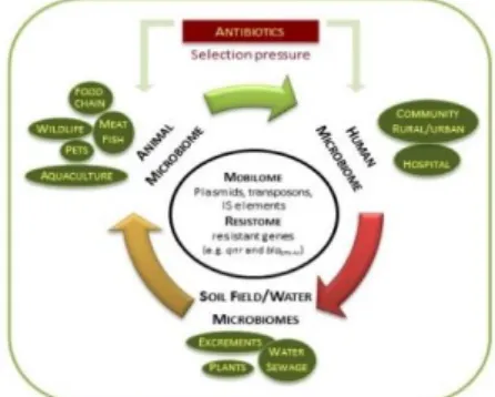
6. DIVERSITY OF ANTIBIOTIC RESISTANCE RESERVOIRS 24
6.1. Animals as reservoirs of antibiotic resistance 25
6.1.1. Food-producing animals 25
6.1.2. Companion animals 26
6.1.3. Wild animals 27
6.1.4. Aquaculture 27
6.2. The role of the environment 28
7. THE IMPORTANCE OF RESEARCH AND MONITORING ANTIBIOTIC RESISTANCE 28
7.1. The current threat 29
7.2. Technological advances in diagnosis 29
7.2.1. Microarrays 29
7.2.2.1. Genomics 30
7.2.2.2. Proteomics and transcriptomics 30
7.2.2.3. The current and future perspectives: veterinomics 31
8. ONE HEALTH 32
CHAPTER 2: OBJECTIVES 33
CHAPTER 3: DYNAMICS OF ANTIBIOTIC RESISTANCE 37
Current perspectives on the dynamic of antibiotic resistance in different reservoirs 39
Abstract 41
3.1. Introduction 41
3.2. Antibiotic residues versus resistome in the environment 43
3.3. Mobilome associated to antibiotic resistance genes 44
3.4. Investigating antibiotic resistance 46
3.5. Strengthening the combat against antibiotic resistance 48
3.6. Conclusions 49
CAPÍTULO 4: SUSCEPTIBILITY AND MOLECULAR CHARACTERISTICS OF ENTEROBACTERIACEAE – ACQUIRED RESISTANCE MECHANISMS TO β-LACTAMS
(ESBL, PMAβ and ESAC) AND FLUOROQUINOLONES (PMQR) 51
4.1.
Antimicrobial Susceptibility of Salmonella enterica isolates from healthy breeder and broiler flocks in Portugal53
Abstract 55
4.1.1. Introduction 55
4.1.2. Material and Methods 56
4.1.3. Results 58
4.1.4. Discussion 62
4.1.5. Conclusion 63
4.2.
Occurrence of extended-spectrum-β-lactamases among isolates of Salmonella enterica subsp. enterica from food-producing animals and food products, in Portugal65
Abstract 67
4.2.1. Introduction 67
4.2.2. Material and Methods 68
4.2.3. Results 71
4.2.4. Discussion 77
4.3. Antimicrobial susceptibility and oxymino-β-lactam resistance mechanisms in Salmonella enterica and Escherichia coli isolates from different animal sources
83
Abstract 85
4.3.1. Introduction 85
4.3.2. Materials and Methods 86
4.3.3. Results 89
4.3.4. Discussion 93
4.4. CTX-M-15–Producing Escherichia coli in dolphin, Portugal 99
Abstract 101
Main text 101
4.5. New insights into resistance to colistin and third-generation cephalosporins of Escherichia coli in poultry, Portugal: novel blaCTX-M-166 and blaESAC genes
105
4.5.3. Results and Discussion 111
4.5.4. Conclusion 119
4.6. Draft genome sequence of an Escherichia coli isolated from a Gallus gallus broiler producing the novel CTX-M-166 variant
121
Abstract 123
4.6.1. Main text 123
4.6.2. Nucleotide sequence accession numbers 124
4.7. Biochemical characterization of CTX-M-166, a new β-lactamase produced by a
commensal Escherichia coli isolate
125
4.7.1. Main text 127
4.7.2. Laboratory procedure 128
4.8. QnrS1 and aac(6’)-Ib-cr-producing Escherichia coli among isolates from animals of different sources: susceptibility and genomic characterization
131
Abstract 133
4.8.1. Introduction 133
4.8.2. Materials and Methods 135
4.8.3. Results 137
4.8.4. Discussion 145
4.9. Draft genomic analysis of an avian multidrug resistant Morganella morganii carrying qnrD1
149
Abstract 151
4.9.1. Introduction 151
4.9.2. Materials and Methods 152
4.9.3. Results and Discussion 154
CHAPTER 5: OTHER RESISTANCE MECHANISMS 159
5.1. Salmonella Enteritidis isolate harboring multiple efflux pumps and pathogenicity factors, shows absence of O antigen polymerase gene
161 Background 163 5.1.1. Methods 164 5.1.2. Results 165 5.1.3. Conclusion 174 5.1.4. DataAccess 174
5.2. The novel MCR-1.9 variant within colistin-resistant Enterobacteriaceae isolates from food-producing animals and meat
175
Abstract 177
5.2.1. Introduction 177
5.2.2. Materials and Methods 179
5.2.3. Results and Discussion 180
5.2.4. Conclusions 184
CHAPTER 6: GENERAL DISCUSSION 185
CHAPTER 7: CONCLUDING REMARKS 197
FIGURES INDEX
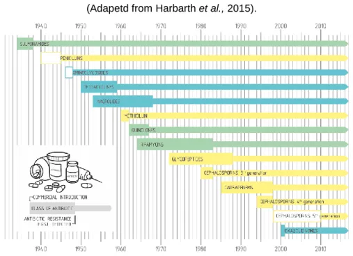
Figure 1.1. The development of new antibiotics is followed by the
emergence of resistance... 3
Figure 1.2. Main antibiotic resistance mechanisms……….
10 Figure 1.3. Hidrolysis of a β-lactam antibiotic by an enzyme with serine on
its active site………... 15
Figure 1.4. Mechanisms involved in horizontal gene transfer…... 21 Figure 1.5. Some transmission pathways of antibiotic resistance, between
different reservoirs: humans, animals and the environment…... 24
Figure 3.1 Crosswalk between resistome and mobiloma among different
environment ………... 46
Figure 4.4.1. Dendogram of PFGE profiles showing the relationship between a clonal strain of Escherichia coli of animal origin (LV143, in
bold), and 22 isolates of Escherichia colifrom humans... 102
Figure 4.5.1. Dendrogram of PFGE profiles of 31 E. coli isolates presenting a non-wild type phenotype to 3rd generation cephalosporins
and/or cefoxitin………... 117
Figure 4.5.2. Schematic representation of CTX-M-166-harboring contig…….... 119 Figure 4.9.1. Examples of contigs containing antibiotic resistance genes in
TABLES INDEX
Table 1.1. Main antibiotics of veterinary importance in Gram negative bacteria. 7 Table 1.2. Some examples of intrinsic resistance and mechanisms………. 12 Table 1.3. Classification of β-lactamases produced by Gram negative
bacteria, with clinical relevance in human and veterinary
practice………. 13
Table 1.4. Comparison of aminoacid substitutions, and the occurrence of the
first cases of MCR-producing isolates………. 22
Table 1.5. Main mobile genetic elements and molecular features……… 23 Table 3.1. Antibiotic resistance is generated by several factors………... 42 Table 4.1.1. Summary of antimicrobial susceptibility of Salmonella enterica
isolated from breeder and broiler flocks (n=333)………... 59
Table 4.1.2. Antimicrobial patterns of decreased susceptibility in Salmonella enterica isolates recovered from breeder and broiler flocks………… .
61 Table 4.2.1. Serotypes of 1120 Salmonella spp. isolates……….. 72 Table 4.2.2. Antimicrobial susceptibility, MIC50 and MIC90 of 1120 Salmonella
enterica isolates……….. 73
Table 4.2.3. MICs among the most common serotypes of Salmonella spp……… 75 Table 4.2.4. Phenotypic and genotypic features of ESBL-producing isolates
(n=5)………... 78
Table 4.3.1. Salmonella spp. (n=562) and Escherichia coli (n=598) isolates……. 88 Table 4.3.2. MIC50 and MIC90 for Salmonella spp. (n=562) and Escherichia coli
(n=598) isolates……….. 91
Table 4.3.3. MIC50 and MIC90 among the most important Salmonella
serotypes………... 92
Table 4.3.4. Characteristics of Salmonella spp. and E. coli isolates displaying non wild-type phenotypes to cefotaxime and or cefoxitin………. 94 Table 4.5.1. MIC50 and MIC90 for 387 Escherichia coli isolates: broilers
(n=202) and turkeys (n=185)……… 112
Table 4.5.2. Characteristics of 15 Escherichia coli isolates displaying non wild-type phenowild-types to 3rd generation cephalosporins and/or
cephamycins………... 115
Table 4.5.3. ampC promoter and attenuator mutations and amino acid sequences of AmpC β-lactamases in the three E. coli
Table 4.5.4. Phenotypic and genotypic context of CTX-M-producing Escherichia
coli clinical isolates, transformants and the recipient strain…………. 118 Table 4.7.1. Kinetic parameters of CTX-M-166 and CTX-M-1 β-Lactamases…… 128 Table 4.8.1. Distribution of the Salmonella enterica(n=89) and Escherichia coli
(n=91) Isolates………... 135
Table 4.8.2. MIC50 and MIC90 for Salmonella enterica (n=89) and Escherichia
coli(n=91) isolates………... 138
Table 4.8.3. Genome analysis of Escherichia coliLV46221, LV46743, LV36464
and LV27950………... 140
Table 4.8.4. General features of PMQR-harboring Escherichia coli isolates
recovered from animals of different sources……….. 142
Table 4.8.5. Representation of intact phage regions detected in the draft
genome of LV46221, LV46743, LV36464 and LV27950... 143
Table 5.1.1. Single nucleotide variants that represent amino acid substitutions in
Salmonella Enteritidis LV60 using Salmonella Enteritidis strain
p125109 as the reference genome……….. 167
Table 5.1.2. Perfect and strict best hit results, by predicted gene, obtained using
the Resistance Gene Identifier (RGI)……….. 169
Table 5.2.1. Comparison of amino acid substitutions, and epidemiology of first
reports of MCR-producing strains……… 178
Table 5.2.2. Escherichia coli (n= 1206), from food-producing animals, meat and
meat products……….. 181
Table 5.2.3. Salmonella spp (n= 634), from food-producing animals, meat, meat
products and animal feed……….. 182
Table 5.2.4. Phenotypic and genotypic context of CTX-M-8 and MCR-1.9
producing E. coliclinical isolate, transformant, transconjugant and
ACRONYMS AND ABBREVIATIONS
Aac(6’)-Ib-cr Aminoglicosyde acetyltransferase variant cr AAC Aminoglicosyde acetyltransferase
AAD, ANT Aminoglicosyde adenyltransferase
ACT AmpC Type
ALA Alanine
APH Aminoglicosyde phosphotransferase
attI Attachment site of the integron
attC Attachment site of the integrons cassette
bla β-Lactamase coding genes
CAT Chloramphenicol Acetyltransferases
cat Chloramphenicol acetyltransferases coding genes
CMY β-Lactamases active on Cephamycins
CTX-M β-Lactamases active on Cefotaxim, isolated for the first time in Munich
dfrA DHFR coding gene
DHFR Dihydrofolate redutase DHPS Dihydropteroate synthase DNA Desoxiribonucleic acid EMA European Medical Agency
ECDC European Center for Prevention and Disease Control ECOFF Epidemiological cut-off values
EFSA European Food Safety Authority
ESAC Extended-spectrum AmpC β-Lactamase ESBL Extended-spectrum β-Lactamase
ESVAC European Surveillance Veterinary Antimicrobial Consumption EUCAST European Committee on Antimicrobial Susceptibility Testing FAO Food and Agriculture Organization of the United Nations
gyr Gyrase coding gene
HGT Horizontal Gene Transfer
ICE Integrative and Conjugative Element IMP
β-Lactamase active on Iminipem
Inc Plasmid Incompatibility group IntI Integron integraseIS Insertion Sequence
ISCR Insertion Sequence Common Region KPC Klebsiella pneumoniae Carbapenemase
MBL Metallo-β-Lactamase
mcr Plasmid-mediated colistin resistance gene
MGE Mobile Genetic Element
MIC Minimum Inhibitory Concentration MLST Multilocus Sequence Typing
mRNA Messenger RNA
MS Mass-Spectrometry
NDM New Delhi Metalo-β-lactamase
NGS Next Generation Sequencing
OIE World Animal Health Organization OMP Outer Membrane Protein
oqxAB Gene coding olanquidox resistance
otr Oxytetracycline resistance coding gene
ORF Open Reading Frame
OriIS Origin Insertion Sequence OXA β-Lactamase active on Oxacillin
PBP Penicillin Binding Protein PBRT PCR-Based Replicon Typing
PintI Promoter of Integrase Integron gene PCR Polymerase Chain Reaction
PFGE Pulsed-Field Gel Electrophoresis PMAβ Plasmid-Mediated AmpC β-lactamase
Pc Promoter of gene cassette
pMLST Plasmid Multilocus Sequence Typing PMQR Plasmid-Mediated Quinolone Resistance PMCR Plasmid-Mediated Colistin Resistance
qepA, Quinolone efflux pump A coding gene
qnr, Quinolone resistance coding genes
QRDR Quinolone-Resistance Determining Region RAST Rapid Annotation using Subsystem Technology
rRNA Ribossomal RNA
sat Streptothricin resistance coding gene
SHV β-Lactamase Sulphydryl Reagent Variable
ST Sequence Type
str Aminoglycoside resistance coding gene
sul Sulphamethoxazole resistance coding gene
TEM β-Lactamase Named after patient Temoniera TerIS Termination Insertion Sequence
tet Tetracycline resistance coding gene
tRNA Transport RNA
UPGMA Unweighted Pair Group Method with Arithmetic Mean
VAL Valine
VIM Verona Integron-encoded Metallo- β-lactamase
THESIS STRUCTURE
Antibiotic resistance is a worldwide problem with serious repercussions on human health, animal health and the economy, and it should be integrated into a global perspective, as bacteria do not respect geographic and species barriers. The selective pressure exerted by the abusive and inadequate use of antibiotics in human and animal clinical therapy, animal and agricultural production, as well as the environmental impact resulting from these activities, are the main cause for the development of antibiotic resistance. In addition to the investigation through the characterization of resistance mechanisms and the mobile genetic elements involved in the emergence and mobilization of antibiotic resistance genes among the different ecosystems and monitoring through the implementation of surveillance programs on antibiotic resistance and consumption, are of crucial importance, in order to obtain the actual picture of the occurrence and trends on antibiotic resistance.
This thesis is based on twelve papers organized in three distinct chapters (3 to 5), eleven of which have already been published and one submitted for publication, in international peer-reviewed journals. Each research article consists of an introduction, materials and methods, results and discussion related to the scope of the study and preceded by a title page describing the reference of the publication and the contributions of each author.
In addition to the articles, this thesis includes a global overview of antibiotic resistance (Chapter 1, General Introduction), followed by the objectives (Chapter 2, Objectives), a general discussion on the results (Chapter 6, General Discussion) and the main conclusions (Chapter 7, Conclusions).
Considering the different types of manuscripts presented in this thesis followed the recommendations of each scientific journal where they were published or submitted for publication, chapters 3, 4 and 5 were formatted in the same style, with all references gathered in a single section (Chapter 8, References).
The numbering of the figures and tables is presented according to the numbers of the chapter and its article.
The specific content of each of the chapters that integrate this PhD thesis consists of:
Chapter 1 consists of a general introduction, where it is intended to reveal the state of the art
in the area of antibiotic resistance focused on Gram negative bacteria. Antibiotic targets and mechanisms of intrinsic and acquired resistance to the main groups of antibiotics, the diversity of antibiotic resistance reservoirs and the main routes of dissemination, are approached. Technological advances and the use of omics in research of antibiotic resistance, is also discussed.
Chapter 3 consists of a brief review on the dynamic and complex process of antibiotic
resistance, and the pathways between the different reservoirs, humans, animals and the environment.
Chapter 4 includes nine publications (4.1 to 4.9), in which the phenotypic susceptibility and
molecular characterization of Enterobacteriaceae strains, regarding, the mechanisms of
acquired resistance to β-lactam antibiotics [Extended-Spectrum β-Lactamases (ESBL),
Plasmid-Mediated AmpC β-lactamases (PMAβ), Extended-Spectrum AmpC β-lactamases
(ESAC]), fluoroquinolones, Plasmid-Mediated Quinolone Resistance (PMQR) and to colistin [Plasmid-Mediated Colistin Resistance (PMCR)], and the presence of mobile genetic elements, were evaluated.
Sub-Chapter 4.1, the phenotypic antibiotic susceptibility patterns of 333 strains of S.
enterica isolated from breeding and broiler flocks, during the period 2009-2011, was evaluated.
Sub-Chapter 4.2, the phenotypic antibiotic susceptibility patterns of 1120 strains of S.
enterica isolated from poultry, swine and food products of animal origin, and the molecular
characterization of ESBL and PMAβ producer strains, mobile genetic elements and genetic
environment, were investigated.
Sub-Chapter 4.3, the phenotypic antibiotic susceptibility of 562 S. enterica strains isolated
from food-producing animals, food products and animal feed, and 598 E. coli strains isolated from several animal species, was evaluated. Molecular characterization of acquired
resistance mechanisms to β-lactam antibiotics (ESBL and PMAβ) and fluoroquinolones
(PMQR), and detection of mobile genetic elements in strains with reduced susceptibility to 3rd
generation cephalosporins and/or cephamycins, were performed.
Sub-Chapter 4.4, the zoonotic potential of an E. coli strain isolated from a captive dolphin
and a set of human clinical strains with similar phenotypic and genotypic characteristics, was assessed.
Sub-Chapter 4.5, the phenotypic antibiotic susceptibility results of 387 strains of E. coli
isolated from broilers and turkeys at slaughter, and the molecular characterization of 15
strains with reduced susceptibility to 3rd generation cephalosporins and or cephamycins,
regarding the detection of ESBL-, ESAC-, PMAβ-, PMQR- and PMCR-encoding genes, were
performed. In addition, sequencing of AmpC-encoding gene in three strains in which only the ampC gene was detected, and genetic relationship between animal and human strains carrying ESBL-encoding genes from the CTX-M, SHV-12 and TEM-52 family by pulsed-field gel electrophoresis technique (PFGE), were performed. The new CTX-M-166 enzyme-producer strain was also analyzed through New Genome Sequencing (NGS) for further
Sub-Chapters 4.6 and 4.7, biochemical characterization of the new CTX-M-166 enzyme,
regarding kinetic parameters, and additional characterization by NGS, concerning antibiotic resistance, virulence and MultiLocus Sequence Typing (MLST), were performed.
Sub-Chapter 4.8, antibiotic susceptibility was determined on 89 strains of S. enterica and 91
E. coli strains isolated from food-producing, companion and zoo animals. Molecular characterization regarding the detection of PMQR, and genomic analysis and comparison of four strains, two carrying qnrS1 gene and two carrying aac(6 ')-Ib-cr gene, was performed through NGS.
Sub-Chapter 4.9, characterization of a multidrug resistant Morganella morganii strain
isolated from broilers through NGS, regarding antibiotic resistance and virulence genes, and genomic analysis of the plasmid carrying qnrD1 gene.
Chapter 5, includes two manuscripts (5.1 and 5.2), concerning antibiotic resistance
mechanisms other than those included in Chapter 4.
Sub-Chapter 5.1, genomic characterization of a S. Enteritidis strain isolated from one
day-old chicks, regarding antibiotic and heavy metals resistance determinants, virulence factors and mobile genetic elements was performed, through NGS.
Sub-Chapter 5.2, phenotypic susceptibility towards colistin and 3rd generation
cephalosporins and/or cephamycins of 1840 Enterobacteriacaeae strains (1206 E. coli and 634 S. enterica), was evaluated. In 138 isolates resistant to colistin, mcr-1 and mcr-1.9
genes were detected; in those strains co-resistant to 3rd generation cephalosporins and/or
cephamycins, genotypic characterization with respect to the detection of ESBL- and/or
PMAβ-encoding genes, was performed. In addition, genotypic characterization of a strain
bearing the new variant of the mcr-1, mcr-1.9 gene, regarding the presence of other antibiotic resistance genes, virulence factors, plasmid’s identification and typing and genetic environment, was performed by using Whole Genome Sequencing (WGS).
Chapter 6 includes a global discussion on the results obtained on the studies performed. Chapter 7, includes the main conclusions of the studies carried out.
Chapters 3, 4 and 5, that can be read in separate, transcribe the contents of the following
publications:
Chapter 3
Manuela Caniça, Vera Manageiro, Daniela Jones-Dias, Lurdes Clemente, Eduarda Gomes-Neves, Patrícia Poeta, Elsa Dias, Eugénia Ferreira. Current perspectives on the dynamic of antibiotic resistance in different reservoirs. Research in Microbiology, 2015, 166(7):594-600.
Chapter 4
4.1. Lurdes Clemente, Ivone Correia, Patrícia Themudo, Isabel Neto, Manuela Caniça, Fernando Bernardo, 2014. Antimicrobial susceptibility of Salmonella enterica isolates from healthy breeder and broiler flocks in Portugal. The Veterinary Journal, 2014, 200(2):276-81.
4.2. Lurdes Clemente, Vera Manageiro, Eugénia Ferreira, Daniela Jones-Dias, Ivone Correia, Patrícia Themudo, Teresa Albuquerque, Manuela Caniça. Occurrence of extended-spectrum β-lactamases among isolates of Salmonella enterica subsp. enterica from food-producing animals and food products, in Portugal. International Journal of Food Microbiology, 2013, 167(2):221-8.
4.3. Lurdes Clemente, Vera Manageiro, Daniela Jones-Dias, Ivone Correia, Patrícia Themudo, Teresa Albuquerque, Margarida Geraldes, Filipa Matos, Cláudia Almendra,
Eugénia Ferreira, Manuela Caniça. Antimicrobial susceptibility and oxymino-β-lactam
resistance mechanisms in Salmonella enterica and Escherichia coli isolates from different animal sources. Research in Microbiology, 2015, 166(7):574-83.
4.4. Vera Manageiro, Lurdes Clemente, Daniela Jones-Dias, Teresa Albuquerque, Eugénia Ferreira, Manuela Caniça. Zoonotic potential of multidrug resistant CTX-M-15-producing Escherichia coli isolate of a marine dolphin, in Portugal. Emerging Infectious Diseases, 2015, 21:2249-51.
4.5. Vera Manageiro, Lurdes Clemente, Ivone Correia, Teresa Albuquerque, Patrícia Themudo, Eugénia Ferreira, Manuela Caniça. New insights into resistance to colistin and
third-generation cephalosporins of Escherichia coli in poultry, Portugal: novel blaCTX-M-166 and
blaESAC genes. International Journal of Food Microbiology, 2017, 263:67-73.
4.6. Vera Manageiro, Lurdes Clemente, Sílvia Duarte, Luís Vieira, Manuela Caniça. Draft genome sequence of an Escherichia coli isolated from a Gallus gallus producing the novel CTX-M-166 variant. Genome Announcement, 2016, 4(5), e0102916.
4.7. Vera Manageiro, Rafael Graça, Eugénia Ferreira, Lurdes Clemente, Richard Bonnet e
Manuela Caniça. 2017. Biochemical characterization of CTX-M-166, a new CTX-M
β-lactamase produced by a commensal Escherichia coli isolate. Journal of Antibiotics, 2017, 70(6):809-810.
4.8. Daniela Jones-Dias, Vera Manageiro, Rafael Graça, Daniel Sampaio, Teresa Albuquerque, Patrícia Themudo, Luís Vieira, Eugénia Ferreira, Lurdes Clemente, Manuela Caniça. QnrS1- and Aac(6’)-Ib-cr-producing Escherichia coli among isolates from animals of different sources: susceptibility and genomic characterization. Frontiers in Microbiology, 2016, 7:671.
4.9. Daniela Jones-Dias, Lurdes Clemente, Inês Barata Moura, Daniel Sampaio, Teresa Albuquerque, Luís Vieira, Vera Manageiro, Manuela Caniça. Draft genomic analysis of an
avian multidrug resistant Morganella morganii isolate carrying qnrD1. Frontiers in Microbiology, 2016, 7:1660.
Capítulo 5
5.1. Daniela Jones-Dias, Lurdes Clemente, Conceição Egas, Hugo Froufe, Daniel Sampaio, Luís Vieira, Maria Fookes, Nicholas Thompson, Vera Manageiro, Manuela Caniça. Salmonella Enteritidis isolate harboring multiple efflux pumps and pathogenicity factors, shows absence of O antigen polymerase gene. Frontiers in Microbiology, 2016, 7:1130.
5.2. Lurdes Clemente, Vera Manageiro, Raquel Romão, Catarina Silva, Luís Vieira, Ana Amaro, Ivone Correia, Teresa Albuquerque, Patrícia Themudo, Eugénia Ferreira, Manuela Caniça. 2017. The novel MCR-1.9 variant within colistin-resistant Enterobacteriaceae isolates from food-producing animals and meat. Submitted to International Journal of Food Microbiology (December 2017).
A ti Walsker
Chapter 1
CHAPTER 1
Chapter 1
1. THE ANTIBIOTICS
"One sometimes finds what one is not looking for"
Sir Alexander Fleming
1.1. History of the antibiotics
The discovery of penicillin by Alexander Fleming in 1929 provided treatment for infectious diseases, becoming an indispensable drug in the therapeutic arsenal after the beginning of its industrial production in 1946 (Dantas & Sommer, 2014).
During the "golden age" of antibiotic development (1940-1960), the discovery of new natural, synthetic and semi-synthetic antibiotics deeply changed human medicine in the field of infectious and oncological chemotherapy, organ transplants, and other invasive surgeries, which could fail without the use of these compounds (Wright, 2010). Later on, the use of antibiotics has also been extended to veterinary medicine.
With the introduction of new antibiotics in the clinical practice, the appearance of resistant bacteria was inevitable. However, has always been counter balanced by the development of new substances by the pharmaceutical industry, which since the 1970s focused mainly on the chemical modification of the existing compounds, instead of the development of new molecules (Dantas & Sommer, 2014) (Figure 1.1).
Figure 1.1. The development of new antibiotics is followed by the emergence of resistance
Chapter 1
1.2. Targets of antibiotics
Antibiotics are classified accordingly to their chemical structure, spectrum of activity and mechanism of action (Table 1.1). Bacterial targets may be associated with metabolic processes or structures essential for growth and survival. Antibiotics inducing cell death are bactericidal, and those inhibiting their growth are bacteriostatic (Bernatova et al., 2013).
1.2.1. Inhibition of nucleic acid synthesis
Quinolones and fluoroquinolones are bactericidal antibiotics, which target topoisomerases II and IV, causing inhibition of the deoxyribonucleic acid (DNA) synthesis and consequently, bacterial growth (Kohanski et al., 2010).
1.2.2. Inhibition of cell wall synthesis
Peptidoglycan is an essential compound of the bacterial cell wall, responsible for maintaining the cellular structure. Synthesis of this compound includes three stages: in the first two stages the peptidoglycan percussors are synthesized and incorporated into the lipid molecule
(lipid II); in the third stage, the molecules are integrated into the existing peptidoglycan.
β-lactam antibiotics are bactericidal and act by blocking peptidoglycan synthesis (Lovering et al., 2012).
1.2.3. Inhibition of protein synthesis
Several antibiotics cause inhibition of protein synthesis, by acting on bacterial ribosomes at different stages of translation (initiation, elongation and termination) (Wright, 2010). Aminoglycosides are bactericidal antibiotics, acting through binding to the 30S subunit of the bacterial ribosome, altering the structure of the complex formed by the aminoacyl-RNA transport (tRNA) and RNA messenger (mRNA) (Kohanski et al., 2010).
Chloramphenicol and florfenicol are bacteriostatic antibiotics and act by preventing the elongation of the peptide chain (Schwarz et al., 2004). Tetracyclines are also bacteriostatic and act by inhibiting the attachment of the aminoacyl-RNA transport complex (tRNA) to the ribosomal receptor A (Chopra & Roberts, 2001).
1.2.4. Inhibitors of folic acid synthesis
Sulfonamides and trimethoprim are bacteriostatic antibiotics acting by competitive inhibition in the synthesis of folic acid. Sulfonamides inhibit the dihydropteroate synthetase enzyme (DHPS), which catalyzes the formation of the dihydrofolate parabenzoic acid, and trimethoprim acts in the next step by inhibiting the enzyme dihydrofolate reductase (DHFR), which catalyzes the synthesis of tetradihydrofolate from dihydrofolate (Huovinen, 2001).
Chapter 1
1.2.5. Inhibitors of cell membrane synthesis
Polymyxins are a group of cationic, bactericidal polypeptide antibiotics that act by binding to the phospholipids of the anionic outer bacterial cell membrane, causing a breakdown in the integrity and permeability of the cell wall (Landman et al., 2008).
2. THE USE OF ANTIBIOTICS IN VETERINARY MEDICINE AND ANIMAL PRODUCTION
Antibiotics are used in veterinary medicine for treatment and prevention of infectious diseases in animals. In livestock production they are also used to improve animal growth and feed efficiency (Marshall & Levy, 2011, Aarestrup, 2015, Economou and Gousia, 2015). Antibiotics can also be administered as a metaphylactic treatment, being applied to entire groups of animals, even if only a few show clinical symptoms of a specific disease. This type of treatment is administered in high doses and for a short period of time, eliminating or minimizing the spread of the disease. In constrast, the prophylactic treatment is administered in a subtherapeutic dosage for a longer period, when there is imminent risk of disease emergence. Prophylactic treatment is usually associated with poor facility management and permanent stress, predisposing animals to infection. This type of treatment shows similar effects to the administration of growth promoters (Aarestrup, 2015; Economou & Gousia, 2015) (Table 1.1). Although growth promoters were abolished in the European Union (EU) in 2006, the consumption of antibiotics did not decrease; in contrast, there was an increase in its use for metaphylactic and prophylactic purposes (Woolhouse et al., 2015).
The values for administration of antibiotics at veterinary level in 29 European Community (EC) and non-European Community countries show that tetracyclines rank first in total sales (33.4%), followed by penicillins (25.5%), sulphonamides (11%) and macrolides (7.5%). Polymyxins, fluoroquinolones and cephalosporins, considered critical important antibiotics for humans and animals represented 6.6%, 1.9% and 0.2% of total sales, respectively. It should be noted that colistin represents more than 99% of the total sales of polymyxins (EMA/ESVAC, 2016).
In Portugal, the total sales of tetracyclines also rank first (38.2%), followed by penicillins (17%) and macrolides (12.2%). The total sales of polymyxins, fluoroquinolones and cephalosporins were 8.7%, 5.6% and 0.2%, respectively (EMA/ESVAC, 2016). Considering the period between 2011 and 2014, there was a decrease in total sales of polymyxins and a
marked increase in fluoroquinolones, with a peak in 2014; the sales of 3rd and 4th generation
cephalosporins remained stable over the same period (EMA/ESVAC, 2016).
Over the past decade, the World Organization for Animal Health (OIE) has been developing relevant work in the field of antibiotic resistance, through the implementation of international standards applicable to the various antibiotics, being its prudent and responsible use the
Chapter 1
main goals. Considering the large number of animal species, it was established by OIE a classification based on the level of importance of antibiotics and according to the following criteria: i) specificity of the infection and lack of alternative therapies; ii:) opinion of the various member countries concerning the antimicrobials of veterinary importance (OIE, 2015).
Table 1.1 lists the classification of the main groups of antibiotics according to the two criteria mentioned above: veterinary critical important antibiotics (VCIA) follow both criteria, veterinary highly important antibiotics (VHIA) follow only one of the criteria, and the veterinary important antibiotics (VIA) are considered if none of the criteria is applicable (OIE, 2015).
Some antibiotics included in the VCIA category, namely 3rd and 4th generation cephalosporins
and fluoroquinolones, are also considered by the World Health Organization (WHO) as critical important in humans. For this reason, i) should not be administered to animals, in food or drinking water, in the absence of clinical signs of disease, ii) should not be used as a first line treatment, unless justified, and after antibiotic susceptibility testing, and iii) should be reserved for use, extra label/off-label, when no alternatives are available (OIE, 201
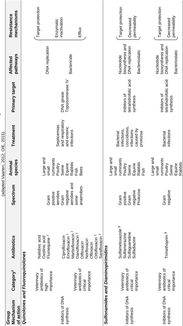
Cha pte r 1 7 T ab le 1.1. M ai n a n ti b io ti c s of v ete ri na ry i m po rtan c e in G ra m ne ga ti v e b ac teria (a d a pt ed Va a rte n , 2 0 1 2 ; O IE, 2 0 1 5 ). G ro u p M ec h anism o f ac tion Cat ego ry 1 A n tibio tic s S p ec tr u m A n im al specie s T rea tmen t P rim ar y tar g et A ff ec ted p ath w ay s Resi stan ce mechan is ms Q u ino lon es and Flu o ro q u ino lon es In ib ito rs o f DN A s y n th e s is Ve te ri n a ry a n ti b io ti c s o f high im p o rta n c e Nal id ix ic a c id O x o lin ic a c id Fl u m e q u in e 2 G ra m p o s iti v e a e ro b e s G ra m n e g a ti v e a e ro b e s a n d s o m e a n a e ro b e s L a rg e a n d s m a ll ru m in a n ts Po u ltry Sw in e Eq u in e Rab b it s Fi s h Be e s Se p ti c e m ia s a n d re s p ir a to ry a n d e n te ri c in fe c ti o n s DN A g ira s e T o p o is o m e ra s e I V DN A re p lic a ti o n Ba c te ri c id e T a rg e t p ro te c ti o n En z y m a ti c in a c ti v a ti o n Eff lu x Ve te ri n a ry a n ti b io ti c s o f c ri ti c a l im p o rta n c e Cip ro fl o x a c in En ro fl o x a c in 2 M a rb o fl o x a c in 2 Dan o fl o x a c in 2 Dif lo x a c in Norf lo x a c in O fl o x a c in O rb ifl o x a c in 2 Sa ra fl o x a c in 2 S u lf o n amid es and Di amin o p ir imid ins In ib ito rs o f DN A s y n th e s is Ve te ri n a ry a n ti b io ti c s o f c ri ti c a l im p o rta n c e Su lfa m e to x a z o le 3 Su lfa m e ra z in e Su lfa g u a n id in e Su lfa d ia z in e G ra m p o s iti v e G ra m n e g a ti v e Pro to z o a L a rg e a n d s m a ll ru m in a n ts Po u ltry Sw in e Eq u in e Rab b it s Fi s h Ba c te ri a l in fe c ti o n s , c o c c id io s is , in fe c ti o n s c a u s e d b y p ro to z o a In ib ito rs o f te tra h id ro fo lic a c id s y n th e s is Nuc le o ti d e b io s y n th e s is a n d DN A re p lic a ti o n Ba c te ri o s ta ti c T a rg e t p ro te c ti o n Dec re a s e d p e rm e a b ili ty In ib ito rs o f DN A s y n th e s is Ve te ri n a ry a n ti b io ti c s o f c ri ti c a l im p o rta n c e T ri m e th o p ri m 3 G ra m n e g a ti v e L a rg e a n d s m a ll ru m in a n ts Po u ltry Sw in e Eq u in e Rab b it s Ba c te ri a l in fe c ti o n s In ib ito rs o f te tra h id ro fo lic a c id s y n th e s is Nuc le o ti d e b io s y n th e s is a n d DN A re p lic a ti o n Ba c te ri o s ta ti c T a rg e t p ro te c ti o n Dec re a s e d p e rm e a b ili ty
pte r 1 8 G ro u p M ec h anism o f ac tion Cat ego ry 1 A n tibio tic s S p ec tr u m A n im al specie s T rea tmen t P rim ar y tar g et A ff ec ted p ath w ay s Resi stan ce mechan is ms -L ac tams h ib ito rs o f c e ll a ll s y n th e s is Ve te ri n a ry a n ti b io ti c s o f high im p o rta n c e Cef a le x in Cef a lo tin Cef a z o lin Cef u ro x im e G ra m p o s iti v e a e ro b e s G ra m n e g a tiv e a e ro b e s a n d a n a e ro b e s L a rg e a n d s m a ll ru m in a n ts Po u ltry Sw in e Eq u in e Rab b its Fi s h Se p ti c e m ia Res p ira to ry in fe c ti o n s Uri n a ry in fe c ti o n s M a s ti ti s Pe n ic illi n -b in d in g p ro te in s Bi o s y n th e s is o f p e p ti d o g lic a n Ba c te ric id e En z y m a ti c in a c ti v a ti o n Eff lu x Ve te ri n a ry a n ti b io ti c s o f c ri ti c a l im p o rta n c e Cef o p e ra z o n e Cef ti o fu r 2 Cef tri a x o n e Cef q u in o m a 2 Ve te ri n a ry a n ti b io ti c s o f c ri ti c a l im p o rta n c e Pe n ic ilin Am p ic ilin Am o x ic ilin M e c ilin a m e Am o x ic ilin /Cl a v u la n a te Am p ic ilin /Su lb a c ta m Clo x a c ilin Dic lo x a c ilin O x a c ilin Dec re a s e d p e rm e a b ility T a rg e t p ro te c ti o lipep tides h ib ito rs o f c e ll a ll s y n th e s is Ve te ri n a ry a n ti b io ti c s o f high im p o rta n c e Ba c itr a c in Po lim ix in Col is tin G ra m n e g a tiv e L a rg e a n d s m a ll ru m in a n ts Po u ltry Sw in e Eq u in e Rab b its Fi s h Se p ti c e m ia Res p ira to ry in fe c ti o n s Uri n a ry in fe c ti o n s In fe c ti o n s o f d ig e s ti v e s y s te m Col ib a c ilo s is Sa lm o n e lo s is Cel l m e m b ra n e Sy n th e s is o f c e ll w a ll Ba c te ric id e Eff lu x Dec re a s e d p e rm e a b ility M CR -1 a n d v a ri a n ts ; M CR M CR -3 ino g lic o side s h ib ito rs o f ro te in y n th e s is Ve te ri n a ry a n ti b io ti c s o f c ri ti c a l im p o rta n c e G e n ta m ic in Stre p to m y c in Ka n a m y c in ; A m ik a c in T o b ra m y c in Neo m y c in Ap ra m ic in 2 Sp e c tin o m y c in 2 G ra m p o s iti v e G ra m n e g a tiv e L a rg e a n d s ma ll ru min a n ts P o u ltr y S w in e E q u in e R a b b its ; F is h ; Se p ti c e m ia Res p ira to ry a n d Uri n a ry in fe c ti o n s In fe c ti o n s o f d ig e s ti v e s y s te m 3 0 S ri b o s s o m a l s u b u n it In h ib iti o n o f p ro te in s y n th e s is Ba c te ric id e En z y m a ti c in a c ti v a ti o n Eff lu x Dec re a s e d p e rm e a b ility
Cha pte r 1 9 H IA , v e te rina ry h igh ly im p o rt a n t a n ti b iot ic s , a c c o m p lis h o n e o f th e c rit e ria e s ta b lis h e d b y OI E ; C IA , v e te rina ry c rit ic a l im p o rt a n t a n ti b iot ic s , a c c o mpli s h b o th c ri te ri a e s ta b lis h e d b y OI E ; [1 . S p e c if ic it y o f th e inf e c tion ; 2 . Op inio n o f m e m b e r c o u n tr ie s ] n tibio ti c s o f v e te rina ry u s e ; a n b e a p p lie d in c o m b ina tion a n d a re b a c te rio s ta ti G ro u p M ec h anism o f ac tion Cat ego ry 1 A n tibio tic s S p ec tr u m A n im al specie s T rea tmen t P rim ar y tar g et A ff ec ted p ath w ay s Resi stan ce mechan is ms T etrac y cli n e s In h ib ito rs o f p ro te in s y n th e s is Ve te ri n a ry a n ti b io ti c s o f c ri ti c a l im p o rta n c e Clo rte tra c ic lin a Dox ic ic lin a O x ite tra c ic lin a T e tra c ic lin a G ra m p o s iti v e G ra m n e g a ti v e L a rg e a n d s m a ll ru m in a n ts Po u ltry Sw in e Eq u in e Rab b it s Fi s h ; Be e s Se p ti c e m ia Res p ir a to ry in fe c ti o n s Uri n a ry in fe c ti o n s In fe c ti o n s o f d ig e s ti v e s y s te m 3 0 S ri b o s s o m a l s u b u n it In h ib iti o n o f p ro te in s y n th e s is Ba c te ri o s ta ti c En z y m a ti c in a c ti v a ti o n Eff lu x Dec re a s e d p e rm e a b ili ty T a rg e t p ro te c ti o n F enicols In h ib ito rs o f p ro te in s y n th e s is Ve te ri n a ry a n ti b io ti c s o f c ri ti c a l im p o rta n c e C h lo ra m p h e n ic o l Fl o rfe n ic o l 2 T ia n fe n ic o l So m e G ra m p o s iti v e G ra m n e g a ti v e L a rg e a n d s m a ll ru m in a n ts Po u ltry Sw in e Eq u in e Rab b it s Fi s h Be e s Res p ir a to ry in fe c ti o n s in b o v in e , s w in e a n d p o u ltry In fe c ti o n s i n f is h 3 0 S ri b o s s o m a l s u b u n it In h ib iti o n o f p ro te in s y n th e s is Ba c te ri o s ta ti c En z y m a ti c in a c ti v a ti o n Eff lu x Dec re a s e d p e rm e a b ili ty T a rg e t p ro te c ti o n
3. MECHANISMS OF ANTIBIOTIC RESISTANCE IN GRAM NEGATIVE BACTERIA
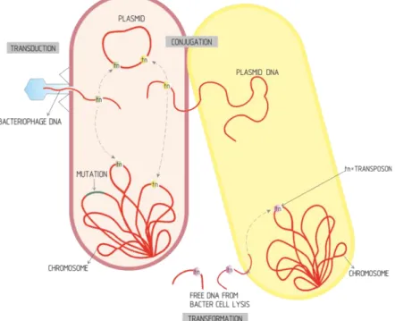
There are four main mechanisms of resistance to antibiotics: i) changes in cell membrane permeability, ii) active efflux, iii) enzymatic modification or inactivation, and iv) target alteration through mutation (Wright, 2010; Blair et al., 2015) (Figure 1.2).
3.1. Decreased cell membrane permeability
The entry of antibiotic molecules in the bacterial cell, including hydrophilic antibiotics
(aminoglycosides, β-lactams and colistin), is achieved by diffusion through the outer
membrane proteins (Omps), depending on its electrical charge, shape and size. Thus, the reduction of the permeability of the outer membrane by decreasing the number of functional porins, or by replacing for more selective pore channels, are limiting factors to the antibiotics entrance into the bacterium (Figure 1.2) (Blair et al., 2015).
OmpF is the most important porin in Escherichia coli and a decrease on its expression
contributes to resistance to various antibiotics, including quinolones, aminoglycosides,
β-lactams. Karczmarczyk et al. (2011) identified it as one of the main mechanisms of resistance to quinolones in strains of E. coli isolated from food-producing animals (Karczmarczyk et al., 2011).
Figura 1.2. Main antibiotic resistance mechanisms. MGE, mobile genetic element. (Adapetd from Levy & Marshall, 2004).
3.2. Active efflux
Efflux pumps are one of the main mechanisms of resistance to antibiotics in Gram-negative bacteria, being responsible for the active transport of antibiotics to the outside of the cell (Figure 1.2) (Blair et al., 2015).
This mechanism occurs frequently in strains of Salmonella enterica and E. coli of animal origin, showing reduced susceptibility to fluoroquinolones, which is frequently associated to mutations in topoisomerase-encoding genes (Randall et al., 2005; Karczmarczyk et al., 2011; Yang et al., 2014).
Active efflux is also the main mechanism of resistance to tetracycline; 30 genetic determinants associated with this resistance mechanism have been identified (Roberts & Schwarz, 2016), of which tetA, tetB, tetC and tetG are the most frequent in strains of S. enterica and E. coli isolated from animals and animal products (Glenn et al., 2013, Gomes-Neves et al., 2014, Chang et al., 2015, Jackson et al., 2015, Shin et al., 2015).
Some efflux pumps act on a specific substrate (Tet tetracycline pumps) (Roberts & Schwarz, 2016), while others, called multidrug resistant (MDR) pumps, act on a larger number of structurally unrelated substrates, such as the OqxAB pump, which is plasmid-mediated and act on quinolones, chloramphenicol and trimethoprim, occurring more frequently in strains of E. coli isolated from animals treated with olanquidox (Jacoby et al., 2014; Yang et al., 2014).
3.3. Modification by enzymatic inactivation
The enzymatic inactivation is the most common mechanism of resistance to β-lactam
antibiotics, through which β-lactamases cause the cleavage of β-lactam ring (Blair et al.,
2015). In Enterobacteriaceae strains of animal origin, β-lactamases belonging to different
families and different hydrolytic profiles are described (Rubin & Pitout, 2014).
Also in E. coli and S. enterica strains of animal origin, enzymatic inactivation is the most important mechanism of resistance to other classes of antibiotics, such as phenicols, through the action of acetyltransferases and phosphotransferases (Schwarz et al., 2004), and aminoglycosides, through acetyltransferases, adenyltransferases and phosphotransferases (Ramirez & Tomalsky, 2010; van Hoek et al., 2011; Frye & Jackson, 2013).
The AAC(6 ')-Ib variant, AAC(6')-Ib-cr, encoded by the aac(6') - Ib-cr gene, is responsible for co-resistance towards aminoglycosides and fluoroquinolones (Ramirez et al., 2013), occurring frequently in strains of animal origin (Veldman et al., 2011; Jones-Dias et al., 2013).
3.4. Alteration of the target by mutation
Target alteration by the acquisition of mutations at the level of gyr (A and B) and par (C and E) genes is the main mechanism of resistance to quinolones and fluoroquinolones, causing protein alteration and avoiding binding of the antibiotic to its target (Figure 1.2) (Wright et al.,
2010). This mechanism is responsible for a high level of resistance to these compounds, and occurs with high frequency in strains of S. enterica and E. coli of animal origin (Tamang et al., 2012a; Jones-Dias et al., 2013, Wasyl, 2014a, Wasyl et al., 2014b).
4. GENETIC SUPPORT OF ANTIBIOTIC RESISTANCE IN DIFFERENT CLASSES OF ANTIBIOTICS
4.1. Intrinsic and acquired resistance
Bacteria can be naturally resistant to some antibiotics, due to the presence of genetic, structural, and functional characteristics, which inactivate the antibiotic, according to the four resistance mechanisms referred in item 3 and Table 1.1 (Cox & Wright, 2013). Some examples are highlighted in Table 1.2.
Acquired resistance includes genetic mutations in structural or regulatory housekeeping genes conferring resistance, and the horizontal acquisition of mobile antibiotic resistance genes (van Hoek et al., 2011). Some examples of acquired resistance to the antibiotics directly related to the present study are described.
Table 1.2. Some examples of intrinsic resistance and mechanisms
Organisms Antibiotic Mechanism
Gram negative bacteria Vancomicin
Daptomicin
Decreased permeability of cell membrane
Klebsiella spp Ampicilin
Enzymatic inactivation
(chromossome-encoded β-lactamase which inactivates the antibiotic)
Escherichia coli Penicillin Cephalosporins Cephamicins and/ or Aztreonam Enzymatic inactivation (chromossome-encoded β-lactamase AmpC which inactivates the
antibiotic) Pseudomonas aeruginosa Sulfamidas Trimetoprim Tetraciclina Cloranfenicol
Decreased permeability of cell membrane
Stenotrophomonas
maltophilia Imipenem
Enzymatic inactivation
(chromossome-encoded metalo-β-lactamase which inactivates the antibiotic)
4.2. Mechanisms of resistance (acquired or intrinsic) in different classes of antibiotics
4.2.1. β-lactam antibiotics
In Enterobacteriaceae, the production of β-lactamases is the most frequent and important
mechanism of resistance, with more than 1000 β-lactamases identified (www.lahey.org)
(Seiffert et al., 2013, Rubin & Pitout, 2014).
These periplasmic enzymes are grouped into four molecular classes (A to D), according to the Ambler classification, which is based on the amino acid sequence homology (Ambler,
1980). Ambler classes A, C and D include β-lactamases with serine at the active site, and
class B include β-lactamases with Zn2+ ion at the active site, that is required as a cofactor in
its catalytic activity (Table 1.3) (Ambler, 1980); these are also called metallo-β-lactamases MBL). According to Bush and Jacoby classification, functional groups 1, 2 and 3, and their subgroups, are designated according to the substrate and inhibition profiles; groups 1 and 2 include serine-β-lactamases, and those of group 3 include the metallo-β-lactamases (Table 1.3) (Bush & Jacoby, 2010).
Class Ambler Functional group or subgroup1 Enzyme families Substrates and inhibition profile Representative enzymes A 2b TEM SHV Penicillins Early cephalosporins (inhibited by β-lactamase inhibitors) TEM-1, -2, -13 SHV-1,-11, -89 2be ESBL TEM SHV CTX-M PER VEB Broad-spectrum cephalosporins (inhibited by β-lactamase inhibitors) TEM-10, -24, -52 SHV-12 1 to CTX-M-172 PER-1 to PER-8 VEB-1 to VEB-16 2br IRT/IRS TEM SHV Penicillins (resistant β-lactamase inhibitors) TEM-30, TEM-31 SHV-72, -84, -107 2ber (CMT) TEM Broad-spectrum cephalosporins Monobactams (resistant β-lactamase inhibitors) TEM-50, -158 2f (Carbapenemases) GES KPC SME Carbapenems Broad-spectrum cephalosporins Cephamycins (variable resistance to β-lactamase inhibitors) GES-2 a GES-27 KPC-2 a KPC-24 SME-1 a SME-5
Tabela 1.3. Classification of β-lactamases produced by Gram negative bacteria, with clinical relevance
1 ESBL, Extended-Spectrum β-Lactamases; IRT, Inhibitor Resistant TEM; IRS, Inibitor Resistant SHV; CMT, Complex Mutant TEM; MBL, Metalo-Lactamases; PMAβ, Plasmid-Mediated AmpC
β-lactamases; ESAC, Extended-Spectrum AmpC β-β-lactamases; CHDL, Carbapenem-Hydrolysing class-D β-Lactamases.
In a general way, β-lactamases promote the cleavage of the β-lactam ring and may act by: 1) using zinc ions in the case of metallo-β-lactamases, and 2) using the ester-serine pathway in the other classes of β-lactamases. In the latter (Figure 1.3), the hydrolysis is triggered in two distinct steps: i) non-covalent bonding of the enzyme (E) to the antibiotic (substrate, S),
resulting in a non-covalent Michaelis complex (E:S), and ii) attack to the β-lactam ring,
through the hydroxyl group of the serine residue, resulting in a covalent acyl ester (ES) bond. The ester hydrolysis will finally release the inactive antibiotic (P) before it reaches the PBPs, and the regenerated enzyme is active and available to hydrolyze other antibiotic molecules
(Bush & Sykes, 1986).
Class Ambler Functional group or subgroup1 Enzyme families Substrates and inhibition profile Representative enzymes Carbapenems Broad-spectrum cephalosporins (resistance to β-lactamase inhibitors) B 3a (MBL) IMP VIM NDM IMP-1 a IMP-53 VIM-1 a VIM-46 NDM-1 a NDM-16 C 1 (PMAβ) CMY DHA Cephalosporins Cephamycins (resistance to β-lactamase inhibitors) CMY-1 a CMY-136 DHA-1 a DHA-23 1e (ESAC) CMY Cephalosporins Cephamycins Increased resistance to ceftazidime (resistance to β-lactamase inhibitors) CMY-10, -19, -37 D 2d OXA Cloxacillin (variable resistance to β-lactamase inhibitors) OXA-1, -2, -10
2de (ESBL) OXA
Cloxacillin Broad-spectrum cephalosporins (variable resistance to β-lactamase inhibitors) OXA-11, -14, -15 2df (CHDL) OXA Cloxacillin Carbapenems (variable resistance to β-lactamase inhibitors) OXA23, 24, 48, 51, -58
Due to the successive identification of new variants of β-lactamases, a characterization of
their kinetic parameters is fundamental. Thus, the catalysis coefficient, kcat, is the best
parameter to describe the hydrolytic activity of β-lactamases and depends on the rates of
formation and hydrolysis of the acyl-enzyme complex. This kinetic parameter allows the identification of any mutations affecting the activity of the free enzyme and the hydrolysis step. However, it is not useful to identify mutations affecting the recognition of substrates by the enzyme (Nordmann & Mammeri, 2007; Page, 2008).
Michaelis constant, Km, is the affinity or semi-saturation constant, that corresponds to an
equilibrium constant equal to the concentration of the substrate to be hydrolyzed at a rate
equal to 0.5 of the maximum velocity (Vmax). Km includes all the rate constants describing the
catalytic steps of β-lactams hydrolysis (Km = k3KS/k2+k3). Thus, it is the most adequate kinetic
parameter for the identification of β-lactamases with mutations associated to extended
spectruns.
The kcat/Km constant does not contribute to any of the steps involved in the hydrolysis and
depends only on the rate constants involved in the formation of the intermediate acyl-enzyme. Although this parameter is said to measure the "catalytic efficiency," it is independent of the enzyme hydrolytic activity; only mutations affecting the recognition of
enzyme substrate, will affect kcat/Km (Nordmann & Mammeri, 2007; Page, 2008).
From the epidemiological point of view, the most important β-lactamases in
Enterobacteriaceae are: i) extended-spectrum β-lactamases (ESBL), of which the TEM, SHV and CTX-M families are highlighted, ii) carbapenemases of class A (e.g. KPC), class B (metallo-β-lactamases, e.g. VIM, IMP and NDM), iii) plasmid-mediated AmpC β-lactamases (PMAβ), and iv) extended-spectrum AmpC β-lactamases (ESAC) (Table 1.3) (Kim et al., 2006; Rubin & Pitout, 2014).
4.2.1.1. ESBL
These enzymes confer resistance to β-lactam antibiotics, including penicillins, 1st, 2nd, 3rd and
4th generation cephalosporins and monobactams; generally, carbapenems and cephamycins
are not hydrolyzed and are inhibited by β-lactamase inhibitors, such as clavulanic acid,
sulbactam, tazobactam and avibactam (Paterson & Bonomo, 2005). There are several families of ESBLs, being the CTX-M family and some variants of the SHV and TEM families
Figure 1.3. Hidrolysis of a β-lactam antibiotic by an enzyme with serine on its active site
(Adapted from Page, 2008).
k-1 k2 k3+H2O
E + S E:S E-S E + P