ABBREVIATIONS
LIST OF TABLES
INTRODUCTION
P ATHOGENESIS
The risk or endometriosis is 7 times greater if a first-degree relative is affected by endometriosis [15, 16]. Attention has been paid to the possible role of dioxins in the pathogenesis of endometriosis, but the issue remains controversial.
F UTURE RESEARCH
In contrast, aromatase is aberrantly expressed in endometriosis, giving rise to very high levels of aromatase activity in the endometriotic tissue. The subclinical pelvic inflammatory status associated with endometriosis is also reflected in the systemic circulation.
P REVALENCE
After attachment of endometrial cells to the peritoneum, subsequent invasion and growth appear to be regulated by matrix metalloproteinases (MMP) and their tissue inhibitors [ 33 , 34 ]. A recent meta-analysis concluded that there is currently insufficient evidence in women or in non-human primates that endometriosis is caused by dioxin exposure [38].
D IAGNOSIS
P AIN
In adolescents, dysmenorrhea may be present after menarche without an interval of painless menstruation. Most studies have not been able to demonstrate a correlation between the degree of pelvic pain and the severity of [52] endometriosis [42].
S UBFERTILITY AND I NFERTILITY
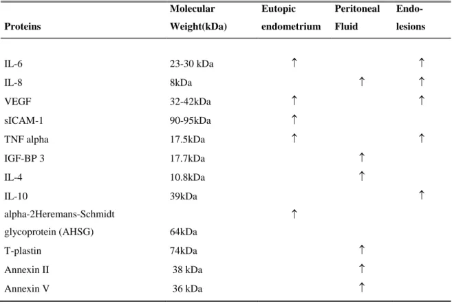
It is a marker of uterine non-receptivity and is abundantly expressed during the implantation window in women with endometriosis and infertility [60]. It was suggested that a direct effect is exerted on the endometrium by inflammatory factors contained in the peritoneal fluid of women with endometriosis.
E NDOCRINOLOGIC DISORDERS
C LINICAL E XAMINATION
Umbilical endometriosis should be suspected when a patient has a palpable mass and cyclical pain in the umbilical region[11]. Although clinical examination is a useful tool in the detection of endometriosis, the diagnosis of endometriosis should be confirmed by biopsy of suspicious lesions obtained laparoscopically.
I MAGING AND E NDOMETRIOSIS
L ABORATORY TESTS
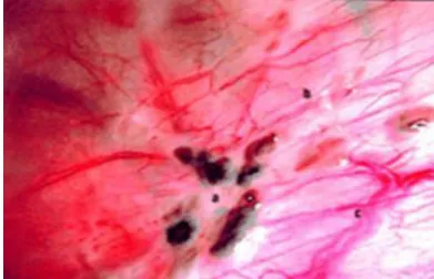
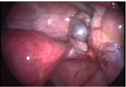
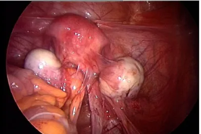
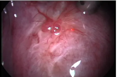
L APAROSCOPIC FINDINGS
There was no significant difference between the nerve fiber densities in women with confirmed minimal endometriosis and those with mild endometriosis p=0.46). High density of small nerve fibers in the functional layer of the endometrium in women with endometriosis.
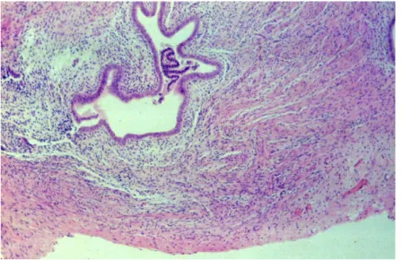
H ISTOLOGIC C ONFÍRMATION
S PONTANEOUS EVOLUTION DURING PREGNANCY
The features of endometriosis are variable during pregnancy, and lesions tend to enlarge during the first trimester but decrease thereafter [91]. Studies in baboons have shown no change in the number or area of endometriotic lesions during the first two trimesters of pregnancy [92].
P REVENTION
These results do not exclude a beneficial effect that may occur during the third trimester or in the immediate postpartum period.
T HERAPY
E NDOMETRIAL SENSORY NERVE FIBRES IN ENDOMETRIOSIS
- Small diameter sensory nerve fibres in endometrium
- Nerve growth factors
It may also be programmed to secrete large amounts of trophic nerve factors, resulting in the remarkable ingrowth of unmyelinated small nerve fibers demonstrated in this study. This can also translate into ingrowth of nerve fibers into new endometriotic plaques on the peritoneum or ovary.
N EED FOR NON - INVASIVE DIAGNOSIS
It is worth noting that surgery often plays an important role in the treatment of endometriosis. Furthermore, it may also be of interest in dealing with other conditions that occur in a similar way (eg tubal infertility).
C ELLS OF PERITONEAL CAVITY
However, it is not known whether red blood cell PF concentration and hemoglobin are higher during menstruation than during nonmenstrual phases of the cycle. However, it is not known whether the PF concentration of white blood cells is higher during menstruation than during nonmenstrual phases of the cycle. The lack of knowledge regarding possible differences in the presence and distribution of PF cell populations during menstruation between women with and without endometriosis is a major obstacle regarding the validity of the Sampson hypothesis.
There is compelling evidence for increased white blood cell counts in the PF of women with endometriosis compared to women without endometriosis [119]. Recent studies suggest that activated macrophages are elevated in the peritoneal fluid of women with endometriosis.
R ETROGRADE MENSTRUATION
- Quality of viable endometrial cells
The phenomenon of adhesion of epithelial cells with a possible endometrial origin may affect the detection of endometrial cells in PF. This explains the observation that among women with Mullerian anomalies, those with outflow obstruction are more likely to have endometriosis than those without (77 vs. 37%)[11]. These data indicate that retrograde transport of viable endometrial cells during menstruation occurs in the majority of women with patent tubes, suggesting that, in addition to the presence of endometrial cell reflux, another factor(s) is critical to the pathogenesis of endometriosis.
It has been hypothesized that the quality of endometrial cells in the PF of women with endometriosis is different from that of women with a normal pelvis. Viable endometrial cells from human endometriotic biopsies, but not from human endometrial biopsies, are invasive in an in vitro collagen invasion assay, probably because they have a higher proportion of potentially invasive E-cadherin-negative epithelial cells [145].
D IAGNOSTIC DELAY IN THE DIAGNOSIS OF ENDOMETRIOSIS
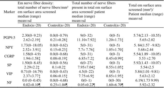
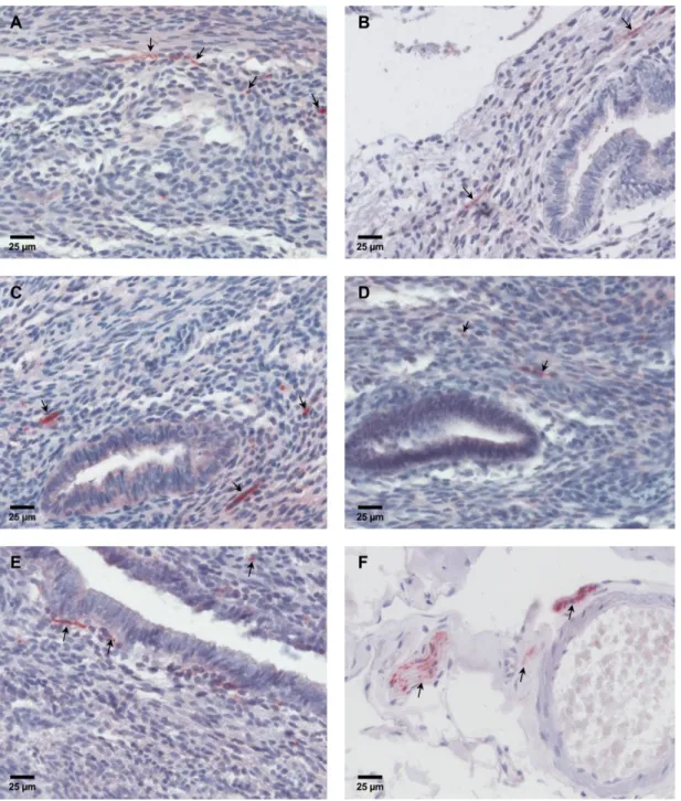
Recent studies have shown a higher density of small unmyelinated nerve fibers in the functional layer of the endometrium in women with confirmed endometriosis compared to women without endometriosis, especially in the secretory phase of the cycle [164, 165]. Indeed, sensory nerve fibers can be identified in the functional layer of the endometrium by immunohistochemical analysis of various neuronal transmitters such as substance P (SP), vasoactive intestinal polypeptide (VIP) or neuronal proteins such as protein gene product 9.5 (PGP9.5), neurofilament ( NF), neuropeptide Y (NPY) and calcitonin gene-related protein (CGRP). Detection of endometrial nerve fibers has been proposed as a diagnostic tool for endometriosis in a recent pilot study [166].
However, this study was limited by the lack of uniform histological confirmation of endometriosis, inclusion of variable numbers of patients from all stages of the disease and by cycle phase-related changes of endometrium. In the present study, we tested the hypothesis that women with minimal and mild endometriosis express a higher density of sensory small-diameter nerve fibers in the functional layer of endometrium than women with a normal pelvis to provide a possible semi-invasive diagnostic tool for minimal to mild endometriosis.
AIMS
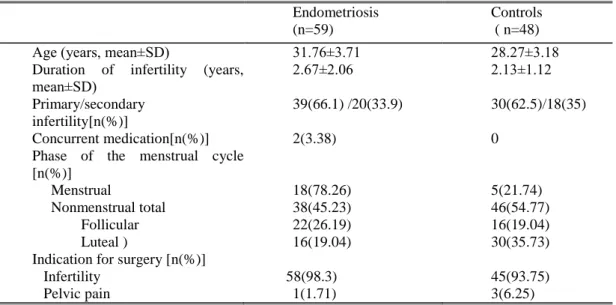
MATERIAL AND METHODS
- T ISSUE COLLECTION
- H ISTOLOGY
- I MMUNOHISTOCHEMISTRY
- S TATISTICAL ANALYSIS
- P ATIENTS AND SAMPLE COLLECTION FOR THE PF STUDY
- C ELL COUNTS
- I MMUNOCYTOCHEMISTRY
- S TATISTICAL ANALYSIS
Rabbit and mouse immunoglobulin fractions were used as respective negative controls, the concentrations being matched to the concentrations of the antibodies. After summing the nerve fiber counts and HPF area values for the entire section, the total number of nerve fibers was divided by the total surface area of the examined endometrium to obtain the nerve fiber density for the current section. In addition, an operating point on the ROC curve was chosen corresponding to the maximum of the sum of sensitivity and specificity.
Samples of PF were collected during the luteal (n=46), follicular (n=38), or menstrual (n=23) phase of the cycle. In these 32 patients, PF samples were collected during the luteal (n=20), follicular (n=5), or menstrual (n=7) phase of the cycle.
RESULTS
R ESULTS OF THE ENDOMETRIAL SENSORY NERVE FIBRE STUDY
Nerve fiber density was markedly skewed, with a few specimens having counts above 30/mm2 and most between 0–10 per mm2. Univariate analysis of different endometrial neural markers for semi-invasive diagnosis of minimal and mild endometriosis. PPV = positive predictive value; NPV = negative predictive value, AUC = area under the curve for diagnostic accuracy.
Selection of the number of neural markers for LS-SVM modeling based on Leave One Out-Cross Validation (LOO-CV). Using Leave-One-Out cross-validation (LOO-CV) analysis with LS-SVM modeling (Table 8.), the best result was obtained by selecting the top 3 neural markers based on their p-value (Mann-Whitney U-test ).
R ESULTS OF THE PERITONEAL FLUID CYTOLOGY STUDY
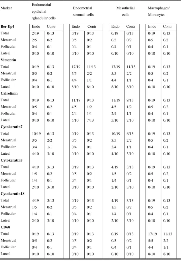
Ber Ep4 Total Menstrual Follicular Luteal Vimentin Total Menstrual Follicular Luteal Calretinin Total Menstrual Follicular Luteal Cytokeratin7 Total Menstrual Follicular Luteal Cytokeratin8 Total Menstrual Follicular Luteal Cytokeratin18 Total. Cells positive for epithelial marker Ber-Ep4 were not observed, except in two patients examined during menstruation who had a few positive cells. In 9/32 patients, the single cell population contained 10-50% cells with very weak, probably non-specific staining for calretinin.
Most single cells present in PF were histiocytes or belonged to the monocyte/macrophage lineage. The prevalence of mesothelial cells and macrophages that stained positive for the antibodies tested (Table 9.) was similar between menstrual and non-menstrual phases of the cycle and between patients with and without endometriosis.
DISCUSSION
In the context of the pathogenesis of endometriosis, retrograde menstruation should be diagnosed not only as an increased presence of erythrocytes in PF, but also as the presence of endometrial cells in PF. In our study, we could not confirm the hypothesis that the incidence and/or quantity of EM cells in PF is increased in women with endometriosis (compared to controls) and increased during menstruation (compared to nonmenstrual phases of the cycle). Third, immunocytological identification of EM cells in PF is not clear as reported in our study and in 3 papers published by 3 other groups of investigators.
It is possible that immunocytological detection of EM cells is facilitated after cell culture compared with analysis of EM cells in native PF. Taking together the evidence from our study and other studies, it is incredible to conclude that to date there is no solid scientific evidence based on analysis of PF fluid cells that retrograde menstruation is associated with an increased presence of endometrial cells in PF.
CONCLUSION
SUMMARY
Az endometriózis gyakori, ösztrogénfüggő krónikus nőgyógyászati megbetegedés, melynek lényege, hogy a méhüregen kívül, általában a medencében endometriumszerű szövetek jelennek meg. Arra azonban nincs bizonyíték, hogy a peritoneális folyadékban az endometriumsejtek, a vörös- és fehérvérsejtek, valamint a hemoglobin koncentrációja magasabb lenne a menstruáció során, mint a menstruációs ciklus többi részében. A retrográd menstruációt vizsgáló kísérletünkben a menstruációs ciklus luteális, follikuláris és menstruációs fázisában endometriózisban szenvedő és egészséges kontrollok medencéjéből vettünk peritoneális folyadékmintákat laparoszkópia során.
Eredményeink azt mutatják, hogy a menstruáció során a peritoneális folyadékban a leukociták, eritrociták és hemoglobin koncentrációja magasabb a menstruációs ciklus többi fázisához képest, ami alátámasztja a retrográd menstruáció elméletét. Eredményeink alapot adnak egy lehetséges félig invazív teszthez, amely lehetővé teszi az endometriózis korai klinikai diagnosztizálását.
Dinulescu DM, Ince TA, Quade BJ, Shafer SA, Crowley D, Jacks T. 2005) Role of K-ras and Pten in the development of mouse models of endometriosis and endometrioid ovarian cancer. Matalliotakis IM, Cakmak H, Fragouli YG, Goumenou AG, Mahutte NG, Arici A. 2008) Epidemiological characteristics in women with and without endometriosis in the Yale series. 1996) Delay in the diagnosis of endometriosis: a survey of women from the USA and the UK.
In-vitro attachment of endometrium to autologous peritoneal membranes:. effect of cycle phase and endometriosis stage. new targets and approaches towards the development of future treatment regimens. Kyama CM, T'Jampens D, Mihalyi A, Simsa P, Debrock S, Waelkens E, Landuyt B, Meuleman C, Fulop V, Mwenda JM, D'Hooghe TM. 2006) ProteinChip technology is a useful method in the pathogenesis and diagnosis of endometriosis: a preliminary study. Kyama CM, Mihalyi A, Simsa P, Mwenda JM, Tomassetti C, Meuleman C, D'Hooghe TM. 2008) Nonsteroidal targets in the diagnosis and treatment of endometriosis.
2001) Effect of menstruation and intrapelvic injection of endometrium on inflammatory parameters of peritoneal fluid in the baboon (Papio anubis and Papio cynocephalus).
LIST OF PUBLICATIONS RELATED TO THE THESIS
The role of the fallopian tube in the development of endometriosis and associated infertility In: Allahbadia G N, Saridogan E, Djahanbakhch O, (editors) Fallopian Tube.
ACKNOWLEDGEMENT