Department of Pathology and Laboratory Medicine (A.T.S., B.Z.) and Department of Medicine (A.T.S.), University of Tennessee, Memphis, Tennessee 38103; Department of Histology (M.A.Z.), Medical University of Gdansk, 80-210 Gdansk, Poland; Center for Skin Sciences (D.J.T.), School of Life Sciences, University of Bradford, Bradford, West Yorkshire BD7 1DP, United Kingdom; Department of Molecular Physiology and Pharmacology, Biochemistry and Internal Medicine (T.C.T.), Tufts University School of Medicine, Boston, Massachusetts 02111; and The Clayton Foundation Laboratories for Peptide Biology (J.R.), The Salk Institute, La Jolla, California 92037. Environmental regulation of skin CRF signaling with emphasis on UV radiation and skin bacteria. Alternative splicing of CRFR-1 and CRFR-2 pre-mRNAs leads to the production of multiple receptor isoforms.

CRF Signaling in the Skin: Its Expression and Organization
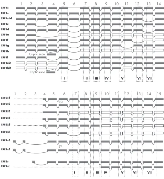
This additional coupling could be responsible for the shapes of the CRF or Urc inhibition curves in HaCaT keratinocytes, determined by the concentration of Ca2⫹ in the growth medium (bell shape in medium containing high Ca stimulates the MAPK signaling pathway with secondary inhibition of vascular endothelial growth factor (VEGF) (259) and IL-18 production (260). The CRFR-1c isoform failed to bind CRF when expressed in the monkey kidney cell line COS-1 (263). Furthermore, the expression of CRFR-1d, CRFR-f, and g with CRFR-1␣ resulted in the accumulation of coexpressed isoforms inside cells (264).
CRF/Urc1CRF/Urc1
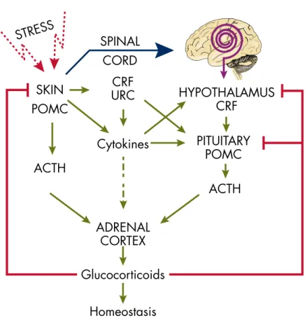
Skin Equivalent of the HPA Axis
Uncovering the chemical nature of the active peptide is important because it determines the final phenotypic effects on the skin. Therefore, the challenge for this field is to define context-dependent processing of POMC in the skin. Unlike the brain and pituitary, in the skin all elements of the HPA axis are synthesized in the same organ and in close proximity, including their regulatory elements such as cytokines (49).
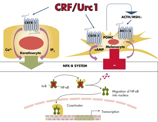
In addition, the alternative ligand for CRFR-1, Urc-1, is also produced in the skin (208) and may therefore also act as a trigger of the CRFR-1-mediated stress response in this organ (40). An example of this mode is the activation of POMC in the skin by chemical peel stress without involvement of CRF (327). For example, the concentration of POMC mRNA in mouse skin is ⱕ10,000 lower than in the pituitary gland (223).
Furthermore, UVR production and secretion of final effectors of the HPA (glucocorticoids) are activated by sequential and/or alternative modes of action originating in the skin that will depend on the wavelength and dose of solar electromagnetic energy. The current hypothesis is that systemic responses to UVR originating in the skin (in addition to the production of vitamin D) also involve pathways that include the activation of the cutaneous and systemic HPA axis (Figure 7). In the first case, UVR regulation of systemic homeostasis via the HPA axis would begin with the stimulation of CRF in the hypothalamus via neutrally transmitted signals (Figure 7).
In the latter case, the skin can activate the HPA axis at different entry points, e.g. the pituitary or adrenal gland, through skin-derived humoral messages such as CRF/Urc-1, cytokines (IL-1, IL-6) , and TNF-. ␣), or ACTH (Figure 7).
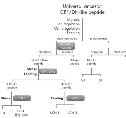
CRF and Urc Function as Pleiotropic Cytokines
Furthermore, the effects of psychological stress in a rat model are reversed by RU-486 (glucocorticosteroid inhibitor) and antalarmin (CRF inhibitor) (484). These data suggest that CRF is acting primarily through CRFR-1 in hair follicle melanocytes. The exact role of the CRF/CRFR-1 system in the differential regulation of skin melanocytes is complicated by the significant heterogeneity of melanocytes in this organ.
In contrast, melanogenesis in the epidermis appears to be continuous (511), although this is further upregulated by UVR. Moreover, at least two fibroblast subtypes exist in the hair follicle, namely the dermal sheath (DS) fibroblast and the dermal papilla fibroblast. This CRFR-1 agonist-mediated inhibition was accompanied by the inhibition of keratinocyte proliferation in the anagen hair bulb and results in the premature deposition of the anagen-growing hair follicle in an apoptosis-driven catagen state.
However, we are still in the early stages of understanding CRFR-1 and CRFR-2 in these processes. Cytokines mediate interactions between cells traditionally accepted as part of the immune system (Langerhans cells, lymphocytes, neutrophils, etc.) and resident cells in the epidermis and dermis. Cutaneous mast cells occupy a strategic position in the brain-skin axis due to their location at the interface of the skin's immune and nervous systems.
However, anti-inflammatory activities of CRF and related Urcs in the periphery have also been described.
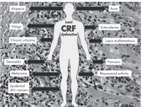
Skin Pathology Associated With Dysregulation of the Cutaneous CRF
There is considerable evidence for the involvement of CRF in RA, a symmetrical arthritis that typically involves the metacarpophalangeal and proximal interphalangeal joints of the hands. In summary, the role of CRF and CRFR-1 in the development of psoriatic lesions is emerging and new theories are proposed on this topic. It is therefore conceivable that the reduction or loss of expression of the POMC peptides␣-MSH and-endorphin in vitiligo (as previously reported in Ref. 564) may be due to disorders related to reactive oxygen species. Ca2⫹-dependent proteolytic activity of this convertase, which could affect the immune status of these lesions.
However, a POMC-independent role of CRF signaling in the regulation of melanin pigmentation is also a viable option (80, 509). The authors interpreted these results on the basis that an up-regulation of the CRF system in aging skin may lead to an exaggerated stress response in this skin (245). The authors interpreted this as suggesting that the observed altered HPA axis activity was a result of the AA-associated immune response.
The authors of recent studies propose the use of specific CRFR-1 antagonists instead of agonists in the treatment of psoriasis. Expression of CRF and CRFR-1 in normal skin, melanoma cells, and effects of CRF1 agonist on melanoma cell proliferation. However, local dysregulation of the HPA axis or defects at receptor levels (for example, generation of alternatively spliced isoforms) may also contribute to tumor progression.
The detailed role of alternative splicing in the development of skin diseases and carcinogenesis remains to be investigated.
Quest for Novel Therapy of Cutaneous Disorders Based on Interventions to the Local
Thus, uncoupling of CRFR-1 from the cutaneous HPA axis or its variants (see above) will lead to sustained pro-inflammatory activity that can self-reinforce leading to skin inflammatory states including psoriasis, acne, AA or allergic reaction. - ties, and maybe vitiligo. An additional instructive model here is the context-dependent coupling of CRF signaling to NF-B (the master regulator of inflammation and context-dependent regulator of differentiation and cell survival) (Figure. We also believe that similar mechanisms contribute to the development of some systemic autoimmune diseases such as RA and we might expect similar contributions in lupus erythematosus and scleroderma.
This can be extended to other organs by proposing that inefficient local attenuation of the CRF signaling system and/or defective coupling to the downstream immunosuppressive regulatory mechanisms may exacerbate or induce local proinflammatory responses leading to inflammatory and/or autoimmune disease processes. This effect can be enhanced by the release of corticosteroids from malignant cells, generating an immunosuppressive environment. In summary, dysregulation of CRF signaling either at the CRFR-1 receptor level or at downstream effectors and feedback mechanism can lead to skin disorders with further systemic implications.
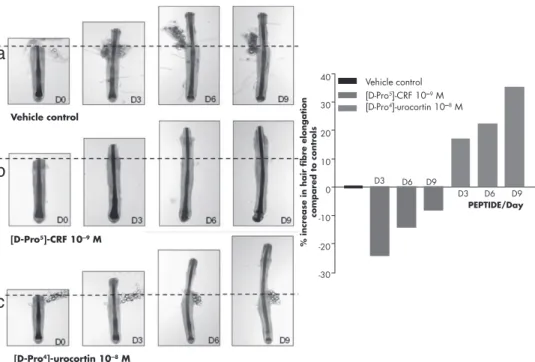
Targeting CRFR-2 may be useful in treating hair cycle disorders or sebaceous gland dysfunction. Agonists at CRFR-2 (eg, Urcs) can stimulate ex vivo organ cultures of anagen-intact scalp hair growth by maintaining the growth-associated anagen phase longer, during which time more too much hair fiber. Their use as targets for pharmacological modifications or activation in the therapy of skin disorders remains to be investigated in appropriate preclinical models.
Finally, the most promising candidates are soluble CRFR-1 isoforms that, by binding with CRF or Urcs, will reduce the availability of the ligands to the receptor, protect the ligands from degradation, or ensure their delivery to the correct compartment. .
Theory on the Origin of CRF-Led Stress Response
Their role, which may be similar but not identical to CRF-binding protein, awaits testing with synthetic proteins.
CRFCRF dysfunction
Final Comments and Future Directions
Slominski A, Wortsman J, Pisarchik A, et al. Cutaneous expression of corticotropin-releasing hormone (CRH), urocortin and CRH receptors. FASEB J. Jin D, He P, You X, et al. Expression of corticotropin-releasing hormone receptor type 1 and type 2 in human pregnant myometrium.Reprod Sci. Kokkotou E, Torres D, Moss AC, et al. Corticotropin-releasing hormone receptor 2-deficient mice have reduced intestinal inflammatory responses.J Immunol.
Slominski A, Wortsman J, Kohn L, et al. Expression of hypothalamic-pituitary-thyroid axis-related genes in the human skin. J Invest Dermatol. Roloff B, Fechner K, Slominski A, et al. Hair cycle-dependent expression of corticotropin-releasing factor (CRF) and CRF receptors in murine skin. FASEB J. Asadi S, Alysandratos KD, Angelidou A, et al. Substance P (SP) induces expression of functional corticotropin-releasing hormone receptor-1 (CRHR-1) in human mast cells.
Rassouli O, Liapakis G, Lazaridis I, fi kkf. Gahee haaraa hormoonii kortikootiroopiin gadhiisu naannoo (CRH) fibroblasts gogaa irratti. PLoS Tokko. Ito N, Sugawara K, Bodo ́ E, fi kanneen biroo. Hormooniin kortikootiroopiin gadhiisu dhaloota seelota mastii bakka jirutti dursitoota meeseenkaayimii foolikula rifeensa namaa keessa jiran irraa kakaasa. Zbytek B, Pikula M, Slominski RM, fi kanneen biroo. Hormooniin kortikootroopiinii gadhiisu HaCaT keratinocytes keessatti garaagarummaa kakaasa. Br J Dermatol jedhamuun beekama.
Park HJ, Kim HJ, Lee JH, et al. Corticotropin-releasing hormone (CRH) regulates interleukin-18 expression in human HaCaT keratinocytes by activation of the p38 mitogen-activated protein kinase (MAPK) pathway. J Invest Dermatol. Licinio J, O'Kirwan F, Irizarry K, et al. Association of a corticotropin-releasing hormone receptor 1 haplotype and antidepressant treatment response in Mexican-Americans. Theoharides TC, Spanos C, Pang X, et al. Stress-induced mast cell intracranial degranulation: a corticotropin-releasing hormone-mediated effect.Endocrinology.