The main purpose of the circadian clock is to plan the body's functions in optimal time windows. The main aim of this study was to investigate how the time of day affects UVB-induced erythema, and the effects of UVB on circadian clock gene expression in the skin.
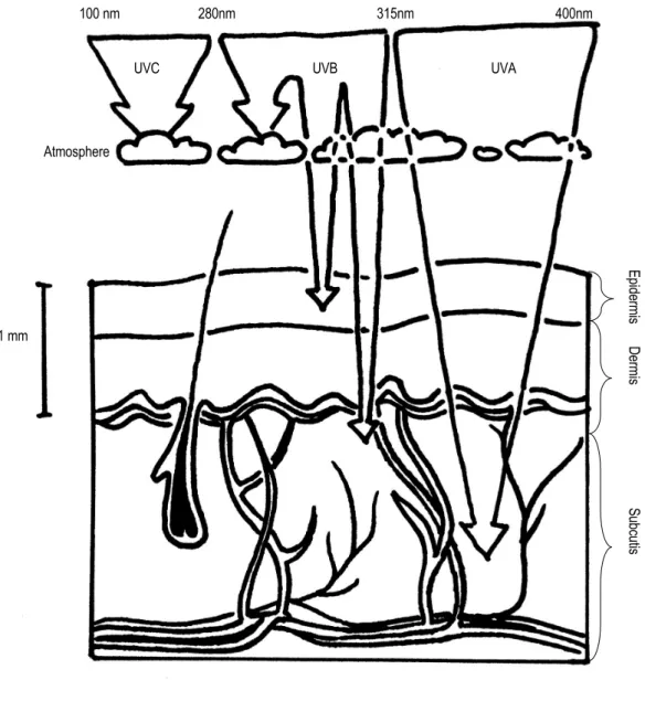
UV radiation and the skin
- Acute effects
- Erythema and pigmentation
- Chronic effects
- Systemic effects
- Population-level effects
Erythema indicates several negative effects of UV irradiation: skin inflammation, damaged DNA and oxidative stress (D'Orazio et al. 2013). Of these, local effects occurring in the irradiated skin are thickening of the epidermis (Lee et al. 2002) and development of skin cancer (Feehan and Shantz 2016).

The circadian clock
- The core circadian clock and Zeitgebers
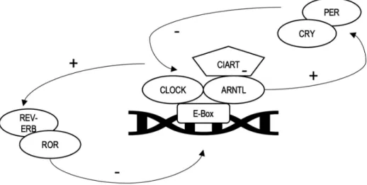
- Clock genes and proteins
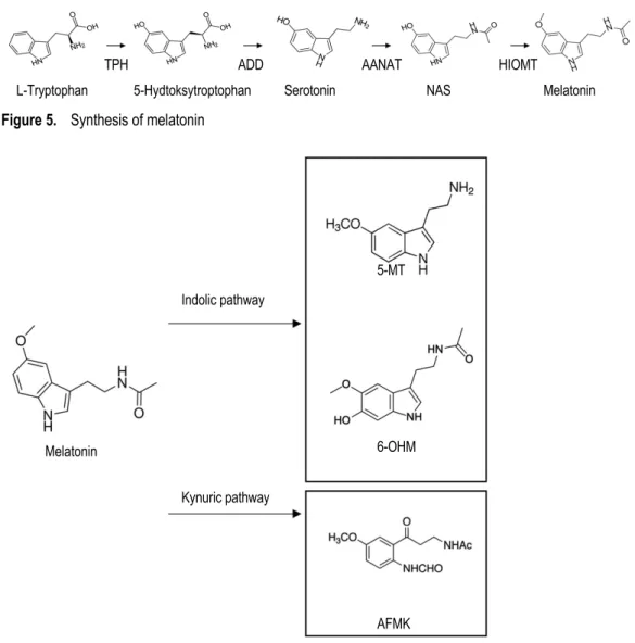
- Melatonin
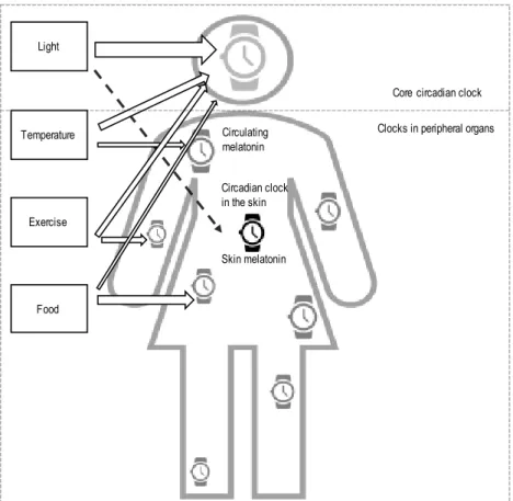
- The clock in the peripheral organs
In humans, contrary to previous assumptions, RORa is not a nuclear melatonin receptor (Slominski et al. 2016). While the core circadian clock is clearly influenced by light, clocks in the peripheral tissues are entrained by various external stimuli (Sherratt et al. 2019).
The circadian clock in the skin
Circadian function and clock genes in the skin
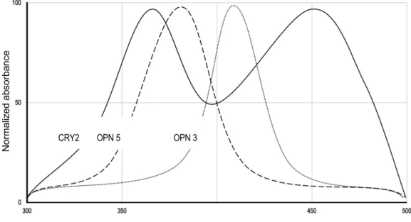
Entrainment of the clock by food intake appears to be independent of the circadian nuclear clock (Wang et al. 2017). OPN1SW, OPN2, OPN3 and OPN5 have been found in human epidermal skin (Haltaufderhyde et al. 2015).
Melatonin in the skin
These protective properties of melatonin may be due not only to melatonin itself, but also to its derivatives and its properties to induce other antioxidants (Reiter et al. 2007; Rusanova et al. 2019). In addition to melatonin, its derivatives AFMK (Onuki et al. 2005) and 4OHM (Pérez-González et al. 2017) are also powerful antioxidants.
The circadian clock and UV- induced erythema
Clock proteins and erythema
These and some other data from animal studies indicated that cryptochromes were involved in clock-controlled apoptosis, cell cycle regulation and NER activity (Sancar et al. 2015). In line with this is the fact that oxidative stress in human skin due to UV radiation is under the control of clock protein functions (Geyfman et al. 2012).
Melatonin and erythema
Although it is known that UV erythema occurs in the human skin in the same context as oxidative stress and the recruitment of protective mechanisms (D'Orazio et al. 2013), knowledge of the circadian actions of erythema is lacking. Their study using cultured human melanocytes showed the involvement of nuclear factor erythroid 2-related factor 2 (NRF2) in a pathway where melatonin protects cells from UVB-induced damage (Janjetovic et al. 2017). Silent information regulator 1 (SIRT1) pathway, induced by melatonin, has also been shown to prevent oxidative stress mediated damage in cultured human keratinocytes (Lee et al. 2016).
Although melatonin, as a small ambiphilic molecule, reaches all parts of the cell, its notable antioxidant activity takes place in the mitochondria (Martin et al. 2000). It has even been suggested that in skin DNA repair, stimulation of NRF2 and reduction of oxidative damage by melatonin would be secondary to its mitochondrial effects (Slominski et al. 2017b). When looking at protection against erythema, circulating melatonin appears to be unimportant compared to melatonin produced in human skin (Slominski et al. 2018a).
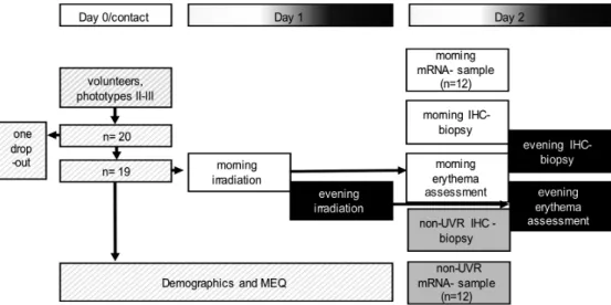
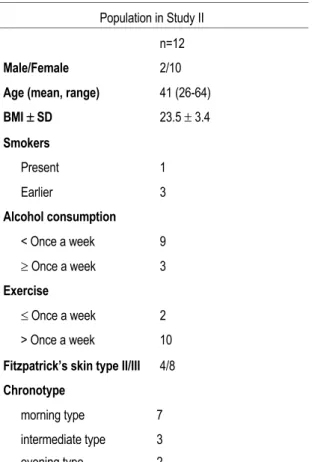
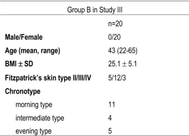
Study protocols and volunteers
Protocols
Regarding the volunteers in Group A, the protocol and target group were the same as in Studies I and II, which were implemented in 2016. The other Group B was recruited later in the winter 2018-2019 with the aim of increasing the size of the total cohort, and in order to enable comparison of the non-irradiated biopsy at different times of the day, namely morning vs. The photosensitivity was equally assessed in both Groups A and B, separately in the morning and in the evening, by performing a photo-test series.
Regarding the sampling of skin biopsies, the groups were treated differently, in group A, one biopsy from each volunteer was obtained from the non-irradiated skin site in the morning, while in group B, a total of two non-irradiated biopsies were obtained, one in the morning and one in the evening.
Volunteers
Methods
- UVB irradiation (I-III)
- Assessment of erythema (I-III)
- Assessment of chronotype (III)
- Skin samples (I-III)
- Immunohistochemistry (I, III)
- RNA analyses (II)
- Statistical analysis
- Ethical aspects
If the MED appeared to be higher than the highest dose of 4 SED in our series, we set the MED as 5.6 SED for further analyses, i.e. the next dose of the geometric UVR dose series, where the incremental factor is Ö2. In Study I, all three samples (two irradiated and one control sample) from 19 volunteers were used in the analyses. The first halves of the 6 mm skin biopsy samples were fixed in 4% formalin, embedded in paraffin and sectioned at 4 µm thickness for further processing.
The microscope and camera settings remained unchanged and the color balance of the images taken based on the ICH findings was not changed. We also studied the gene expression of clock proteins, cytokines, proopiomelanocortin and melanocortin receptors by analyzing mRNA from non-irradiated morning control samples and 4 morning samples irradiated with SED, a total of 12 healthy volunteers. Cytokine and circadian clock gene mRNA expression was studied by TagMan 7500 Fast System (Applied Biosystems) with PerfeCTa qPCR FastMIx (Quanta Biosciences, Gaithersburg, MD, USA).
Time of the day effects on UVB- induced erythema (I)
Impact of time of day and NB-UVB dose on erythema (a) and the effect of CRY2 (b).

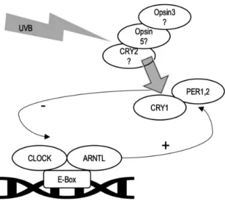
Effects of UVB radiation on clock genes (II)
Classification of the population of Study I by immunohistochemical staining of cryptochromes in control (non-irradiated) biopsies and the population of Study II by changes of mRNA expressions of CRY1, CRY2 and CIART genes after irradiation.
Effects of skin melatonin on UVB-induced erythema (III)
The skin is an important organ that protects the human body from potentially dangerous electromagnetic exposure from the sun and other sources. The current dissertation study is one of the first studies to evaluate the interactions between biologically active electromagnetic radiation and the circadian clock in human skin in vivo. We have established a research protocol proposing that UVB radiation applied to the skin is a potential pacemaker of the circadian clock of the epidermal cells, and that UVB may also interact with the internal circadian clock through the skin.
In other words, we proposed that UVB could create an interaction with the circadian clock that is quite similar to the interaction of visible light, which produces its main effect through the eyes (Roenneberg et al. We also hypothesized that the physiological response of the skin to UVR would vary with knowledge from this study could be used, for example, in optimizing the timing of phototherapy, in other skin disease treatments, skin cancer treatments, and more.
Circadian time and erythema (I)
However, it was shown in the early stages of human circadian clock research that CRY1 and 2 cannot act directly as enzymes of DNA repair when UVR-related damage occurs in humans (Hsu et al. 1996). In Study I we detected more p53 protein in irradiated morning samples than in evening samples and Study II showed that CRY2 mRNA decreases in the upper layers of the skin as a result of irradiation. Unfortunately, we do not know whether UVB would initiate less CRY2 mRNA production in the evening when p53 accumulated in smaller amounts in epidermis, because we did not detect CRY2 mRNA changes due to UVB in the evening.
However, suboptimal function of the circadian clock generally leads to sensitivity to UVR-induced stress (Kawamura et al. 2018), and skin clock entrainment by UVB is possible according to our findings in Study II. In light of these facts, NB-UVB phototherapy could be more effective with fewer adverse effects when administered in the morning hours. Going forward, as the best benefit and minimal side effects are warranted in the field of dermatological treatments, chronopharmacology should not be disregarded.
UVB- radiation and clock proteins (II)
Based on the signaling mechanisms of cryptochrome (Vieira et al. 2012), it is possible that they may even serve as UVB sensing agents in the skin themselves. The fact that UVB can induce the production of these cytokines in the outer layers of the skin is not a new finding (Slominski and Wortsman 2000). Therefore, we believe that the inflammation was mediated by circadian clock cytokines in the subcutaneous fat.
Aspects affecting clock entrainment include, among others, individual differences in the effectiveness of different Zeitgebers, free-running circadian time period length, and synchronization (entrainment) of Zeitgebers with internal time (Roenneberg et al. 2003a). Human adipose tissue appears to contain a circadian clock that controls its metabolism (Christou et al. 2019). Changes in the subcutaneous adipose tissue clock may even predispose an individual to metabolic syndrome (Hernandez-Morante et al. 2012).
Skin melatonin and erythema (III)
This fluctuation resembles the pattern of pineal expression of melatonin that begins in the evening and peaks at night (Reiter 1991). However, extra-pineal sites can express melatonin oscillation that does not vary in a circadian manner (Venegas et al. 2012) and pineal melatonin has only limited access from blood to the skin due to its degradation in the liver (Slominski et al. . 2017a) ). In an earlier human study, melatonin was found in keratinocytes and is cytoplasmic in the skin of the scalp (Slominski 2005).
Serotonin, the precursor of melatonin, was also cytoplasmic and membranous in the same anatomical location (Slominski et al. 2020). In both tissues, there was a significant amount of melatonin in the nuclei (Venegas et al. 2012). In the scalp, melatonin may have effects related to hair growth via mitochondria (Rusanova et al. 2019).
Chronotype and erythema (III)
Limitations and strengths
For the first time, we showed the difference in the expression of epidermal melatonin to vary depending on the time of day. Like the circadian clock in general, the clock in the skin and in the subcutis was also adjustable. Ultraviolet B radiation affected mRNA expression in the epidermal/dermal skin and in the subcutaneous adipose tissue.
We showed for the first time that UVB radiation affects circadian gene expression in subcutaneous adipose tissue. Genes involved in regulating and maintaining circadian rhythms are called clock genes. There is a daily rhythm in the expression of the circadian-associated repressor of transcription (CIART) gene, and it is also regulated by stress responses (20).
No significant changes in the subcutaneous adipose tissue were seen in the cytokine expressions studied (Figure 4b). In addition to epidermal/dermal skin, UVB radiation also appears to affect mRNA expression in the subcutaneous adipose tissue.