Specifically, Poly(ethylene terephthalate), Nylon-6 and cotton/PET blends were "activated" through a limited surface treatment using enzymes. After immobilization via the EDC/NHS coupling system, the functionalized cotton/PET blends were tested against Gram positive and negative bacteria.
Aim of the thesis
Textile functionalization
- Poly(ethylene terephthalate) (PET)
Unlike natural fibers, most synthetic polymers (i.e., polyesters and polyamides) used in textile applications show high hydrophobicity, which is also responsible for the reduced possibility of applying finishing agents (such as antistatic agents or antimicrobials and dyes), due to the minimal presence of reactive. groups on the surface of the polymer [5], [6]. Among these various polymers[10] PET (also commonly called polyester) is considered one of the most important man-made polymers.
Enzymatic functionalization of PET
Nylon 6
When a certain concentration of aminocaproic acid has been produced, further CL units are attached to the already growing chain to produce a polymer with 2 or more repeats.
Enzymatic treatment of PA
Flame retardant applications
The phosphoric acid leads to carbon formation and inhibits the pyrolysis necessary for the growth of the flame. These compounds can give off ammonia during combustion, which can lead to a dilution in the gas phase resulting in flame extinction (Figure 6C).
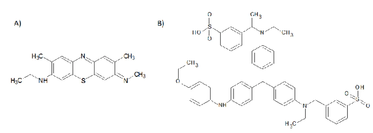
DNA as flame retardant
Antimicrobial textiles
Due to their large surface area and ability to retain moisture, textiles are known to be conducive to the growth of microorganisms, such as bacteria and fungi. The infections acquired in hospitals can be caused by several species, such as Escherichia coli, Klebsiella pneumonia, Pseudomonas aeruginosa and Acinetobacter baumannii.
Protein-based nanoparticles
Human serum albumin
Silk fibroin
Eugenol
Textile waste recycling
- Protein based fibres
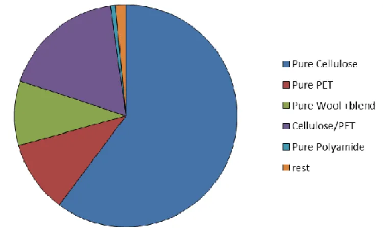
In 2007, the total consumption of textiles, including clothing and household textiles such as bedding and carpets, in the EU was 9.55 million tonnes. A recent assessment by the Waste and Resources Action Program (WRAP UK) mentions 6.4 million tonnes of clothing in the EU in 2015[2].
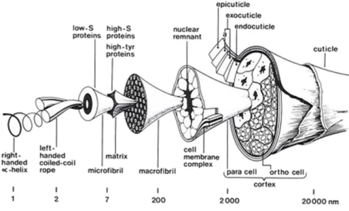
Enzymatic applications on wool fibres
Cotton
The specific composition of cotton varies depending on species and environmental conditions and has an impact on fiber length. The secondary cell wall is made of higher molecular weight cellulose with a degree of polymerization of up to 14,000.
Enzymatic degradation of cellulose
Value-added products
Moreover, this process involves and promotes innovation throughout the value chain instead of relying only on the "end-of-life products" solution.
Amino acids
Glucose
PET monomers
Biundo et al., “Characterization of a lipase hydrolyzing poly(butylene adipate-co-terephthalate) from Pelosinus fermentans,” Appl. Ribitsch et al., “Enhanced cutinase-catalyzed hydrolysis of polyethylene terephthalate by covalent fusion to hydrophobins,” Appl.
Increased flame retardancy of enzymatic functionalized PET and Nylon fabrics via DNA immobilization
Abstract
Introduction
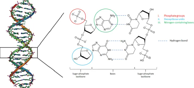
In short, the application of Flame Retardant Compounds (FRC) prevents the ignition or leads to the self-extinguishing of the flame. Together with a deoxyribose unit and a phosphate group, they built up the sugar phosphate backbone of the DNA.
Materials and Methods
- Chemicals, substrates and enzymes
- Biochemical characterization of HiC
- Enzymatic functionalization of PET and nylon 6 .1 Enzymatic functionalization
- High Performance Liquid Chromatography (HPLC)
- DNA immobilization on surface activated polymers
- DNA characterization
- DNA cross-linking methods
- Fourier-transformed Infrared Spectroscopy (FT-IR)
- Environmental Scanning Electron Microscopy (ESEM)
- Durability of washing
- Flame retardant assessment
- Flammability test
- Thermogravimetric Analysis (TGA)
A ColorLite sph850 Spectrophotometer – Color Measuring Instrument (Innovac, Germany) was used to determine the difference in color intensity of PET and nylon samples after acid/base dye treatment. To coat with dopamine or tyrosine, 3 mL of the solutions were pipetted onto enzyme-treated PET and nylon-6 samples and incubated for 24 hours at 250 rpm and 21 °C.
Results
- Enzymatic functionalization
- DNA immobilization
- Flame retardant tests
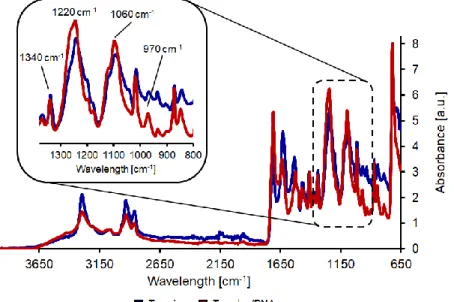
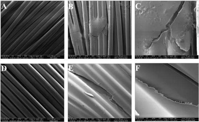
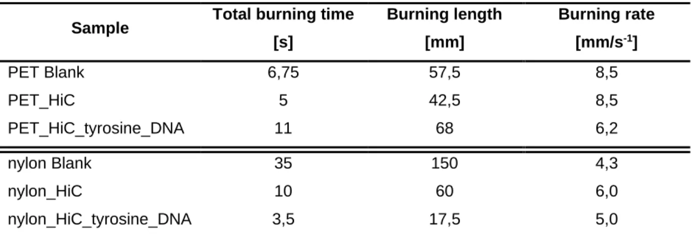
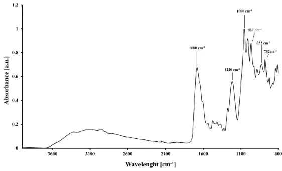
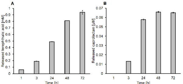
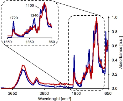
The enzymatic hydrolysis of PET showed a reduction and a slight shift of the band at 1721 cm-1 (indicative of the carbonyl stretching group). Tyrosine/DNA treated PET samples resulted in a decreased burning rate and length of the burned sample. An increase in the char formation during combustion of the tyrosine/DNA treated sample also indicates the presence of DNA (Figure 7).
Conclusions
The weight loss due to thermal degradation between 450–625 °C was significantly lower compared to the nylon sample treated with tyrosine alone showing a complete weight loss at 516 °C ( Figure 10B ).
Author Contributions
ACKNOWLEDGMENTS
Van Kuppevelt, “Development of tailored collagen} glycosaminoglycan matrices: EDC / NHS crosslinking and ultrastructural aspects,” Vol. Closing the loop: enzymatic synthesis and functionalization of biobased polyesters,” Trends Biotechnol., vol. Cavaco-paulo, “Effect of mechanical mixing on the activity of cutinases and proteases against polyamide substrates,” vol.
Supporting information
Smart Textiles in Wound Care: Functionalization of Cotton/PET Blends with Antimicrobial Nanocapsules
Introduction
Acute wound infections as well as chronic infected wounds are a serious complication worldwide with wound care becoming one of the most costly sectors of healthcare. In this regard, the prevention of the development of possible antibiotic resistance in wound colonizing bacteria can be overcome.[7] In general, widespread misuse of antibiotics in wound care led to new virus occurrence and the appearance of multidrug-resistant bacteria such as vancomycin-resistant Enterococcus (VRE) or methicillin-resistant Staphylococcus aureus (MRSA)[8]. In this context, the purpose of the use of natural components as antimicrobial and antiseptic agents in wound care has grown steadily.
Material and Methods
- Materials, Chemicals and Enzymes
- Biochemical characterization of enzymes
- Enzymatic treatment of cotton/PET blends
- High-Performance Liquid Chromatography (HPLC)
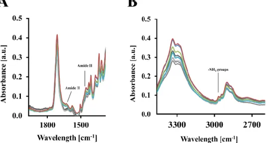
- Surface analysis using attenuated total reflection Fourier transform infrared spectroscopy and scanning electron microscopy
- Extraction of silk fibroin from Bombyx mori cocoons
- Immobilization of loaded HSA/SF nanocapsules
For eugenol loaded HSA/SF nanocapsules, 1 mg ml-1 (1%) eugenol was added to the organic phase before sonication. For each sample of eugenol loaded HSA/SF nanocapsules, release experiments were performed in triplicate. Cytotoxicity of unloaded and eugenol-loaded HSA/SF nanocapsules was analyzed using a commercial MTT assay (3-(4,5-dimethylthiazol-2-yl)-2,5-diphenyltetrazolium bromide; Sigma Aldrich, USA) .
![Table 1. Compositions and pH-values of artificial sweats (ISO pH 5.5, ISO pH 8.0 and EN pH 6.5) published by Callewaert et al.[17]](https://thumb-eu.123doks.com/thumbv2/pubdocorg/274324.42866/86.918.116.772.570.900/table-compositions-values-artificial-sweats-iso-published-callewaert.webp)
Results
- Enzymatic surface activation of cotton/PET blends
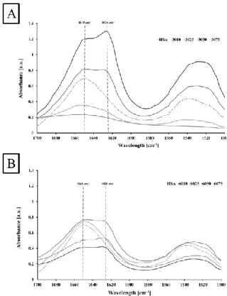
- HSA/SF nanocapsules production and formulation properties
Characterization of monodisperse eugenol-loaded HSA/SF nanocapsules composed of different percentages of SF (10–75%) and two different levels of degradation (30 or 60 mb). To ensure the safety of the produced eugenol-loaded, unloaded HSA/SF nanocapsules, cytotoxicity studies on THP-1 human monocytic cells were performed similar to the method reported by Tallian et al.[16] (Figure 5). Effect of SF content, HSA concentration and HSA/SF nanocapsules on cell viability of THP-1 human monocytic cells.
HSA/SF nanocapsules immobilization on cotton/PET textiles and antimicrobial activity
95 Additionally, SEM analysis was performed to investigate the successful immobilization of eugenol-loaded HSA/SF nanocapsules on cotton/PET blends ( Figure 7 ). 96 In Figure 8, cotton/PET blends functionalized with different formulations of eugenol-loaded HSA/SF nanocapsules have shown higher inhibition against S. In general, in gram+ bacteria the presence of eugenol leads to bacterial membrane damage, possibly because of its load. and interaction with lipids.
Conclusions
Conflicts of interest
Acknowledgements
Weinberger et al., "Enzymatic surface hydrolysis of thin films of poly(ethylene furanoate) with different crystallinities", Green Chem., vol. Quartinello et al., "Synergistic Chemoenzymatic Hydrolysis of Poly(ethylene Terephthalate) from Textile Waste." Microb. Ali et al., "Antimicrobial Activities of Eugenol and Cinnamaldehyde Against the Human Gastric Pathogen Helicobacter pylori," Ann.
Supporting information
3050 with 30 mb SF and a 50% HSA/SF ratio, HSA refers to formulations that do not contain silk fibroin. Hydrodynamic radius data based on dynamic light scattering in nm over time for a measurement period of 30 minutes of eugenol-loaded HSA/SF nanocapsules composed of two different levels of SF degradation (30 or 60 mb) and different SF concentrations (10–75%) evaluated for the initial 10 minutes (t1), the measurement time between 10-30 min (monodisperse phase, t2) and the entire measurement time (t3). Cumulative release of eugenol from HSA and HSA/SF nanocapsules with a lower degradation rate (30 mb) and 10 SF over a time period of 168 h in artificial saliva prepared as described in Callewaert et al. [17] (EN pH 6.0).
Highly selective enzymatic recovery of building blocks from wool-cotton-polyester textile waste blends
- Introduction
- Materials and Methods
- Chemicals, substrates and enzymes
- Enzymatic hydrolysis
- Protein hydrolysate characterization
- Cellulose hydrolysis
- Poly(ethylene terephthalate) characterization
- Results
- Step-wise enzymatic extraction of textile building blocks
- Protein hydrolysate characterization
- Cellulose based material hydrolysis and ethanol production
- Poly(ethylene terephthalate) characterization
- Conclusions
- Author contributions
After the hydrolysis of the cellulose residues, each sample was filtered and dried as described above. However, almost 80% of the building blocks of cellulose fibers were recovered from real textile waste after treatment with cellulase. The amount of glucose released after hydrolysis of the textile waste samples by cellulases was dependent on the initial cellulose content.
Notes
Enzymatic hydrolysis of wool- and cellulose-based fibers led to yields of approximately 95% and 85%, respectively. The purity of the resulting poly(ethylene terephthalate) was comparable to pure PET as indicated by FT-IR measurements, allowing recycling. Furthermore, in line with circular economy concepts, the building blocks recovered from wool and cellulose fiber components in blended textiles can also be reused.
Acknowledgments
Cavaco-Paulo, “Enzymes are big: surface hydrolysis and functionalization of synthetic polymers,” Trends Biotechnol., vol. Gamerith et al., “Enzymatic processing of polyester building blocks from polymer blends,” Process Biochem., vol. Bhavsar et al., Hydrolysis of waste wool with superheated water in a semi-industrial nitrogen fertilizer recovery reactor, vol.
Supporting information
128 Figure S2 Volumetric protease activity with (B) and without SDS and sodium bisulfite (Bc) at pH 8, 9, and 10 with the azocasein assay.

Synergistic chemo-enzymatic hydrolysis of poly(ethylene terephthalate) from textile waste
Introduction
Previously, various studies have shown that a class of enzymes belonging to the α/β hydrolase family, namely cutinases, is capable of hydrolyzing the ester bonds of PET and several other polyesters [12]–[14]. Among them, cutinase is currently being investigated for the bioprocessing of PET textiles on an industrial scale[15]. In this work, we propose an innovative synergistic chemo-enzymatic hydrolysis of PET to produce TA of high purity (97%) while avoiding harsh chemical treatments.
Materials and Methods
- Chemicals, substrates and enzymes
- Water-based PET hydrolysis
- ATR FT-IR analysis
- FT-Raman analysis
- Protein quantification and SDS-PAGE analysis
- Esterase activity assay
- Enzymatic hydrolysis of PET oligomers
- High Performance Liquid Chromatography
- H-NMR analysis
Then, 200 µL of prepared Bio-Rad reagent solution (BioRad reagent diluted 1:5 with MQ water) was added. Enzymatic hydrolysis of PET powder resulting from chemical treatments was performed as previously described with some modifications[7], [12]. To determine the percentage of TA in the mixture, 10 mg of PET powder resulting from before and after enzymatic hydrolysis was diluted in 100 mL of 100 mM Tris-HCl buffer pH 7 and analyzed as previously described.
Results
- FT-Raman analysis
- Water-based PET hydrolysis
- Enzymatic PET hydrolysis
- FT-Raman detection of depolymerized PET
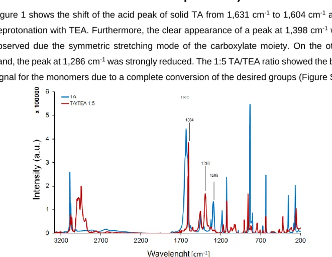
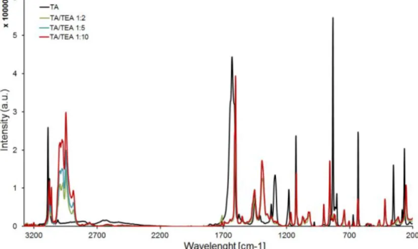
FT-Raman analysis shows the deprotonation of the carboxylic acid groups of the TA via incubation with different TA/TEA ratios. Spectra were normalized between the cm-1 region. Unfortunately, further variation of the reaction conditions, including addition of zinc acetate, did not lead to a higher yield of TA. Similarly, H spectra (included in DMSO-d6) indicate the presence of some longer oligomers in the chemically pretreated sample, while the spectra of the reaction product after subsequent enzymatic hydrolysis suggest their conversion to TA.
Conclusions
Author contributions
Pellis et al., "Biocatalyzed approach for surface functionalization of poly(L-lactic acid) films using hydrolytic enzymes," Biotechnol. Eberl et al., "Enzymatic surface hydrolysis of poly(ethylene terephthalate) and bis(benzoyloxyethyl) terephthalate by lipase and cutinase in the presence of surface-active molecules," J. Enzymatic Hydrolysis of Poly(ethylene terephthalate) Applied and Analyzed by Pore Modification of membranes engraved in traces", N.
Supporting information
This excess must be either as end group or as free TA, so ~28% (i.e. the TA units are assumed to be end groups, but due to their excess over EG. Since some of the EG are end groups despite the excess of TA calculated above, there must therefore be additional TA units that are also end-groups (i.e., those due to EG becoming end-groups, releasing more carboxylic acid end-groups) We had already established that ~28% of all TA were end-group because it was too many, but another 41% of the remaining (72%) TA is also end group.
General Conclusion
Various eugenol release studies were performed and evaluated in different artificial sweats varying in pH (5.5, 6.5 and 8.00) to simulate the contribution of the sweat gland to wound re-epithelialization. Following a circular economy view, artificial and real textile blends were used to extract to recover value-added products from wool (with 95% efficiency) and cellulose-based materials (85% yield). Amino acids and oligomers. The deprotonation ability of the free carboxylic group of TPA was used to monitor the rate of the depolymerization process, in the presence of triethylamine (ratio 1:5) using FT-Raman.
Appendix
- List of figures Chapter 2
- List of tables Chapter 3
- Scientific publications Papers
- Oral presentations as presenting author
- Autex Conference (Corfu´, Greece-2017)
- ICET Conference (Wuxi, China-2018) Enzymatic separation of polymers from textile waste
- Poster presentations (most relevant)
- Autex Conference (Ljubliana, Slovenia- 2016)
- ECAB Conference (Florence, Italy- 2019)
- Acknowledgments
34;Enhanced Flame Retardancy of Enzymatically Functionalized PET and Nylon Fabrics via DNA Immobilization." Frontiers in Chemistry. 34;Smart Textiles in Wound Care: Functionalization of Cotton/PET Blends with Antimicrobial Nanocapsules." Journal of Materials Chemistry B 7, no. 34; Environmentally Friendly Covalent Coupling of Proteins on Oxidized Cellulosic Materials." New Journal of Chemistry 43, no.
Simone: Oh my dear, there are no words how I can say Thank you for all these years and pleasant time with you. I can easily say that through a twisted connection a deep friendship was born, then I am happy to have "Le Arancine" (Valentina and Alessandra) and Matteo in my life. 170 I declare that I have written this thesis independently, that I have not used other than the stated sources/resources, and that I have expressly marked all material that has been quoted either literally or by content from the source used.