Volume 2013, Article ID 795916,7pages http://dx.doi.org/10.1155/2013/795916
Research Article
Antioxidant Effects of Quercetin and Naringenin Are Associated
with Impaired Neutrophil Microbicidal Activity
Francielli de Cássia Yukari Nishimura,
1Ana Carolina de Almeida,
2Bianca Altrão Ratti,
1Tânia Ueda-Nakamura,
1Celso Vataru Nakamura,
1Valdecir Farias Ximenes,
2and Sueli de Oliveira Silva
11Departamento de Ciˆencias B´asicas da Sa´ude, Universidade Estadual de Maring´a, Avenida Colombo 5.790,
17020-900 Maring´a, PR, Brazil
2Departamento de Qu´ımica, Faculdade de Ciˆencias, Universidade Estadual Paulista, Avenida Eng. Luiz Edmundo Carrijo Coube 14-01,
17033-360 Bauru, SP, Brazil
Correspondence should be addressed to Sueli de Oliveira Silva; lautenschlager@uem.br
Received 3 April 2013; Revised 16 May 2013; Accepted 18 June 2013
Academic Editor: Chong-Zhi Wang
Copyright © 2013 Francielli de C´assia Yukari Nishimura et al. This is an open access article distributed under the Creative Commons Attribution License, which permits unrestricted use, distribution, and reproduction in any medium, provided the original work is properly cited.
Naringenin and quercetin are considered antioxidant compounds with promising activity against oxidative damage in human cells. However, no reports have described their effects on reactive oxygen species (ROS) production by phagocytes during microbicidal activity. Thus, the present study evaluated the effects of naringenin and quercetin on ROS production, specifically hypochlorous acid (HOCl), and their involvement in the microbicidal activity of neutrophils. Naringenin and quercetin inhibited HOCl production through different systems, but this inhibition was more pronounced for quercetin, even in the cell-free systems. With regard to
the microbicidal activity of neutrophils, both naringenin and quercetin completely inhibited the killing ofStaphylococcus aureus.
Altogether, these data indicate that the decrease in the oxidant activity of neutrophils induced by these compounds directly impaired the microbicidal activity of neutrophils. Naringenin and quercetin exerted their effects by controlling the effector mechanisms of ROS production, with both positive and negative effects of these antioxidant agents in oxidative stress conditions and on ROS in the microbicidal activity of phagocytes. The present results challenge the traditional view of antioxidants as improvers of pathological conditions.
1. Introduction
Accumulating evidence indicates the involvement of reactive oxygen species (ROS) in different physiological functions and various cell signaling processes, including reproduction, cell migration, stem cell proliferation, neurogenesis, and phago-cytosis [1–5]. Depending on the intracellular concentration of ROS, they can contribute to both physiological and patholog-ical conditions. The long-term exposure of cells to enhanced levels of ROS is involved in the pathogenesis of many human diseases, including chronic inflammation, neurodegenera-tive disorders, and some cancers, by damaging essential molecules, such as lipids, proteins, and DNA [6–9]. Thus, maintaining an appropriate balance between ROS and antiox-idant enzymes is important to avoid deleterious processes.
New effective therapies based on exogenous antioxidants have been sought [10, 11]. Although the literature presents various compounds obtained from plants with promising antioxidant effects, few studies have examined the side effects of these substances on physiological functions that depend on ROS.
hypochlorous acid (HOCl), produced by phagocytes through NADPH oxidase in microbicidal activity [14,15]. In addition to microbicidal activity, ROS produced by NADPH oxidase has emerged as an important messenger of several cellular signaling pathways, including the activation of nuclear tran-scription factors such as NF-𝜅B and AP-1 that are associated with physiological functions involving, respectively, inflam-matory responses and the expression of protective genes that repair damaged DNA, [16].
Considering that the production of ROS by the NADPH oxidase system is an initial and critical event for the onset of oxidative stress conditions and microbicidal activity, the present study investigated the effects of quercetin and narin-genin on the production of ROS, especially HOCl, and their involvement in the microbicidal activity of neutrophils. We sought to determine whether the antioxidant effects of quercetin and naringenin are associated with impaired neutrophil function.
2. Materials and Methods
2.1. Chemicals. Quercetin, naringenin, dextran, taurine, 3,3,5,5-tetramethylbenzidine (TMB), hydrogen peroxide, MPO, catalase, phorbol 12-myristate 13-acetate (PMA), dimethyl sulfoxide (DMSO), and Histopaque were obtained from Sigma (St. Louis, MO, USA). Naringenin and quercetin stock solutions (2.5 mM) were prepared in DMSO, stored at 8∘C, and used within 1 week. Dimethyl sulfoxide was added at the same concentration in all of the samples including the controls at a final concentration of 0.2%, a concentration that has been shown to not affect neutrophil viability.
2.2. Neutrophils and Total Leukocytes. Neutrophils and total leukocytes were isolated from peripheral venous blood obtained from healthy volunteers by centrifugation over a Ficoll-Hypaque gradient (Histopaque; 𝑑 = 1.077) [17,
18]. Cell concentration and viability were determined in a Neubauer chamber. Neutrophils (2.5×10⁶cells/mL) and total leukocytes (2.0 × 10⁶cells/mL) were suspended in 10 mM phosphate-buffered saline (PBS; pH 7.4) supplemented with 1 mg/mL glucose, 1 mM CaCl2, and 0.5 mM MgCl2.
2.3. Effects of Quercetin and Naringenin on HOCl Production.
The concentration of HOCl produced in cellular and cell-free systems was evaluated according to the method described by Dypbukt et al. [19]. Briefly, HOCl was trapped as the less reactive and stable taurine chloramine. Taurine chloramine in the supernatant was then quantified by the oxidation of TMB (10 mM in 1 : 1 [v/v] dimethylformamide/0.8 M acetic acid, containing 100𝜇M potassium iodide) to a blue product with maximum absorbance at 655 nm. A calibra-tion curve that consisted of pure HOCl was generated to calculate the production of the oxidant. The analyses were performed in a final volume of 250𝜇L using a microplate reader spectrophotometer (Biotec power-WaveX5, USA). The effects of naringenin and quercetin on HOCl pro-duction were studied using three different experimental models.
2.3.1. Cell-Free System: Antioxidant Effects of Naringenin and Quercetin. A 96-well culture plate that contained 50𝜇M HOCl and 5 mM taurine in supplemented PBS was incubated in the presence or absence of naringenin and quercetin (25 and 50𝜇M) for 10 min at 25∘C. The final volume was 200𝜇L, and the reactions were triggered by adding HOCl. The TMB solution (50𝜇L) was then added to measure the remaining taurine chloramine.
2.3.2. Cell-Free System: Effects of Naringenin and Quercetin on HOCl Production by MPO/Hydrogen Peroxide (H2O2)/Cl−. A 96-well culture plate that contained MPO (65 nM), 5 mM tau-rine, and 50𝜇M H2O2in supplemented PBS was incubated in the presence or absence of quercetin and naringenin (25 and 50𝜇M) for 10 min at25∘C. The reactions were triggered by adding H2O2and stopped by adding catalase (65𝜇g/mL). The final volume was 200𝜇L. The TMB solution (50𝜇L) was then added to the samples, and HOCl production was quantified. The positive control, without the tested substances, was used to calculate the inhibitory effect.
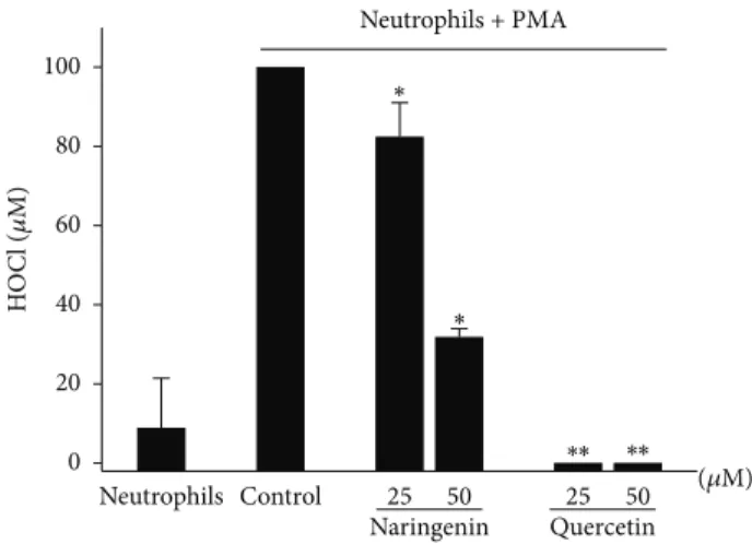
2.3.3. Effects of Naringenin and Quercetin on HOCl Pro-duction by Neutrophils. Neutrophils (1.0 × 10⁶cells/mL) were preincubated in the presence or absence of quercetin and naringenin (25 and 50𝜇M) in supplemented PBS that contained 5 mM taurine for 10 min at37∘C. All of the samples were then incubated with PMA (6.65𝜇g/mL) for 30 min at 37∘C. Afterward, the reactions were stopped by adding catalase (65𝜇g/mL). The final volume was 200𝜇L. The cells were pelleted by centrifugation (1,200 ×g for 10 min at24∘C), and HOCl production was quantified as stated previously.
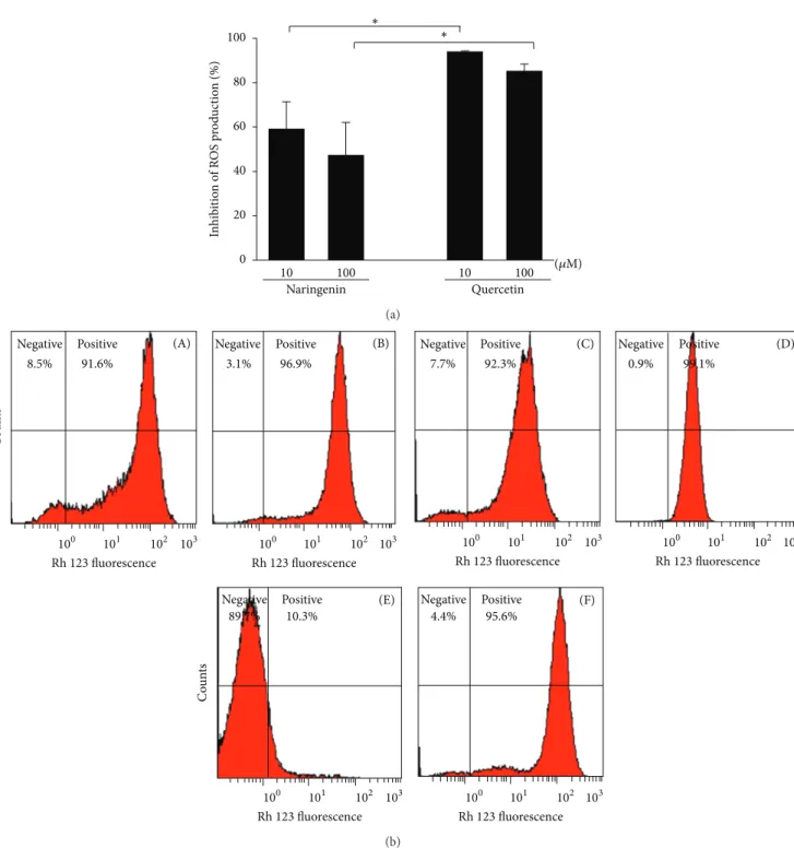
2.4. Effects of Naringenin and Quercetin on ROS Production by Leukocytes. Dihydrorhodamine 123 (DHR) is widely used for the detection of intracellular oxidant species production by cell systems [20]. The oxidation of DHR by ROS results in the formation of rhodamine, a highly fluorescent component. Total leukocytes (2.0 × 10⁶cells/mL) were incubated with quercetin or naringenin (10 and 100𝜇M) for 2 h and then stimulated with PMA (400 nM) for 10 min. After PMA stim-ulation, the cells were incubated with DHR (10 mg/mL) for 5 min, washed once with PBS, and suspended in PBS/bovine serum albumin/azide buffer. The fluorescence of gated neu-trophils was detected at FL1, counting 30,000 events/gate, in a FACS Canto Flow Cytometer (BD, Franklin Lakes, NJ, USA). The data were analyzed using Flow Cytometry Analysis software (Treestar, Ashland, OR, USA), and the results are expressed as the fluorescence intensity and percentage of positive cells in the sample.
2.5. Effects of Naringenin and Quercetin on Microbicidal Assay
(2.0 × 10⁷cells/mL) were opsonized with 10% serum (v/v, final concentration) from healthy donors for 30 min at 37∘C with constant and moderate agitation and used for the killing assay.
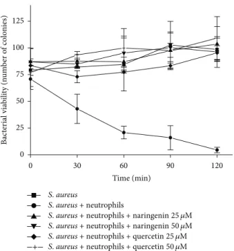
2.5.2. Bacterial Killing. Neutrophils (2.0 × 10⁶cells/mL per assay) were suspended in RPMI 1640 and incubated with opsonized bacteria (2.0 × 10⁷cells/mL) in a final volume of 1.0 mL. Killing activity was monitored in the presence or absence of quercetin and naringenin (25 and 50𝜇M). The samples were maintained at 37∘C with moderate shaking. Killing activity was determined by aseptically removing the samples at intervals of 0, 30, 60, 90, and 120 min. These samples were then diluted in sterile distilled water (1 : 10), whirlmixed for 5 min to lyse neutrophils, and subsequently diluted in sterile saline (1 : 500). The number of viable bacteria was evaluated by spread-plating suitable diluted samples on nutrient agar and incubating them at 37∘C for 24 h [21].
2.6. Statistical Analysis. Comparisons were made using one-way analysis of variance (ANOVA) and the Dunnett multiple comparisons test. The results are expressed as the mean± standard error of the mean (SEM) of at least three indepen-dent experiments. The data were analyzed using BioEstat 5.0 software. Values of𝑃 < 0.05 were considered statistically significant.
3. Results
The present study investigated the antioxidant activity of naringenin and quercetin in three different systems of HOCl formation.
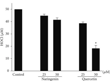
3.1. Cell-Free System: Direct HOCl Antioxidant Effects of Naringenin and Quercetin. In our first cell-free system, quercetin but not naringenin functioned as a HOCl scav-enger. The scavenging action of quercetin depended on its concentration. Quercetin at a concentration of 50𝜇M decreased HOCl by greater than 50% compared with the control group (Figure 1).
3.2. Cell-Free System: Effects of Naringenin and Quercetin on HOCl Production by MPO/H2O2/Cl−. We also evaluated the effects of naringenin and quercetin in a cell-free system that contained MPO/H2O2/Cl−. In this experimental model, HOCl is directly produced by the enzymatic system. Both quercetin and naringenin significantly and dose-dependently decreased HOCl production compared with the control group (Figure 2). Naringenin was less effective than quercetin in inhibiting HOCl formation, but this difference was not significant. A decrease in HOCl production by more than 60% was observed with the higher concentration of quercetin (50𝜇M), whereas the decrease induced by naringenin was approximately 50% at the same concentration. One sam-ple with 5-fluortryptamine (FTR), an MPO inhibitor [22], was assayed to compare the potential of the flavonoids as inhibitors of the chlorinating activity of MPO. The flavonoids were less effective than FTR.
0 10 20 30 40 50
∗
(𝜇M)
Quercetin Naringenin
25 50 25 50
Control
HO
C
l (
𝜇
M)
Figure 1: Antioxidant effects of naringenin and quercetin on HOCl.
A 96-well culture plate that contained HOCl (50𝜇M) and taurine
(5 mM) was incubated in the presence or absence of naringenin and
quercetin (25 and 50𝜇M). The TMB solution (50𝜇L) was then added
to the samples, and HOCl was quantified. The data are expressed as
the mean±SEM of three experiments.∗𝑃 < 0.05, compared with
control group.
+ MPO
0 10 20 30 40 50 60
(𝜇M)
Quercetin Naringenin
25 50 25 50
Control
HO
C
l (
𝜇
M)
2,0 FTR
∗
∗∗ ∗∗
∗ ∗∗
∗ ∗∗
Figure 2: Effects of naringenin and quercetin on myeloperoxidase (MPO)-HOCl production. A 96-well culture plate that contained
MPO (65 nM), taurine (5 mM), and H2O2(50𝜇M) was incubated in
the presence or absence of naringenin and quercetin (25 and 50𝜇M).
The TMB solution (50𝜇L) was then added to the samples, and HOCl
production was quantified. The positive control (i.e., without the tested substances) was used to calculate the inhibitory effect. The
data are expressed as the mean±SEM of three experiments. ∗𝑃 <
0.05,∗∗𝑃 < 0.005, and∗∗∗𝑃 < 0.001, compared with control group.
(𝜇M) Neutrophils + PMA
0 20 40 60 80 100
Control 25 50 25 50
Neutrophils
Quercetin Naringenin
HO
C
l (
𝜇
M)
∗
∗
∗∗ ∗∗
Figure 3: Effects of naringenin and quercetin on HOCl production
by activated neutrophils. Neutrophils (1.0 × 10⁶cells/mL) were
preincubated in the presence or absence of naringenin and quercetin
(25 and 50𝜇M) with 5 mM taurine. All of the samples were then
incubated with PMA (6.65𝜇g/mL). The TMB solution (50𝜇L) was
then added to the samples, and HOCl production was quantified.
The data are expressed as the mean±SEM of three experiments.
∗𝑃 < 0.05,∗∗𝑃 < 0.01compared with control group.
tested, causing approximately 100% decreases in HOCl pro-duction. However, naringenin exerted a significant effect only at 50𝜇M, inhibiting HOCl production by approximately 60%.
3.4. Effects of Naringenin and Quercetin on ROS Production by Leukocytes. As a second step, we compared the ability of quercetin and naringenin to inhibit intracellular ROS production assessed by flow cytometry, in which the nonfluo-rescent DHR is oxidized by ROS, producing fluononfluo-rescent rho-damine. Again, quercetin was more efficient than naringenin
(Figure 4). Quercetin at both tested concentrations inhibited
ROS by more than 80%. Naringenin at higher concentrations inhibited ROS by approximately 50%.
3.5. Effects of Naringenin and Quercetin on Microbicidal Activity. We showed that quercetin and, to a lesser extent, naringenin affected HOCl production by PMA-activated neutrophils. HOCl is a toxic metabolite responsible for the microbicidal activity of phagocytes [14,15]. We expected that these compounds would have different effects on neutrophil microbicidal activity. Thus, we studied the effects of these flavonoids on the microbicidal activity of neutrophils by spread-platingS. aureusonto a nutrient-agar medium after incubation with neutrophils. In contrast to the previous results of the present study mentioned above, both quercetin and naringenin (25 and 50𝜇M) completely inhibited neu-trophil microbicidal activity compared with the control group
(Figure 5).
4. Discussion
Numerous compounds with potential antioxidant effects and promising activity against many human diseases associated
with oxidative damage have been studied over the past years [23,24]. These compounds include naringenin and quercetin, two flavonoids with antioxidant effects that act as ROS scavengers [11, 12] and inhibit the activity of ROS-forming enzymes (e.g., NADPH oxidase) [13]. However, no reports have described their action on the microbicidal response of neutrophils. The microbicidal activity of phagocytes is well known to depend on ROS, and HOCl plays an important role in this process [25]. The present study sought to further elucidate the effects of naringenin and quercetin on the neutrophil response, especially with regard to microbicidal activity.
We first investigated the antioxidant effects of naringenin and quercetin on HOCl production by cellular and cell-free systems. The effects of naringenin observed in the MPO model and in PMA-activated neutrophils, compared with the first cell-free system, indicated that significant inhibition of MPO chlorinating activity can be induced by this compound. The production of HOCl might be reduced in the presence of flavonoids that act as MPO inhibitors [26]. Thus, the 15% (cell-free system) to 50% (MPO system) increases in the inhibition of HOCl production induced by 50𝜇M naringenin might be related to direct inhibition of MPO chlorinating activity. Quercetin exhibited the same pattern of inhibition in the cell-free systems, suggesting that quercetin is a better scavenger of HOCl and poor inhibitor of MPO. Quercetin also markedly inhibited HOCl production in PMA-activated neutrophils. In this system, PMA activated the NADPH oxidase complex, which is responsible for the production of superoxide anions and, after a cascade of reactions, produces H2O2 and HOCl [27]. Therefore, quercetin, a well-known antioxidant, could react with all ROS formed in the cellular system, consequently disrupting HOCl formation through a scavenging effect [11, 12]. To support this possibility, the DHR assay confirmed that quercetin is an efficient scavenger of ROS generated by activated neutrophils.
We showed that naringenin and quercetin also inhibited the microbicidal activity of neutrophils. These results may reflect the antioxidant activity of these compounds, which consume both HOCl and ROS precursors of HOCl, thus inhibiting the formation of HOCl derivatives with high microbicidal activity, such as singlet oxygen. The suppression of these ROS induced a direct effect on microbicidal activity [28].
0 20 40 60 80 100
(𝜇M)
Quercetin
10 100 10 100
Naringenin
∗ ∗
In
hib
it
io
n o
f R
OS p
ro
d
uc
tio
n
(%)
(a)
103
102 101 100
3.1% 96.9%
Rh 123 fluorescence (B) Positive
Negative
8.5% 91.6%
Co
u
n
ts
103
102 101 100
Rh 123 fluorescence (A) Positive
Negative
103
102 101 100
0.9% 99.1%
Rh 123 fluorescence (D) Positive
Negative
103
102 101 100
7.7% 92.3%
Rh 123 fluorescence (C) Positive
Negative
103
102 101 100
4.4% 95.6%
Rh 123 fluorescence (F) Positive
Negative
Co
u
n
ts
103
102 101 100
89.7% 10.3%
Rh 123 fluorescence (E) Positive
Negative
(b)
Figure 4: Effects of naringenin and quercetin on ROS production by neutrophils. (a) Neutrophils were preincubated in the presence or
absence of naringenin and quercetin (10 and 100𝜇M) and stimulated with PMA (400 nM), and intracellular ROS production was determined
by flow cytometry using DHR as a probe. (b) Representative histograms are shown in logarithmic scale. Neutrophils were preincubated in
the presence of naringenin 10𝜇M (A) and 100𝜇M (B) and activated with PMA. Neutrophils were preincubated in the presence of quercetin
10𝜇M (C) and 100𝜇M (D) and activated with PMA. Neutrophils (negative control) (E); neutrophils activated with PMA (positive control)
(F). The percentage of inhibition of ROS production by naringenin and quercetin was calculated and compared with the positive group. The
data are expressed as the mean±SEM of four experiments. ∗𝑃 < 0.05, 10𝜇M naringenin versus 10𝜇M quercetin and 100𝜇M naringenin
0 30 60 90 120 0
25 50 75 100 125
Time (min)
B
ac
ter
ial via
b
ili
ty (n
um
b
er o
f co
lo
nies)
S.aureus
S.aureus+ neutrophils
S.aureus+ neutrophils + naringenin25 𝜇M S.aureus+ neutrophils + naringenin50 𝜇M S.aureus+ neutrophils + quercetin25 𝜇M S.aureus+ neutrophils + quercetin50 𝜇M
Figure 5: Effects of naringenin and quercetin on the kinetics
of the killing of S. aureus by neutrophils. Neutrophils (2.0 ×
106cells/mL per assay) were incubated with opsonized bacteria
(2.0 × 10⁷cells/mL). Microbicidal activity was monitored in the
presence and absence of 25𝜇M () and 50𝜇M () naringenin and
25𝜇M (Q) and 50𝜇M (+) quercetin. Killing activity was determined
by aseptically removing the samples at intervals of 0, 30, 60, 90, and 120 min. The number of viable bacteria was evaluated by spread-plating suitable diluted samples on nutrient agar. The controls
included bacteria alone (◼) and bacteria plus neutrophils (∙). The
data are expressed as the mean±SEM of three experiments.
always associated with improvements in pathological con-ditions [31,32]. This view must be revisited, especially with regard to infectious diseases and the development of certain cancers.
Conflict of Interests
There is no conflict of interests declared by the authors.
Acknowledgments
This study was supported by Conselho Nacional de Desen-volvimento Cient´ıfico e Tecnol´ogico (CNPq), Coordenac¸˜ao de Aperfeic¸oamento de Pessoal de N´ıvel Superior (Capes), and Fundac¸˜ao Arauc´aria.
References
[1] M. J. Morgan and Z.-G. Liu, “Reactive oxygen species in TNF𝛼
-induced signaling and cell death,”Molecules and Cells, vol. 30,
no. 1, pp. 1–12, 2010.
[2] B. C. Dickinson and C. J. Chang, “Chemistry and biology of
reactive oxygen species in signaling or stress responses,”Nature
Chemical Biology, vol. 7, no. 8, pp. 504–511, 2011.
[3] A. Rizzo, M. T. Roscino, F. Binetti, and R. L. Sciorsci, “Roles of
reactive oxygen species in female reproduction,”Reproduction
in Domestic Animals, vol. 47, no. 2, pp. 344–352, 2012.
[4] J.-S. Kim, T. Y. Huang, and G. M. Bokoch, “Reactive oxygen
species regulate a slingshot-cofilin activation pathway,”
Molec-ular Biology of the Cell, vol. 20, no. 11, pp. 2650–2660, 2009. [5] B. C. Dickinson, J. Peltier, D. Stone, D. V. Schaffer, and
C. J. Chang, “Nox2 redox signaling maintains essential cell
populations in the brain,”Nature Chemical Biology, vol. 7, no.
2, pp. 106–112, 2011.
[6] A. L. P. Chapman, M. B. Hampton, R. Senthilmohan, C. C. Winterbourn, and A. J. Kettle, “Chlorination of bacterial and neutrophil proteins during phagocytosis and killing of
Staphylococcus aureus,”The Journal of Biological Chemistry, vol. 277, no. 12, pp. 9757–9762, 2002.
[7] R. S. Frey, M. Ushio-Fukai, and A. B. Malik, “NADPH oxidase-dependent signaling in endothelial cells: role in physiology and
pathophysiology,”Antioxidants & Redox Signaling, vol. 11, no. 4,
pp. 791–810, 2009.
[8] S. C. Correia, R. X. Santos, G. Perry, X. Zhu, P. I. Moreira, and M. A. Smith, “Mitochondrial importance in Alzheimer’s,
Huntington’s and Parkinson’s diseases,”Advances in
Experimen-tal Medicine and Biology, vol. 724, pp. 205–221, 2012.
[9] P. Lonkar and P. C. Dedon, “Reactive species and DNA damage in chronic inflammation: reconciling chemical mechanisms and
biological fates,”International Journal of Cancer, vol. 128, no. 9,
pp. 1999–2009, 2011.
[10] A. K. Tiwari, “Antioxidants: new-generation therapeutic base
for treatment of polygenic disorders,”Current Science, vol. 86,
no. 8, pp. 1092–1102, 2004.
[11] F. Ursini, M. Maiorino, P. Morazzoni, A. Roveri, and G. Pifferi, “A novel antioxidant flavonoid (IdB 1031) affecting molecular
mechanisms of cellular activation,” Free Radical Biology and
Medicine, vol. 16, no. 5, pp. 547–553, 1994.
[12] G. Agati, E. Azzarello, S. Pollastri, and M. Tattini, “Flavonoids as antioxidants in plants: location and functional significance,”
Plant Science, vol. 196, pp. 67–76, 2012.
[13] M. Ciz, P. Denev, M. Kratchanova, O. Vasicek, G. Ambrozova, and A. Lojek, “Flavonoids inhibit the respiratory burst of
neutrophils in mammals,” Oxidative Medicine and Cellular
Longevity, vol. 2012, Article ID 181295, 6 pages, 2012.
[14] L. Wang, M. Bassiri, R. Najafi et al., “Hypochlorous acid as a
potential wound care agent,”Journal of Burns and Wounds, vol.
6, no. 5, pp. 65–79, 2007.
[15] L. Gebicka and E. Banasiak, “Hypochlorous acid-induced heme
damage of hemoglobin and its inhibition by flavonoids,”
Toxi-cology in Vitro, vol. 26, no. 6, pp. 924–929, 2012.
[16] M. Valko, D. Leibfritz, J. Moncol, M. T. D. Cronin, M. Mazur, and J. Telser, “Free radicals and antioxidants in normal
physio-logical functions and human disease,”The International Journal
of Biochemistry and Cell Biology, vol. 39, no. 1, pp. 44–84, 2007. [17] A. Bøyum, “Isolation of lymphocytes, granulocytes and
macrophages,”Scandinavian Journal of Immunology, vol. 5, pp.
9–15, 1976.
[18] A. B¨oyum, “Isolation of leucocytes from human blood. A two-phase system for removal of red cells with methylcellulose as
erythrocyte-aggregating agent,”Scandinavian Journal of
Clini-cal and Laboratory Investigation, Supplement, vol. 97, pp. 9–29, 1968.
production by myeloperoxidase,” Free Radical Biology and Medicine, vol. 39, no. 11, pp. 1468–1477, 2005.
[20] J. P. Crow, “Dichlorodihydrofluorescein and dihydrorhodamine 123 are sensitive indicators of peroxynitrite in vitro: implications for intracellular measurement of reactive nitrogen and oxygen
species,”Nitric Oxide: Biology and Chemistry, vol. 1, no. 2, pp.
145–157, 1997.
[21] M. B. Hampton and C. C. Winterbourn, “Methods for quanti-fying phagocytosis and bacterial killing by human neutrophils,”
Journal of Immunological Methods, vol. 232, no. 1-2, pp. 15–22, 1999.
[22] M. L. Zeraik, V. F. Ximenes, L. O. Regasini et al., “4
-Aminochalcones as novel inhibitors of the chlorinating activity
of myeloperoxidase,”Current Medicinal Chemistry, vol. 19, no.
31, pp. 5405–5413, 2012.
[23] H. Yagi, J. Tan, and R. S. Tuan, “Polyphenols suppress hydro-gen peroxide-induced oxidative stress in human bone-marrow
derived mesenchymal stem cells,”Journal Cellular Chemistry,
vol. 28, pp. 1–38, 2012.
[24] M. Zhang, S. G. Swarts, L. Yin et al., “Antioxidant properties of
quercetin,”Advances in Experimental Medicine and Biology, vol.
701, pp. 283–289, 2011.
[25] C. C. Winterbourn, M. B. Hampton, J. H. Livesey, and A. J. Kettle, “Modeling the reactions of superoxide and myeloperox-idase in the neutrophil phagosome: implications for microbial
killing,”The Journal of Biological Chemistry, vol. 281, no. 52, pp.
39860–39869, 2006.
[26] V. A. Kostyuk, T. Kraemer, H. Sies, and T. Schewe,
“Myeloperoxidase/nitrite-mediated lipid peroxidation of
low-density lipoprotein as modulated by flavonoids,” FEBS
Letters, vol. 537, no. 1-3, pp. 146–150, 2003.
[27] J. M. Robinson, “Reactive oxygen species in phagocytic
leuko-cytes,”Histochemistry and Cell Biology, vol. 130, no. 2, pp. 281–
297, 2008.
[28] C. C. Winterbourn and A. J. Kettle, “Redox reactions and
microbial killing in the neutrophil phagosome,”Antioxidants &
Redox Signaling, vol. 18, no. 6, pp. 642–660, 2013.
[29] D. Trachootham, J. Alexandre, and P. Huang, “Targeting can-cer cells by ROS-mediated mechanisms: a radical therapeutic
approach?”Nature Reviews Drug Discovery, vol. 8, no. 7, pp.
579–591, 2009.
[30] G. M. DeNicola, F. A. Karreth, T. J. Humpton et al., “Oncogene-induced Nrf2 transcription promotes ROS detoxification and
tumorigenesis,”Nature, vol. 475, no. 7354, pp. 106–110, 2011.
[31] S. C. Bischoff, “Quercetin: potentials in the prevention and
therapy of disease,”Current Opinion in Clinical Nutrition and
Metabolic Care, vol. 11, no. 6, pp. 733–740, 2008.
[32] Z.-P. Xiao, Z.-Y. Peng, M.-J. Peng, W.-B. Yan, Y.-Z. Ouyang, and H.-L. Zhu, “Flavonoids health benefits and their molecular
mechanism,”Mini-Reviews in Medicinal Chemistry, vol. 11, no.
Submit your manuscripts at
http://www.hindawi.com
Stem Cells
International
Hindawi Publishing Corporation
http://www.hindawi.com Volume 2014
Hindawi Publishing Corporation
http://www.hindawi.com Volume 2014
INFLAMMATION
Hindawi Publishing Corporation
http://www.hindawi.com Volume 2014
Behavioural
Neurology
International Journal of
Endocrinology
Hindawi Publishing Corporation
http://www.hindawi.com Volume 2014
Hindawi Publishing Corporation
http://www.hindawi.com Volume 2014
Disease Markers
BioMed Research International
Hindawi Publishing Corporation
http://www.hindawi.com Volume 2014
Oncology
Journal ofHindawi Publishing Corporation
http://www.hindawi.com Volume 2014
Hindawi Publishing Corporation
http://www.hindawi.com Volume 2014
Oxidative Medicine and Cellular Longevity
PPAR
R e s e a r c h
Hindawi Publishing Corporation
http://www.hindawi.com Volume 2014
The Scientific
World Journal
Hindawi Publishing Corporationhttp://www.hindawi.com Volume 2014
Immunology Research Hindawi Publishing Corporation
http://www.hindawi.com Volume 2014
Journal of
Obesity
Journal ofHindawi Publishing Corporation
http://www.hindawi.com Volume 2014
Hindawi Publishing Corporation
http://www.hindawi.com Volume 2014
Computational and Mathematical Methods in Medicine
Ophthalmology
Journal ofHindawi Publishing Corporation
http://www.hindawi.com Volume 2014
Diabetes ResearchJournal of
Hindawi Publishing Corporation
http://www.hindawi.com Volume 2014
Hindawi Publishing Corporation
http://www.hindawi.com Volume 2014
Research and Treatment
AIDS
Hindawi Publishing Corporation
http://www.hindawi.com Volume 2014
Gastroenterology Research and Practice
Parkinson’s Disease
Hindawi Publishing Corporation
http://www.hindawi.com Volume 2014
Evidence-Based Complementary and Alternative Medicine
Volume 2014 Hindawi Publishing Corporation