Article
J. Braz. Chem. Soc., Vol. 25, No. 4, 639-647, 2014. Printed in Brazil - ©2014 Sociedade Brasileira de Química
0103 - 5053 $6.00+0.00
A
http://dx.doi.org/10.5935/0103-5053.20140011*e-mail: [email protected]
#Present address: Embrapa Agroindústria Tropical, Rua Dra. Sara
Mes-quita, 2270, Pici, CP 3761, 60511-110 Fortaleza-CE, Brazil
Amburanins A and B from
Amburana cearensis
: Daphnodorin-Type Biflavonoids
that Modulate Human Neutrophil Degranulation
Kirley M. Canuto,#,a Luzia K. A. M. Leal,b Amanda A. Lopes,b Christina M. Coleman,c Daneel Ferreirac and Edilberto R. Silveira*,a
aDepartamento de Química Orgânica e Inorgânica, Universidade Federal do Ceará,
Campus do Pici, Bloco 940, Pici, 60451-970 Fortaleza-CE, Brazil.
bCentro de Estudos Farmacêuticos e Cosméticos (CEFAC), Departamento de Farmácia,
Universidade Federal do Ceará, Rua Cap. Francisco Pedro, 1210, Rodolfo Teófilo, 60430-170 Fortaleza-CE, Brazil
cDepartment of Pharmacognosy and the Research Institute of Pharmaceutical Sciences, School of
Pharmacy, The University of Mississippi, P.O. Box 1848, University MS 38677-1848, USA
Dois novos biflavonoides 3,5,7,4’-tetraidroxiflavanona-(2→O →4’:3→3’)-2’,4’,6’,4-tetraidroxidiidrochalcona (1) e 3,5,7,4’-tetraidroxiflavanona-(2→O →7:3→8)-3,4’,5,7-tetraidroxiflavona (2), denominados amburanina A e amburanina B, respectivamente, foram isolados da casca do caule de Amburana cearensis, e as estruturas deles foram elucidadas com base em análises espectroscópicas e por comparação com dados da literatura. Os efeitos de 1 e 2 sobre a resposta pro-inflamatória de neutrófilos humanos foram investigados (0,1; 1; 25; 50 e 100 µg mL-1). Ambos compostos inibiram em torno de 92% a degranulação de neutrófilos a partir da concentração de 25 µg mL-1, e reduziram em até 53% a atividade da mieloperoxidase humana, indicando o potencial deles como substâncias anti-inflamatórias.
Two new biflavonoids 3,5,7,4’-tetrahydroxyflavanone-(2→O →4’:3→3’)-2’,4’,6’,4-tetrahydroxydihydrochalcone (1) and 3,5,7,4’-tetrahydroxyflavanone-(2→O →7:3→8)-3,4’,5,7-tetrahydroxyflavone (2), named as amburanin A and amburanin B, respectively, were isolated from the trunk bark of Amburana cearensis, and their structures elucidated on the basis of spectroscopic analysis and by comparison with literature data. The effects of 1 and 2 on the pro-inflammatory response of human neutrophils were investigated (0.1; 1; 25; 50 e 100 µg mL-1). At concentration higher than 25 µg mL-1, both compounds suppressed nearly 92% of the neutrophil degranulation and 53% of myeloperoxidase activity, thus indicating that they are potential anti-inflammatory lead compounds.
Keywords: Amburana cearensis, anti-inflammatory, myeloperoxidase, biflavonoids, amburanins
Introduction
Amburana cearensis A. C. Smith (syn.: Torresea cearensis Fr. All.), commonly known as “cumaru” or “imburana-de-cheiro”, is a native tree from northeastern Brazil. Popularly, the trunk bark and seeds are utilized either as a household medicine (“lambedô”) or as a syrup commercially available to treat respiratory
conditions (asthma and bronchitis).1 Previously, the
aqueous alcohol extract of the trunk bark showed bronchodilatory, anti-inflammatory and analgesic properties.2 One of its constituents, amburoside A
[(4-O-β-D-glucopyranosylbenzyl) protocatechuate], showed neuroprotective and hepatoprotective effects,3,4
while a second compound, isokaempferide, exhibited bronchodilator and antiproliferative activities.5,6 Both
amburoside A and isokaempferide displayed anti-inflammatory activity.7 Coumarin, the most abundant
P r e v i o u s p h y t o c h e m i c a l s t u d i e s o f t h e trunk bark of A. cearensis revealed several other k n o w n p h e n o l i c c o m p o u n d s : a m b u r o s i d e B [ ( 4 -O-β- D - g l u c o p y r a n o s y l b e n z y l ) va n i l l a t e ] ,8
vanillic acid (4-hydroxy-3-methoxybenzoic acid), protocatechuic acid (3,4-dihydroxybenzoic acid), afrormosin (7-hydroxy-4’,6-dimethoxyisoflavone), kaempferol (4’,5,7-trihydroxyflavonol), quercetin (3’,4’,5,7-tetrahydroxyflavonol) and 4’-O-methylfisetin (3’,7-dihydroxy-4’-methoxyflavonol).9 Later on, novel
compounds such as glucosylprotocatechuate derivatives (amburosides C-H) and 6-coumaryl protocatechuate were reported from either the trunk bark or seeds of A. cearensis,
besides 6-hydroxycoumarin and formononetin.10
Furthermore, 3’,4’-dimethoxy-1’-(7-methoxy-4-oxo-4H -chromen-3-yl)benzo-2’,5’-quinone along with five known flavonoids were isolated from resin of A. cearensis.11
Phytochemical analyses associated with pharmacological studies revealed that cultivated young plants of A. cearensis
exhibited similar chemical and pharmacological profiles as the wild plant.12,13 Recently, we have also studied the
physicochemical characteristics of the spray-dried ethanol extract for obtaining A. cearensis powders with better pharmacological and technological properties.14
This report will discuss the isolation and structural elucidation of two daphnodorin-type biflavonoids from
A. cearensis trunk bark (Figure 1), as well as their
in vitro effect on human neutrophil degranulation and myeloperoxidase (MPO) activity.
Experimental
General experimental procedure
Melting points (uncorr.) were determined with a Mettler Toledo FP82HT apparatus (Columbus, OH, USA), with a heating rate of 2 °C min-1. Optical rotations were
measured on a Perkin-Elmer 341 polarimeter (at 589 nm)
in MeOH at 20 °C. Electronic circular dichroism (ECD) spectra were recorded in high purity MeOH on a JASCO J-815 CD spectrometer (Easton, MD, USA) at 25 °C using a 1 cm path length quartz cuvette. ECD spectra for both compounds were obtained at concentrations of 0.10, 0.08, 0.06, 0.04, and 0.02 mmol mL-1 in order to determine the
concentration which produced the highest quality data. UV spectra were obtained on a Varian Cary 50 Conc UV-Visible spectrophotometer (Mulgrave, Australia). Infrared (IR) spectra were recorded with a Perkin-Elmer FT-IR 1000 spectrometer (Waltham, MA, USA), using KBr pellets. Nuclear magnetic resonance (NMR) experiments were performed on a Bruker DRX-500 spectrometer (Bruker Biospin, Rheinstetten, Germany) at room temperature, using dry DMSO-d6 as solvent (Cambridge Isotope
Laboratories), and were internally referenced to residual undeuterated solvent signals (dH2.50 and dC 39.51 ppm).
High resolution mass spectra (HR-MS) were recorded on a Q-TOF Xevo mass spectrometer (Waters, Milford, USA), utilizing an electrospray ionization (ESI) source as ionization method. Column chromatography was run on silica gel 60 (70-230 mesh, VETEC) and Sephadex LH-20 (Amersham Pharmacia Biotech). Thin layer chromatography (TLC) was performed on precoated silica gel polyester sheets (Merck) and monitored by UV detection and vanillin-perchloric acid reagent detection. High performance liquid chromatography (HPLC) purification was performed on a Waters 1525 (Milford, MA, USA) chromatograph, equipped with a binary pump, Rheodyne injector (200 µL loop), and photodiode-array detector (Waters-2996 PDA), using a Waters X-Terra RP-18 column (250 × 4.6 mm, 5 µm) at 35 ºC in a thermostatic oven. HPLC MeOH was purchased from Tedia Co, and HPLC grade water (18 mΩ) obtained by a Milli-Q purification system (Millipore, Bedford, MA, USA).
Plant material
Trunk bark of A. cearensis was collected in the Quixeramobim region, Ceará State, Northeastern Brazil, in September 2002. Voucher specimens (# 837 and 847) were deposited at the Prisco Bezerra Herbarium, and identified by Dr. Afrânio G. Fernandes, a botanist at the Departamento de Biologia, Universidade Federal do Ceará.
Extraction and isolation
A. cearensis was submitted to a similar procedure as that previously described by Canuto et al..10 Briefly,
powdered dried trunk bark (4.0 kg) was extracted with EtOH in a Soxhlet apparatus, yielding 192.5 g (4.8% m/m)
Canuto et al. 641 Vol. 25, No. 4, 2014
of a viscous brown extract, which was suspended in H2O
(350 mL) and partitioned with EtOAc (150 mL, 3×). Afterwards, the fractions were evaporated and lyophilized. The EtOAc fraction (82.6 g) was dissolved in MeOH (200 mL) and defatted with hexane (100 mL, 3×) to provide 64.5 and 16.6 g of the MeOH (ACM, brown solid) and hexane (ACH, green solid) residues, respectively. The latter fraction was discarded, whereas ACM was adsorbed onto 65 g of silica gel and eluted with CHCl3 (0.5 L);
CHCl3/EtOAc 1:1 (3.5 L); EtOAc (2.0 L), and finally
MeOH (0.5 L) to afford 2.1, 22.7, 25.3, and 12.3 g fractions, respectively. The CHCl3-EtOAc (1:1) fraction was adsorbed
onto 160.2 g of silica gel in a glass column (500 mL) and chromatographed, collecting fractions of 75 mL with the following eluents: CHCl3 (F1-6), CHCl3-EtOAc (9:1)
(F7-18), CHCl3-EtOAc (8:2) (F19-25), CHCl3-EtOAc (6:4)
(F26-29), CHCl3-EtOAc (1:1) (F30-31), EtOAc (F32), and
MeOH (F33). Fractions F1-2, F3-7, F8-15, F16-19, F20-23, and F24-31 were each pooled together after TLC analysis. F24-31 (4.0 g) was subjected to Sephadex LH-20 (60 g) column chromatography with MeOH as the eluent, resulting in 34 fractions of 75 mL each. The fractions were analyzed by TLC and pooled to yield F’1, F’2-8, F’9-18, F’19-33, and F’34. F’34 was identified as quercetin (40 mg, dark yellow solid). F’2-8 was re-subjected to Sephadex LH-20 column chromatography under the same conditions as used previously, resulting in the isolation of protocatechuic acid (F’’4-6: 186 mg, yellow crystals), amburoside A (F’’8-13: 954 mg, pink solid) and vanillic acid (F’’14-15: 47 mg, yellowish crystals). F’9-18 (1.5 g) was subjected to successive Sephadex LH-20 column chromatography, eluted with MeOH to yield compound 1 (61 mg, yellow powder). F’19-33 was also chromatographed on Sephadex LH-20 (8.0 g) with MeOH, yielding 15 fractions of 3 mL each. Fractions F’’5 to F’’12 were pooled. F’’5-12 (162 mg) was purified by HPLC/PDA (loop = 200 µL) on a 250 × 10 mm i.d. X-Terra RP-18 column, using H2O-MeOH
as the mobile phase at a flow rate of 4.5 mL min-1. The
elution gradient varied from 60 to 75% MeOH over 10 min total run time. The chromatogram was observed at 284 nm and showed one of the peaks corresponding to compound 2
(tR = 8.8 min, 53 mg, yellow powder).
Amburanin A (1)
Yellow powder (MeOH); m.p. > 300 °C; [α]D20−15.9
(c 0.24, MeOH); UV (MeOH) λmax/nm (log ε) 291 (4.96),
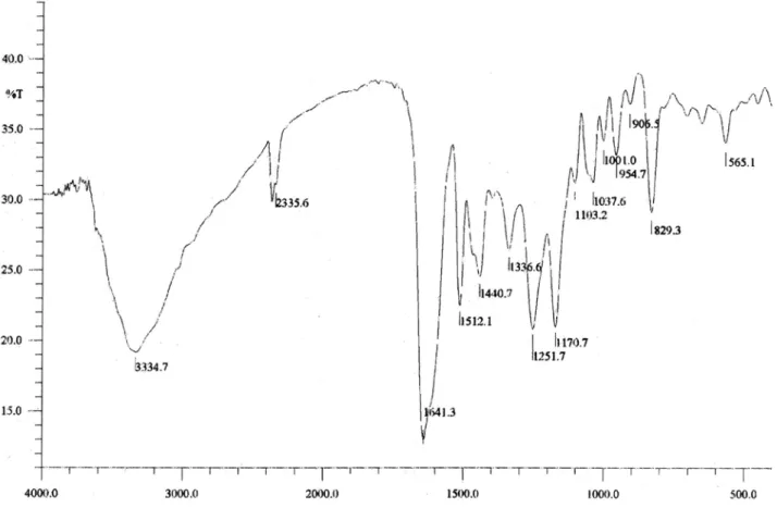
230 (5.18), 208 (5.26); IR (KBr) νmax/cm-1 3335 (O–H),
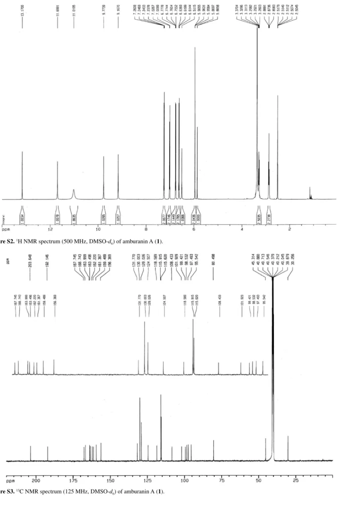
1641 (>C=O), 1512, 1440, 1252, 1171, 955. For 1H and 13C NMR spectroscopic data, see Table 1. HRESIMS
m/z 581.1060 [M+Na]+ (calcd. for C
30H22O11Na, 581.1063);
m/z 557.1066 [M–H]– (calcd. for C
30H21O11, 557.1084).
Amburanin B(2)
Yellow powder (MeOH); m.p. > 300 °C, [α]D 20−12.4
(c 0.21, MeOH); UV (MeOH) λmax/nm (log ε) 285 (4.73),
227 (4.98), 208 (5.06); IR (KBr) νmax/cm
-1 3433 (O–H),
1640 (>C=O), 1517, 1473, 1329, 1258, 1168. For 1H and 13C NMR spectroscopic data, see Table 1. HRESIMS
m/z 593.0679 [M+Na]+ (calcd. for C
30H18O12Na, 593.0696);
m/z 571.0853 [M+H]+ (calcd. for C
30H19O12, 571.0877).
Human neutrophil degranulation assay
Human neutrophils from peripheral blood of healthy volunteers were isolated using the method described by Lucisano and Mantovani.15 The cell preparations presented
90% neutrophils with 97.7 ± 0.94% viability, determined by the Trypan blue test. According to the procedure reported by Boyum,16 aliquots of 5 × 10⁶ cells mL-1 were suspended
in buffered Hank’s balanced salt solution (HBSS) and then incubated at 37 °C with compounds 1 and 2 (0.1, 1, 25, 50, and 100 mg mL-1) or indomethacin (36 mg mL-1,
standard compound) for 15 min at 37 ºC. The cells were then stimulated with phorbol-myristate-acetate (PMA, 0.1 mg mL-1) for 15 min at 37 ºC. The resulting suspension
was centrifuged for 10 min at 2000 × g at 4 ºC and aliquots (50 µL) of the supernatants were then added to phosphate-buffered saline (100 µL), sodium phosphate buffer (6.1 mmol L-1 Na
2HPO4 and 74 mmol L-1 Na2HPO4.2H2O,
50 µL, pH 5.4) and H2O2 (0.012%). After 5 min at 37 ºC,
3,3’,5,5’-tetramethylbenzidine (TMB, 1.5 mmol L-1, 20 µL)
was added and the reaction was stopped by 30 µL of NaOAc (1.5 mol L-1, pH 3.0). Absorbance was recorded at 620 nm.
Myeloperoxidase activity assay
The aforementioned method16 was modified to evaluate
the effect of the bioflavonoids on MPO activity. Human neutrophils were stimulated with PMA, and the MPO-rich supernatant was separated by centrifugation. Aliquots of the supernatant were incubated at 37 °C with compounds 1
and 2 (25, 50, and 100 µg mL-1), DMSO (1% v/v, vehicle)
or HBSS (untreated cells) for 15 min, before determining MPO activity.
Cytotoxic assay
Human neutrophils (5 × 10⁶ cells mL-1) were incubated at
37 °C with the compounds 1 and 2 (25, 50 and 100 µg mL-1),
After 3 h under a 5% CO2 atmosphere, the cells were
washed with PBS, and the formazan product was dissolved in 100 µL of DMSO. The absorbance was recorded at 550 nm;17 its values decreased as a function of cell death.
Results and Discussion
The ethanol extract of A. cearensis trunk bark was subjected to liquid-liquid partitioning (EtOAc/H2O) to
yield an EtOAc phase that was further separated by either silica gel or Sephadex LH-20 column chromatography. Subsequent HPLC and spectroscopic analysis of purified compounds led to the isolation and structural characterization of biflavonoids 1 and 2, along with known compounds vanillic acid, protocatechuic acid, quercetin and amburoside A.9
Compound 1,a yellow powder, showed quasi-molecular ions at m/z 581.1060 [M+Na]+ (calcd. for C
30H22O11Na,
581.1063) and 557.1066 [M-H]– (calcd. for C
30H21O11,
557.1084) in its HRESIMS spectrum. A broad and intense IR absorption band centered at 3335 cm-1 confirmed the
presence of hydroxy groups, while an intense band with a shoulder at 1641 cm-1 suggested the presence of conjugated
carbonyl functionalities. Additional IR absorption bands at 1252 and 1171 cm-1 were attributed to phenolic and tertiary
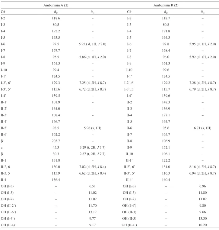
alcohol C-O stretching frequencies, respectively. The 1H NMR spectrum for compound 1 in dry DMSO-d
6
displayed two methylene proton triplets at dH2.87and 3.29
(t, 2H, J 7.7 Hz, H-IIβ and H-IIα),and two m-coupled doublets at dH5.86and 5.95(d, 1H, J 2.0 Hz, H-I8 and
H-I6), the latter characteristic of the I-A-ring aromatic protons. Five sharp singlets at dΗ6.51, 9.17, 9.77, 11.70 and 13.17 (s, 1H), in addition to a broad singlet at dH11.02
(s, 2H) characterized seven hydroxy groups. Four pairs of two-proton doublets at dH6.62, 7.02 (d, 2H, J 8.4 Hz, H-II3,
5 and H-II2, 6), 6.72 and 7.25 (d, 2H, J 8.7 Hz, H-I3’, 5’ and H-I2’, 6’) were characteristic of two para-disubstituted aromatic rings, while a singlet at dH5.96 (s, 1H, H-II5’) was
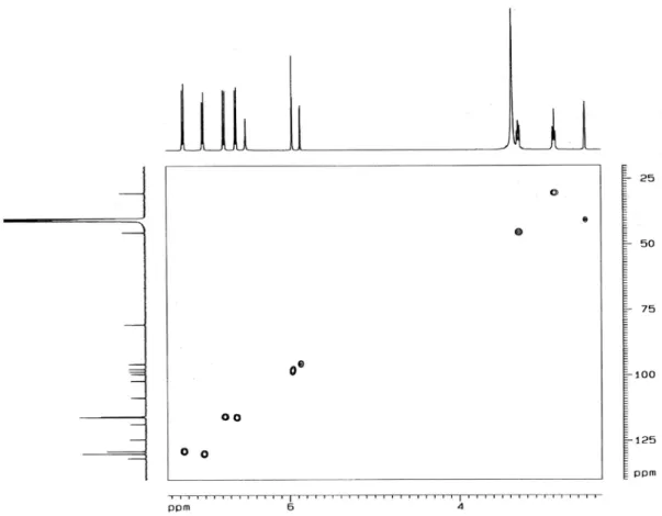
associated with the single hydrogen on the pentasubstituted benzene ring. A gradient-selective correlation spectroscopy (gs-COSY) spectrum showed, as expected, scalar coupling between the two aliphatic methylenes and hydrogen at
dH6.62 (d, 2H, J 8.4 Hz, H-II3, 5) and the hydrogens at
dH7.02 (d, 2H, J 8.4 Hz, H-II2, 6), as well as between the
hydrogens at dH6.72 (d, 2H, J 8.7 Hz, H-I3’, 5’) and those
at dH7.25 (d, 2H, J 8.7 Hz, H-I2’, 6’).
The 13C NMR composite pulse decoupling (CPD)
spectrum showed 26 resonances, four of which had intensities indicative of the two sets of carbons on
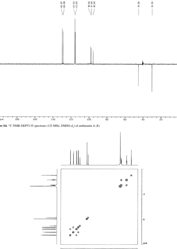
para-disubstituted aromatic rings. The distortionless enhancement by polarization transfer (DEPT) 135 spectrum
confirmed the presence of two saturated methylene carbons at dC30.3 (C-IIβ) and 45.3 (C-IIα), the methine carbons of
the p-disubstituted aromatic rings at dC115.6 (C-I3’, 5’),
115.9 (C-II3, 5), 129.3 (C-I2’, 6’) and 130.0 (CII-2, 6), and three methine sp2 carbons at d95.5 (C-I8), 97.5 (C-I6) and
98.5 (C-II5’).
The 13C NMR data (Table 1) showed the presence of
two carbonyl carbons; one at dC203.7 (C-IIβ’), related
to a dihydrochalcone as suggested by the presence of the two methylene carbons, and one at dC 192.2 (C-I4),
characteristic of a dihydroflavonol moiety. The presence of eight oxygenated aromatic carbons was inferred from the carbon resonances at dC156.4 (C-II4), 159.5 (C-I4’),
161.3 (C-I9), 162.2 (C-II6’), 163.5 (C-I5), 164.0 (C-II2’), 166.7 (C-II4’) and 167.7 (C-I7). A further conclusion from the chemical shifts of these carbons is that there were not oxygenated ortho-carbons, as such species would lead to more shielded chemical shifts (144 ≤dC≤ 148).
18
This suggested that the A-ring oxygenation pattern followed the common phloroglucinol A-ring theme for flavonoids.18 A saturated quaternary carbon bearing an
oxygen atom atdC 80.5 (C-I3)was readily characterized,
while the dioxygenated carbon at d 118.6 (C-I2) was identified by comparison with the chemical shifts of dihydroflavonol moieties from the daphnodorins isolated from Daphne odora Thunb.19
The heteronuclear single quantum coherence (HMQC) spectrum permitted the correlation of all protonated carbons (Table 1). The heteronuclear multiple-bond correlation (HMBC) data were of crucial importance for confirming structural assignments and correctly positioning the hydroxy groups and all non-protonated carbons. From Table 1 it is evident that the assignments of C-6 and C-8 could mistakenly be interchanged, as C-6 is slightly more deshielded than C-8.18 Unambiguous assignment was,
however, possible due to several key HMBC correlations. The hydrogen at dH 5.86 (H-I8) showed two- and
three-bond correlations to the carbons at dC99.4 (C-I10) and
161.3 (C-I9), respectively, while the one at dH 5.95 (H-I6)
exhibited correlations to the carbons at dC163.5 (C-I5)
and 95.5 (C-I8). The doublet at dH7.25 (H-I2’, 6’) showed
a three bond correlation with the dioxygenated carbon at dC118.6 (C-I2), whereas the one at dH 7.02 (H-II2,
6) correlated to the monoxygenated carbon at dC 156.4 (C-II4). The respective correlations involving the methylene hydrogens at dH 3.29 (H-IIα) and 2.87 (H-IIβ) with the
methylene carbon at dC 30.3 (C-IIβ) and the carbonyl
carbon at dC 203.7 (C-IIb’) ratified the presence of the
dihydrochalcone moiety. The hydrogen at dH5.96 (H-II5’)
additionally presented “W”-coupling (4J) with the C II-β’
Canuto et al. 643 Vol. 25, No. 4, 2014
and 166.7 (C-II4’). The linkage between the two flavonoid moieties via a dihydrofuran ring was confirmed by the coupling of the hydroxy hydrogen at dH6.51 (HO–C-I3)
with the carbons at dC 118.6 (C-I2) and 108.4 (C-II3’).
Long-range correlations for the hydroxy hydrogens further corroborated the suggested structure. The hydrogen-bonded hydroxy group at dH 13.17 correlated to carbon resonances
at dC162.2 (C-II6’,
2J),101.9(C-II1’, 3J) and 98.5 (C-II5’, 3J), while the hydroxy proton at d
H 11.70 (H-II2’) correlated
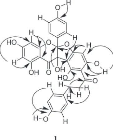
to d 164.0 (C-II2’). Figure 2shows additional important C-H correlations observed in the HMBC spectrum.
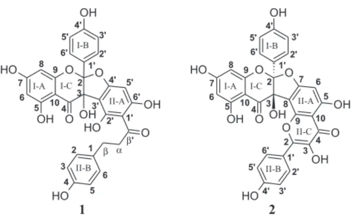
T h u s , t h e s t r u c t u r e o f 1 w a s a s s i g n e d a s 3,5,7,4’-tetrahydroxyflavanone-(2→O→4’:3→ 3’)-2’,4’,6’,4-tetrahydroxydihydrochalcone, an adduct of the flavonol kaempferol and the dihydrochalcone phloretin, and named amburanin A. Comparison of the 13C NMR
chemical shifts reported for these two monomers with those of compound 1, shows agreement, taking into account the
Table 1.1H and 13C NMR data (din ppm, J in Hz) of compounds 1 and 2 (500/125 MHz, respectively, DMSO-d 6)a
Amburanin A (1) Amburanin B (2)
C# dC dH C# dC dH
I-2 118.6 – I-2 118.7 –
I-3 80.5 – I-3 80.8 –
I-4 192.2 – I-4 191.8 –
I-5 163.5 – I-5 164.3 –
I-6 97.5 5.95 ( d, 1H, J 2.0) I-6 97.8 5.95 (d, 1H, J 2.0)
I-7 167.7 – I-7 168.4 –
I-8 95.5 5.86 (d, 1H, J 2.0) I-8 96.0 5.92 (d, 1H, J 2.0)
I-9 161.3 – I-9 161.3 –
I-10 99.4 – I-10 99.6 –
I-1’ 124.5 – I-1’ 124.5 –
I-2’, 6’ 129.3 7.25 (d, 2H, J 8.7) I-2’, 6’ 129.2 7.28 (d, 2H, J 8.7)
I-3’, 5’ 115.6 6.72 (d, 2H, J 8.7) I-3’, 5’ 115.7 6.79 (d, 2H, J 8.7)
I-4’ 159.5 – I-4’ 159.6 –
II-1’ 101.9 – II-2 148.3 –
II-2’ 164.0 – II-3 136.9 –
II-3’ 108.4 – II-4 177.1 –
II-4’ 166.7 – II-5 164.7 –
II-5’ 98.5 5.96 (s, 1H) II-6 95.6 6.71 (s, 1H)
II-6’ 162.2 – II-7 165.7 –
β' 203.7 – II-8 106.9 –
a 45.3 3.29 (t, 2H, J 7.7) II-9 152.1 –
β 30.3 2.87 (t, 2H, J 7.7) II-10 106.1 –
II-1 131.8 – II-1’ 122.2 –
II-2, 6 130.0 7.02 (d, 2H, J 8.4) II-2’, 6’ 131.0 8.16 (d, 2H, J 8.7)
II-3, 5 115.9 6.62 (d, 2H, J 8.4) II-3’, 5’ 116.3 6.94 (d, 2H, J 8.7)
II-4 156.4 – II-4’ 160.4 –
OH (I-3) – 6.51 OH (I-3) – 6.96
OH (I-5) – 11.02 OH (I-5) – 11.80
OH (I-7) – 11.02 OH (I-7) – 11.02
OH (II-2’) – 11.70 OH (I-4’) – 9.80
OH (II-6’) – 13.17 OH (II-3) – 9.66
OH (I-4’) – 9.77 OH (II-5) – 13.30
OH (II-4) – 9.17 OH (II-4’) – 10.20
empirical influence of the substitution effect.18 This effect
is particularly noticeable for C-II3’ as it is deshielded by 12.7 ppm due to the “ipso” effect of C-I3, and for C-I3 as it is deshielded by 6.8 ppm due to the α-effect of C-II3’.18
Although compound 1 possessed a negative specific rotation, ([α]D
20−15.9, c 0.24, MeOH), the experimental
ECD spectrum (not shown) lacked distinct Cotton effects (CEs) at all concentrations examined, indicating a lack of enantiomeric purity.
Compound 2 was obtained as a yellow powder, with a molecular formula of C30H18O12 as revealed by a HREIMS
quasi-molecular ions at m/z 593.0679 [M+Na]+ (calcd. for
C30H18O12Na, 593.0696); m/z 571.0853 [M+H]+ (calcd. for
C30H19O12, 571.0877). Its FT-IR spectrum exhibited a broad
absorption band centered at 3433 cm-1, characteristic of
hydroxy groups, and an absorption band with a shoulder at 1640 cm-1 characteristic of C=O stretching. Absorption
bands at 1258 cm-1 and 1168 cm-1 were characteristic of
phenolic and tertiary alcohol C–O stretching frequencies, similar to those observed for 1.
The 1H NMR spectrum displayed two m-coupled
one-proton doublets at dH5.92 and 5.95 (d, 1H, J 2.0 Hz, H-I8
and H-I6), a one-proton singlet at dH6.71 (s, 1H, H-II6) and
two pairs of o-coupled two-proton doublets at dH6.79 (d,
2H, J 8.7 Hz, H-I3’,5’), 6.94 (d, 2H, J 8.7 Hz, H-II3’,5’), 7.28 (d, 2H, J 8.7 Hz, H-I2’,6’) and 8.16 (d, 2H, J 8.7 Hz, H-II2’,6’). The o-coupled two-proton doublets showed the same relative coupling patterns (dH6.79 with 7.28 and dH
6.94 with 8.16) as observed for compound 1.
The 13C NMR (CPD) spectrum of 2 was similar to that
of compound 1 andshowed 26 resonances between dC80
and 192, two with prominent intensities. From Table 1, it is evident that the dihydroflavonol moiety is the same for both compounds. The chemical shift of the second carbonyl absorption (dC177.1, C-II4), however, indicated that the
second C6–C3–C6 unit was a flavonol moiety instead of a
dihydrochalcone. This was supported by the lack of the
two saturated methylene carbons seen in compound 1. Two sp2 carbons at d 136.9 (C-II3) and 148.3 (C-II2) were
instead present, further supporting the assignment of a flavonol moiety.
The HMQC and HMBC experiments corroborated the suggested structure. The most important C–H long-range correlations in the HMBC spectrum are depicted in Figure 3. Thus, compound 2 was structurally characterized as 3,5,7,4’-tetrahydroxyflavanone-(2→O→7:3→ 8)-3,4’,5,7-tetrahydroxyflavone, an adduct of two kaempferol molecules, and named amburanin B. Since the ring closure of the flavonol moiety could have occurred via either the C-II9 or C-II5 hydroxy group, the HMBC experiment was again of crucial importance. As depicted in Figure 3, the hydrogen-bonded hydroxy proton at d13.3 (HO–C-II5) correlated to the carbons at dC95.6 (C-II6,
3J), 106.1
(C-II10, 3J) and 164.7 (C-II5, 2J), unequivocally confirming
structure 2.
Although compound 2 possessed a low magnitude negative specific rotation of [α]D20−12.4 (c 0.21, MeOH),
the experimental ECD spectrum (Figure S17 in the Supplementary Information section) contained distinct CEs which appeared to correspond to the multiple UV maxima for the compound. Attempts to interpret these CEs based on the precedent of the daphnodorins,19,20 however,
revealed a series of significant differences between ECD data for the daphnodorins with their flavan and flavan-3-ol constituents units, and compound 2. However, the negative CE at ca. 320 nm presumably indicates (2S, 3S) absolute configuration for this compound based on the ECD data of daphnodorins F and H with similar configuration at C-2 and C-3. The low magnitude of the specific rotation of compound 2 presumably reflects a low degree of enantiomeric purity when compared to the reported values for daphnodorins F and H (–120.0 and –170.4, respectively).16,19
Figure 2. HMBC correlations (H→ C) observed for compound 1.
Canuto et al. 645 Vol. 25, No. 4, 2014
Although the two new amburanins A and B (1 and 2) are here described for the first time, analogous dihydrofuran-type biflavonoids, the daphnodorins, have been reported from Glycyrrhiza glabra L. (Fabaceae)21 and several
species of the genus Daphne (Thymelaeaceae) including
D. odora,18,20 D. acutiloba Rehd.,22D. genkwa Sieb. et
Zucc.,23D. giraldii Nitsche24 and D. tangutica Maxim.25
These compounds have shown pharmacological activities including in vitro and in vivo cytotoxicity,23,25-27 antifungal
and anti-HIV activities,28 and inhibitory effects on human
chymase and 12-lipoxygenase.27-30
In previous studies,2,4,7,13,31 it has been shown that
hydroalcohol extract, coumarin, isokaempferide, amburoside A, vanillic acid and afrormosin from
A. cearensis display various pharmacological properties. These molecules showed anti-inflammatory activity in rodents by inhibiting paw edema and the accumulation of inflammatory cells into the peritoneal cavity of mice. In addition, isokaempferide and amburoside A were able to inhibit human neutrophil degranulation, myeloperoxidase activity and the secretion of TNF-α by human neutrophils, showing no cytotoxicity at the concentrations investigated. Other studies have shown the anti-inflammatory properties of vanillic and protocatechuic acids.31-33 Additional
investigations into the possible anti-inflammatory activities of amburanins A and B were therefore deemed worthwhile, as these compounds could contribute to the biological activities of A. cearensis.
The recruitment and activation of polymorphonuclear leukocytes (PMNs) is considered one of the main defense mechanisms of innate immunity. Chemical mediators subsequently released by activated cells play an important role in the pathophysiology of several inflammatory diseases, such as rheumatoid arthritis, pulmonary emphysema and asthma.34-36 As part of the first line of
host defense, PMNs possess bactericidal activities and are involved in pathogen adherence, chemotaxis towards infected cells, and phagocytosis of pathogens and damaged cells.37 Activation of various PMNs during inflammatory
processes results in the release of complement components, reactive oxygen species and lysosomal enzymes, including MPO. MPO levels can therefore serve as an index of neutrophil recruitment and activation and also display traditional cytokine-like properties that can serve to modulate the activation state of leukocytes in the inflammatory process.38-40 In addition, MPO has direct
effects on endothelial cells since its internalization by these cells is followed by the intracellular production of oxidants.41
The possible effects of biflavonoids 1 and 2 on the neutrophil degranulation were assessed by measuring
MPO release by PMA-stimulated cells. Exposure of neutrophils to compounds 1 and 2 (0.1-100 µg mL-1)
caused an inhibition of MPO release (Figure 4) that was comparable to the effects of indomethacin (36 µg mL-1), a
non-selective cyclooxygenase inhibitor used as reference compound. At concentrations higher than 10 µg mL-1,
biflavonoids 1 and 2 inhibited MPO activity by 45.5 and 52.7%, respectively; at 36 µg mL-1, indomethacin inhibited
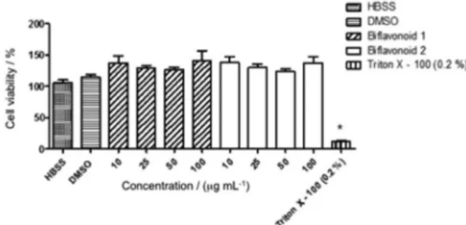
MPO activity by 45% (Figure 5). Compared with the control group (115.2 ± 3.8%), 1 and 2 did not interfere significantly in the cell viability at concentrations ranging from 10 to 100 µg mL-1; the reference compound Triton
X-100 reduced the cell viability to 12.1% (Figure 6).
Figure 4. Effects of biflavonoids 1 and 2 on the human neutrophil degranulation assayed using release MPO as marker. Freshly isolated cells (5 × 10⁶) were preincubated with the indicated concentration of 1 or 2 prior
to the addition of PMA (0.1 mg mL-1). Data are expressed as percentage
of inhibition of MPO release by the tested compounds. Numbers represent mean ± standard error of the mean (SEM).
Figure 5. Effects of biflavonoids 1 and 2 on the activity of human neutrophil myeloperoxidase (MPO). The MPO-rich supernatant obtained from PMA-stimulated neutrophils was incubated with compound 1 or 2 at the concentrations indicated in the graph. Data are expressed as percentages of inhibition of MPO activity by the tested compounds. Numbers represent mean ± SEM.
These results provide evidence that the biflavonoids amburanins A and B have anti-inflammatory potential through the modulation of human neutrophil degranulation, but this effect seems is related at least in part to a direct effect of the biflavonoids on the MPO activity. Furthermore, the anti-inflammatory activity of these biflavonoids seems not to be related to a cytotoxic effect, since these compounds did not affect the neutrophil viability, as assayed by the MTT assay. In this context, because neutrophils play important roles in the pathophysiology of several inflammatory diseases and the MPO activity correlates well with leukocytes infiltration in inflamed regions,42 it is likely that pharmacological activities of
amburanins A and B reported here have beneficial effects in the treatment of inflammatory diseases.
Conclusions
The chemical investigation of Amburana cearensis trunk bark permitted the isolation of two unusual biflavonoids, amburanins A and B (compounds 1 and 2).These compounds displayed inhibition of human neutrophil pro-inflammatory responses, such as degranulation and MPO activity, suggesting potential anti-inflammatory effects similar to those of co-occurring amburoside A, isokaempferide and afrormosin.7,31 These findings corroborate the folk usage of
this plant, justifying its pharmacological potential for the treatment of inflammatory diseases such as asthma.
Supplementary Information
Supplementary data of the compounds 1 and 2 (NMR, IR and HR-MS spectra, Figures S1-S17) are available free of charge at http://jbcs.sbq.org.br as PDF file.
Acknowledgments
The authors are grateful to CNPq for the fellowships, to CAPES, PRONEX, FINEP, FUNCAP, BNB and the USDA (Agricultural Research Service Specific Cooperative Agreement no. 58-6408-2-0009) for financial support and to L. L. Machado (Universidade Federal do Ceará) for recording the UV spectra. We would like to thank Drs. Yuanqing Ding and Xing Cong Li for their efforts computing the theoretical ECD spectra for compound 2 and the Mississippi Center for Supercomputing Research for use of the computational facilities to perform the necessary theoretical calculations. We would also like to thank the Department of Chemistry and Biochemistry at Mississippi College, Clinton, MS, USA, especially Dr. Dale Rosado, for the use of the JASCO J-815 Spectrometer.
References
1. Matos, F. J. A.; Farmácias Vivas -Sistema de Utilização de Plantas Medicinais Projetado para Pequenas Comunidades; Editora UFC: Fortaleza, 2002.
2. Leal, L. K. A. M.; Nechio, M.; Silveira, E. R.; Canuto, K. M.; Fontenele, J. B.; Ribeiro, R. A.; Viana, G. S. B.; Phytother. Res.
2003, 17, 335.
3. Leal, L. K. A. M.; Costa, M. F.; Pitombeira, M.; Barroso, V. M.; Silveira, E. R.; Canuto, K. M.; Viana, G. S. B.; Life Sci.2006,
79, 98.
4. Leal, L. K. A. M.; Fonseca, F. N.; Pereira, F. A.; Canuto, K. M.; Felipe, C. F. B.; Fontenele, J. B.; Pitombeira, M. V.; Silveira, E. R.; Viana, G. S. B.; Planta Med.2008, 74, 497.
5. Leal, L. K. A. M.; Nobre, H. V.; Cunha, G. M. A.; Moraes, M. O.; Pessoa, C.; Oliveira, R. A.; Silveira, E. R.; Canuto, K. M.; Viana, G. S. B.; Neurosci. Lett.2005, 388, 86. 6. Costa-Lotufo, L. V.; Jimenez, P. C.; Wilke, D. V.; Leal,
L. K. A. M.; Cunha, G. M. A.; Silveira, E. R.; Canuto, K. M.; Viana, G. S. B.; Moraes, M. E. A.; Moraes, M. O.; Pessoa, C.;
Z. Naturforsch., C: J. Biosci.2003, 58, 675.
7. Leal, L. K. A. M.; Canuto, K. M.; Costa, K. C. S.; Nobre-Júnior, H. V.; Vasconcelos, S. M.; Silveira, E. R.; Ferreira, M. V. P.; Fontenele, J. B.; Andrade, G. M.; Viana, G. S. B.; Basic Clin. Pharmacol. Toxicol.2009, 104, 198.
8. Bravo, J. A.; Sauvain, M.; Gimenez, A.; Muñoz, V.; Callapa, J.; Le Men-Olivier, L.; Massiot, G.; Lavaud, C.; Phytochemistry
1999, 50, 71.
9. Canuto, K. M.; Silveira, E. R.; Quim. Nova2006, 29, 1241. 10. Canuto, K. M.; Lima, M. A. S.; Silveira, E. R.; J. Braz. Chem.
Soc.2010, 21, 1746.
11. Bandeira, P. N.; Farias, S. S.; Lemos, T. L. G.; Braz-Filho, R.; Santos, H. S.; Albuquerque, M. R. J. R.; Costa, S. M. O.; J. Braz. Chem. Soc.2011, 22, 372.
12. Canuto, K. M.; Silveira, E. R.; Bezerra, A. M. E.; Quim. Nova
2010, 33, 662.
13. Leal, L. K. A. M.; Pierdoná, T. M.; Góes, J. G. S.; Fonsêca, K. S.; Canuto, K. M.; Silveira, E. R.; Bezerra, A. M. E.; Viana, G. S. B.; Phytomedicine2011, 18, 230.
14. Araruna, S. M.; Silva, A. H.; Canuto, K. M.; Silveira, E. R.; Leal, L. K. A. M.;Braz. J. Pharmacogn.2013, 23, 132. 15. Lucisano, Y. M.; Mantovani B.; J. Immunol.1984, 132, 2015. 16. Boyum, A.;Scand. J. Clin. Lab. Invest. 1968, 21, 77. 17. Mosmann, T.; J. Immunol. Methods1983, 65, 55.
18. Agrawal, P. K.; Carbon-13 of Flavonoids; Elsevier: New York, 1989.
19. Taniguchi, M.; Baba, K.; Phytochemistry1996, 42, 1447. 20. Baba, K.; Yoshikawa, M.; Taniguchi, M.; Kozawa, M.;
Phytochemistry1995, 38, 1021.
Supplementary Information
S
I
J. Braz. Chem. Soc., Vol. 25, No. 4, S1-S9, 2014. Printed in Brazil - ©2014 Sociedade Brasileira de Química 0103 - 5053 $6.00+0.00
*e-mail: [email protected]
#Present address: Embrapa Agroindústria Tropical, Rua Dra. Sara
Mes-quita, 2270, Pici, CP 3761, 60511-110 Fortaleza-CE, Brazil
Amburanins A and B from
Amburana cearensis
: Daphnodorin-Type Biflavonoids
that Modulate Human Neutrophil Degranulation
Kirley M. Canuto,#,a Luzia K. A. M. Leal,b Amanda A. Lopes,b Christina M. Coleman,c
Daneel Ferreirac and Edilberto R. Silveira*,a
aDepartamento de Química Orgânica e Inorgânica, Universidade Federal do Ceará,
Campus do Pici, Bloco 940, Pici, 60451-970 Fortaleza-CE, Brazil.
bCentro de Estudos Farmacêuticos e Cosméticos (CEFAC), Departamento de Farmácia,
Universidade Federal do Ceará, Rua Cap. Francisco Pedro, 1210, Rodolfo Teófilo, 60430-170 Fortaleza-CE, Brazil
cDepartment of Pharmacognosy and the Research Institute of Pharmaceutical Sciences, School of
Pharmacy, The University of Mississippi, P.O. Box 1848, University MS 38677-1848, USA
Figure S2. 1H NMR spectrum (500 MHz, DMSO-d
6) of amburanin A (1).
Figure S3.13C NMR spectrum (125 MHz, DMSO-d
Canuto et al. S3 Vol. 25, No. 4, 2014
Figure S4.13C NMR-DEPT135 spectrum (125 MHz, DMSO-d
6) of amburanin A (1).
Figure S5.1H,1H COSY-NMR spectrum (500
Figure S6. 1H, 13C HMQC-NMR spectrum (500
× 125 MHz, DMSO-d6) of Amburanin A (1).
Figure S7. 1H, 13C HMBC-NMR spectrum (500
Canuto et al. S5 Vol. 25, No. 4, 2014
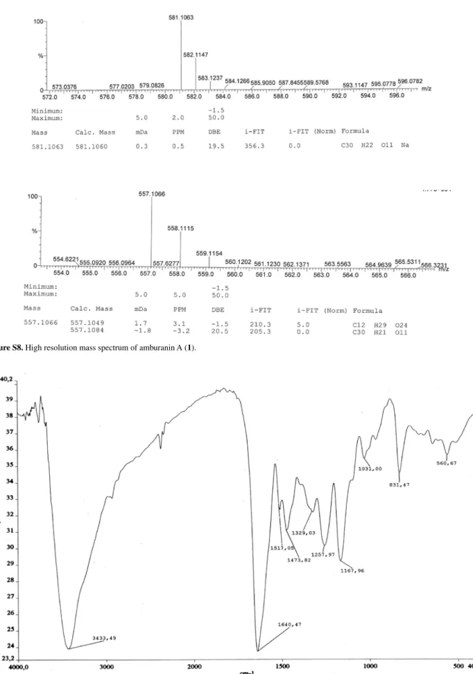
Figure S8. High resolution mass spectrum of amburanin A (1).
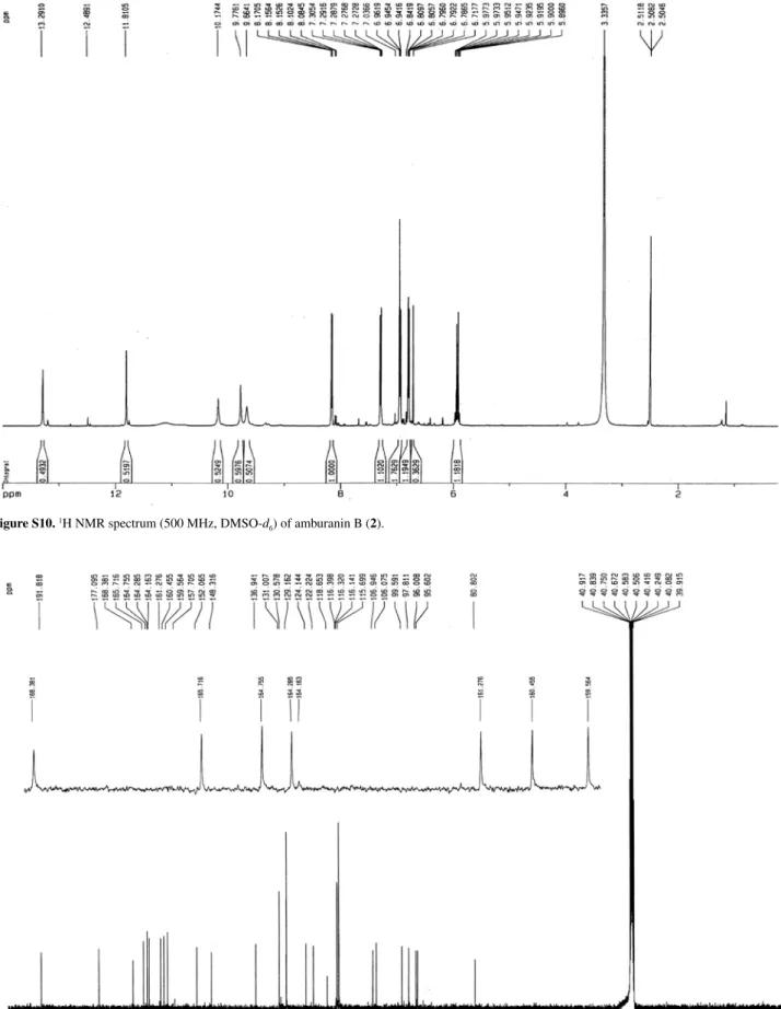
Figure S10. 1H NMR spectrum (500 MHz, DMSO-d
6) of amburanin B (2).
Figure S11. 13C NMR spectrum (125 MHz, DMSO-d
Canuto et al. S7 Vol. 25, No. 4, 2014
Figure S12. 13C NMR-DEPT135 spectrum (125 MHz, DMSO-d
6) of amburanin B (2).
Figure S13. 1H,1H COSY-NMR- spectrum (500
Figure S14. 1H, 13C HSQC-NMR spectrum (500
× 125 MHz, DMSO-d6) of amburanin B (2).
Figure S15. 1H, 13C HMBC-NMR spectrum (500
Canuto et al. S9 Vol. 25, No. 4, 2014
Figure S16. High resolution mass spectrum of amburanin B (2).
22. Taniguchi, M.; Fujiwara, A.; Baba, K.; Wang, H.-N.;
Phytochemistry1998, 49, 863.
23. Zheng, W.; Gao, X.; Chen, C.; Tan, R.; Int. Immunopharmacol.
2007, 7, 117.
24. Zhang, Q.; Ye, N.; Sun, W.-X.; Zhang, K.-M.; Jiang, J.-Q.;
Biochem. Syst. Ecol.2008, 36, 63.
25. Zhang, W.; Shen, Y. H.; Lou, Z. Y.; Liu, R. H.; Zhang, C.; Fu, P.; Shan, L.; Zhang, W. D.; Nat. Prod. Res.2007, 21, 1021. 26. Zhang, W.; Zhang, W. D.; Zhang, C.; Liu, R. H.; Li, T. Z.; Fu, P.;
Shan, L.; Phytother. Res.2007, 21, 1113.
27. Zheng, W.; Gao, X.; Gu, Q.; Chen, C.; Wei, Z.; Shi, F.; Int. Immunopharmacol.2007, 7, 128.
28. Hu, K.; Kobayashi, H.; Dong, A.; Iwasaki, S.; Yao, X. S.; Planta Med.2000, 66, 564.
29. Sakaguchi, M.; Yamamoto, D.; Takai, S.; Jin, D.; Taniguchi, M.; Baba, K.; Miyazaki, M.; Biochem. Biophys. Res. Commun.
2001, 283, 831.
30. Takai, S.; Jin, D.; Sakaguchi, M.; Kirimura, K.; Ikeda, J.; Baba, K.; Fujita, T.; Miyazaki, M.; Prostaglandins, Leukotrienes Essent. Fatty Acids1999, 60, 135.
31. Lopes, A. A.; Magalhães, T. R.; Uchôa, D. E.; Silveira, E. R.; Azzolini, A. E. C. S.; Kabeya, L. M.; Lucisano-Valim, Y. M.; Vasconcelos, S. M.; Viana, G. S. B.; Leal, L. K. A. M.; Basic Clin. Pharmacol. Toxicol.2013, 113, 363.
32. Chiang, L. C.; Ng, L. T.; Chiang, W.; Chang, M. Y.; Lin, C. C.;
Planta Med.2003, 69, 600.
33. Min, S.-W.; Ryu, S.-N.; Kim, D.-H.; Int. Immunopharmacol.
2010, 10, 959.
34. Weiss, S. J.; N. Engl. J. Med.1989, 320, 365.
35. Sibile, Y.; Reynolds, H. Y.; Am. Rev. Respir. Dis.1990, 141, 471.
36. Botha, A. J.; Moore, F. A.; Moore, E. Z. E.; Sauaia, A.; Banerjee, A.; Peterson, V. M.; J. Trauma1995, 39, 411. 37. Miyasaki, K. T.; Wilson, M. E.; Brunetti, A. D.; Genco, R. J.;
Infect. Immun.1986, 53, 154.
38. Dale, C. D.; Boxer, L.; Liles, W. C.; Blood J.2008, 112, 935. 39. Lau, D.; Mollnau, H.; Eiserich, J. P.; Freeman, B. A.; Daiber, A.;
Gehling, U. M.; Brümmer, J.; Rudolph, V.; Münzel, T.; Heitzer, T.; Meinertz, T.; Baldus, S.; Proc. Natl. Acad. Sci. USA
2005, 102, 431.
40. Hazen, S. L.; Hsu, F. F.; Duffin, K.; Heinecke, J. W.; J. Biol. Chem.1996, 271, 23080.
41. Yang, J. J.; Preston, G. A.; Pendergraft, W. F.; Segelmark, M.; Heeringa, P.; Hogan, S. L.; Jennette, J. C.; Falk, R. J.; Am. J. Pathol.2001, 158, 581.
42 Nakamura, Y.; Murakami, A.; Ohto, Y.; Torikai, K.; Tanaka, T.; Ohigashi, H.; Cancer Res.1998, 58, 4832.
Submitted: November 2, 2013