319
Artigo
ISSN 0102-695X Brazilian Journal of Pharmacognosy
17(3): 319-324, Jul./Set. 2007 Received 05/14/07. Accepted 07/28/07
Bioactive
fl
avone dimers from Ouratea multi
fl
ora (Ochnaceae)
Carlos Alberto Carbonezi
1, Lidilhone Hamerski
1, A. A. Leslie Gunatilaka
2, Alberto
Cavalheiro
1, Ian Castro-Gamboa
1, Dulce Helena Siqueira Silva
1, Maysa Furlan
1,
Maria Claudia Marx Young
3, Marcia Nasser Lopes
1, Vanderlan da Silva Bolzani
1*
1Instituto de Química, Universidade Estadual Paulista, UNESP, CP 355, 14801-970, Araraquara, SP, Brazil, 2Arid Land Research Institute, University of Arizona, Tucson, AZ 85706-6800, USA,
3Seção de Fisiologia e Bioquímica de Plantas, Instituto de Botânica, 01061-970, São Paulo, SP, Brazil
RESUMO: “Dímeros fl avônicos bioativos de Ouratea multifl ora (Ochnaceae)”. O fracionamento cromatográfi co do extrato orgânico das folhas de Ouratea multifl ora forneceu os fl avonóides diméricos, heveafl avona, 7′′,4′′′-dimetilamentofl avona, podocarpusfl avona-A e amentofl avona. Suas estruturas foram elucidadas com base nos dados espectrais, incluindo experimentos bidimensionais de RMN, das substâncias naturais. A atividade antibiótica de todos os isolados foi avaliada, usando-se as bacterias Gram-positivas Staphylococcus aureus and Bacillus subtilis. Teste de citotoxicidade nas linhagens de linfoma de ratos (L5178) e KB também foram conduzidos para avaliar os extratos e os fl avonóides isolados. a triagem biológica para a avaliação de atividade antioxidante e inibidora de acetil colinesterase foram conduzidas pela técnica da bioautografi a com DPPH e teste pelo teste de Ellman respectivamente.
Unitermos: Ochnaceae, Ouratea multifl ora, bifl avonoids, antibacterial activity.
ABSTRACT: Chromatographic fractionation of the organic extract from leaves of Ouratea multifl ora afforded the fl avone dimers heveafl avone, amentofl avone-7′′,4′′′-dimethyl eter, podocarpusfl avone-A and amentofl avone. Their structures were elucidated from spectral data, including 2D-NMR experiments of the natural substances. Biological activities of all isolates were evaluated, using antimicrobial assay against Gram-positive bacteria Staphylococcus aureus and Bacillus subtilis, cytotoxicity assay against mouse lymphoma (L5178) and KB cell lines, TLC screening for acetylcholinesterase inhibitors and antioxidant activity measured by DPPH test.
Keywords: Ochnaceae, Ouratea multifl ora, bifl avonoids, antibacterial activity.
INTRODUCTION
Species of the Ochnaceae family are distributed in tropical and subtropical zones throughout the World (Hegnauer, 1969; Carvalho et al., 2000) This family is characterized by the presence of fl avonoids and bifl avonoids and terpenoids as main secondary metabolites (Oliveira et al., 2002; Estevam et al 2005). Several members of the Ouratea genus are employed for the extraction of edible oil, and are used as medicinal plants (Moreira et al., 1994; Agra et al., 2007). As part of our research program on the bioactive constituents of Atlantic Forest plant species, we have investigated Ouratea multifl ora collected in Juréia Reserve, São Paulo State. As result, four fl avonoid dimers: heveafl avone (Geiger, 1986) amentofl avone-7′′,4′′′-dimethyl-ether (Geiger, 1986), podocarpusfl avone-A (Geiger, 1986; Markham et al., 1987) and amentofl avone (Geiger, 1986; Markham et al., 1987; Felício et al., 2001), were isolated from the ethanolic extract of the leaves. Additionally, the biological activity of the isolates was also evaluated.
MATERIAL AND METHODS
General procedure
1H and 13C NMR, inverse heteronuclear
HMQC and HMBC as well as COSY experiments were performed on a Varian Inova 500 MHz instrument operating at 500 MHz for hydrogen, and 125 MHz for carbon, respectively. Pyridine-d5 or DMSO-d6 were used as solvents and TMS as internal standard. TLC was performed on precoated aluminum sheets (silica F254, 0.25 mm, Merck, Parmtadt, Germany) with detection provided by UV light (254 and 366 nm) and by spraying with anisaldeyde reagent followed by heating (120 °C). Silica gel (230-400 mesh ASTM, particle size 0.040-0.063 µM, Merck) or Sephaex LH-20 (Pharmacia) were used in the CC fractionations.
Plant material
voucher was placed at the Botanical Institute, São Paulo State, Brazil.
Extraction and isolation
Dried powdered leaves of Ouratea multifl ora (118.0 g) were extracted by maceration using ethanol at room temperature. The extract was fi ltered and the solvent was removed under vacuum. The crude extract (3.44 g) was dissolved in n-BuOH. Subsequent addition of water afforded two phases which were evaporated to dryness under reduced pressure. The n-BuOH fraction (2.10 g) was solubilized with methanol/water (8:2 v/v) and partitioned with n-hexane, chloroform and ethyl acetate, successively. The ethyl acetate phase after concentration of the solvent (980.0 mg), was chromatographed on a Sephadex LH-20 column with a gradient elution of MeOH/H2O. The separation was monitored by TLC, and eluted fractions exhibiting similar appearances were combined, yielding 20 fractions. Fractions 7 and 8 showed a solid precipitation. The solid was washed with cold methanol to afford heveafl avone(Geiger, 1986) (1) (21.2 mg). Fraction 13 was chromatographed on silica gel yielding amentofl avone 7”, 4”’-dimethyl ether (2) (10.8 mg). Fraction 15 gave a powder after precipitation. This powder was washed with cold methanol to afford podocarpusfl avone-A 3 (Markam et al., 1987) (8.9 mg). Fraction 17 was chromatographed on silica gel using CHCl3:MeOH (4:6) yielding amentofl avone 4 (Geiger, 1986; Markham et al., 1987) (15.0 mg).
Antimicrobial assay
Sterile fi lter paper disks were impregnated with 20 mg of samples using DMSO as carrier solvent. The impregnated disks were then placed on agar plates previously inoculated with Staphylococcus aureus (ATCC 2592) and Bacillus subtilis (DSM 2105). Solvent controls were incubated at 37 oC for each organism,
and after incubate time of 24 h at 37 oC, antimicrobial
activity was recorded as clear zones (in mm) of inhibition surrounding the disk. The sample was considered active when the inhibition surrounding the disk was greater than 7 mm.
Cytotoxicity assay
Antiproliferative activity was examined against two cell lines and was determined through an MTT assay as described earlier (Edrada et al., 1996).
TLC screening for acetylcholinesterase inhibitors
The protocol adopted for this in vitro assay is described by Ellman and co-workers (Ellman et al., 1961).
Determination of the radical-scavenging activity
The determination of the antioxidant activity was tested according to a protocol described elsewhere (Cardoso et al., 2004; So; Lewis, 2002).
H 1 (δH, J in Hz) 2 (δH, J in Hz) 3 (δH, J in Hz) 4 (δH, J in Hz)
3 6.63 (s) 6.82 (s) 6.81 (s) 6.82 (s)
6 6.23 (d, 2.5) 6.17 (d, 2.5) 6.17 (d, 1.5) 6.18 (d, 2.5)
8 6.49 (d, 2.5) 6.45 (d, 2.5) 6.45 (d, 1.5) 6.45 (d, 2.5)
2’ 7.82 (d, 2.5) 7.98 (d, 2.5) 7.98 (d, 2.5) 8.00 (d, 2.5)
5’ 7.13 (d, 9.0) 7.14 (d, 8.5) 7.15 (d, 8.5) 7.14 (d, 9.0)
6’ 7.86 (dd, 9.0, 2.5) 8.01 (dd, 8.5, 2.5) 8.00 (dd, 8.5, 2.5) 7.99 (dd, 9.0, 2.5)
3” 6.67 (s) 6.92 (s) 6.86 (s) 6.78 (s)
6” 6.51 (s) 6.67 (s) 6.41 (s) 6.39 (s)
2”’, 6’’’ 7.55 (d, 9.0) 7.66 (d, 9.5) 7.65 (d, 9.0) 7.56 (d, 9.0)
3”’, 5’’’ 6.82 (d, 9.0) 6.91 (d, 9.5) 6.91 (d, 9.0) 6.70 (d, 9.0)
MeO-4’’’ 3.81 (s) 3.83 (s) - -
MeO-7 3.77 (s) 3.75 (s) 3.74 (s) -
MeO-7’’ 3.72 (s) - - -
OH-5 13.93 (s) 12.94 13.05 13.09
OH-5’’ 12.93 (s) 13.20 12.95 12.96
Table 1.1H NMR data for compounds 1, 2, 3 and 4 (500MHz, DMSO-d
321
fl
Rev. Bras. Farmacogn. RESULTS AND DISCUSSION
The chromatographic fractionation of the ethanol extract from the leaves of O. multifl ora afforded heveafl avone (1), amentofl avone-7′′,4′′′-dimethyl-ether (2), podocarpusfl avone-A (3), and amentofl avone (4).
The 13C NMR spectrum of compound 1
showed 31 signals, which were attributed to twelve sp2 CH, including signals at δ
CH 114.1 and 127.6, each
representing two carbon atoms, three sp3 carbons (δ CH3
55.1, 55.5 and 55.9), sixteen sp2 quaternary carbons and
two carbonyl groups (δC 181.6 and 182.2) (Table 2).
C 1 (δ) 2 (δ) 3 (δ) 4 (δ)
2 163.9 163.6 163.8 163.9
3 103.1 103.1 103.0 102.9
4 181.6 181.8 181.7 181.8
5 162.0 161.4 161.5 162.1
6 97.7 98.8 98.8 98.9
7 164.8 164.1 164.1 164.2
8 92.0 94.1 94.0 94.1
9 157.1 157.4 157.4 157.4
10 104.6 103.7 103.7 103.7
1’ 120.9 121.2 121.1 120.9
2’ 130.9 131.3 131.4 131.4
3’ 119.4 119.6 119.9 120.1
4’ 159.3 159.4 159.5 159.7
5’ 116.0 116.2 116.2 116.2
6’ 127.5 128.0 127.9 127.8
2” 163.4 163.7 163.2 163.7
3” 102.9 103.2 103.2 102.6
4” 182.2 182.4 182.2 182.2
5” 161.5 161.4 160.6 161.1
6” 95.0 95.6 98.7 98.7
7” 161.2 162.8 161.9 160.6
8” 104.7 105.0 104.0 104.1
9” 153.6 153.7 154.5 154.5
10” 104.2 104.2 103.7 103.6
1’” 122.8 122.9 123.0 121.4
2’” 127.6 128.1 128.0 128.2
3’” 114.1 114.5 114.5 115.8
4’” 162.4 162.3 162.2 161.5
5’” 114.1 114.5 114.5 115.8
6’” 127.6 128.1 128.0 128.2
CH3O-4’’’ 55.9 56.5 55.5 -
CH3O-7’’ 55.5 55.5 - -
CH3O-7 55.1 - - -
Table 2.13C NMR spectral data for compounds 1, 2, 3 and 4 (125MHz, DMSO-d
The 1H NMR spectrum showed signals for two chelated
hydroxyls at δH 13.19 and 12.93 (OH-5 and OH-5”), three aromatic methoxyl groups, a 1,3,4-trisubstituted aromatic ring (B ring) and a 1,4-disubstituted (B’ ring). The 1H NMR spectra [1D and 2D (1H-1H-COSY)] showed
a para-substituted aromatic ring at δH 7.55 (d, J 9.0 Hz, H-2”’, H-6”’), δH 6.82 (d, J 9.0 Hz, H-3”’, H-5”’), two meta aromatic hydrogens at δH 6.23 (d, J 2.5 Hz, H-6), 6.49 (d, J 2.5 Hz, H-8), commonly observed in a fl avone nucleous. The structure of 1 was established on the basisi of HMBC spectra, which showed heteronuclear long-range couplings of quaternary carbons C-7’’ (δC 161.2), C-7 (δC 164.8) and C-4”’ (δC 162.4) with the hydrogens of methoxyl groups at δH 3.72 (OMe-7’’), 3.77 (OMe-7), and 3.81 (OMe-4”’), respectively, and by comparison of their spectral data with those reported for heveafl avone (Geiger, 1986).
Compound 2 also showed spectral features of a fl avonoid dimer derivative (Table 2). The 1H MNR
spectrum (Table 1) of 2 showed the presence of to chelated hydroxyl and at δH 3.83 and δH 3.75 two methoxyl groups. The 1H-1H-COSY spectrum of
compound 2 showed a set of doublets at δH 7.66 (d, J 9.5 Hz, H-2”’, H-6”’) and δH 6.91 (d, J 9.5 Hz, H-3”’, H-5”’) of an AA’BB’ system, besides the signals at δH 7.98 (d, J 2.5 Hz, H-2’), 7.14 (d, J 8.5, H-5’) and 8.01 (dd, J 8.5; 2.5, H-6’) attributed to a 1,3,4-trisubstituted aromatic ring. Furthermore, the 1H-1H-COSY spectrum showed
signals with a meta coupling pattern at δH 6.17 (d, J 2.5 Hz, H-6) and 6.45 (d, J 2.5 Hz, H-8) corresponding to an A ring of a fl avone. The HMBC spectrum of 2 revealed correlations of the hydrogen-bonded OH-5 (δH 12.94) with C-5 (δC 161.4), C-6 (δC 98.8) and C-10 (δC 103.7), while OH-5” (δH 13.20) correlated with C-5” (δC 161.4), C-6” (δC 95.6) and C-10” (δC 104.2). From the long range coupling of H-6 (δH 6.17) and H-3 (δH 6.82) with C-10 (δC 103.7); H-3”’ and H-5”’ (δH 6.91) with C-1”’ (δC 122.9); and H-3” (δH 6.92) with C-10” (δC 104.2) thus, the structure of 2 was established to be a dimer. A literature search confi rmed the structure of 2 as 7”,4”’-dimethoxy-amentofl avone (Geiger, 1986).
1H and 13C NMR spectra of 3 showed signals at δ H
13.05, δC 182.2, δH 12.95, and δC 181.8 of two hydrogen of chelated hydroxyl groups, and an O-methyl group at δH 3.74 (δC 55.5). A detailed analysis of cross peaks from the 1H-1H COSY spectrum showed two doublets
for a mono-oxygenated para disubstituted system at
δH 7.65 (d, J 9.0 Hz, H-2”’, H-6”’) and 6.91 (d, J 9.0 Hz, H-3”’, H-5”’), one trioxygenated tetra substituted ring with alternating oxy-substituents at δH 6.17 (d, J 1.5 Hz, H-6) and δH 6.45 (d, J 1.5 Hz, H-8), and one mono-oxygenated trisubstituted system at δH 7.98 (d, J 2.5 Hz, H-2’), 7.15 (d, J 8.5 Hz, H-5’) and 8.00 (dd, J 8.5; 2.5 Hz, H-6’). Additional 1D and 2D NMR data indicated a pentasubstituted aromatic ring with a 5,7-dioxygenation pattern for the A’ ring of a fl avone (δH 6.41, H-6”) and two singlets (δ 6.81 (H-3), δ 6.86
(H-3’’) of both fl avones units in compound 3. Analysis of HMBC spectra revealed characteristic correlations which defi ned the positions of aromatic rings and their substitution patterns. Correlations of H-3 (δ 6.81) with C-2 (δ 163.8), C-4 (δ 181.7), C-10 (δ 103.7), C-1’ (δ 121.1) and H-3” (δ 6.86) with C-2” (δ 163.2), C-4” (δ 182.2), C-10” (δ 103.7), C-1”’ (δ 123.0) established the linkages between rings C/B and C”/B”, respectively. Moreover, the correlation between H-2’ (δ 7.98) and C-8” (δ 104.0) showed the linkage between C-3’and C-8”. The ROESY correlation observed between methoxyl group at δH 3.74 and the aromatic protons at δH 6.91 (d, J 9.0 Hz, H-3”’, H-5”’) justify the location of the methoxyl group at C-4”’ of the B’ aromatic ring. These data are in agreement with those published for podocarpusfl avone-A (Geiger, 1986; Markham, et al., 1987).
1H NMR spectrum of 4 showed two signals at δ H
13.09 and 12.96 due to chelated phenolic hydroxyls, and two doublets at δH 7.56 (J 9.0 Hz, H-2”’, H-6”’), and 6.70 (J 9.0 Hz, H-3”’, H-5”’) assigned to an AA’BB’ aromatic system. Signals of a trisubstituted aromatic ring containing one oxygenated carbon were observed at δH 8.00 (d, J 2.5 Hz, H-2’), 7.14 (d, J 9.0 Hz, H-5’), and 7.99 (dd, J 9.0; 2.5 Hz H-6’), along with doublets of a tetrasubstituted aromatic ring containing three oxygenated carbons at δH 6.18 (J 2.5 Hz, H-6) and 6.45 (J 2.5 Hz, H-8). Analysis of the HMBC spectra evidenced correlations of the hydroxyl group with hydrogen bonded by cross peak at δH 13.09 (OH-5) with C-5” (δ 161.1), C-6” (δ 98.7), and C-10” (δ 103.6) and δH 12.96 (OH-5”) with C-5 (δ 162.1), C-6 (δ 98.9) and C-10 (δ 103.7), which are in agreement to those of amentofl avone (Geiger, 1986; Markham et al., 1987).
Previous studies on 13C NMR data for
amenthofl avone derivative dimers indicated a close relationship between substituent effects and chemical shift for the inter-fl avonoid linkage: a) amenthofl avones; I-3’ (+ 6 ppm), II-8’’ (+ 10 ppm), b) dihydroamenthofl avone; I-3’(+ 4 ppm), II-8” (+ 9 ppm), which can be diagnostic values in the recognition of new bifl avones in the amenthofl avone series (Markhan, et al., 1987; Felício, et al., 2001). The values of 13C NMR spectra of 1-4 (Table
2) followed logically the average found to the series reported and indicated that O-methylation induced shifts that are in the same direction as those reported for monofl avonoids (Markham, 1978).
323
fl
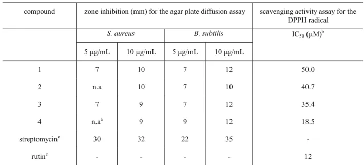
Rev. Bras. Farmacogn. compound zone inhibition (mm) for the agar plate diffusion assay scavenging activity assay for the
DPPH radical
S. aureus B. subtilis IC
50 (µM) b
5 µg/mL 10 µg/mL 5 µg/mL 10 µg/mL
1 7 10 7 12 50.0
2 n.a 10 7 10 40.7
3 7 9 7 12 35.4
4 n.aa 9 9 12 18.5
streptomycinc 30 32 22 35 -
rutinc - - - - 12
Table 3. Biological activity of compounds 1-4.
an.a (not active). bConcentration in µM required to scavenge 50% DPPH free radical. cStreptomycin sulfate and rutin
O
O OH R1O
OH
O
O
OH OR3
R2O
R1 R2 R3
CH3 CH3 CH3
H CH3 CH3
H H CH3
H H H
1 2 3 4
2
3 4 5
6 7
8
9
10
4'
2"
5" 7"
4'"
1'
3' 8"
acetylcholinesterae (AchE) inhibitors (Ellman et al., 1961), in which none inhibited the enzyme at 0.1 and 1.0 µM concentrations. The antioxidant activities of bifl avones 1-4 were tested toward DPPH radical (Table 3) (Cardoso et al. 2004). Compound 4 showed moderate scavenging activity (IC50 18.5 µg/mL) in this test (So; Lewis, 2002), while 1-3 showed weak scavenging activity which confi rms the dependence of antioxidant activity with the number of free aromatic hydroxyl groups of tested compounds.
ACKNOWLEDGMENT
This work was funded by grants of the Fundação de Amparo à Pesquisa do Estado de São Paulo (FAPESP) as part of the Biota-FAPESP - The Biodiversity Virtual Institute Program (www.biotasp.org.br), grant n° 03/02176-7 awarded to V. da S. Bolzani, Principal Investigator. V. da S. B., M.F. and C.A.C acknowledge CNPq and CAPES for researcher and Ph.D. fellowships. The authors wish to thank Arid Land Research Institute University of Arizona (Tucson, USA) by complementation of the fellowship awarded to C.A.C .
REFERENCES
Agra MF, França PF, Barbosa-Filho JM 2007. Synopsis of the plants known as medicinal and poisonous in Northeast of Brazil. Rev Bras Farmacogn 17: 114-140.
Cardoso CL, Castro-Gamboa I, Silva DHS, Furlan M, Epifanio RA, Pinto AC, Rezende CM, Lima JAL, Bolzani VS 2004. Indole glucoalkaloids from Chimarrhis turbinata and their evaluation as antioxidant agents and acetylcholinesterase inhibitors. J Nat Prod 67: 1882-1885.
Carvalho MG, Carvalho GJA, Braz-Filho R 2000. Chemical constituents from Ouratea fl oribunda: complete 1H
and 13C RMN assignments of atranorin and its new
acetylated derivative. J Braz Chem Soc 11: 143-147. Edrada RA, Proksch P, Wray V, Witte L, Müller WEG, Van
Soest RWM 1996. Four new bioactive manzamine-type alkaloids from the Philippine marine sponge Xestospongia ashmorica. J Nat Prod 59: 1056-1060. Ellman GL, Courtney KD, Andres V, Featherstone RM 1961.
A new and rapid colorimetric determination of acetylcholinesterase activity. Biochem Pharmacol 7: 88-90.
Estevam CS, Oliveira FM, Conserva LM, Lima LF, Barros ECP, Barros SCP, Rocha EMM, Andrade EHA 2005. Constituintes químicos e avaliação preliminar in vivo da atividade antimalárica de Ouratea nitida Aubl (Ochnaceae). Rev Bras Farmacogn 15: 195-198. Felicio JD, Rossi MH, Park HR, Gonçalez E, Braggio MM,
David JM, Cordeiro I 2001. Bifl avonoids from Ouratea multifl ora. Fitoterapia 72: 453-455. Geiger H 1986. The Flavonoids: Advances in Research since
1986. London Chapman and Hall.
Hegnauer R 1969. Chemotaxonomie der Pfl anzen, Vol. V. Basel: Stuttgart.
Markham KR, Ternai B, Stanley R, Geiger H, Mabry TR 1978.
13C NMR studies of
fl avonoids 3 naturally occurring fl avonoid glycosides and their acylated derivatives. Tetrahedron 34: 1389-1397.
Markham KR, Sheppard C, Geiger H 1987. 13C NMR of
fl avonoids 4 13C NMR studies of some naturally
occurring amentoflavone and hinokifl avone bifl avonoids. Phytochemistry 26: 3335-3337. Moreira IC, Sobrinho DC, Carvalho MG, Braz-Filho R 1994.
Isofl avone dimers A, hexaspermone-B and hexaspermone-C from Ouratea hexasperma. Phytochemistry 35: 1567-1572.
Oliveira MCC, Carvalho MG, Silva CJ, Werle AA 2002. New bifl avonoid and other constituents from Luxemburgia nobilis EICHL. J Braz Chem Soc 13: 119-123. So S, Lewis BA 2002. Free radical scavenging and antioxidative