In vitro
Manganese-Dependent Cross-Talk
between
Streptococcus mutans
VicK and
GcrR: Implications for Overlapping Stress
Response Pathways
Jennifer S. Downey1, Lauren Mashburn-Warren2, Eduardo A. Ayala1, Dilani B. Senadheera3, Whitney K. Hendrickson4, Lathan W. McCall4, Julie G. Sweet4, Dennis G. Cvitkovitch3, Grace A. Spatafora4*, Steven D. Goodman2*
1.Division of Biomedical Sciences at the Herman Ostrow School of Dentistry of the University of Southern California, Los Angeles, California, United States of America,2.Center for Microbial Pathogenesis, The Research Institute at Nationwide Children’s Hospital, Columbus, Ohio, United States of America,3.Dental Research Institute, Faculty of Dentistry, University of Toronto, Toronto, Ontario, Canada,4.Middlebury College, Department of Biology, Middlebury, Vermont, United States of America
*[email protected] (SDG);[email protected] (GAS)
Abstract
Streptococcus mutans, a major acidogenic component of the dental plaque biofilm, has a key role in caries etiology. Previously, we demonstrated that the VicRK two-component signal transduction system modulates biofilm formation, oxidative stress and acid tolerance responses inS. mutans. Using in vitrophosphorylation assays, here we demonstrate for the first time, that in addition to activating its cognate response regulator protein, the sensor kinase, VicK can transphosphorylate a non-cognate stress regulatory response regulator, GcrR, in the presence of manganese. Manganese is an important micronutrient that has been previously correlated with caries incidence, and which serves as an effector of SloR-mediated metalloregulation inS. mutans. Our findings supporting regulatory effects of manganese on the VicRK, GcrR and SloR, and the cross-regulatory networks formed by these components are more complex than previously appreciated. Using DNaseI footprinting we observed overlapping DNA binding specificities for VicR and GcrR in native promoters, consistent with these proteins being part of the same transcriptional regulon. Our results also support a role for SloR as a positive regulator of thevicRKtwo component signaling system, since its transcription was drastically reduced in a SloR-deficient mutant. These findings demonstrate the regulatory complexities observed with theS. mutans manganese-dependent response, which involves cross-talk between non-cognate signal transduction systems (VicRK and GcrR) to modulate stress response pathways.
OPEN ACCESS
Citation:Downey JS, Mashburn-Warren L, Ayala EA, Senadheera DB, Hendrickson WK, et al. (2014)In vitro
Manganese-Dependent Cross-Talk between
Streptococcus mutansVicK and GcrR: Implications for Overlapping Stress Response Pathways. PLoS ONE 9(12): e115975. doi:10.1371/journal.pone. 0115975
Editor:Roy Martin Roop II, East Carolina University School of Medicine, United States of America
Received:July 23, 2013
Accepted:December 3, 2014
Published:December 23, 2014
Copyright:ß2014 Downey et al. This is an open-access article distributed under the terms of the
Creative Commons Attribution License, which permits unrestricted use, distribution, and repro-duction in any medium, provided the original author and source are credited.
Funding:This work was supported by the following grants funded by the National Institutes 6 of Health: 5R01DE013230-09 to S. D. G. & D. G. C. and 5R01DE014711-09 to G. A. S. & S. D. G. The funders had no role in study design, data collection and analysis, decision to publish, or preparation of the mansucript.
Introduction
Streptococcus mutans, one of the primary etiological agents of dental caries, can metabolize dietary carbohydrates and produce lactic acid as a fermentative end-product [1]. In addition to its acidogenicity, S. mutans is also aciduric, owing partly to an acid tolerance response [2] that allows it to adapt to conditions of low pH in the plaque environment. Part of this adaptive response is facilitated by differential regulation of genes under acid stress that include those whose products mediate proton extrusion (e.g. atpE/A), alter membrane composition (e.g. fabM, ffh) and assist with DNA repair (e.g.uvrA, recA) [3–8].
In previous work, we defined a role for GcrR (also known as CovR), in theS. mutans ATR and noted that gcrR expression was subject to metalloregulatory control by SloR [4]. The SloR metalloregulator in S. mutans is a DtxR homolog that controls the expression of a plethora of genes in response to metal ion availability, particularly manganese [9]. Manganese is an important micronutrient that has been correlated with streptococcal virulence and caries incidence [10]. The SloR regulon controls manganese-responsive genes that encode sucrose-independent and –dependent adherence (spaP, gbpC, gtfB andgtfC, genetic competence (comD/E), and oxidative stress tolerance (sod), all of which were shown to be up-regulated by SloR [4,9,11,12]. More recently, it was
demonstrated that manganese limitation increased gcrRexpression in a SloR-dependent manner [4].
In contrast with what is found in other closely related streptococci, inS. mutans gcrR is not part of a two-component system (TCS) but rather is an orphan response regulator (RR), meaning it is not genetically linked to a cognate histidine kinase [13] [14]. A typical TCS is comprised of a membrane-bound HK, which autophosphorylates when activated by an environmental stimulus and subse-quently transphosphorylates a cytosolic RR, which is often co-transcribed as an operon with the HK. The phosphorylation of the RR typically results in its activation, thus facilitating the RRs binding to DNA promoter/operator regions to modulate the expression of genes under the control of the TCS [15]. Unlike inS. mutans, the GcrR ortholog inStreptococcus pyogenes (designated CovR), is genetically linked to its cognate HK, CovS. In fact, CovRS has been extensively studied in group A streptococci (GAS) and more than 15% of the GAS genome, including those genes that mediate growth, virulence, biofilm formation, and stress tolerance are controlled by the CovRS TCS [16–22].
therefore is tightly regulated by multiple signal transduction systems at the transcriptional and/or translational levels [24–28]. One such system is the VicRK TCS, that has a drastic effect on the biofilm phenotype of S. mutans, and was shown to have a positive regulatory impact on gtf expression [29].
Of 14 TCSs present inS. mutans, the VicRK TCS is noteworthy as the only signaling system that is essential for its viability. VicRK has been shown to be essential for survival and virulence in a wide range of bacteria, including the streptococci, bacilli and the staphylococci [29–32]. Depending on the bacterial species, either both VicK and VicR or just VicR alone are essential (the latter is true in S. mutans). The VicRK homologs (also known as YycGF and WalKR) in
Staphylococcus aureus and Bacillus subtilishave been implicated as the master regulatory system for cell wall metabolism by positively regulating autolysin synthesis and biofilm formation [32,33].
Previous work has demonstrated that in addition to modulating thegtfB, gtfC, andgtfDgenes for biofilm formation,S. mutansVicRK is also involved in genetic competence development, acid production, cell viability and tolerance of
oxidative and acidic stressors in this organism [29,34–39].
Despite these findings, the genetic basis forS. mutansVicRK-modulated stress tolerance is not well-understood and the signal(s) that stimulate VicK activation remain unknown. Therefore, an improved understanding of how VicRK
modulates these various stress responses through gene expression could provide insight into how bacterial TCSs might be manipulated, thereby fostering the development of therapeutics against bacterial infections.
In the present study, we report thatS. mutansVicK can transphosphorylate not only its cognate RR, VicR, but also the orphan RR, GcrR; the latter is only demonstrable in vitro in the presence of manganese. We also demonstrate that while autophosphorylation and transphosphorylation reactions were enhanced by manganese, the specificity for a given RR appears specific for just VicR and GcrR. In addition, we present evidence to support a role for both VicK and SloR ingcrR
transcriptional control. Finally, we show that the DNA binding sites of VicR and GcrR overlap in common downstream gene promoters further integrating the two RRs. A model is proposed for the biological basis for cross-talk between VicK, VicR and GcrR.
Methods
Bacterial strains, plasmids and growth conditions
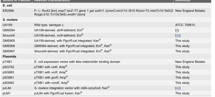
The bacterial strains and plasmids used in this study are described in Table 1.
Escherichia coli was grown overnight at 37
˚
C in Luria–Bertani broth (also known as LB broth, Difco) with gentle aeration, or on Luria-Bertani medium containing 1.5% w/v agar (LB agar). Kanamycin sulfate and ampicillin were added to these media when appropriate, each at a final concentration of 100 mg ml21. S. mutanswas grown as standing overnight cultures at 37
˚
C and 5% CO2 in Todd-HewittKanamycin (700 mg ml21) was added to THYE to maintain selection for the S. mutans GMS905, GMS906 and GMS907 fusion strains. S. mutans GMS584 and SmuvicK, isogenic mutants of the wild-type UA159 strain, were grown in THYE supplemented with erythromycin at a final concentration of 10 mg ml21, when needed.
Cloning and purification of VicK, GcrR & ComE
To generate a tagless version of VicK we used the Impact Kit (New England Biolabs) and generated a C-terminal VicK-Intein fusion protein by PCR
amplifying the vicK coding sequence fromS. mutans UA159 chromosomal DNA with oligonucleotides oSG548 and oSG550 (S1 Table). For the Intein tag to be removed with greater efficiency, the last amino acid of VicK was changed from serine to alanine using primer oSG550. The purified amplicon was digested with
NdeI and SapI and ligated into the expression plasmid pTXB1 (New England Biolabs) according to the supplier’s protocol. The ligation mixture was
transformed into E. coli ER2566 cells, selected for ampicillin resistance, and the construct was confirmed by DNA sequencing. To overexpress VicK-Intein, cells grown to mid-logarithmic phase (OD600 nm 0.3–0.5) were induced with 1 mM
IPTG for 3 hr at 37
˚
C with aeration before being harvested by centrifugation (4˚
C, 5,000 x g, 15 min) and frozen at 220˚
C. To purify tagless VicK, cell pellets were thawed and resuspended in Intein column buffer (20 mM Tris-HCl, pH 8.0, 0.5 M NaCl) before lysozyme was added to 1 mg ml21. After 30 min on ice, theTable 1.Bacterial strains and plasmids used in this study.
Strains or Plasmid Relevant characteristics Reference
E. coli
ER2566 F-l-fhuA2[lon]ompT lacZ::T7 gene 1gal sulA11D(mcrC-mrr)114::IS10 R(mcr-73::miniTn10-TetS)2 R(zgb-210::Tn10)(TetS)endA1[dcm]
New England Biolabs
S. mutans
UA159 Wild type, serotype c ATCC 700610
GMS584 UA159-derived,sloR-deficient; EmR [9]
SmuvicK UA159-derived,vicK-deficient; EmR [29]
GMS905 UA159-derived, with PgcrR:catintegrated; KanR This study
GMS906 GMS584-derived, with PgcrR:catintegrated; EmR, KanR This study GMS907 SmuvicK-derived, with PgcrR:catintegrated; EmR, KanR This study
Plasmids
pTXB1 E. coliexpression vector withMxeintein/chitin binding domain New England Biolabs
pSG752 pTXB1 withvicK; AmpR This study
pSG893 pTXB1 withvicR; AmpR This study
pSG901 pTXB1 withgcrR; AmpR This study
pSG892 pTXB1 withcomE; AmpR This study
pJL84 S. mutansintegration vector withmtlA-cat-phnA; KanR [44]
pLM1 pJL84 with PgcrR:catfusion; KanR This study
cells were lysed by sonication and centrifuged (10,000 x g, 30 min, 4
˚
C). The insoluble pellet was resuspended in column buffer containing 0.5% w/v sarkosyl and incubated at 4˚
C for 1 h prior to centrifugation (10,000 x g, 30 min, 4˚
C). The resulting supernatant was diluted in column buffer to a final sarkosylconcentration of 0.05% w/v, applied to a Chitin column (New England Biolabs) and washed with Intein column buffer. To cleave VicK from the Intein tag, the column was incubated in Intein column buffer with 50 mM DTT at 4
˚
C for 16– 40 h prior to elution with additional Intein column buffer. Fractions containing tagless VicK were visualized by SDS-PAGE and the protein quantified using a Bio-Rad Protein Assay and bovine serum albumin as a standard. The purified protein was stored in 25% v/v glycerol at 280˚
C.The coding sequence ofS. mutans vicR (smu.1517) was amplified as described above forvicKwith oligonucleotides oSG726 and oSG727 (S1 Table). To improve cleavage of the Intein tag by DTT, alanine was substituted for thevicRstop codon in the oSG727 primer. The vicR coding sequence was then cloned into pTXB1, confirmed by sequencing as described above, and transformed into ER2566 cells for protein expression. Freshly transformed cells were used to inoculate an overnight culture for VicR overexpression, which was induced with 1 mM IPTG as described above. The purification was carried out as described for tagless VicK except that the pH of the binding buffer was adjusted to pH 9.0 and the insoluble pellet was incubated with 0.65% sarkosyl for 1 hr to release the fusion protein into the soluble fraction. Pure tagless VicR was visualized by SDS-PAGE, quantified as described above and stored in 25% glycerol at 280
˚
C.The coding sequence ofS. mutans gcrR(smu.1924) was amplified by PCR with primers oSG741 and oSG742 (S1 Table). To facilitate cleavage of the Intein tag by DTT, glutamine was substituted for the gcrRstop codon in the oSG741 primer. ThegcrRcoding sequence was digested with FauI and PvuII and then cloned into the NdeI and SapI sites on pTXB1 before being confirmed by sequencing as described above. Tagless GcrR was purified as described for tagless VicK with one exception; the pH of the column buffer was adjusted to 8.5. Pure tagless GcrR was visualized by SDS-PAGE, quantified as described above and stored in 25% glycerol at 280
˚
C.The coding sequence ofS. mutans comE(smu.1917) was amplified by PCR with primers oSG691 and oSG692, cloned into pTXB1 and confirmed by sequencing as described above. Tagless ComE was purified as described for VicR except 0.7% sarkosyl was used to solubilize the fusion protein. Pure tagless ComE was visualized by SDS-PAGE, quantified as described above, and stored in 25% glycerol at 280
˚
C.Phosphorylation assays
Reactions were stopped by adding 2X SDS sample buffer (120 mM Tris-HCl, pH 7.4, 20% v/v glycerol, 4% w/v SDS, 10% v/v ß-mercaptoethanol and 0.1% w/v bromophenol blue) and resolved on a 4–20% Tris-Glycine gel (Invitrogen) run at approximately 18.75 V cm ml21for 1.5 h. The gels were dried and scanned with a Pharos FX imaging system (Bio-Rad) and quantified using ImageQuant 5.0 (Molecular Dynamics).
For transphosphorylation reactions, 1mM VicK was incubated for 15 minutes at room temperature with excess cold ATP (10 mM), to ensure ATP was not limiting, and 0.2 mM [c-32P] ATP, 100 mM Tris-HCl pH 7.5, 50 mM NH4Cl, and
1 mM MgCl2. Then GcrR, VicR, or ComE RRs were added to 1mM to the the
VicK autophosphorylation reaction for 1 hr at room temperature. The
phosphoryation reactions were diluted 1:1 in 2x SDS loading buffer (125 mM Tris-HCl pH 7.4, 0.005% bromophenol blue, 4% SDS, 20% glycerol). A 4–20% acrylamide Tris-glycine SDS PAGE Novex gel (Invitrogen), the X-Cell SureLock Mini-Cell protein electrophoresis chamber and the Tris-glyine running buffer were pre-chilled at 4
˚
C for ,3 hrs. The reactions were separated by SDS-PAGE for ,3 hrs at 150 V at 4˚
C. The resulting gels were dried, exposed to a phosphor screen [26] and scanned using a Typhoon phosphorimager (GE Healthcare).For transphosphorylation reactions in the presence of MnCl2, 1 mM VicK was
incubated in the presence of excess cold ATP (10 mM), 0.2 [c-32P] ATP mM, 100 mM Tris-HCl pH 7.5, and 1 mM MnCl2 for 15 min at room temperature.
The response regulator (1 mM) was added and the reactions were incubated for 1 hr at room temperature before being diluted 1:1 in 2x SDS loading buffer. As described above, the buffers and gels were pre-chilled at 4
˚
C. The transpho-sphorylations reactions were separated on 4–20% acrylamide Tris-glycine SDS PAGE Novex gels for 2.5 hrs at 128 V and then for 1 hr at 150 V at 4˚
C. The resulting gels were dried, exposed to a phosphor screen [26] and scanned using a Typhoon phosphorimager (GE Healthcare). The Phos-Tag mobility shift assay was performed as described previously [41], with the exception that a 12% gel was prepared and visualized by silver staining [42].DNaseI footprinting
DNaseI footprinting analysis was performed as previously described [43]. For protein concentrations used, refer to the figure legend.
Construction of the
S. mutans
P
gcrR:cat
fusion strains GMS905,
GMS906 and GMS907
The gcrRpromoter region was amplified by PCR from the S. mutans UA159 chromosome using primers gcrR_356.FV.F and gcrR_356.FV.R (S1 Table).SacI and BamHI digested amplicons were then cloned into the integration vector pJL84, which contains theS. aureus cat andS. mutans phnAandmtlAgenes [44]. Kanamycin-resistant transformants were selected and the presence of the gcrR
pLM1, was transformed into S. mutans UA159, GMS584 and SmuvicK in the presence of competence stimulating peptide (CSP) according to established protocols, to generate strains GMS905, GMS906 and GMS907, respectively [9,29,45]. Integration of the PgcrR:catfusion via allelic exchange was mediated by the S. mutans phnAand mtlAgenes that are resident on the pJL84 plasmid [44]. PCR amplification and nucleotide sequencing were used to confirm the double allelic crossover event at the desired locus on the S. mutans chromosome.
CAT assay
Overnight cultures ofS. mutansGMS905, GMS906 and GMS907 were diluted 1:10 in pre-warmed THYE and grown to an OD600 nm0.6–0.7. Cells were harvested by
centrifugation, resuspended in 1 ml of 10 mM Tris-HCl at pH 7.8 and lysed by mechanical disruption in a BIO101 Savant FastPrep (Thermo Savant) for 1.5 min with intermittent cooling on ice. Unlysed cells and debris were removed by centrifugation (9,300 x g, 4 min, 4
˚
C) and the resulting cell lysates were stored at220
˚
C for subsequent protein determination with a BCA protein assay kit (Pierce) and chloramphenicol acetyltransferase [35] assays. CAT assays were performed according to the method described by Shaw [46]. Briefly, a 360 ml reaction mixture consisting of 100 mM Tris-HCl pH 7.8, 0.4 mg ml21 5,59 -dithiobis(2-nitrobenzoic acid) (DTNB, (e412 nm513.6 mM21cm21), and 0.1 mMacetyl-CoA was mixed with 40 ml of whole cell lysate. To monitor CAT specific activity, 0.1 mM chloramphenicol (CM) was added and absorbance readings at OD412were obtained every 10 sec over a five min interval in a Synergy HT
Microtiter Plate Reader (BioTek). To assess background activity, absorbance readings for wells that contained only the reaction mixture and the whole cell lysate were obtained in parallel. The rate of change due to addition of CM was determined by subtracting the background activity from the rate of change after the addition of CM. This value was divided by 0.0136 (extinction coefficient of DTNB) to yield CAT activity and then subsequently divided by the total protein concentration to express the CAT specific activity result in nM min21 mg21. All CAT assays were performed as three independent experiments each in triplicate.
Quantitative real time PCR (qRT-PCR)
Overnight cultures of S. mutans UA159 and SmuvicK were grown to mid-exponential phase (OD600 nm,0.4) in Tryptone (Bioshop) Yeast Extract Glucose
medium (TYEG; 10% w/v tryptone, 5% w/v yeast extract, 17.2 mM K2HPO4,
0.5% w/v glucose, pH 7.5). Following incubation, the cells were pelleted,
resuspended in TYEG at pH 7.5 or pH 5.5 and incubated for 1 h at 37
˚
C and 5% CO2. The cells were harvested by centrifugation, snap frozen in liquid nitrogeninvariable under the experimental test conditions (data not shown). Relative expression of the target genes was calculated using results from 3 independent experiments, according to the method of Pfaffl et al [47].
To determine vicRexpression in a sloR-deficient (GMS584) mutant vs. wild-type UA159, overnight cultures of both strains were grown in a semi-defined medium (SDM) [48] to OD600 nm,0.6 before the cells were pelleted and snap
frozen as described above. RNA was isolated from the pellets and reverse
transcribed as previously described [4]. The expression ofvicRwas normalized to that ofgyrA, which did not change under the experimental conditions tested (data not shown). These qRT-PCR experiments were performed in triplicate in each of three independent experiments.
Results
Autophosphorylation of VicK is facilitated by manganese and
inhibited by iron
To demonstrate that VicK can undergo autophosphorylation, VicK was purified to.90% homogeneity as determined by Coomassie blue staining of SDS-PAGE gels (data not shown) and subsequently used in phosphorylation assays. Initial phosphorylation assay conditions were based on results described previously by Clausen et al [40]. VicK was readily phosphorylated when incubated at room temperature for 15 min in the presence of NH4Cl, MgCl2and [c-32P] ATP (data
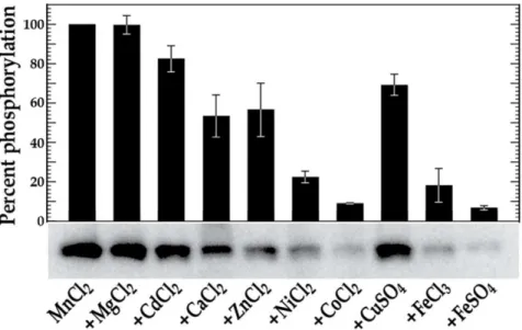
not shown). We next tested a variety of different divalent metal cations (1 mM MgCl2, CdCl2, CaCl2, MnCl2, ZnCl2, NiCl2, CoCl2, CuSO4, FeCl3, or FeSO4) in
lieu of NH4Cl and MgCl2 to assess their potential impact on VicK
phosphoryla-tion. As shown in Fig. 1, VicK was able to autophosphorylate at relatively low levels in the presence of MgCl2, CaCl2, CoCl2, or FeCl3, and at significantly
increased levels in the presence of MnCl2(an over 3-fold increase compared to the
next highest phosphorylation condition that was observed with MgCl2).
To determine whether any of the metal cations tested above might inhibit VicK autophosphorylation in the presence of Mn2+, we repeated the
autopho-sphorylation assays with reaction mixtures containing VicK, MnCl2and
equimolar amounts of MgCl2, CdCl2, CaCl2, ZnCl2, NiCl2, CoCl2, CuSO4, FeCl3,
or FeSO4. CaCl2, NiCl2, and ZnCl2inhibited VicK autophosphorylation by 46%,
77% and 43%, respectively, relative to the 100% phosphorylation of VicK that we observed in the presence of Mn2+alone (Fig. 2). Interestingly, CoCl
2, FeCl3 and
FeSO4 had an even more pronounced effect on VicK phosphorylation in the
presence of Mn2+, diminishing activity to 9%, 18% and 7%, respectively (Fig. 2). Importantly, the addition of either FeCl3or FeSO4(freshly prepared before use to
limit oxidation) did not alter the overall pH of the phosphorylation reaction, consistent with a direct inhibitory effect for iron on VicK phosphorylation. Collectively, these autophosphorylation experiments demonstrate that while standard conditions (NH4Cl/MgCl2) and Mn2+ can readily stimulate
VicK differentially transphosphorylates VicR and GcrR in the
presence of manganese
S. mutans VicK’s ability to transphosphorylate VicR has recently been
demonstrated [41,49,50]. To further explore this in the presence of different divalent metal cations, we phosphorylated VicK in the presence of excess ATP, and buffer containing MgCl2 and NH4Cl and then supplemented the reaction
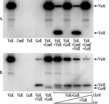
mixture with various combinations of the VicR, ComE or GcrR responder proteins. ComE and GcrR were selected since both have overlapping pathways with the VicRK TCS based on transcriptome analysis and regulation of common phenotypes that include genetic competence, acid tolerance and biofilm formation [29,36,39,51–53]. Despite this knowledge cross-talk with VicK has not been demonstrated. Each RR was allowed to incubate individually with VicK. As seen in
Fig. 3A (lanes 5–7), VicR was the only RR that was phosphorylated under these conditions. As an additional control, each RR was added to phosphorylation reactions in the absence of VicK and no phosphorylation was observed (Fig. 3A, lanes 2–4). ComE was also added to the transphosphorylation reaction containing either VicR or GcrR. Even with the addition of ComE, only VicR was efficiently phosphorylated by VicK (Fig. 3A, lane 8). Likewise no detectable transpho-sphorylation was noted when GcrR and ComE were combined (Fig. 3A, lane 9).
Fig. 1.in vitrophosphorylation of VicK in the presence of various metal cations.VicK (1mM) was incubated in 100 mM Tris-HCl, pH 7.5 containing 1 mM of the designated cations and 0.10mM [c-32P] ATP at room temperature for 15 minutes. The relative autophosphorylation of VicK was quantified using Image Quant 5.0 software (Molecular Dynamics) and is represented by the histogram above the scanned gel. The gels shown are representative of at least three independent experiments. Error bars represent¡std. errors of the average phosphorylation values derived from at least 3 independent experiments.
Previous studies have shown that the GcrR regulon is manganese-responsive [4], and here we demonstrate that VicK autophosphorylation is stimulated by manganese. To date, no cross-talk has ever been observed between any of the S. mutans HKs tested (VicK, CiaH and LiaS) and their noncognate RRs (CiaR and LiaR) [49]; albeit these experiments were performed in the absence of divalent cations. We explored whether VicR or the GcrR orphan RR could be
phosphorylated by VicK-P in the presence of manganese. The autophosphoryla-tion and transphosphorylaautophosphoryla-tion reacautophosphoryla-tions were allowed to proceed in the presence of excess ATP, and buffer containing MnCl2. As seen in Fig. 3Blane 4, VicR was
phosphorylated under these conditions, although at slightly lower levels than those observed previously in transphosphorylation reactions lacking manganese. In contrast to Fig. 3Aand in the presence of manganese, VicK was able to phosphorylate the non-cognateS. mutansresponse regulator, GcrR (Fig. 3B, lane 5). Importantly, neither VicR nor GcrR were capable of autophosphorylation under these conditions (Fig. 3B, lanes 2–3).
To determine whether VicK has a preference for the phosphorylation of VicR and/or GcrR in the presence of manganese, the reaction containing 1 mM GcrR was repeated in the presence of 0.01–1 mM VicR (Fig. 3B, lanes 6–9). For these experiments VicR was added to the reaction mixture after GcrR was added. The addition of even 0.02 mM VicR outcompeted GcrR as a substrate for VicK transphosphorylation (Fig. 3B, lane 7) although measurable GcrR
phosphoryla-Fig. 2.in vitrophosphorylation of VicK in the combined presence of Mn2+and other various metal
cations.VicK (1mM) was incubated in 100 mM Tris-HCl, pH 7.5 containing 1 mM MnCl2plus 1 mM of the
designated cation and 0.10mM [c-32P] ATP at room temperature for 15 minutes. The relative
autophosphorylation of VicK was quantified using Image Quant 5.0 software (Molecular Dynamics). The sample containing only VicK and MnCl2was set at 100% for comparison and the results are shown in the
histogram above the scanned gel. The gels shown are representative of at least three independent experiments. Error bars represent¡std. errors of the average phosphorylation values derived from at least 3 independent experiments.
tion persists under these conditions through VicR concentrations as high as 0.04 mM (Fig. 3B, lane 8). To demonstrate that this competition was specific for VicR and GcrR, we transphosphorylated VicR and GcrR in the presence of manganese and 1 mM ComE. As shown inS1 Fig. as much as 1 mM ComE had no effect on the phosphorylation state of either VicR or GcrR. To confirm that the bands shown in Fig. 3b are the proteins indicated, we performed transpho-sphorylation assays in the presence of MnCl2followed by Phos-Tag mobility shift
analysis [41] and silver staining with purified VicK (51.7 kDa), VicR (26.9 kDa), and GcrR (26.7 kDa) under the same conditions described above (S2 Fig.). The protein migration patterns shown in S2 Fig. correspond to those seen inFig. 3b, demonstrating that these protein bands are the specified proteins.
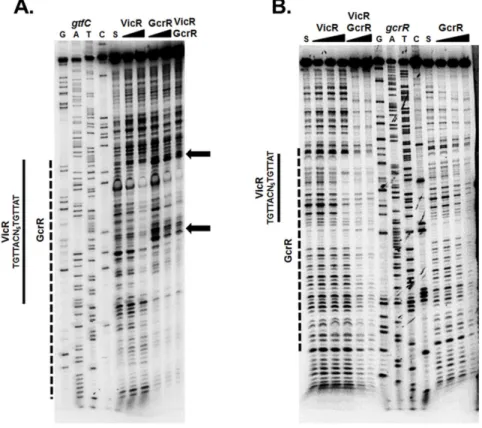
GcrR and VicR binding sites overlap at co-regulated genes
VicRK and GcrR regulate a number of overlapping genes including gtfC
[2,11,12,29,52,54]. We recently further characterized theconsensus sequence of VicR [43] and sought to gain a better understanding of the direct regulation of VicR and GcrR. Specifically, we wanted to examine the binding site boundaries of VicR and GcrR particularly since a GcrR binding consensus has proven elusive. To
Fig. 3.in vitrotransphosphorylation of VicR and GcrR by VicK.A) Phosphorylation of VicR and GcrR by VicK in the presence of MgCl2. For each reaction 1mM of each of the following proteins were included in the
reaction: Lane 1: VicK; Lane 2: ComE; Lane 3: VicR; Lane 4: GcrR; Lane 5: VicK and ComE; Lane 6: VicK and VicR; Lane 7: VicK and GcrR; Lane 8: VicK, ComE and VicR; Lane 9: VicK, ComE and GcrR. B)
Phosphorylation of VicR and GcrR by VicK in the presence of MnCl2. For each reaction 1mM of each of the
following proteins were included in the reaction unless otherwise indicated: Lane 1: VicK; Lane 2: VicR; Lane 3: GcrR; Lane 4: VicK and VicR; Lane 5: VicK and GcrR; Lane 6: VicK, GcrR and 0.01mM VicR; Lane 7: VicK, GcrR and 0.02mM VicR; Lane 8: VicK, GcrR and 0.04mM VicR; Lane 9: VicK, GcrR and 1mM VicR. The gels shown are a representative set of replicate gels run for each experiment.
explore this, we used DNaseI footprinting with VicR and GcrR individually or both RRs present at an equimolar amount, with thegtfCpromoter region as DNA substrate. As shown in Fig. 4A, VicR and GcrR independently displayed
protection that overlapped the VicR consensus sequence (indicated by the solid and dashed lines respectively). Unlike VicR, the GcrR footprint exhibited
enhanced cleavage (indicated by the arrows) at two specific locations. These sites correspond to the bases 59-TGTG and 59-GTGT that flank the VicR consensus sequence. Such ‘‘hypersensitive sites’’ reflect an increased accessibility of specific phosphodiester linkages to DNase I cleavage, often indicative of DNA bending. The same enhanced cleavage was also observed when VicR and GcrR were both present in the assay at an equimolar amount, whereas VicR alone displayed protection at this site (Fig. 4A, solid line). This indicates that not only do VicR and GcrR have overlapping binding sites but that GcrR under equimolar conditions exhibits greater binding affinity for gtfC compared to VicR.
To determine if GcrR demonstrated DNA binding dominance at another gene locus (in addition togtfC), we also investigated the binding affinities of VicR and GcrR to the gcrRpromoter region. To examine this we used DNaseI footprinting by amplifying a 160 bp fragment upstream of thegcrRstart codon [43]. As shown previously, VicR protected a region that overlapped its consensus sequence [43] clearly seen with the highest concentration of VicR (0.5 mM) (Fig. 4B, solid line). In contrast, GcrR displayed protection that overlapped that of VicR, but the binding affinity was much stronger with protection observed at 0.25 mM (dashed line). When VicR and GcrR were present simultaneously at equimolar amounts, protected regions again overlapped but resembled that of GcrR compared to VicR, suggesting that GcrR demonstrates stronger binding affinity for thegcrRsubstrate (Fig. 4B). These results provide evidence that further integrate the functions of VicR and GcrR at select promoter regions.
SloR positively regulates
vicRKX
expression
Transcription of
gcrR
is subject to VicK and SloR control
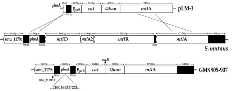
To further elucidate the impact of VicK and SloR on gcrR expression, PgcrR:cat
fusions were constructed inS. mutanswild-type (GMS905) and mutant (GMS906 and GMS907 which harbor sloR and vicK insertion-deletion mutations,
respectively) backgrounds (Fig. 5). The resulting fusion strains were confirmed by nucleotide sequencing with Smu_1179c- and cat-specific primers (S1 Table). For cat assays, whole cell lysates were prepared from mid-exponential phase cultures of each strain so that CAT-specific activity could be monitored according to the spectrophotometric assay of Shaw [46]. Importantly, we observed CAT-specific activity for the wild-type GMS905 strain (5.84¡1.24 nM min21 mg21), that was 3-fold greater than that of the GMS906 SloR-deficient derivative (1.78¡0.74 nM min21mg21), and 2.5-fold greater than that of the VicK-deficient strain, GMS907 (2.30¡0.69 nM min21 mg21). These findings support a role for both SloR and VicK in modulation of the S. mutans gcrR gene in vivo.
Fig. 4. DNaseI footprinting of thegtfCandgcrRpromoter regions.(A) VicR or GcrR at increasing concentrations (0.25 and 0.5mM) or a combination of VicR and GcrR at an equimolar concentration (0.5mM) were incubated with labeledgtfCDNA substrate. The S above the fifth lane indicates the DNA substrate incubated in the absence of VicR/GcrR. The arrows designate the areas of enhanced cleavage by DNaseI. (B) LabeledgcrRDNA substrate was incubated with increasing concentrations of VicR or GcrR (0.125, 0.25, and 0.5mM) or a mixture of VicR and GcrR at equimolar concentrations (0.25 and 0.5mM). The S above the first and eleventh lanes indicates the DNA substrate incubated in the absence of VicR/GcrR. The solid line represents the region of protected nucleotides by VicR and the dashed line represents the region of protection by GcrR. The VicR consensus sequence is shown to the left of the solid lines.
VicK positively regulates
gcrR
and ATR related gene transcription
under low pH
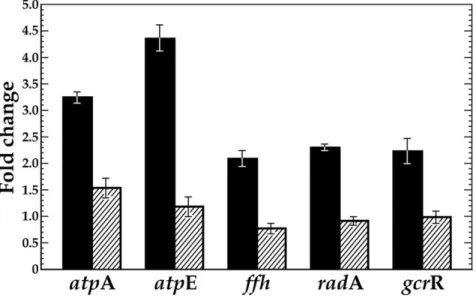
VicK has previously been implicated in facilitating S. mutans ability to respond and adapt to low pH [39]. To further examine the role of VicK in regulating theS. mutansATR, we compared the expression of known ATR genes,atpE/A,ffh, radA
andgcrR, in the UA159 wild-type strain and avicK-knockout derivative, SmuvicK [29]. Exposure of wild-type UA159 cells to a sub-lethal acid challenge (pH 5.5) resulted in over a 2-fold increase in expression at these loci compared to cells grown at pH 7.5 (data not shown). The greatest induction of gene expression in the UA159 wild-type strain was observed for atpAand atpE (.3-fold), which encode the alpha and c subunits of the FoF1 membrane-bound
proton-translocating ATPases, respectively. Not surprisingly, the involvement of these ATPase subunits in the S. mutans ATR is well established in the literature [4,6,7,55,56].
In contrast, loss of VicK failed to induce any of these genes at low pH, suggesting a requirement for VicK in modulating their transcription under conditions of acid stress (Fig. 6). In addition, all of the genes were significantly down-regulated in the VicK mutant relative to that in wild type (p,0.001, with the exception of gcrRat pH 7.5), further supporting a positive regulatory role for VicK in their transcription (S2 Table).
Discussion
In this report, we demonstrate thatin vitroautophosphorylation of theS. mutans
VicK HK is enhanced by manganese and inhibited by ferrous iron, suggesting two
Fig. 5. Construction of theS. mutansfusion strains GMS905, GMS906, and GMS907.The integration of the PgcrR:catfusion that is resident on plasmid pLM1 occurred via a double cross-over event into the chromosome ofS. mutansUA159, GMS584 and SmuvicK at thephnAandmtlAloci. Sequencing across theS. mutanschromosome-pLM1 junction confirmed appropriate insertion of the PgcrR:catfusion.
potential roles for VicRK in sensing manganese availability and conditions of redox. We further demonstrate that VicK, in addition to phosphorylating its cognate RR, VicR, facilitatesin vitrocross-talk by phosphorylating the orphan RR GcrR, in the presence of manganese albeit only when VicR is present at
comparatively low relative concentration. Our findings are similar to those of Guckes et al who report similar cross talk in the form of one HK being able to transphosphorylate specific non-cognate RRs, but only under specific conditions [57]. While VicK-facilitated phosphorylation of VicR and GcrR appears to be specific in vitro, it remains to be seen to what extent this phenomenon occurs in vivo. Interestingly, Stippet alrecently demonstrated that VicRK and GcrR work in concert to form structurally stable biofilms by coordinating surface biogenesis and cell division in S. mutans [54]. However their model did not show any direct interaction between VicK and the orphan RR GcrR. Liang et alidentified a CovS mutant inS. pyogenes that retained CovR-mediated virulence gene regulation, via an unidentified alternate pathway for CovR phosphorylation [58].
Here we provide insight into the communication pathways of theS. mutans
SloR, VicRK and GcrR regulators. Cross-regulation of GcrR by the VicK sensor kinase, may explain the overlap between the VicRK-regulon and that of GcrR, in modulating genes whose products contribute to the oxidative stress and acid tolerance responses of S. mutans. Work conducted previously by Dunninget al
showed that SloR interacts directly with thegcrRpromoter region to facilitate its expression [4]. Here we demonstrate that SloR also positively regulates expression of thevicRKXoperon, but whether its impact onvicRKXtranscription is direct or
Fig. 6. VicK has a significant impact on transcription of known ATR-related genes inS. mutans. qRT-PCR was performed to reveal fold-change in gene expression at pH 5.5 versus 7.5 with cDNAs derived from
S. mutansUA159 (solid black bars) and avicKinsertion-deletion mutant (SmuvicK) (striped bars). Error bars represent¡std. errors of the average expression values derived from at least 3 independent experiments. Student t-tests confirm that all genes are significantly down-regulated in the VicK mutant relative to the UA159 wild-type progenitor strain (p,0.001).
indirect remains to be determined. Moreover, CAT-specific activity observed in the S. mutans catfusion strains support gcrRexpression that is subject to both SloR and VicRK control.
Results of phosphorylation assays highlight a role for manganese in integrating the VicRK, GcrR and SloR regulatory pathways. Manganese is an essential micronutrient that affects S. mutans genes whose products are conducive to its virulence, which include those that mediate adherence and biofilm formation [59–62]. In fact, manganese functions as a cofactor for a S. mutans superoxide dismutase (SOD), which converts damaging reactive oxygen species, into less toxic substances [63,64]. Reports in the literature describe a correlation between intracellular manganese and iron concentrations with the sensitivity of bacteria to oxidative stress [65,66]. It has been suggested that bacteria may utilize manganese instead of ferrous iron to avoid redox conditions that are associated with Fenton chemistry [67]. This could be especially important in a biofilm environment where extracellular manganese can reach mM concentrations in the oral cavity [68].
A novel aspect of this study is our demonstration of VicK’s ability to transphosphorylate GcrR under low VicR concentrations in the presence of manganese (Fig. 3B). Further, the presence of VicR causes the turnover of phosphorylated VicK, something that is not seen with other tested RRs. This effect may be important biologically as VicR-VicK interactions may activate VicK phosphatase activity reducing the net steady state levels of phosphorylated VicK; this effect seems enhanced with increasing concentrations of manganese. In contrast, we also show that iron inhibits VicK phosphorylation especially in the presence of manganese (Figs. 1 and 2). These findings are consistent with GcrR regulation by the VicRK system as well as by the metal ion-dependent SloR metalloregulator that binds manganese preferentially over iron. It is worth pointing out that we have used tagless but not necessarily native proteins for these phosphotransfer reactions. As yet to be determined, post translational modifica-tions to each protein may be critical in each of the aforementioned reacmodifica-tions.
We previously described modulation of theS. mutansATR by SloR with GcrR as an essential intermediary [4]. We also confirmedsloABC expression that is responsive to SloR and manganese concentrations that are physiological
conditions likely representative of feast (.10 mM) or famine (,0.1mM) [4]. Our
expression analysis to determine whethersloABC transcription was dependent on VicK revealed that loss of VicK did not significantly affect expression of sloABC
(data not shown); suggesting that VicRK does not modulate metal ion uptake via the sloABC transport system. Given the in vitro effect of manganese on steady-state levels of GcrR and VicR phosphorylation, it is possible that VicK, like SloR, may use GcrR as an intermediary to modulate acid tolerance in S. mutans by responding to manganese.
known as CovR) GcrR is co-transcribed along with a cognate HK, CovS. Interestingly, extracellular Mg2+stimulates covRS expression in group A
streptococci (GAS) and increasing concentrations of exogenous Mg2+ have been shown to increasegcrRexpression [70,71]. Moreover, CovS can dephosphorylate (and thus inactivate) CovR in GAS under stress-inducing conditions including high temperature, low pH, high salt and iron starvation [17,72]. It would be interesting to investigate a potential physical interaction between VicR and GcrR, and characterize the VicR- GcrR- and SloR-binding sites to validate potential cross-regulation between their respective regulons in S. mutans. In fact, reports in the literature supported the binding of VicR and GcrR to overlapping sequences upstream of S. mutans gtfB/C, encoding sucrose-dependent glucosyltransferases that are critical determinants of colonization and subsequent virulence [11,29]. Indeed, we confirmed this hypothesis using DNaseI footprinting analysis. We demonstrated that both GcrR and VicR bind to the same regions upstream ofgtfC
andgcrR. When incubated together at equimolar amounts, GcrR displayed higher DNA binding affinity than VicR suggesting that in vitro, GcrR predominates under these conditions. Although these studies were performed using unpho-sphorylated forms of the RRs in vitro, the phosphorylation states of these RRs likely play an important role in their regulation in vivo. Additional studies and genomic analyses need to be performed in vivo to support our results.
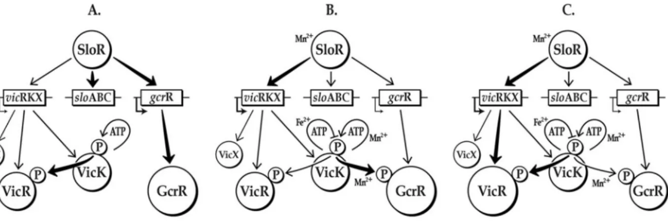
Based on our results, we propose that it is highly likely that manganese is the common denominator for cross-communication between the VicK, GcrR, and SloR regulatory networks (Fig. 7). Both VicK and SloR activation are manganese-dependent, whereas VicK has been shown to respond to pH, oxidative and cell wall stresses [32,33,39,73]. Thus, we hypothesize that VicK autophosphorylation and the subsequent transphosphorylation of VicR and GcrR (the latter in the presence of manganese) leads to modulation of numerous S. mutans virulence genes that facilitates bacterial survival in the presence of reactive oxygen species.
To ensure that the cross-talk between VicK and GcrR is tightly regulated, we propose that S. mutans regulates expression of VicR so that it may compete for phosphorylation by VicK. In the presence of manganese SloR represses gcrR
expression and enhances VicR expression while manganese also makes GcrR a substrate for VicK transphosphorylation. In addition, when VicK, SloR or manganese are limiting there is increased expression of GcrR. It is possible that in the absence of manganese there is sufficiently more GcrR than VicR inS. mutans
during this brief period when GcrR is phosphorylated that it altersS. mutansgene expression to accommodate the transition to manganese rich conditions.
Further studies are needed to determine if the phosphorylated forms of these RRs are the active or inactive forms. These additional studies are necessary to dissect the complex interactions that occur between VicK, VicR, GcrR and SloR and the larger role these regulators likely have in governing the cellular physiology of S. mutans.
Supporting Information
S1 Fig. ComE has no effect on the phosphorylation state of VicR or GcrR. Phosphorylation of VicR and GcrR by VicK in the presence of MnCl2and ComE.
For each reaction 1 mM of each of the following proteins were included in the reaction: Lane 1: VicK; Lane 2: ComE; Lane 3: VicR; Lane 4: GcrR; Lane 5: VicK and ComE; Lane 6: VicK, VicR; Lane 7: VicK and GcrR; Lane 8: VicK, ComE and VicR; Lane 9: VicK, ComE and GcrR. The gel shown is a representative of replicate gels run for each experiment.
doi:10.1371/journal.pone.0115975.s001 (DOCX)
S2 Fig. Phos-Tag mobility shift assay of in vitrotransphosphorylation of VicR
and GcrR by VicK. Transphosphorylation of VicR and GcrR by VicK in the presence of MnCl2was performed as described in Materials and Methods followed
by Phos-Tag SDS-PAGE analysis and silver staining. The protein amounts included in the reaction are indicated above each lane.
doi:10.1371/journal.pone.0115975.s002 (DOCX)
S1 Table. Primers used for PCR in this study. doi:10.1371/journal.pone.0115975.s003 (DOC)
Fig. 7.In vitromodel of manganese-independent (A) and –dependent cross-regulation involvingS. mutansSloR, VicRK and GcrR.A) In the absence of Mn2+(approximating conditions of free-floating planktonic cells)S. mutans gcrRexpression is de-repressed (VicR expression is not induced in the absence of Mn2+). Even though GcrR is the more abundant substrate, VicR is the favored species for VicK phosphorylation under these conditions. B) During this so-called ‘‘transition stage’’ (approximating conditions of an early biofilm) a ‘‘spike’’ in Mn2+renders GcrR (still the predominant species) the favored substrate for phosphorylation by VicK, but only transiently. C) As the biofilm matures and Mn2+concentrations increase, SloR is activated to repress
gcrRexpression, thereby reducing the availability of GcrR as a substrate. The activated SloR-Mn2+complex encouragesvicRexpression, and hence VicR becomes the favored substrate for VicK phosphorylation once again.
S2 Table. Differences in ATR gene expression in Smuvick compared to wildtype.
doi:10.1371/journal.pone.0115975.s004 (DOCX)
Acknowledgments
We thank Gary Nelson for figure preparation and Kirsten Krastel for assistance with qRT-PCR.
Author Contributions
Conceived and designed the experiments: JSD EAA DBS DGC GAS SDG. Performed the experiments: JSD EAA DBS WKH LWM JGS. Analyzed the data: JSD LMW EAA DBS WKH LWM JGS DGC GAS SDG. Contributed reagents/ materials/analysis tools: DBS DGC GAS SDG. Wrote the paper: JSD LMW EAA DBS WKH LWM JGS DGC GAS SDG.
References
1. Banas J(2004) Virulence properties ofStreptococcus mutans. Front Biosci 1: 1267–1277.
2. Dmitriev A, Mohapatra SS, Chong P, Neely M, Biswas S, et al. (2011) CovR-controlled global regulation of gene expression in Streptococcus mutans. PLoS One 6: e20127.
3. Hanna MN, Ferguson RJ, Li YH, Cvitkovitch DG(2001)uvrAis an acid-inducible gene involved in the adaptive response to low pH inStreptococcus mutans. J Bacteriol 183: 5964–5973.
4. Dunning DW, McCall LW, Powell WF Jr, Arscott WT, McConocha EM, et al.(2008) SloR modulation of the Streptococcus mutans acid tolerance response involves the GcrR response regulator as an essential intermediary. Microbiology 154: 1132–1143.
5. Fozo EM, Quivey RG Jr(2004) ThefabMgene product ofStreptococcus mutansis responsible for the synthesis of monounsaturated fatty acids and is necessary for survival at low pH. J Bacteriol 186: 4152– 4158.
6. Hamilton IR, Svensater G(1998) Acid-regulated proteins induced byStreptococcus mutansand other oral bacteria during acid shock. Oral Microbiol Immunol 13: 292–300.
7. Kobayashi H, Suzuki T, Unemoto T(1986) Streptococcal cytoplasmic pH is regulated by changes in amount and activity of a proton-translocating ATPase. J Biol Chem 261: 627–630.
8. Kremer BA, van der Kraan M, Crowley PJ, Hamilton IR, Brady LJ, et al.(2001) Characterization of thesatoperon inStreptococcus mutans: evidence for a role of Ffh in acid tolerance. J Bact 183: 2543– 2552.
9. Rolerson E, Swick A, Newlon L, Palmer C, Pan Y, et al. (2006) The SloR/Dlg metalloregulator modulatesStreptococcus mutansvirulence gene expression. J Bacteriol 188: 5033–5044.
10. Lu L, Singh JS, Galperin MY, Drake D, Taylor KG, et al.(1992) Chelating agents inhibit activity and prevent expression of streptococcal glucan-binding lectins. Infect Immun 60: 3807–3813.
11. Biswas S, Biswas I (2006) Regulation of the glucosyltransferase (gtfBC) operon by CovR in Streptococcus mutans. J Bacteriol 188: 988–998.
12. Idone V, Brendtro S, Gillespie R, Kocaj S, Peterson E, et al.(2003) Effect of an orphan response regulator on Streptococcus mutans sucrose-dependent adherence and cariogenesis. Infect Immun 71: 4351–4360.
14. Ajdic D, McShan WM, McLaughlin RE, Savic G, Chang J, et al. (2002) Genome sequence of
Streptococcus mutansUA159, a cariogenic dental pathogen. PNAS 99: 14434–14439.
15. Hoch JA, Silhavy TJ(1995) Two-component signal transduction. Washington, D.C.: ASM Press. 488 p.
16. Cho KH, Caparon MG(2005) Patterns of virulence gene expression differ between biofilm and tissue communities ofStreptococcus pyogenes. Mol Microbiol 57: 1545–1556.
17. Dalton TL, Scott JR(2004) CovS inactivates CovR and is required for growth under conditions of general stress in Streptococcus pyogenes. J Bacteriol 186: 3928–3937.
18. Graham MR, Virtaneva K, Porcella SF, Barry WT, Gowen BB, et al.(2005) Group A Streptococcus transcriptome dynamics during growth in human blood reveals bacterial adaptive and survival strategies. Am J Pathol 166: 455–465.
19. Sumby P, Whitney AR, Graviss EA, DeLeo FR, Musser JM(2006) Genome-wide analysis of group a streptococci reveals a mutation that modulates global phenotype and disease specificity. PLoS Pathog 2: e5.
20. Virtaneva K, Porcella SF, Graham MR, Ireland RM, Johnson CA, et al.(2005) Longitudinal analysis of the group A Streptococcus transcriptome in experimental pharyngitis in cynomolgus macaques. Proc Natl Acad Sci U S A 102: 9014–9019.
21. Dalton TL, Collins JT, Barnett TC, Scott JR (2006) RscA, a member of the MDR1 family of transporters, is repressed by CovR and required for growth ofStreptococcus pyogenesunder heat stress. J Bacteriol 188: 77–85.
22. Graham MR, Smoot LM, Migliaccio CA, Virtaneva K, Sturdevant DE, et al.(2002) Virulence control in group A Streptococcus by a two-component gene regulatory system: global expression profiling andin vivoinfection modeling. PNAS 99: 13855–13860.
23. Mattos-Graner RO, Napimoga MH, Fukushima K, Duncan MJ, Smith DJ (2004) Comparative analysis of Gtf isozyme production and diversity in isolates ofStreptococcus mutanswith different biofilm growth phenotypes. J Clin Microbiol 42: 4586–4592.
24. Goodman SD, Gao Q (2000) Characterization of the gtfB andgtfCpromoters from Streptococcus mutansGS-5. Plasmid 43: 85–98.
25. Monchois V, Willemot RM, Monsan P(1999) Glucansucrases: mechanism of action and structure-function relationships. FEMS Microbiol Rev 23: 131–151.
26. Fujiwara T, Hoshino T, Ooshima T, Hamada S(2002) Differential and quantitative analyses of mRNA expression of glucosyltransferases fromStreptococcus mutansMT8148. J Dent Res 81: 109–113.
27. Li Y, Burne RA(2001) Regulation of thegtfBCandftfgenes ofStreptococcus mutansin biofilms in response to pH and carbohydrate. Microbiology 147: 2841–2848.
28. Wexler DL, Hudson MC, Burne RA (1993) Streptococcus mutans fructosyltransferase (ftf) and glucosyltransferase (gtfBC) operon fusion strains in continuous culture. Infect Immun 61: 1259–1267.
29. Senadheera MD, Guggenheim B, Spatafora GA, Huang Y-CC, Choi J, et al.(2005) A VicR/K signal transduction system inStreptococcus mutansaffectsgtfB/C/D, gbpBandftfexpression, biofilm formation and genetic competence development. J Bacteriol 187: 4064–4076.
30. Fabret C, Hoch JA (1989) A Two-Component Signal Transduction System Essential for Growth of
Bacillus subtilis: Implications for Anti-Infective Therapy. J Bacteriol 180: 6375–6383.
31. Wagner C, de Saizieu A, Schonfeld H-J, Kamber M, Lange R, et al. (2002) Genetic analysis and functional characterization of theStreptococcus pneumoniae vicoperon. Infect Immun 70: 6121–6128.
32. Dubrac S, Bisicchia P, Devine KM, Msadek T(2008) A matter of life and death: cell wall homeostasis and the WalKR (YycGF) essential signal transduction pathway. Mol Microbiol 70: 1307–1322.
33. Dubrac S, Boneca IG, Poupel O, Msadek T(2007) New insights into the WalK/WalR (YycG/YycF) essential signal transduction pathway reveal a major role in controlling cell wall metabolism and biofilm formation inStaphylococcus aureus. J Bacteriol 189: 8257–8269.
34. Ahn S-J, Burne RA (2007) Effects of oxygen on biofilm formation and the AtlA Autolysin of
Streptococcus mutans. J Bacteriol 189: 6293–6302.
35. Deng DM, Liu MJ, ten Cate JM, Crielaard W(2007) The VicRK system ofStreptococcus mutans
36. Senadheera MD, Lee AW, Hung DC, Spatafora GA, Goodman SD, et al.(2007) The Streptococcus mutans vicX gene product modulates gtfB/C expression, biofilm formation, genetic competence, and oxidative stress tolerance. J Bacteriol 189: 1451–1458.
37. Ahn SJ, Wen ZT, Burne RA(2007) Effects of oxygen on virulence traits ofStreptococcus mutans. J Bacteriol 189: 8519–8527.
38. Mattos-Graner RO, Porter KA, Smith DJ, Hosogi Y, Duncan MJ(2006) Functional Analysis of Glucan Binding Protein B fromStreptococcus mutans. J Bacteriol 188: 3813–3825.
39. Senadheera D, Krastel K, Mair R, Persadmehr A, Abranches J, et al.(2009) Inactivation of VicK affects acid production and acid survival ofStreptococcus mutans. J Bacteriol 191: 6415–6424.
40. Clausen V, Bae W, Throup J, Burnham MKR, Rosenberg M, et al. (2003) Biochemical characterization of the first essential two-component signal transduction system fromStaphylococcus aureusandStreptococcus pneumoniae. J Mol Microb Biotech 5: 252–260.
41. Wang C, Sang J, Wang J, Su M, Downey JS, et al.(2013) Mechanistic insights revealed by the crystal structure of a histidine kinase with signal transducer and sensor domains. PLoS Biol 11: e1001493.
42. Chevallet M, Luche S, Rabilloud T(2006) Silver staining of proteins in polyacrylamide gels. Nature protocols 1: 1852–1858.
43. Ayala E, Downey JS, Mashburn-Warren L, Senadheera DB, Cvitkovitch DG, et al. (2014) A Biochemical Characterization of the DNA Binding Activity of the Response Regulator VicR from Streptococcus mutans. PLoS One 9: e108027.
44. Zeng L, Burne RA (2008) Multiple sugar: phosphotransferase system permeases participate in catabolite modification of gene expression inStreptococcus mutans. Mol Microbiol 70: 197–208.
45. Li YH, Lau PC, Lee JH, Ellen RP, Cvitkovitch DG (2001) Natural genetic transformation of
Streptococcus mutansgrowing in biofilms. J Bacteriol 183: 897–908.
46. Shaw WV(1975) Chloramphenicol acetyltransferase from chloramphenicol-resistant bacteria. Methods Enzymol 43: 737–755.
47. Pfaffl MW(2001) A new mathematical model for relative quantification in real-time RT-PCR. Nucleic Acids Res 29: e45.
48. Li YH, Lau PC, Tang N, Svensater G, Ellen RP, et al.(2002) Novel two-component regulatory system involved in biofilm formation and acid resistance in Streptococcus mutans. J Bacteriol 184: 6333–6342.
49. Eguchi Y, Kubo N, Matsunaga H, Igarashi M, Utsumi R(2011) Development of an antivirulence drug againstStreptococcus mutans: repression of biofilm formation, acid tolerance, and competence by a histidine kinase inhibitor, walkmycin C. Antimicrob Agents Chemother 55: 1475–1484.
50. Wayne KJ, Li S, Kazmierczak KM, Tsui HC, Winkler ME (2012) Involvement of WalK (VicK) phosphatase activity in setting WalR (VicR) response regulator phosphorylation level and limiting cross-talk inStreptococcus pneumoniaeD39 cells. Mol Microbiol 86: 645–660.
51. Ahn SJ, Burne RA(2007) Effects of oxygen on biofilm formation and the AtlA autolysin of Streptococcus mutans. J Bacteriol 189: 6293–6302.
52. Duque C, Stipp RN, Wang B, Smith DJ, Hofling JF, et al. (2011) Downregulation of GbpB, a component of the VicRK regulon, affects biofilm formation and cell surface characteristics of
Streptococcus mutans. Infect Immun 79: 786–796.
53. Senadheera DB, Cordova M, Ayala EA, Chavez de Paz LE, Singh K, et al.(2012) Regulation of bacteriocin production and cell death by the VicRK signaling system in Streptococcus mutans. J Bacteriol 194: 1307–1316.
54. Stipp RN, Boisvert H, Smith DJ, Hofling JF, Duncan MJ, et al.(2013) CovR and VicRK Regulate Cell Surface Biogenesis Genes Required for Biofilm Formation inStreptococcus mutans. PLoS One 8: e58271.
55. Belli WA, Marquis RE(1991) Adaptation of Streptococcus mutans and Enterococcus hirae to acid stress in continuous culture. Appl Environ Microbiol 57: 1134–1138.
57. Guckes KR, Kostakioti M, Breland EJ, Gu AP, Shaffer CL, et al. (2013) Strong cross-system interactions drive the activation of the QseB response regulator in the absence of its cognate sensor. Proc Natl Acad Sci U S A 110: 16592–16597.
58. Liang Z, Zhang Y, Agrahari G, Chandrahas V, Glinton K, et al.(2013) A natural inactivating mutation in the CovS component of the CovRS regulatory operon in a pattern DStreptococcal pyogenesstrain influences virulence-associated genes. J Biol Chem 288: 6561–6573.
59. Adkins BL, Losee FL(1970) A study of the covariation of dental caries prevalence and multiple trace element content of water supplies. N Y State Dent J 36: 618–622.
60. Kitten T, Munro CL, Michalek SM, Macrina FL(2000) Genetic characterization of aStreptococcus mutansLraI family operon and role in virulence. Infect Immun 68: 4441–4451.
61. Paik S, Brown A, Munro CL, Cornelissen CN, Kitten T(2003) ThesloABCRoperon ofStreptococcus mutansencodes an Mn and Fe transport system required for endocarditis virulence and its Mn-dependent repressor. J Bacteriol 185: 5967–5975.
62. Spatafora G, Moore M, Landgren S, Stonehouse E, Michalek S(2001) Expression ofStreptococcus mutans fimAis iron-responsive and regulated by a DtxR homologue. Microbiology 147: 1599–1610.
63. Jakubovics NS, Jenkinson HF (2001) Out of the iron age: new insights into the critical role of manganese homeostasis in bacteria. Microbiology 147: 1709–1718.
64. Jakubovics NS, Smith AW, Jenkinson HF(2002) Oxidative stress tolerance is manganese (Mn(2+)) regulated inStreptococcus gordonii. Microbiology 148: 3255–3263.
65. Archibald FS, Fridovich I(1981) Manganese, superoxide dismutase, and oxygen tolerance in some lactic acid bacteria. J Bacteriol 146: 928–936.
66. Daly MJ(2009) A new perspective on radiation resistance based onDeinococcus radiodurans. Nat Rev Microbiol 7: 237–245.
67. Anjem A, Varghese S, Imlay JA(2009) Manganese import is a key element of the OxyR response to hydrogen peroxide inEscherichia coli. Mol Microbiol 72: 844–858.
68. Arirachakaran P, Luengpailin S, Banas JA, Mazurkiewicz JE, Benjavongkulchai E(2007) Effects of manganese on Streptococcus mutans planktonic and biofilm growth. Caries Res 41: 497–502.
69. Ong CL, Potter AJ, Trappetti C, Walker MJ, Jennings MP, et al.(2013) Interplay between manganese and iron in pneumococcal pathogenesis: role of the orphan response regulator RitR. Infect Immun 81: 421–429.
70. Chong P, Drake L, Biswas I(2008) Modulation ofcovRexpression inStreptococcus mutansUA159. J Bacteriol 190: 4478–4488.
71. Gryllos I, Levin JC, Wessels MR (2003) The CsrR/CsrS two-component system of group A Streptococcus responds to environmental Mg2+. PNAS 100: 4227–4232.
72. Froehlich BJ, Bates C, Scott JR (2009)Streptococcus pyogenes CovRS mediates growth in iron starvation and in the presence of the human cationic antimicrobial peptide LL-37. J Bacteriol 191: 673– 677.
![Fig. 1. in vitro phosphorylation of VicK in the presence of various metal cations. VicK (1 mM) was incubated in 100 mM Tris-HCl, pH 7.5 containing 1 mM of the designated cations and 0.10 mM [c- 32 P] ATP at room temperature for 15 minutes](https://thumb-eu.123doks.com/thumbv2/123dok_br/16326768.187900/9.918.300.772.112.475/phosphorylation-presence-incubated-containing-designated-cations-temperature-minutes.webp)