Alterations in biochemical components in mesta
plants infected with yellow vein mosaic disease
arpita chatterjee* and subrata K. ghosh
Division of Crop Protection, Central Research Institute for Jute and Allied Fibres, Barrackpore, Kolkata – 700 120, India. *Corresponding author: [email protected].
Received: 03 June 2008; Returned for revision: 30 July 2008; Accepted: 26 November 2008.
ALTERATIONS IN BIOCHEMICAL COMPONENTS IN MESTA PLANTS: Yellow vein mosaic disease of mesta (kenaf, Hibiscus cannabinus L.; and roselle, H. sabdariffa L.) is a new entrant to the disease scenario and it is associated with a novel monopartite Begomovirus. Changes in different biochemical parameters in diseased mesta plants were observed as compared to healthy ones. Isozyme pattern and assays of different enzymes, namely catalase, acid phosphatase, peroxidase, esterase, polyphenol oxidase and superoxide dismutase, revealed lower activities of catalase, acid phosphatase and peroxidase enzymes and enhanced activities of esterase, polyphenol oxidase and superoxide dismutase in diseased plants as compared to healthy ones. Due to the infection, chlorophyll content, phenolics and total soluble protein decreased whereas free amino acid, proline and disease-related proteins increased in the host plants. Differential responses of polyacetylene and isoflavone content as well as SDS-PAGE band profiling of total soluble proteins were also observed in plants due to the infection.
Key Words: Begomovirus, chlorophyll, Hibiscus, isozyme pattern, phenolics, proline.
introDUction
Plants in nature are constantly challenged by a diverse array of pathogenic microorganisms. In many cases, their protective mechanisms involve an inducible defense system. The ability of plants to invoke such defense reactions is presumed to be mediated by an initial recognition process that involves detection of certain unique signal molecules of incompatible pathogens by receptor-like molecules in plants, resulting in a cascade of biochemical events that leads to the expression of resistance and susceptibility to a disease(Ryals et al., 1994). Host-pathogen interactions are presumed to generate signals that activate nuclear genes involved in plant defense responses leading to the induction of stress-related enzymes, differential expression of proteins and release of free amino acids and the associated accumulation of high levels of phenolic compounds. Antimicrobial phytoalexins such as sesquiterpenoids, isoflavanoids, coumarins, acetylenic and
phenolic compounds also contribute to multilayered plant defense systems(Keen, 1992).
remain unknown. Hence, the present investigation was undertaken with the diseased plants in order to determine the patho-physiological changes that take place.
Materials anD MethoDs
Mesta plants (H. cannabinus cv. HC-583 and H. sabdariffa cv. HS-4288) were raised using seeds in healthy conditions in a glasshouse. Leaves from infected mesta plants showing the typical yellow vein mosaic disease symptom were used as a source of inoculum. Artificial inoculation of healthy plants was carried out using viruliferous whiteflies, the natural vector of this disease. The inoculated plants, along with their respective healthy controls, were then maintained in insect-proof wooden cages kept at 30oC in a temperature
controlled glasshouse under a photoperiod of 18/6 h (light/ dark) and 60% RH. After the development of symptoms in infected plants the experiment was terminated and the plants harvested for analysis.
Concentration of chlorophyll in leaves from diseased mesta plants and the respective healthy plant controls was determined at 30-d intervals using a standard procedure (Sadasivam and Manickam, 1992). Phenolic compounds (bound phenols, ortho-dihydric phenols and total phenols) (Malick and Singh, 1980), total free amino acids (Misra et al., 1975), proline (Bates et al., 1973), and total protein (Lowry et al., 1951) were estimated using standard protocols after day 110 of inoculation. For protein, a standard curve was prepared from a stock standard solution of BSA (200 µg protein mL-1). Polyacrylamide gel
electrophoresis (SDS-PAGE) of total soluble protein was conducted using a 12% resolving gel and 5% stacking gel in tris-glycine-SDS buffer following the protocol of Laemmli (1970). Analysis of disease-related proteins (Mitra et al., 1990), and extraction and identification of polyacetylenes and isoflavones by thin-layer chromatography (TLC) (Harborne, 1973) were performed from healthy and diseased mesta plants. Activity assays of catalase (CAT, EC. 1.11.1.6) (Braber, 1980), esterase (EST, EC 3.1.1.8 (Thimmaiah, 1999), acid phosphatase (ACP, EC. 3.1.3.2) and peroxidase (POD, EC. 1.11.1.7) (Malik and Singh, 1980), polyphenol oxidase (PPO, EC 1.14.18.1) (Sarvesh and Reddy, 1988) and superoxide dismutase (SOD, EC
1.15.1.1) (Oberley and Spitz, 1985) were performed as described for diseased and healthy mesta plants after day 110 of inoculation. In the enzyme assays, H2O2 was used
as the substrate for CAT (assay pH 7.0), indophenyl acetate for EST (assay pH 5.5), p-nitrophenyl phosphate for ACP (assay pH 5.2), orthodianisidine for POD (assay pH 6.0), o-catechol for PPO (assay pH 6.8), and diethylenetriamine pentaacetic acid, nitroblue tetrazolium and xanthine for SOD (pH 7.8). The isoenzyme profiles of CAT (Woodbury et al., 1971), EST (Brewbaker et al., 1968), ACP (Murray and Collier, 1977), POD (Sheen and Calvert, 1969), PPO (De Ascensao and Dubery, 2000) and SOD (Chen and Pan, 1996) were examined by native PAGE. Each experiment was replicated five times. Data are depicted as a histogram and values represent the means of five observations (n = 5). The vertical bar above the mean represents SD.
resUlts anD DiscUssion
0
0.5
1
1.5
2
2.5
3
3.5
4
4.5
Chlorophyll a
Chlorophyll b Total chlorophyll
C
ho
ro
ph
yl
l (
g
kg
-1
FW
)
K1 K2 K3 K4 R1 R2 R3 R4
figure 1. Chlorophyll concentration of healthy and diseased Hibiscus cannabinus (kenaf) and H. sabdariffa (roselle) at different ages. K1 = healthy kenaf before inoculation, K2 = diseased kenaf after one month of inoculation, K3 = diseased kenaf after two months of inoculation, K4 = diseased kenaf after three months of inoculation; R1 = healthy roselle before inoculation, R2 = diseased roselle after one month of inoculation, R3 = diseased roselle after two months of inoculation,
R4 = diseased roselle after three months of inoculation. Each bar represents the mean (Figure 2 n = 5) ± SD.
0
10
20
30
40
50
Total phenol
Bound phenol
Ortho-dihydric
phenol
Ph
en
ol
ic
s
(g
c
at
ec
ho
l e
qu
iv
k
g
-1
F
W
)
Healthy kenaf Diseased kenaf Healthy roselle Diseased roselle
Figure 2. Phenolic (bound phenols, ortho-dihydric phenols and total phenols) concentrations from healthy and diseased Hibiscus cannabinus (kenaf) and H. sabdariffa (roselle) plants (after 110 d of inoculation). Each bar represents the mean (n = 5) ± SD.
The protein concentration was low in diseased plants of both species compared to controls (Figure 3A). In diseased leaves after 110 d of inoculation the free amino acid concentration was greater than in the control (Figure 3B). A higher amount of proline in diseased material was also observed compared to the respective controls (Figure 3C). Low protein content and higher free amino acid content in diseased samples indicate that the disease might have
species (ROS) that induce programmed cell death in the plant cells surrounding the infection site to effectively wall off the pathogen and terminate the disease process (Apel and Hirt, 2004). The amino acid proline may act as a potent scavenger of ROS and this property of proline might prevent the induction of programmed cell death by ROS (Chen and
Dickman, 2005). Proline may also function as a protein-compatible hydrotrope (Srinivas and Balasubramanian, 1995), and as a hydroxyl radical scavenger (Smirnoff and Cumbes, 1989). In any case, the higher proline accumulation in diseased tissue as noted in the present study might be related to pathological disorder (Stewart, 1980).
Healthy kenaf; Diseased kenaf; Healthy roselle; Diseased roselle.
Figure 3.
(A) (B)
46 48 50 52 54 56 58 60 62 Pr ot ei n (m g g
-1 F
W ) 0 20 40 60 80 100 120 140 160 Fr ee a m in o ac id (m g g
-1 o
f p
ro
te
in
)
(C) (D)
0 5 10 15 20 25 30 35 40 Pr ol in e (m g g
-1 o
f p ro te in ) 0 0.1 0.2 0.3 0.4 0.5 D is ea se re la te d pr ot ei n (m g g
-1 F
W
)
figure 3. Concentrations of protein (a), free amino acid (b), proline (c) and disease-related protein (D) from healthy and diseased Hibiscus cannabinus (kenaf) and
H. sabdariffa (roselle) plants (after 110 d of inoculation). Each bar represents the mean (n = 5) ± SD.
The SDS-PAGE protein profile of total soluble proteins from diseased leaves of both H. cannabinus and H. sabdariffa showed differences in band patterns when compared with their respective healthy plants (Figure 4). In H. cannabinus the virus infection caused the disappearance of protein bands at 27 kDa and near 85 kDa which were present in the healthy plant, while some new protein bands at 49 kDa, 170 kDa and 175 kDa were observed in diseased samples that were absent in the healthy sample. Additionally, one hypersensitive 20 kDa protein band was present in healthy H. cannabinus that was absent in the
figure 4.SDS-PAGE profile of total soluble proteins from healthy and diseased
leaves of Hibiscus cannabinus (kenaf) and H. sabdariffa (roselle) plants. A
= diseased H. cannabinus, B = healthy H. cannabinus, C = diseased H.
sabdariffa, D = healthy H. sabdariffa. M = Protein Ladder (10-200 kDa).
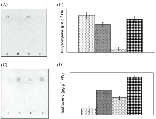
Analysis of disease-related proteins revealed that the content of such proteins was greater in diseased H. cannabinus and H. sabdariffa than in the respective controls (Figure 3D). In the present investigation, TLC separation and Uv-spectrum analysis revealed the presence of a higher amount of polyacetylenes in healthy than in diseased plants, whereas lower concentrations of isoflavones were found in the healthy plants compared to the diseased ones (Figure 5). Plants have flexible detection systems and probably employ several recognition and signal transduction pathways to activate their defense (Johal et al., 1995). Overall, precise temporal and spatial coordination of induced defense responses are required to successfully kill or restrict the invading microbe while simultaneously minimizing the damage to host tissue (Hammond-Kosack and Jones, 1996).
Healthy kenaf; Diseased kenaf; Healthy roselle; Diseased roselle.
(A) (B)
Po
ly
ac
et
yl
en
e
(
M
g
-1 F
W
)
(C) (D)
Is
ofl
av
on
e
(
g
g
-1 FW
)
figure 5. Estimation of polyacetylenes and isoflavones from healthy and diseased Hibiscus cannabinus (kenaf) and H. sabdariffa (roselle) plants (after 110 d of
inoculation); thin-layer chromatographic separation (a) and Uv-spectrum analysis of polyacetylenes (b), thin-layer chromatographic separation (c) and Uv-spectrum
Incompatible host-pathogen interaction results in the synthesis of inhibitors, known as phytoalexins, which have different structures according to the plant source, such as sesquiterpenoid, isoflavanoid, acetylenic or phenolic. The results thus indicate that the possible variation in balance of such defense-related components like polyacetylenes and isoflavones might be one of the factors for establishment of this disease in host plants.
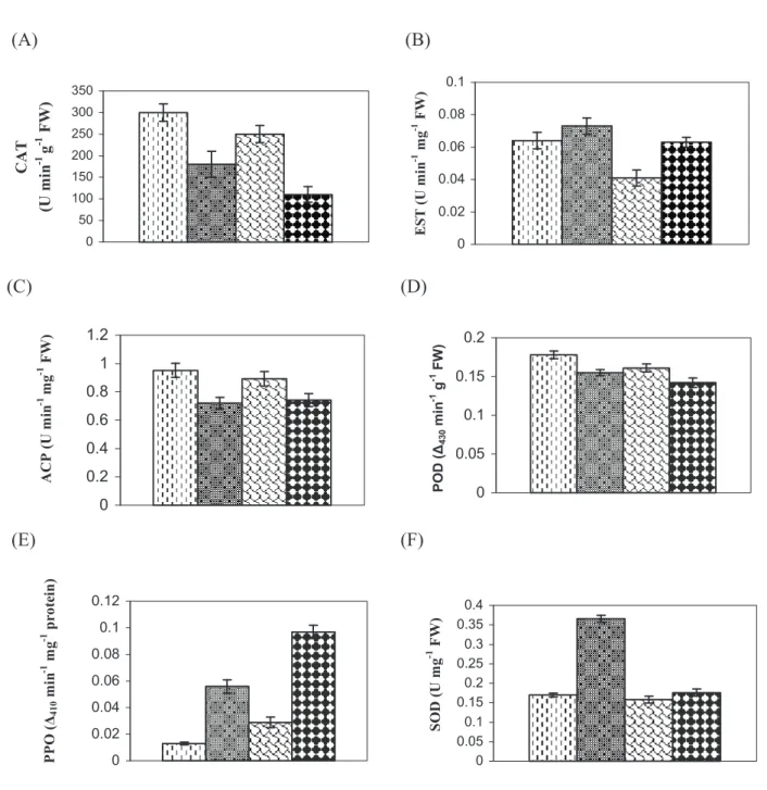
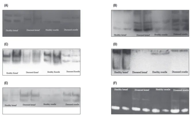
Analyses of isozyme patterns and activities of CAT, EST, ACP, POD, PPO and SOD in both the species, as shown in Figure 6, indicated alteration in activities of different enzymes due to the infection. Enzyme assays revealed lower activity of CAT, ACP and POD enzymes in diseased plants in comparison with healthy ones; in contrast, a marked increase in activities of EST, PPO and SOD was found in diseased plants as compared with the respective healthy plants. The isozyme patterns of these enzymes from diseased and healthy mesta plants produced similar types of band pattern in the case of ACP, PPO and SOD (Figure 7). For PPO and SOD the bands were found to be hyperactive in diseased plants in comparison with control plants, whereas in case of ACP the reverse was true. In the case of EST band profiling a clear extra band was found in diseased plants, and the other hyperactive bands observed in diseased plants indicate higher enzyme activity compared with their respective healthy plants. The isozyme profile of POD revealed the disappearance of some bands in diseased material which were present in their respective controls. In the case of CAT the isozyme pattern of diseased mesta was different from the healthy plant; a new band was noted in diseased material and some other bands were pronounced in healthy material but missing in the diseased plants.
Since enzymes control biochemical reactions, and their syntheses are under the control of specific gene(s), any change in the activity of an enzyme would reflect the pattern of gene expression and corresponding metabolic events in the cell. Hence, enzymes can be used as tools to study the induced responses of plants showing disease symptoms at the biochemical level (Neog et al., 2004). In addition, phenol-oxidizing enzymes such as POD and PPO are associated with many diseases (Pegg, 1985). In the
Healthy kenaf; Diseased kenaf; Healthy roselle; Diseased roselle.
(A)
(B)
0 50 100 150 200 250 300 350 C A T (U m in -1 g
-1 F
W ) 0 0.02 0.04 0.06 0.08 0.1 E S T ( U m in
-1 m
g
-1 FW)
(C)
(D)
0 0.2 0.4 0.6 0.8 1 1.2 A C P ( U m in -1 m g
-1 F
W ) 0 0.05 0.1 0.15 0.2 PO D ( 43 0 m in
-1 g -1 F
W
)
(E)
(F)
0 0.02 0.04 0.06 0.08 0.1 0.12 P P O ( 4 1 0 m in
-1 m
g
-1 p
ro te in ) 0 0.05 0.1 0.15 0.2 0.25 0.3 0.35 0.4 S O D ( U m g
-1 F
W
)
figure 6. Activities of different enzymes from healthy and diseased leaves of Hibiscus cannabinus (kenaf) and H. sabdariffa (roselle) plants (after 110 d of
inoculation). Catalase (a), esterase (b), acid phosphatase (c), peroxidase (D), polyphenol oxidase (e) and superoxide dismutase (f). Each bar represents the
(a) (b)
(c) (D)
(e) (f)
figure 7. Isozyme polymorphism profiles from healthy and diseased leaves of Hibiscus cannabinus (kenaf) and H. sabdariffa (roselle) plants. Catalase (a), esterase
(b), acid phosphatase (c), peroxidase (D), polyphenol oxidase (e) and superoxide dismutase (f).
acknowledgement: Authors are grateful to the Director of the Central Research Institute for Jute and Allied Fibres for his keen interest in the present investigation. The first author is also grateful to ICAR for financial assistance during the tenure of which this work was carried out.
references
Apel K, Hirt H (2004) Reactive Oxygen Species: Metabolism, oxidative stress and signal transduction. Annu. Rev. Plant Biol. 55:373-399.
Bates LS, Waldeen RP, Teare ID (1973) Rapid determination of free proline for water stress studies. Plant Soil 39:205-207.
Braber JM (1980) Catalase and peroxidase in primary bean leaves during development and senescence. Z. Pflanzenpkysiol. 97:135-144.
Brewbaker JL, Upadhya MD, Makinen Y, MacDonald T (1968) Isozyme polymorphism in flowering plants III. Gel Electrophoretic methods and applications. Physiol. Plant. 21:930-940.
Chatterjee A, Ghosh SK (2007a) A new monopartite begomovirus isolated
from Hibiscus cannabinus L.in India. Arch. virol. 152:2113-2118.
Chatterjee A, Ghosh SK (2007b) Association of a satellite DNA β molecule
with mesta yellow vein mosaic disease. virus Genes 35:835-844. Chatterjee A, Roy A, Ghosh SK (2006) Yellow vein Mosaic Disease of Kenaf. In: Rao GP, PaulKhurana SM, Lenardon SL (eds), Characterization, Diagnosis and Management of Plant viruses: vol. 1: Industrial Crops, pp.497-505. Studium Press, Texax, USA.
Chen C, Dickman MB (2005) Proline suppresses apoptosis in the fungal
pathogen Colletotrichum trifolii. Proc. Natl. Acad. Sci. USA. 102:3459-3464.
Chen CN, Pan SM (1996) Assay of superoxide dismutase activity by combining electrophoresis and densitometry. Bot. Bull. Acad. Sin. 37:107-111. Datta K, Muthukrishnan S, Datta SK (1999). Expression and function of PR-proteins genes in transgenic plants. In: Datta SK (ed), Pathogenesis-Related Proteins in Plants, pp.261-291. CRC Press, Boca Raton.
De Ascensao ARDCF, Dubery IA (2000) Panama disease: cell wall
reinforcement in banana roots in response to elicitors from Fusarium
oxysporum f. sp. cubense race four. Phytopathology. 90:1173-1180. Endo T, Okuda T, Tamura M, Yasuoka Y (2000) Estimation of net photosynthetic rate based on in-situ hyperspectral data. (Access: http://yasulab.iis.u-tokyo. ac.jp/pdf/endo_acrs).
Fernandez MR, Heath MC (1989) Interaction of the nonhost French bean plant (Phaseolus vulgaris) with parasitic and saprophytic fungi. III. Cytologically detectable responses. Can. J. Bot. 67:676-686.
Hammond-Kosack KE, Jones JDG (1996) Resistance gene dependent plant defense responses. Plant Cell. 8:1773-1791.
Harborne JB (1973) Phytochemical Methods: A Guide to Modern Techniques of Plant Analysis. Chapman and Hall, London.
Johal GS, Gray J, Briggs SP (1995) Convergent insights into mechanisms determining disease and resistance response in plant-fungal interactions. Can. J. Bot. 73(Suppl.):468-474.
Keen NT (1992) The molecular biology of disease resistance. Plant Mol Biol.19:109-122.
Laemmli UK (1970) Cleavage and structural proteins during the assembly of the head of bacteriophage T4. Nature. 227:680-685.
Lowry OH, Roserbrough NJ, Farr AL, Randall RJ (1951) Protein measurement with folin-phenol reagent. J. Biol. Chem. 193:256-275.
Mace ME, Wilson EM (1964) Phenol oxidases and their relation to vascular
browning in Fusarium infected banana roots. Phytopathology. 54:840-842.
Malik CP, Singh MB (1980) Plant Enzymology and Histoenzymology. Kalyani Publications, New Delhi.
Misra PS, Mertz ET, Glover Dv (1975) Studies on corn proteins: vIII. Free amino acid content of opaque-2 and double mutants. Cereal Chem. 52:844-848. Mitra R, Gadgil JD, Bhatia CR (1990) Host proteins associated with disease resistance. In: Sinha SK, Sane Pv, Bhargava SC, Agarwal PK (eds), Proceedings of the International Congress of Plant Physiology. Society for Plant Physiology and Biochemistry, pp.660-667. IARI, New Delhi.
Murray DR, Collier MD (1977) Acid phosphatase activities in developing
seeds of Pisum sativum L. Aust. J. Plant Physiol. 4:843-848.
Neog B, Yadav RNS, Singh ID (2004) Peroxidase, polyphenol oxidase and
acid phosphatase activities in the stigma-style tissue of Camellia sinensis
(L) O. Kuntze following compatible and incompatible pollination. J Indian Inst Sci. 84:47-52.
Oberley LW, Spitz DR (1985) Nitroblue tetrazolium. In: GreenwaldRA (ed),
CRC Handbook of Methods for Oxygen Radical Research, pp.217-220. CRC Press, Boca Raton.
Pegg GF (1985) Life in a black hole: The micro-environment of the vascular pathogen. Trans. Br. Mycol. Soc. 85:1-20.
Rathi YPS, Bhatt A, Singh US (1986) Biochemical changes in pigeonpea (Cajanus cajan (L.) Millsp.) leaves in relation to resistance against sterility mosaic disease. J. Biosci. 10:467-474.
Ryals J, Uknes S, Ward E (1994) Systemic acquired resistance. Plant Physiol. 104:1109-1112.
Sadasivam S, Manickam (1992) Biochemical Methods for Agricultural Sciences, pp.184-185. Wiley Eastern Ltd., New Delhi.
Sarvesh A, Reddy TP (1988) Peroxidase, polyphenol oxidase, acid phosphatase and alkaline inorganic pyrophosphatase activities during leaf
senescence in varieties of castor (Ricinus communis L.). Indian J. Exp. Biol.
26:133-136.
Sela I (1981) Plant-virus interactions related to resistance and localization of viral infections. Adv. virus Res. 26:201-237.
Sheen SL, Calvert J (1969) Studies on polyphenol content, activities and isozymes of Polyphenol oxidase and peroxidase during air-curing in three tobacco types. Plant Physiol. 44:199-204.
Smirnoff N, Cumbes QJ (1989) Hydroxyl radical scavenging activity of compatible solutes. Phytochemistry. 28:1057-1060.
Srinivas v, Balasubramanian D (1995) Proline is a protein-compatible hydrotrope. Langmuir. 11:2830-2833.
Sterjiades R, Dean JFD, Gamble G, Himmelsbach DS, Eriksson KEL
(1993) Extracellular laccases and peroxidases from sycamore maple (Acer
pseudoplatamus) cell-suspension cultures: Reactions with monolignols and lignin model compounds. Planta. 190:75-87.
Stewart CR (1980) The mechanism of abscisic acid-induced proline accumulation in barley leaves. Plant Physiol. 66:230-233.
Thimmaiah SR (1999) Standard Methods of Biochemical Analysis, pp.230-231. Kalyani Publishers, New Delhi.