UNIVERSIDADE ESTADUAL PAULISTA JULIO DE MESQUITA FILHO INSTITUTO DE BIOCIÊNCIAS
CÂMPUS DE BOTUCATU
TOTAL BACTERIAL COUNTING AND THE RESPONSE TO
TREATMENT OF BACTERIAL VAGINOSIS WITH
METRONIDAZOLE
Heloise Ranucci Luchiari
Monografia na forma de Artigo Científico apresentada ao Instituto de Biociências, Campus de Botucatu, para obtenção do título de Bacharel em Ciências Biomédicas.
BOTUCATU – SP 2013
Instituto de Biociências – Gabinete do Diretor
UNIVERSIDADE ESTADUAL PAULISTA JULIO DE MESQUITA FILHO INSTITUTO DE BIOCIÊNCIAS
CÂMPUS DE BOTUCATU
TOTAL BACTERIAL COUNTING AND THE RESPONSE TO
TREATMENT OF BACTERIAL VAGINOSIS WITH
METRONIDAZOLE
Aluna: Heloise Ranucci Luchiari Orientadora: Dra. Camila Marconi
Co-orientadora: Prof. Dra. Márcia Guimarães da Silva Supervisora: Prof. Dra. Maria de Lourdes Ribeiro de Souza da Cunha
Monografia na forma de Artigo Científico apresentada ao Instituto de Biociências, Campus de Botucatu, para obtenção do título de Bacharel em Ciências Biomédicas.
BOTUCATU – SP 2013
Instituto de Biociências – Gabinete do Diretor
Total bacterial counting and the response to treatment of bacterial vaginosis with metronidazole*
Heloise R. Luchiari 1, Márjorie A. Golim2, Márcia G. da Silva1, Camila Marconi1.
1Department of Pathology, Botucatu Medical School, UNESP – Univ Estadual
Paulista, Brazil
2Blood Center, Botucatu Medical School, UNESP - Univ Estadual Paulista, Brazil
Reprint requests and corresponding author:
Camila Marconi, e-mail: marconi.cml@gmail.com
Department of Pathology, Botucatu Medical School, UNESP – Univ Estadual
Paulista Botucatu, São Paulo, Brazil. Zip code: 18618-970. Phone: +55 14
3880-1655; Fax: +55 14 38152348.
Key words: Bacterial vaginosis, metronidazole, flow cytometry, total bacterial count
*
Trabalho Científico apresentado segundo as normas do periódico
Abstract
Objective: To evaluate if the total bacterial count of vaginal samples with bacterial
vaginosis assessed by flow cytometry influences the response to treatment with
metronidazol. Methods: In this cross-sectional study, 273 low-risk reproductive aged
women were enrolled. Vaginal samples were taken to evaluate the pattern of vaginal
flora according to Nugent’s criteria, as well as the presence of trichomoniasis and
candidosis. Cases identified of bacterial vaginosis were treated with metronidazole
and controlled after 45 days. Cervical infection by Chlamydia trachomatis and
Neisseria gonorrhoeae were also assessed. Flow cytometry for total bacterial
counting was performed in propidium iodide stained cervicovaginal samples, using
fluorescent beads at a known concentration. Non-parametric Mann-Whitney test was
used to compare total bacterial count between groups of interest, at p<0.05.
Results: From the total of 273 women enrolled, 50 were excluded as they presented
at least one of the infections investigated. Bacterial vaginosis was detected in 79
women (35.4%), of which 33 (41.8%) returned for re-evaluation after treatment,
being 21 cases successfully treated and 12 with persistent abnormal vaginal flora.
Flow cytometric data showed that total bacterial counting does not differ between
normal flora and bacterial vaginosis samples (p=0.14). Also, no difference was found
between the cases of treated and persistent bacterial vaginosis (p=0.48).
Conclusion: Total bacterial counting does not influence the response to
Introduction
Vaginal flora is typically defined as normal when Lactobacillus sp.
predominates over the numerous other bacterial species that can be found in this
environment [1]. Bacterial vaginosis is the most common type of abnormal vaginal
flora (AVF) and is characterized by the total or partial replacement of the local
lactobacilli by other bacteria, anaerobes mostly [2]. In reproductive aged-women, this
condition is particularly important as it is associated with poor pregnancy outcomes
and increased risk for acquisition and transmission of sexually transmitted infections
[3-5].
Although bacterial vaginosis has deleterious effects on women’s reproductive
health, the treatment of this condition remain a challenge for clinical practice. Current
recommended treatment for this AVF consists in a 7-day regimen with metronidazole
[6], with treatment failure rate of 36.0% one month after treatment cessation [7].
Although the treatment failure is frequent and the recurrence rate after six months of
treatment is more than 50% [8] the microbiological aspects involved in this difficulty
on flora restoration remain to be defined.
It is well established that women with bacterial vaginosis present a
significantly increased diversity of bacterial species in their vaginal flora [9,10]. In
addition to flora disruption, some studies have shown that bacterial vaginosis is
accompanied by an increase in the number of vaginal bacteria [11,12]. Corroborating
with this idea, bacterial biofilms are more frequently found in vaginal samples from
women with bacterial vaginosis when compared to those with normal flora, which
contribute for increases in bacterial number [13,14]. Considering that most vaginal
bacterial species are fastidious or non-cultivable, the determination of the total
methods.[10] Recently, the use of flow cytometry for total bacterial counting on
vaginal fluids was successfully demonstrated [15].
Therefore, considering that the microbiologic aspects of bacterial vaginosis
linked to therapeutic failure or resistance remain under investigation, the aim of the
current study was to evaluate if the total bacterial count in vaginal samples
influences the response to treatment of bacterial vaginosis with metronidazole.
Methods
Study population
From September 2012 to October 2013, a total of 273 reproductive-aged
women attending one unity of primary medical care in Botucatu-SP for routine
pap-test were invited to participate of this cross-sectional study. Women that reported
vaginal bleeding, urinary loss, recent sexual intercourse (<72h), puerperium (<3
months after delivery), use of intrauterine device (IUD), antibiotics (<30 days) and
confirmed/probable pregnancy were not included. Demographic, behavioral and
clinical data were obtained by interview using a standard questionnaire. Study aims
and procedures were explained and all women enrolled provided written informed
consent. Study was reviewed and approved by the Ethics Board of Botucatu Medical
School (Protocol 306.547).
Sampling procedures
During physical examination, using sterile speculum, vaginal pH was
assessed by pressing the commercial pH strips (4.0-7.0, Merck, Darmstadt,
Germany) for approximately 1 minute against the vaginal wall and then comparing
by adding 10% KOH solution to the vaginal swabs and the results were interpreted
as positive, doubtful or negative by the practitioner.
For microscopic evaluation of the vaginal flora, samples were taken using
sterile cotton swabs from mid-lateral vaginal wall, rolled on microscope slides and
Gram stained for classification according Nugent et al. (1991) [16] in normal,
intermediate flora or bacterial vaginosis. Another vaginal smear was prepared for wet
mount evaluation with addition of 0,9%NaCl solution and examined under light
microscope at 400 magnification (Olympus CX31, Tokyo, Japan) to detect the
presence of Trichomonas vaginalis and Candida sp. morphotypes. Additionally,
cervical samples were taken using cervical brush for assessment of Chlamydia
trachomatis and Neisseria gonorrhoeae infection by PCR.
Finally, cervicovaginal rinsings were performed by allowing the contact of 3mL
of sterile 0.9% NaCl with the lateral vaginal wall and ectocervix and recovering the
total liquid volume with sterile pipettes. Rinsing samples were stored at -80oC until
analysis and discarded if volume recovered was inferior to 3mL or in the presence of
blood.
Detection of endocervicitis
Cervical samples were submitted to DNA extraction using the commercial
Tissue & cells genomicPrep Mini Spin kit (GE Healthcare, Buckinghamshire, UK),
following the manufacturer’s protocol for Extraction of genomic DNA from animal
tissue. Efficiency of the extraction was confirmed by amplification of the constitutive
gene of β-globin using the primers PCO4 (5’-CAACTTCATCCACGTTCACC-3’) and
GH20 (5’-GAAGAGCCAAGGACAGGTAC-3’) for a 268bp product [17]. Reaction was
performed following parameters previously established in our laboratory consisting of
annealing at 52°C for 1 min and extension at 72°C for 1 min, and final extension at
72°C for 5 min. The products of the reactions were observed after electrophoresis in
agarose gel 1.5% stained with GelRed™ Nucleic Acid Gel Stain (Biotium, Hayward,
CA). Once the amplification of the β-globin sequence was confirmed, samples were
tested for C. trachomatis and N. gonorroheae.
Detection of C. trachomatis was performed by multiplex end-point PCR using
GoTaq Green Master Mix, 2X (Promega, Madison, WI) and primers CTP1
(5’-TAGTAACTGCCACTTCATCA-3’), CTP2 (5’-TTCCCCTTGTAATTCGTTGC-3’),
PL61 (5’-AGAGTACATCGGTCAACGA-3’), PL62
(5’-TCACAGCGGTTGCTCGAAGCA-3’) [18], that resulted in products of 201bp and
130bp, respectively. Cycling protocol used was denaturation at 95°C for 5 min,
followed by 40 cycles of 95°C for 1 min, 55°C for 1 min and 72°C at 1 min, with a
final extension at 72°C for 5 min. For all reactions, negative (DNAse-free water) and
positive (extracted DNA from McCoy cells infected by C. trachomatis) controls were
used. Presence of C. trachomatis was confirmed when observing both PCR products
on 1.5% agarose gel after electrophoresis.
Neisseria gonorroheae was detected by real time PCR using Maxima™ SYBR
Green/ROX PCR Master Mix 2X (Fermentas®, Thermo Scientific Inc, Waltham, MA)
and primers OH1 GCTACGCATACCCGCGTTGC-3’) and OH3
(5’-CGAAGACCTTCGAGCAGACA-3’) [19] resulting in a 390bp product. Melting
temperature of 83oC was used to determine the presence of N. gonorrhoeae
amplicons. For all reactions, negative (DNAse-free water) and positive controls (DNA
obtained by pure culture of N. gonorrhoeae ATCC® 19424) were used. Reactions
were performed on Line-gene K Real-time PCR Detection System (Bioer
Technology, China) and Line-gene K software (Bioer Technology, China) was used
by 45 cycles of 95°C for 15 s, 60°C for 30 s and 72°C for 30 s. Finally, fragments
were melted from 65 to 95°C at a rate of 0.5 °C/s.
Treatment of bacterial vaginosis and control visit
Treatment for bacterial vaginosis consisted in two-daily doses of
metronidazole 500mg for 7 days as recommended by the Center of Disease Control
and Prevention.[6] Women were scheduled for a control visit 45 days after the end of
treatment. At return, vaginal swabs of mid-lateral vagina wall were taken to a new
evaluation of vaginal flora as previously described. It allowed identifying cases of
success or failure of the treatment for bacterial vaginosis.
Total bacterial counting by flow cytometry
Cervicovaginal samples were thawed at -80°C and processed 24h before
cytometric analyses. Aliquots of 400µL were taken from all samples and fixed with
200µL of paraphormaldehyde 5% overnight at 4°C. To the total volume of 600µL of
the fixed samples, 8µL of 0,002% propidium iodide (BD, San Jose, CA) were added
and incubated at 37°C for 15 minutes. Samples were briefly centrifuged at 800rpm to
remove the largest residues and 500µL of the supernatants were transferred to 5mL
cytometry tubes. Prior to analysis, 5µL of fluorescent beads (Bacteria Counting Kit,
Invitrogen, Carlsbad, CA) were added to each sample to reach the density of 1.0 x
106 beads/mL.
Flow cytometric analyses were performed at FACScalibur (BD, San Jose,
CA). Data were acquired and processed using CellQuest Pro (Version 5.2, San
Jose, CA) with double threshold set on 280 and forward scatter (FSC) as primary
parameter and side scatter (SSC) as second parameter, both in logarithmic scale.
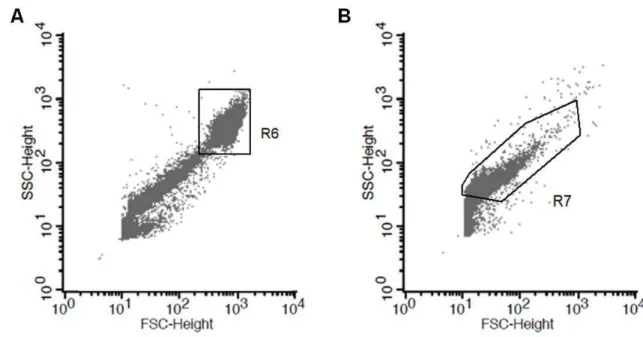
According to methodology previously described by Schellenberg et al. (2008)[15], a
samples (Figure 1A). The upper population consists of host’s shed epithelial cells
while the lower represents the total of bacterial cells in the sample. The position of
this lowest population consisting of bacterial cells on the dot plot was confirmed by
evaluating an overnight culture of Staphylococcus aureus (ATCC 19095) in filtered
tripticase soy broth, as observed in Figure 1B.
For bacterial counting, total of 10000 events at the beads were recorded for
each sample. As observed in Figure 2, gates were created for both bacterial cells
population and beads (R5). Considering the initial density of the fluorescent beads, a
10000 beads count is equivalent to 10µL sample volume, which allows determining
the total of bacterial cell units per µL of sample based on the events observed on
gate R6.
Statistical Analysis
Comparison of discrete variables between the groups of women with normal
and AVF was performed by Chi-squared test, while the continuous variables were
compared by Mann-Whitney test. For data on bacterial counting, comparison was
performed by Mann-Whitney test, between the groups with normal and bacterial
vaginosis at baseline, and between those women that presented effective and
treatment failure. All statistical tests were performed using GraphPad Prism 5.0
software (GraphPad, San Diego, CA) and P<0.05 was considered as significant.
Results
From the total of 273 women initially included in the study, we excluded
those cases of C. trachomatis (n=21, 9.4%), N. gonorroheae (n=2, 0.9%),
concomitant C. trachomatis and N. gonorrhoeae (n=1, 0.5%), Trichomonas vaginalis
gynecological characteristics of the 223 women finally included in the study are
shown in Table 1, according to the vaginal flora classification using Nugent’s scoring
system in normal (scores 0-3) and AVF (scores 4-10). Married women were less
likely to present flora alterations than single women (p=0.02). Self-reported previous
episode of bacterial vaginosis, but not sexually transmitted infections, showed
association with current AVF (p=0.02). Considering the gynecological data, women
with AVF were more likely to present higher vaginal pH (p<0.0001) and positive or
doubtful whiff test (p<0.0001).
Flora classification of the total of 223 women included showed that 132
(59.2%) had normal vaginal flora, while 91 (40.8%) presented AVF of which 12
(5.4%) were classified as intermediate flora and 79 (35.4%) as bacterial vaginosis
(BV). From the 79 women with bacterial vaginosis, 33 (41.8%) opt to treat BV and
returned to visit for control 45 days after conclusion of treatment. At return visit,
evaluation of the vaginal flora showed that in 21 (63.6%) women the treatment was
effective, as they returned with normal vaginal flora, while 12 (36.4%) women still
presented AVF.
Bacterial counting by flow cytometry was performed in 32 samples of
women with normal flora and 33 cases of BV, of which 21 were successfully treated
after metronidazole therapy and 12 failed to restore the lactobacilli-predominant
flora, as shown in Figure 3. Total bacterial counting, expressed in bacterial cell units
per µL of cervicovaginal samples, does not differ significantly between women who
had normal flora (median: 366.5, range: 46.0-10530.0) and women with bacterial
vaginosis (median: 277.0, range: 7.0-1232.0), (p=0.14). When comparing the results
on bacterial counting in first visit samples from women who had flora restoration after
effective (median: 236.5, range: 21.0-762.0), no statistical difference was observed
(p= 0.48).
Discussion
The evaluation of the demographic and behavioral data from the population
enrolled showed that both groups of study, normal and AVF, were similar for most of
the variables investigated. Although AVF is commonly associated with smoking
habit, number of sex partners and ethnicity [20,21], this study failed to demonstrate
such associations. This might be explained by the study design, as its main goal was
not to evaluate the characteristics associated with vaginal flora patterns. However, in
agreement with findings from Koumans et al., (2007) [21], the current data show that
being married is a protector factor for AVF, although the number of sex partners in
the last 12 months is not associated with this condition. The report of previous
episode of bacterial vaginosis was associated with AVF, which is in agreement with
the literature that shows that women with bacterial vaginosis have increased risk to
develop more episodes of this condition during their lifetime [8]. The association of
clinical findings as pH and positive or doubtful whiff test with an abnormal pattern
vaginal flora was well established [22] and could be confirmed by our results.
Bacterial counting by flow cytometry was already described and proposed as
a fast tool to evaluate microbiological changes in vaginal flora over the time [15].
Although in this previous study by Schellenberg et al. [15] it was showed that total
bacterial counting is increased in normal flora when compared with bacterial
vaginosis, our results failed to confirm these findings, as no difference was observed
between the two groups in the current study. Of noteworthy, Schellenberg et al. [15]
evaluated samples from mid-vagina, while the current study was performed using
studies. Nevertheless, these findings have been a matter of discussion, since data
from cultures of vaginal swabs show the opposite results, as the number of colony
forming units (CFU) correlate positively with Nugent score [11]. This finding is
supported by the presence of bacterial biofilms that are frequently detected in cases
of bacterial vaginosis [13] and contribute positively for bacterial growth.
Regarding the treatment of bacterial vaginosis, no difference in total bacterial
counting was found at baseline samples from women that returned with normal flora
against those women who had AVF in the second visit. This finding suggests that
total bacterial counting should not be a determining feature on treatment resistance
of bacterial vaginosis. Therefore other microbiological features might be linked to
treatment failures, such as the presence of metronidazole resistant Atopobium
vaginae strains[23]. Moreover, in relation to recurrence, women with the concomitant
detection of A. vaginae and G. vaginalis have higher rates of recurrence when
compared to those with the detection of G. vaginalis alone [24].
Conclusion
Present data show that total bacterial counting assessed by flow cytometry
does not influence the response to metronidazole treatment of bacterial vaginosis. It
suggests that therapeutic failures might be related to other microbiological aspects of
the vaginal flora, such as the bacterial composition and pathogenic potential of the
strains.
Acknowledgments: This study was supported by the Fundação de Amparo à
Pesquisa do Estado de São Paulo (Grants: #2012/16800-3, # 2012/10403-2 and #
2013/01750-3).
References
1. Spiegel CA. Bacterial vaginosis. Clin Microbiol Rev 1991;4:485-502.
2. Hillier SL, Holmes KK, Marrazzo JM. Bacterial Vaginosis. In: Sexually
Transmitted Diseases. 4th ed. McGraw-Hill, Health Professions Division, New
York; 2008:737-68.
3. Leitich H, Bodner-Adler B, Brunbauer M, Kaider A, Egarter C, Husslein P.
Bacterial vaginosis as a risk factor for preterm delivery: a meta-analysis. Am J
Obstet Gynecol 2003;189:139-47.
4. Gallo MF, Macaluso M, Warner L, Fleenor ME, Hook EW 3rd, Brill I et al.
Bacterial Vaginosis, Gonorrhea, and Chlamydial Infection Among Women
Attending a Sexually Transmitted Disease Clinic: A Longitudinal Analysis of
Possible Causal Links. Ann Epidemiol 2012;22:213-20
5. Cohen CR, Lingappa JR, Baeten JM, Ngayo MO, Spiegel CA, Hong T et al.
Bacterial vaginosis associated with increased risk of female-to-male HIV-1
transmission: a prospective cohort analysis among African couples. PLoS Med
2012;9:e1001251.
6. Workowski KA, Berman S. Centers for Disease Control and Prevention. Sexually
Transmitted Diseases Treatment Guidelines 2010. MMWR 2006;55(No. RR-11).
7. Schwebke JR, Desmond RA. Tinidazole vs metronidazole for the treatment of
bacterial vaginosis. Am J Obstet Gynecol 2011;204:211.e1-6.
8. Bradshaw CS, Morton AN, Hocking J, Garland SM, Morris MB, Moss LM et al.
High recurrence rates of bacterial vaginosis over the course of 12 months after
oral metronidazole therapy and factors associated with recurrence. J Infect Dis
2006;193:1478-86.
9. Diao Y, Fang X, Xia Q, Chen S, Li H, Yang Y et al. Organism diversity between
women with and without bacterial vaginosis as determined by polymerase chain
reaction denaturing gradient gel electrophoresis and 16S rRNA gene sequence. J
10. Ravel J, Gajer P, Abdo Z, Schneider GM, Koenig SS, McCulle SL et al. Vaginal
microbiome of reproductive-age women. Proc Natl Acad Sci USA
2011;108:4680-7.
11. Nikolaitchouk N, Andersch B, Falsen E, Strömbeck L, Mattsby-Baltzer I. The
lower genital tract microbiota in relation to cytokine-, SLPI- and endotoxin levels:
application of checkerboard DNA-DNA hybridization (CDH). APMIS
2008;116:263-77.
12. Delaney ML, Oderdonk AB. Nugent score related to vaginal culture in pregnant
women. Obstet Gynecol 2001;98:79-84.
13. Swidsinski A, Mendling W, Loening-Baucke V, Swidsinski S, Dörffel Y, Scholze J
et al. An adherent Gardnerella vaginalis biofilm persists on the vaginal epithelium
after standard therapy with oral metronidazole. Am J Obstet Gynecol
2008;198:97.e1-6.
14. Costerton W, Veeh R, Shirtliff M, Pasmore M, Post C, Ehrlich G. The application
of biofilm science to the study and control of chronic bacterial infections. J Clin
Invest 2003;112:1466-77.
15. Schellenberg J, Blake Ball T, Lane M, Cheang M, Plummer F. Flow cytometric
quantification of bacteria in vaginal swab samples self-collected by adolescents
attending a gynecology clinic .J Microbiol Methods 2008;73:216-26.
16. Nugent RP, Krohn MA, Hillier SL. Reability of diagnosing bacterial vaginosis is
improved by a standardized method of gram stain interpretation. J Clin Microbiol
1991;29:297-301.
17. Bauer HM, Ting Y, Greer CE, Chambers JC, Tashiro CJ, Chimera J et al. Genital
human papillomavirus infection in female university students as determined by a
PCR-based method. JAMA 1991;265:472-7.
18. Morré SA, Sillekens P, Jacobs MV, van Aarle P, de Blok S, van Gemen B et al.
RNA amplification by nucleic acid sequence-based amplification with an internal
standard enables reliable detection of Chlamydia trachomatis in cervical
19. Ho BSW, Feng WG, Wong BKC, Egglestone SI. Polymerase chain reaction for
the detection of Neisseria gonorrhoeae in clinical samples. J Clin Pathol
1992;45:439-42.
20. Marconi C, Donders GG, Parada CM, Giraldo PC, da Silva MG. Do Atopobium
vaginae, Megasphaera sp. and Leptotrichia sp. change the local innate immune
response and sialidase activity in bacterial vaginosis? Sex Transm Infect
2013;89:167-73
21. Koumans EH, Sternberg M, Bruce C, McQuillan G, Kendrick J, Sutton M et al.
The prevalence of bacterial vaginosis in the United States, 2001-2004;
associations with symptoms, sexual behaviors, and reproductive health. Sex
Transm Dis 2007;34:864-9.
22. Amsel R, Totten PA, Spiegel CA, Chen KC, Eschenbach D, Holmes KK.
Nonspecific vaginitis. Diagnostic criteria and microbial and epidemiologic
associations. Am J Med 1983;74:14–22.
23. De Backer E, Verhelst R, Verstraelen H, Claeys G, Verschraegen G,
Temmerman M et al. Antibiotic susceptibility of Atopobium vaginae. BMC Infect
Dis 2006;6:51.
24. Bradshaw CS, Tabrizi SN, Fairley CK, Morton AN, Rudland E, Garland SM. The
association of Atopobium vaginae and Gardnerella vaginalis with bacterial
vaginosis and recurrence after oral metronidazole therapy. J Infect Dis
Table 1. Demographic, behavioral and clinical characteristics of the 223 women included in the study, in relation to the classification of the vaginal flora in normal (scores 0-3) and abnormal vaginal flora (AVF, scores 4-10), according to Nugent’s system [16].
Characteristics Normal
(n=132)
AVF
(n=91) p
Age* 34 [18-50] 33 [18-49] 0.92
Ethnicity**
White 82 (62.1%) 47 (51.7%) 0.12
Non-White 50 (37.9%) 44 (48.3%)
Marital status**
Single 34 (25.8%) 37 (40.7%) 0.02¥
Married/living together 98 (74.2%) 54 (59.3%)
Years at school* 9 [0-16] 8 [0-15] 0.10
Remunerated Activity (n/total)** 71 (53.8%) 49 (53.8%) 0.99
Smoking Habit (n/total)** 21 (15.9%) 20 (22.0%) 0.25
Number of sex partner (1 year) **
0 or 1 120 (90.9%) 77 (84.6%) 0.15
2 or more 12 (9.1%) 14 (15.4%)
Number of vaginal
intercourse/week*
2 [0-7] 2 [0-7] 0.70
Previous BV** 55 (41.7%) 52 (57.1%) 0.02¥
Previous STD** 11 (8.3%) 10 (11.0%) 0.50
Consistent Condom Use** 22 (16.7%) 23 (25.3%) 0.11
Hormonal contraceptive use** 64 (48.5%) 33 (36.3%) 0.07
Vaginal pH* 4.4 [4.0-5.0] 4.7 [4.0-7.0] <0.0001¥
Cervical ectopy** 50 (38.0%) 38 (41.8%) 0.56
Whiff test**
Positive or doubtful 71 (53.8%) 77 (84.6%) <0.0001¥
Negative 61 (46.2%) 14 (15.4%)
AVF: Abnormal vaginal flora, BV: Bacterial vaginosis; STD: Sexually transmitted infection.
Figure 1. A. Scattering profile of a vaginal sample resembling the findings of
Schellenberg et al. (2008) [15] with two distinct populations observed.
Upper population (R6) consisted of host’s epithelial or inflammatory
cells and lower of bacterial cells. B. Distribution of the bacterial cells
on the dot-plots was confirmed using a pure culture of Staphylococcus
aureus (ATCC 19095) (R7).
Figure 2. Scattering profiles at FSC/SSC and FL3/SSC showing gates that were
created for both bacterial cells population and beads (R5) and bacterial
cells (R6) exclusively. Bacterial cells units were determined based on
the events observed on gate R6 when 10000 bead-events were
Figure 3. A. Total bacterial counting in cervicovaginal lavages from women with
normal flora (n=32) and bacterial vaginosis (n=33) and B. from
women with bacterial vaginosis successfully treated after
metronidazole therapy (n=21) and those with persistence of abnormal