BRAZ TAVARES DA HORA JÚNIOR
Molecular phylogeny and population genetics of Microcyclus ulei, causal agent of the South American leaf blight of Hevea brasiliensis
VIÇOSA
MINAS GERAIS – BRASIL 2012
Aos meus pais, Braz Tavares e Liana
Maria, e ao meu filho Vitor da Hora,
AGRADECIMENTOS
Ao Prof. Dr. Eduardo Seiti Gomide Mizubuti, meu orientador, pela
oportunidade, confiança e amizade.
Aos pesquisadores da Michelin, Carlos Raimundo Reis Mattos e Eric
Cavaloc pela oportunidade, confiança e amizade.
Aos pesquisadores do Cirad, em especial ao Dr. Dominique Garcia, Vincent
Le Guen e Frank Rivano pelo apoio e amizade.
Ao Prof. Dr. Robert Weingart Barreto e ao Dr. Harold Charles Evans, pela
orientação e amizade.
A Profa. Dra. Karla Suemy Clemente Yotoko, pelo apoio e amizade.
Aos amigos das Plantações Michelin da Bahia pela amizade, pelos
ensinamentos e pela colaboração nos trabalhos.
Aos meus colegas e amigos do Laboratório de Biologia de Populações de
Fitopatógenos, pela amizade, pelos ensinamentos e pelo convívio.
Aos meus colegas e amigos do PPGFito/UFV, pela amizade, pelos
ensinamentos e pelo convívio.
Aos professores e técnicos do PPGFito/UFV, pelos ensinamentos e pela
amizade.
À Carine Rezende Cardoso pelo amor e incentivo.
À minha família, em especial meus irmãos André e Diogo e minha sobrinha
Letícia, Helenice e Vinícius, pela compreensão, amor e incentivo.
À CAPES e ao CNPq, pela concessão da bolsa de estudo, e à Plantações
Michelin da Bahia pelo apoio financeiro a este projeto.
SUMÁRIO
SUMÁRIO ... iii
RESUMO ... vi
ABSTRACT ... viii
INTRODUÇÃO GERAL ... 10
REFERENCES ... 13
CAPÍTULO 1 ... 15
Unraveling critical characteristics of the life cycle of Microcyclus ulei, a highly destructive fungal pathogen of the rubber tree (Hevea brasiliensis) ... 15
ABSTRACT ... 16
INTRODUCTION ... 18
MATERIAL AND METHODS ... 22
RESULTS ... 27
DISCUSSION ... 29
TAXONOMY ... 34
ACKNOWLEDGEMENTS ... 35
REFERENCES ... 36
FIGURE LEGENDS ... 49
SUPPORTING INFORMATION ... 58
CAPÍTULO 2 ... 69
Spatial pattern and population biology of Microcyclus ulei in Hevea agricultural landscapes in Brazil ... 69
ABSTRACT ... 70
INTRODUCTION ... 72
MATERIAL AND METHODS ... 76
RESULTS ... 80
DISCUSSION ... 83
REFERENCES ... 90
FIGURE LEGENDS ... 99
SUPPORTING INFORMATION ... 108
CAPÍTULO 3 ... 111
Influence of hosts with partial resistance on the genetic structure of the pathogen Microcyclus ulei in Hevea spp. ... 111
ABSTRACT ... 112
INTRODUCTION ... 114
MATERIAL AND METHODS ... 118
RESULTS ... 123
DISCUSSION ... 127
ACKNOWLEDGEMENTS ... 131
REFERENCES ... 132
FIGURE LEGENDS ... 141
SUPPORTING INFORMATION ... 151
CONCLUSÃO GERAL ... 158
RESUMO
HORA JÚNIOR, Braz Tavares, D.Sc., Universidade Federal de Viçosa,
Agosto de 2012. Filogenia molecular e genética de populações
de Microcyclus ulei, agente causal do mal das folhas de Hevea brasiliensis. Orientador: Eduardo Seiti Gomide Mizubuti.
Co-orientadores: Luiz Antonio Maffia e Robert Weingart Barreto.
O mal das folhas da seringueira, doença causada pelo fungo Microcyclus
ulei, impediu o desenvolvimento de Hevea spp. em áreas de monoculturas
na região amazônica e, consequentemente, acarretou perdas na produção
de borracha natural em sistemas agrícolas na América Tropical. O uso de
clones de seringueira com resistência parcial ao mal das folhas é a melhor
opção para o manejo da doença. Embora o mal das folhas seja conhecido
desde o início do século 20, aspectos básicos da classificação filogenética e
da estrutura genética da população do patógeno são desconhecidos. Quatro
regiões genômicas (LSU rRNA, mtSSU, MCM7 e ITS) foram usadas em
estudos de reconstrução filogenética que suportam a classificação de M. ulei
na família Mycosphaerellaceae s. str., ordem Capnodiales, em Ascomycota,
proximamente relacionado às espécies de Mycosphaerella. Da mesma forma
o seu anamorfo Fusicladium heveae é melhor acomodado em
Pseudocercospora s. str. A partir desta perspectiva evolutiva, propomos um modelo de ciclo de vida que inicia com espermogônias em folhas próximas à
maturidade levando a ascósporos maduros em pseudotécios dentro dos
estromas. A variabilidade genética do patógeno em plantações comerciais
de seringueiras foi analisada em grande e pequena escalas, utilizando 17
produtoras no Brasil foram analisadas em larga escala. Constatou-se alta
diversidade gênica e associação aleatória de alelos. Algumas populações
geograficamente distantes foram geneticamente relacionadas. Isolamento
por distância foi evidente apenas para as populações da região amazônica.
Os padrões espaciais da variação genética de M. ulei são o resultado de
fluxo gênico, provavelmente, afetado por fatores antrópicos e deriva genética
por falha de conectividade entre os seringais. O estudo em pequena escala
foi feito para caracterizar a variabilidade intraespecífica da população do
patógeno a partir de isolados coletados de clones de seringueira suscetíveis
e resistentes. A análise bayesiana de agrupamento revelou agrupamento de
isolados de acordo com o nível de resistência sugerindo que seleção
desempenha um papel importante na evolução do M. ulei. Além disso, houve
diferenciação genética elevada entre populações simpátricas do patógeno
em hospedeiros suscetíveis e parcialmente resistentes. A especificidade a
ABSTRACT
HORA JÚNIOR, Braz Tavares, D.Sc., Universidade Federal de Viçosa, August, 2012. Molecular phylogeny and population genetics of
Microcyclus ulei, causal agent of the South American leaf blight of
Hevea brasiliensis Adviser: Eduardo Seiti Gomide Mizubuti. Co-advisers:
Luiz Antonio Maffia and Robert Weingart Barreto.
South American leaf blight (SALB) caused by Microcyclus ulei prevented the
development of large-scale Hevea spp. monoculture areas in the Amazon
region and consequently led to failure of natural rubber production in
managed landscapes in Tropical America. Planting of clones with partial
resistance to SALB is the best option to manage the disease. Although SALB
has been known since the beginning of the 20th century, basic aspects of its
phylogenetic classification and the genetic structure of the pathogen
population are unknown. Four genomic regions (LSU rRNA, mtSSU, MCM7
and ITS) were used for reconstructing the molecular phylogeny. The results
of these analyses support the classification of M. ulei in the family
Mycosphaerellaceae s. str, order Capnodiales, in the Ascomycota, closely
related to Mycosphaerella-like species. Similarly, the phylogeny of its
anamorph, Fusicladium heveae, placed this species in a different order and it was better accommodated in Pseudocercospora s. str. From this evolutionary perspective, we propose a model of the life cycle of the pathogen starting
with spermogonia in the near-mature leaf stage and leading to mature
ascospores in pseudothecia within stromata. Genetic variability of the
pathogen in large and small scale commercial rubber tree plantations was
Amazon and the main producing areas in Brazil were analyzed in large scale.
The pathogen has high gene diversity and random association of alleles
across loci. Some geographically distant populations were genetically
related. Isolation by distance was evident only for populations from Amazon
region. The spatial patterns of the genetic variation of M. ulei are the result of gene flow probably affected by anthropogenic factors and genetic drift
enhanced by the lack of connectivity between rubber plantations from the
eastern and western regions of Brazil. A small scale study was conducted to
characterize the intraspecific variability of the pathogen population
associated with susceptible and resistant rubber tree clones. Bayesian
clustering analyses revealed that isolates could be grouped according to the
clone resistance level suggesting that directional selection plays a role in
shaping the evolution of M. ulei. Additionally, it was observed a high genetic differentiation between sympatric populations from susceptible and partially
resistant clones. In areas where susceptible and partially resistant clones are
cultivated in close proximity, clone specificity probably acts as an efficient
INTRODUÇÃO GERAL
A borracha natural é uma commodity de alto valor e é a matéria-prima de produtos essenciais e amplamente utilizados em diversos setores.
Fatores físico-químicos como elasticidade, plasticidade, resistência ao uso,
isolamento e impermeabilidade a líquidos e gases, fazem da borracha
natural um produto insubstituível em muitas aplicações (LIEBEREI, 2007).
Consequentemente, existe uma demanda crescente por esta commodity. As
maiores plantações de seringueiras encontram-se instaladas no sudeste da
Ásia e 92% da produção mundial de borracha natural mundial provêm da
Tailândia, Indonésia, Malásia, Vietnã, Índia e China. Em 2010, o Brasil
produziu aproximadamente 220.000 toneladas, o que corresponde a 2% da
produção mundial, e importou 260.000 toneladas para atender sua demanda
interna (FAO, 2012; MDIC, 2012).
A previsão é que a demanda mundial em 2020 seja de
aproximadamente 16,4 milhões de toneladas contra os 10,4 milhões de 2011
(IRSG, 2012). Comparado a outros países, o Brasil possui áreas extensas,
propícias ao plantio de seringueiras, mas o mal das folhas, doença causada
pelo fungo Microcyclus ulei (Henn.) von Arx (Ascomycota) está presente em todas as regiões onde a seringueira é cultivada (GASPAROTTO et al.,
1997).
As epidemias do mal das folhas foram e continuam sendo
responsáveis pela baixa produção de borracha natural em áreas úmidas da
América do Sul além de reduzirem a longevidade dos seringais implantados
induz a desfolha e até a morte de clones suscetíveis (HOLLIDAY, 1970;
CHEE e HOLLIDAY, 1986; LIEBEREI, 2007).
As estratégias de controle disponíveis são limitadas e a utilização de
variedades resistentes é a melhor opção para o manejo da doença (GARCIA
et al., 2004). Plantios de clones resistentes e produtivos possibilitam o cultivo
de seringueiras em zonas de ocorrência endêmica da doença. A resistência
ao mal das folhas da seringueira é o objetivo de vários programas de
melhoramento na América Tropical, África e Ásia, mas existem poucos
clones comerciais disponíveis por causa da instabilidade da resistência e
baixa produção dos clones resistentes selecionados. A variabilidade genética
da população do patógeno é um fator que explica o comportamento variável
de clones resistentes plantados em diversas áreas (PERALTA et al., 1990;
GARCIA et al., 2002).
Atualmente, o enfoque dos programas de melhoramento, como por
exemplo, o projeto CIRAD-Michelin-Brazil (CMB), é a obtenção de clones
produtivos com resistência quantitativa. Os clones resistentes obtidos são
tão produtivos quanto os clones asiáticos e têm a vantagem de poder ser
usados em áreas afetadas pelo mal das folhas. Além disso, estes clones
seriam interessantes para área indenes como as do sudeste asiático e
África, como uma estratégia preventiva para reduzir o potencial impacto
econômico da introdução do M. ulei (GARCIA et al., 2004).
Apesar de programas de melhoramento da seringueira para
resistência ao mal das folhas estarem sendo conduzidos, não existem
informações sobre a variabilidade genética na população de M. ulei e a
estrutura genética da população do patógeno pode ser usado para entender
o desenvolvimento da doença, prever a evolução do mal das folhas e
desenvolver estratégias efetivas de melhoramento visando resistência à
doença, e, principalmente, prolongar a vida útil dos materiais resistentes
selecionados (MILGROOM e FRY, 1997). Estudos sobre a epidemiologia do
mal das folhas foram recentemente finalizados (GUYOT et al., 2008;
HONORATO JÚNIOR, 2010), mas até o momento, não existem estudos
relacionados aos aspectos filogenéticos de M. ulei e sobre a genética
molecular da população do patógeno. Assim, os objetivos do presente
trabalho foram: i. Investigar a filogenia de M. ulei e, ii. Determinar a estrutura genética da população do patógeno no Brasil em grande e pequena escala
REFERENCES
CHEE, K. H. and HOLLIDAY, P. 1986. South American leaf blight of Hevea
rubber. Malaysian Rubber Research and Development Board. Malaysian
Rubber Research and Development Board Monograph No. 13, 50 pp.
FAO. 2012. http://faostat.fao.org/site/339/default.aspx
GARCIA, D., LE GUEN, V., MATTOS, C. R. R., GONÇALVES, P. and
CLÉMENT-DEMANGE, A. 2002b. Genetic parameter estimations of three
traits used to evaluate South American leaf blight (SALB) in rubber tree. Crop Breed. Appl. Biotechnol., 2: 453-462.
GARCIA, D., MATTOS, C. R. R., GONÇALVES, P. S. and LE GUEN, V.
2004. Selection of rubber clones for resistance to South American leaf blight
and latex yield in the germplasm of the Michelin Plantation of Bahia (Brazil).
J. Rubb. Res., 7: 188-198.
GASPAROTTO, L., SANTOS, A. F., PEREIRA, J. C. R. and FERREIRA, F.
A. 1997. Doenças da Seringueira no Brasil. Embrapa-SPI: Manaus:
Embrapa-CPAA.
GUYOT, J., CILAS, C. and SACHE, I. 2008. Influence of host resistance and
phenology on South American leaf blight of the rubber tree with special
consideration of temporal dynamics. Eur. J. Plant Pathol., 120: 111-124.
HOLLIDAY, P. 1970. South American leaf blight (Microcyclus ulei) of Hevea brasiliensis. Commonwealth Mycological Institute. Phytopath. Pap., 12: 1-31.
HONORATO JÚNIOR, J. 2010. Mal-das-folhas da seringueira: dinâmica de
inóculo do patógeno, progresso e danos, em três condições topográficas.
IRSG. 2012. International Rubber Study Group. http://www.rubberstudy.com
LIEBEREI, R. 2007. South American leaf blight of the rubber tree (Hevea
spp.): New steps in plant domestication using physiological features and
molecular markers. Ann. Bot., 100: 1-18.
MDIC. 2012. Ministério do Desenvolvimento, Indústria e Comércio Exterior.
http://www.desenvolvimento.gov.br
MILGROOM, M. G., and FRY, W. E. 1997. Contributions of population
genetics to plant disease epidemiology and management. Adv. Bot. Res., 24: 1-30.
PERALTA, A. M., FURTADO. E. L., AMORIM, L., MENTEN, J. O. M. and
BERGAMIN FILHO, A. 1990. Melhoramento genético da seringueira para a
resistência ao mal das folhas (Microcyclus ulei). Revisão. Summa
CAPÍTULO 1
ABSTRACT
Four genomic regions (LSU rRNA, mtSSU, MCM7 and ITS) were used for
reconstructing the molecular phylogeny of Microcyclus ulei, a pleomorphic
fungus that causes South American leaf blight (SALB) in Hevea spp.
Classification based on the teleomorphic and anamorphic morphological
traits do not reflect proper evolutionary relationships. The molecular
phylogeny of the teleomorph supports the classification of M. ulei in the
family Mycosphaerellaceae s. str., order Capnodiales (Ascomycota), closely
related to Mycosphaerella-like species. However, the phylogeny of the
anamorph, Fusicladium heveae, suggests a different order and it is better
accommodated in Pseudocercospora s. str. Based on these findings, the life cycle of the pathogen was revisited. Pathogen development was monitored in
inoculated leaves under field conditions. ‘F. heveae’ was observed from the young leaves (B2 stage) and decreased in the mature leaves (D stage).
‘Aposphaeria ulei’ structures were seen in the near mature leaves (C/D
stage) preceding M. ulei which was fully formed in the mature leaves.
‘Pycnospores’ did not germinate and were not infective. We propose a model
of sexual cycle beginning with ‘A. ulei’ as spermogonia in the C/D leaf stage leading to mature ascospores in pseudothecia within pronounced, erumpent
ascostromata of Mycosphaerella-type.
Key words: South American leaf blight, Fusicladium, Aposphaeria,
INTRODUCTION
Molecular phylogenetic approaches have become key tools for the
classification of plant pathogens and new features of biological and
epidemiological relevance have emerged from studies that used these
techniques. DNA polymorphism data provide additional characters for the
classification of microorganisms, allowing for the application of the
phylogenetic species concept and contributing to improving the
understanding of the evolutionary relationships among species from the
application of high-resolution analyses (AVISE and WOLLENBERG, 1997;
TAYLOR et al. 2000; HIBBETT et al., 2007). Traditionally, the phylogenetic
relationships among fungal species have been investigated mainly using
morphological characters, however ultrastructural, biochemical and genomic
traits can also be used and often contribute to increasing the power of the
analyses (MCLAUGHLIN et al. 2009).
Recent studies provide strong evidence that molecular phylogenetic
trees accurately reflect the evolutionary history of the Fungi and a consensus
classification for many groups is possible (HIBBETT et al., 2007). This is
particularly useful for members of an important group of ascomycete fungi,
the Dothideomycetes (SCHOCH et al. 2009a; CROUS et al., 2009a; ZHANG
et al., 2009a). The molecular phylogenies of several plant and human
pathogenic, endophytic, saprophytic and epiphytic species of the
Dothideomycetes have been studied. However, the classification and
evolutionary issues related to the tropical plant pathogenic Dothideomycetes
South American leaf blight (SALB) of rubber tree caused by the plant
pathogenic Dothideomycete Microcyclus ulei (Henn.) von Arx is recognized
as the most serious threat to the natural rubber industry worldwide (van
BEILEN and POIRIER, 2007). Epidemics of SALB led to the failure of rubber
cultivation in managed landscapes in tropical America in the early 20th
century (GRANDIN, 2009) and, because of the potential serious economic
consequences, there are quarantine measures for preventing SALB in the
rubber in the Palaeotropics, especially in southeast Asia, a SALB-free zone
(GASPAROTTO et al., 1997; LIEBEREI, 2007). The fungus infects young
leaves, stems and fruit tissues of Hevea brasiliensis (Willd. ex A. L. Juss.) Muell.-Arg., H. benthamiana Muell.-Arg., H. spruceana (Benth.) Muell.- Arg.,
H. guianensis Aublet and H. camporum Ducke (CHEE and HOLLIDAY, 1986). Immature leaves are the only susceptible stage and lesions are formed 5 to 7
days after infection and the asexual spores (conidia) of the fungus are
observed on immature leaves, mainly on the lower side of the leaf, shortly
after. The pycnidial form and sexual structures (stromata) appear on the
margins of the necrotic lesions once the leaves reach maturity (HOLLIDAY,
1970).
Although SALB has been known since the beginning of the 20th
century, some critical aspects of the life cycle of the fungus remain unknown,
especially the function of the pycnidial stage and its role in the development
of sexual structures. Koch’s postulates were performed by LANGFORD
(1945) and confirmed recently by GUYOT and DOARÉ (2010). In both
studies, the authors inoculated conidia and ascospores to reproduce the
remains unclear.
The fungus was first observed in 1900 in the Amazon rainforest in
Peru and Brazil and was later described by HENNINGS (1904). Initially, two
spore stages were described: the teleomorph as Dothidella ulei and a
pycnidial form (Coelomycetes) as Aposphaeria ulei. The conidial stage
(Hyphomycetes) was described by J. Kuyper in Surinam in 1912 as
Fusicladium macrosporum. In 1917, G. Stahel observed the connection of hyphae from different fungal structures within the leaf tissue and linked the
teleomorph and anamorph stages of the fungus and renamed the teleomorph
as Melanopsammopsis ulei (HOLLIDAY, 1970). Later, MÜLLER and von ARX (1962) transferred the teleomorph to the genus Microcyclus and suggested a
close relationship with the genus Mycosphaerella based on the morphology
of the Passalora-type conidia. In Microcyclus, the development of erumpent stromata on living leaves (or other plant parts) from a substrate is due to the
proliferation of a foot-like hypostroma and some of the Mycosphaerella spp. associated with pine trees have similar prominent ascostromata (EVANS,
1984).
The taxonomy of the causal agent of SALB is confusing and the lack
of DNA sequence data for all three stages of the life cycle has prevented
better elucidation of its classification. The genus Microcyclus used to be classified as belonging to the Mycosphaerellaceae (order Capnodiales) as a
stromatic counterpart of the family (von ARX and MÜLLER 1975; ERICSSON
and HAWKSWORTH 1993), but in 1996 it was re-classified in the
Planistromellaceae (order Dothideales) (BARR 1996; LUMBSCH and
stage, the anamorphic name was changed to Fusicladium heveae, a species that belongs to the Venturiaceae family (order Venturiales) (ZHANG et al.
2011a). The anamorphic genus Aposphaeria is recognized as a member of
the family Lophiostomataceae (order Pleosporales) as a well-supported
group (MUGAMBI and HUHNDORF, 2009; ZHANG et al., 2011b).
Thus, there are questionable issues regarding the classification of the
teleomorph and anarmorph stages at the genus, family and order levels of
the causal agent of SALB. Additionally, knowledge about the evolutionary
history of the pathogen and of related species is scarce. Molecular studies
could help to resolve the true affinity of this fungus (EVANS, 2002;
SCHUBERT et al., 2003; KIRK et al., 2008) and the objective of this study
was to investigate the phylogenetic relationships of M. ulei using molecular approaches to shed light on its classification and on the various stages of its
MATERIAL AND METHODS
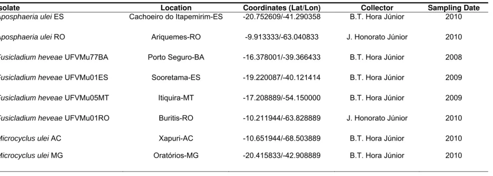
Sampling, Isolation and DNA extraction
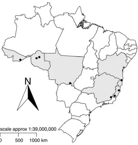
Leaves with typical lesions of South American leaf blight were sampled
in commercial fields of rubber in Brazil. Sampling was aimed at areas with
records of high incidence of SALB in the states of Acre, Rondônia, Mato
Grosso, Minas Gerais, Espírito Santo and Bahia between 2008 and 2010
(Figure 01 and Table 01). Single conidia were transferred from fungal
structures formed on lesions to culture media, using a sterilized fine-needle
under a dissecting microscope. Monosporic cultures of F. heveae were grown on potato sucrose agar (PSA) containing sucrose (30mM), potassium
phosphate monobasic (14.7mM), in double distilled water to 1000 ml (pH 5.0
± 0.2) supplemented with cysteine (10000 ppm), tryptophan (2500 ppm),
threonine (2500 ppm) and chloramphenicol (50µg/mL), for 20 days at 24
±1ºC in the dark (CARLOS MATTOS, personal communication). Isolates
were cultivated on M4 culture medium (JUNQUEIRA et al., 1984) in the dark
for 2 months at 24 ± 1 ºC. Pycnidial stromata of A. ulei and ascostromata of
M. ulei were excised from one lesion of an infected leaf with a sterilized razor blade. Each structure was examined under the microscope to check for
possible contamination by other fungi and these stromata were transferred to
a microtube (1.5 mL). The procedure was repeated from another lesion on
the same leaf. This method was chosen in order to avoid contamination of
the sample with other fungal species and plant material. In order to break up
the melanised cell walls, the microtubes containing fungal material
macerated using a micropestle. DNA extraction was carried out following
standard cetyltrimethyl ammonium bromide extraction procedures (DOYLE
and DOYLE, 1990).
DNA phylogeny
All phylogenetic analyses were performed using molecular loci of
nuclear ribosomal as the first 600 bp at the 5’ end of the 28S rRNA gene
(LSU) using primers LR1 and LR4 (O’DONNELL 1992) and the first internal
transcribed spacer (ITS1), the 5.8S rRNA gene, and the second ITS region
(ITS2) using primers ITS1 and ITS4 (WHITE et al., 1990).; mitochondrial
ribosomal gene as the 650 bp internal region of the mtSSU-rDNA using
primers NMS1 and NMS2 (LI et al.,1994); and the single-copy protein-coding
MCM7 genes using the primers Mcm7-709for and Mcm7-1384rev
(AGUILETA et al., 2008; SCHMITT et al., 2009; RAJA et al., 2011).
The polymerase chain reaction (PCR) reaction was done with a
mixture containing 20 g of DNA, 0.2 µM of each primer and 1X of
DreamTaq™ DNA Polymerase Master mix as described by the manufacturer
(Thermo Fisher Scientific). PCR cycles were carried out in a PTC100 thermal
cycler (MJResearch, Incline Village, NV) and consisted of a 5min
denaturation step at 94 ºC, followed by 35 cycles of 30 s at 94 ºC, 30 s at 60
ºC for LSU, mtSSU and ITS primers or 57 ºC for MCM7 primers and 1min at
72 ºC with a final extension of 10min at 72 ºC. PCR products were visualized
by ultra- violet fluorescence following 1% agarose gel electrophoresis in 1X
TBE buffer and GelRed™ (Biotium) staining. Single-band products were
concentration was measured by NanoDrop 2000 Spectrophotometer
(Thermo Fisher Scientific). The same primers used for PCR amplification
were used for the sequencing reactions using the DYEnamic ET Terminator
Cycle Sequencing Kit (GE Healthcare) according to the manufacturer’s
recommendations. The purified PCR products were sequenced using a
MegaBACETM 1000 DNA Sequencing System (GE Healthcare). Sequences
were manually edited with The Staden Package, ver. 1.6.0 (STADEN, 1996)
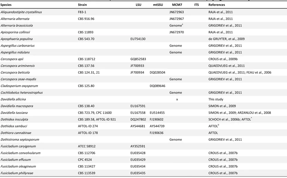
to generate a consensus sequence. Additional sequences used in the
analyses were obtained from GenBank and Fungal Genomics Portal of Joint
Genome Institute (GRIGORIEV et al., 2011) (Table S1). Sequences were
aligned with the Muscle® v. 3.6 software (EDGAR, 2004) implemented in the
Mega 5.0 program (TAMURA et al. 2007). The final data matrices comprised
of the LSU dataset with 62 taxa with 551 unambiguously aligned characters,
mtSSU dataset with 43 taxa and 621 aligned characters, MCM7 dataset with
37 taxa and 468 aligned characters and the ITS dataset with 45 taxa and 462
aligned characters.
The software Modeltest v.3.7 (POSADA and CRANDALL, 1998) was
used to determine the best-fit model of evolution for Maximum Likelihood
(ML) analyses and model selection was based on the Akaike Information
Criterion. The ML analyses were carried out for each of the four datasets
using RAxML (STAMATAKIS, 2006) employing GTR+I+G model of evolution
and bootstrap support was obtained by running 1000 pseudo replicates
(STAMATAKIS et al., 2008). Bayesian analysis was conducted with MrBayes
v.3.1.2 (RONQUIST and HUELSENBECK, 2003) on the same aligned
the GTR+I+G as the best nucleotide substitution model. The Markov Chain
Monte Carlo (MCMC) analysis used four chains that started with a heating
parameter of 0.2 from a random tree topology and lasted 50 million
generations. Trees were saved each 100 generations, resulting in 500,000
saved trees. Burn-in was set at 12,500,000 generations after which the
likelihood values were stationary, leaving 375,000 trees from which the 50 %
majority rule consensus trees and posterior probabilities were calculated.
Quality of mixing and convergence to the stationary distribution were
assessed from three independent runs using Tracer v1.5 (RAMBAUT and
DRUMMOND, 2007).
Assessments of the pleomorphic development of Microcyclus ulei
under natural conditions.
At the Michelin Plantation of Bahia (Brazil), 93 leaves at the B2
developmental stage (HALLÉ and MARTIN, 1968) of 8 rubber trees of the
RO38 clone were tagged with a label and observations were made until
maturity (stage D), from December 15, 2011 to February 24, 2012. A total of
1,353 assessments were made during the experiment.Scoring of sporulation
in lesions naturally infected was performed at every four days using a 1–6
scale for sporulation intensity of the anamorph (Fusicladium) adapted from JUNQUEIRA et al. (1988), where 1 = necrotic non-sporulating lesions, 2 =
chlorotic non-sporulating lesions, 3 = slight sporulation on lower side of the
leaflets, 4 = moderate sporulation on lower side of the leaflets, 5 = high
sporulation on lower side of the leaflets, and 6 = high sporulation on both
same time interval using a 0–4 scale where 0 = no stroma, 1 = 1–5 stromata
per leaflet, 2 = 6–15 stromata per leaflet, 3 = 16–50 stromata per leaflet, and
4 = more than 50 stromata per leaflet.
Test of infectivity and germination of pycnosporos of Microcyclus ulei
under controlled conditions
Suspension of pycnospores was obtained from pycnidia formed in C/D
leaves of the RO38 rubber clone. There were no conidia or ascospores. The
lower surface of three young leaves from the Fx 3864 rubber tree clone were
spray-inoculated with an inoculum suspension of pycnospores or conidia
separately using a HS Airbrush Complete set (Paasche Airbrush company) in
an inoculation chamber with a temperature maintained at 24°C, relative
humidity superior to 85%, artificial daylight of 2.000 lux and 12 h photoperiod.
The inoculum suspension was made of 2x10⁵ spore/ml of water plus Tween
80 at 0.05%. Water with 0.05% Tween 80 was used as a negative control.
Sporulation was scored after 12 days on all inoculated leaves. The
suspensions of pycnospores and conidia were incubated in the dark at 25 ºC
on water agar PSA supplemented as described above. Germination
assessments were conducted at 6, 12, 24 and 120 h of incubation at 24
RESULTS
Phylogeny
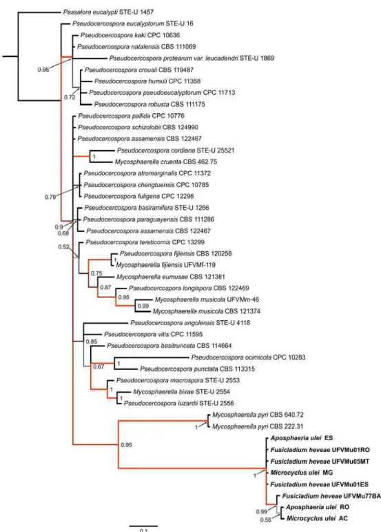
Analyses using maximum likelihood and bayesian methods with
Dothideomycetes members resulted in phylogenies with similar topologies.
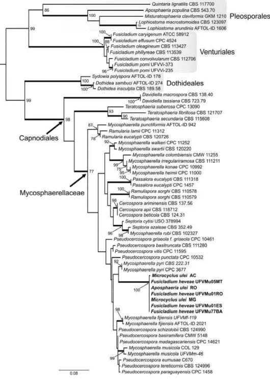
Strongly supported clades confirmed that the holomorph M. ulei belongs to
the family Mycosphaerellaceae s. str., order Capnodiales (Figure 02).
The RAxML search of the partial LSU alignment yielded a most likely
tree (Figure 02) with a log likelihood value of -4021.9. The alignment of the
62 OTUs had 551 sites including alignment gaps, of which 203 sites were
parsimony-informative, 32 were variable and parsimony-uninformative, and
311 were constant. Members of the Lophiostomataceae (Pleosporales),
among them Aposphaeria populina, and species of the genus Fusicladium
(Venturiaceae, Venturiales) formed well-supported monophyletic groups,
while the anamorphs A. ulei and F. heveae were grouped together in the
Pseudocercospora s. str. clade of the Mycosphaerellaceae with
Mycosphaerella pyri as the nearest relative.
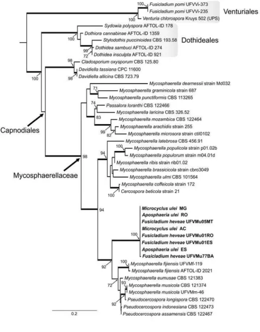
The dataset for the mtSSU sequences had 43 taxa and 621 characters
(202 sites parsimony-informative and 35 singletons) and the log likelihood
value of the most likely tree was -3620.4 (Figure 03). In this analysis, the
holomorph M. ulei had as nearest relatives members of the genus
Pseudocercospora. The phylogeny reconstructed with the partial sequence of the MCM7 region (Figure 04) (log likelihood of -6817.7) was based on a
dataset with 468 characters (244 parsimony-informative sites and 256
clade. The Bayesian analysis of LSU, mtSSU, MCM7 and ITS are shown in
Supplementary Figures (S1 to S4).
Function of intermediate pycnidial stage in the life cycle of Microcyclus
ulei.
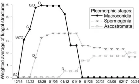
F. heveae was found in 7.0% of the leaves in the B2 stage, 100% in B2/C, C and C/D; and 38.8% in D leaves (Figure 05; Table S2). Most
commonly, the occurrence of F. heveae was recorded after January 19th. A. ulei first emerged from the upper side of infected leaves in the C/D stage in December 26th and it was found in 41.8% of the leaves in this stage and in
97.4% in D stage leaves. Ascostromata arose on January 12th and were
found only in the upper side of D leaves in 62.6% of the observations. Only
during the period of January 12th to January 19th, the three types of pathogen
structures were concomitantly observed.
The main weather descriptors during the course of the experiment (72
days) were: average maximum temperature 29 ºC and average minimum of
22 ºC, the average relative humidity during the day and at night were 72%
and 94%, respectively. Total (cumulative) rainfall was 267 mm.
The suspension of pycnospores did not cause lesions (Figure S4A),
but the pathogen sign was visible with infection from conidia after 12 days of
inoculation (Figure S4B). The pycnospores did not germinate in in vitro
assays, while conidia germination started at 6 h of incubation (data not
shown).
DISCUSSION
DNA sequences of the three stages of the life cycle of M. ulei collected in a wide geographic area in Brazil confirmed the anamorph-teleomorph
connection of this fungal species, but the current classification of the
pathogen in the Planistromellaceae (BARR 1996; LUMBSCH and
HUHNDORF, 2007) was not supported by any of the phylogenies. The
analysis conducted in the present study based on nuclear and mitochondrial
ribosomal rDNA as well as protein-coding gene support the classification of
M. ulei in the family Mycosphaerellaceae s. str., in the order Capnodiales of the phylum Ascomycota. Mycosphaerellaceae is a well-supported family
within the Capnodiales with Mycosphaerella punctiformis as the type species
(VERKLEY et al. 2004; CROUS et al., 2009a). DNA sequence data of M.
punctiformis were included in all our analyses, corroborating the classification of the pathogen at the family level and revealing a close relationship of M. ulei with Mycosphaerella. Currently, robust multi-gene phylogenetic analysis
support Mycosphaerella as a polyphyletic group (CROUS et al., 2007a,
2009b, SCHOCH et al., 2009a, b), suggesting that Mycosphaerella s. l.
should be subdivided to reflect natural groups (genera) as defined by their
anamorphs since Mycosphaerella s. str. is today restricted to species with
Ramularia anamorphs (CROUS et al. 2009b).
The classification of Microcyclus as a Mycosphaerella-like organism
was discussed previously (CANNON, 1995). The Microcyclus genus has
ellipsoidal, hyaline, 1-septate ascospore in clavate, bitunicate asci typical of
stromatic tissue in Microcyclus appears to be the only character that
contributes to its separation from the genus Mycosphaerella (CANNON,
1995). However, some Mycosphaerella-like species have similar erumpent
ascostromata (EVANS, 1984, 2002). As already demonstrated for
Mycosphaerella, the genus Microcyclus,as currently circumscribed, may also be polyphyletic, given the variety of anamorphs associated with the assigned
species (CANNON, 1995).
The conidial stage of M. ulei, F. heveae, is morphologically
indistinguishable from the anamorphic stage of Venturia (SCHUBERT et al.
2003). Other species of Fusicladium s. l. (including Spilocaea and Pollaccia)
and the teleomorphic genus Venturia form a monophyletic group in
Venturiaceae (BECK et al., 2005; ZHANG, et al. 2011a), but some
Fusicladium-like species belong to Sympoventuriaceae (ZHANG, et al.
2011a). Although F. heveae has already been treated as a species of the
Passalora-type and, therefore, a cercosporoid fungus (CROUS and BRAUN, 2003), our molecular data demonstrate that this species is better
accommodated in Pseudocercospora s. str., within the Clade 16 of CROUS
et al. (2009b) and Clade 14 in CROUS et al. (2012) in which the type
species, Pseudocercospora vitis, resides (Figure 02 and S1). Whilst in the Mycosphaerellaceae, many anamorph forms evolved in more than one clade
and represent different genera (CROUS, et al., 2007a; CROUS et al., 2009b)
the anamorph convergence of ‘F. heveae’ is at the order level. This
observation is also evident for ‘A. ulei’(Figure 02).
(CROUS et al., 2009b; CROUS et al., 2012). Pseudocercospora is now recognized as a holomorphic genus, and several species have
Mycosphaerella-like teleomorphs. As observed for ‘F. heveae’ in the present study, when the phylogenetic species concept is applied to other species of
the genera Paracercospora, Cercostigmina, Phaeoisariopsis and Stigmina,
they are reduced to synonymy with the genus Pseudocercospora (STEWART
et al. 1999, CROUS et al. 2001a, BRAUN and HILL 2002, CROUS et al.
2006a).
Recognizing that the anamorphic form of causal agent of SALB
belongs to Pseudocercospora s. st. has important implications, at least the fact that it is a close relative of well-known highly destructive plant pathogens
that affect important crops worldwide (CROUS et al., 2012). This fact allows
for the adoption of a comparative epidemiology and genomics approaches,
using better studied pathogenic species such as P. fijiensis, the causal agent of the black leaf streak disease of banana (CHURCHILL, 2011).
From an evolutionary perspective, and based on the molecular
phylogenetic evidence, the life cycle of M. ulei was re-examined with special attention to its intermediate pycnidial stage. After the proof of the
anamorph-teleomorph connection provided by molecular phylogenetic analyses, the
development of the pathogen in the rubber leaf was monitored under
environmental conditions favorable to the development of SALB.
Physiological data indicate that leaves at the B and C stages act as sinks
with high respiration rates and are almost lignin-free (LIEBEREI, 2007). The
conidial lesions are the first signal of the disease and fertile pycnidia occur
(LANGFORD, 1945; CHEE and HOLLIDAY, 1986). Ascocarps become
mature at about four to six weeks and the formation of ascospores is
correlated with effete pycnidia (HOLLIDAY, 1970). In the present study,
mature pycnidia were seen after three weeks on the upper surface of leaves
in the C/D and D stages in the area occupied by the conidial
(Pseudocercospora) lesions. After five to six weeks, the stromata become more visible and increase in number and size.
In contrast to a previous finding (HOLLIDAY, 1970), but in accordance
to another study (LANGFORD, 1945), our results confirm that the
pycnospores do not germinate in vitro and fail to infect rubber leaves. These observations corroborate the hypothesis that the supposedly erumpent
pycnidia structures are in fact spermogonia and they are involved in the initial
stages of the sexual cycle (LANGFORD, 1945; CHEE and HOLLIDAY, 1986;
EVANS, 2002). Generally, fungi in the Mycosphaerellaceae produce
spermogonia and the spermatia are thought to act as male sexual elements
because of their small size, inability to germinate and infect the host plant,
and pseudothecial development beginning from protoascomata concurrently
(or a few days later) with spermogonia and the two structures are similar in
size and shape (HIGGINS, 1920; SNYDER, 1946; DRING, 1961; INMAN et
al., 1991). The production of spermatia is reported to occur in P. fijiensis
(LIBERATO et al., 2009), and they are considered as male gametes, formed
in spermogonia, which usually develop from the substomatal chambers
before the formation of pseudothecia, although the cytological details of
spermatization and ascospore development have not yet been fully described
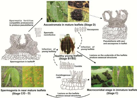
Based on a highly likely phylogenetic position of the pathogen, a
revised version of the life cycle of this pleomorphic fungus that causes SALB
of the rubber tree is presented (Figure 06). Only one anamorphic stage,
which belongs to Pseudocercospora s. str., is present and it infects young leaves being responsible for the secondary cycles of the disease in the field.
The sexual cycle begins with spermogonial developing in the leaf (from stage
C/D) and finishes with mature ascospores in pseudothecia within
TAXONOMY
Pseudocercospora ulei (Kuyper) Authors to be defined later comb. nov.
≡ Fusicladium heveae K. Schub. & U. Braun, in Crous & Braun,
Mycosphaerella and its anamorphs: 1. Names published in Cercospora and
Passalora. CBS Biodiversity Series 1: 481 (2003)
≡ Fusicladium macrosporum Kuyper, Recueil Trav. Bot. Néerl. 8: 374 (1911).
= ?Passalora heveae Massee (nom. nud.) sensu Stahel, Bull. Dept. Landb. Suriname 34: 34 (1917).
Teleomorph: Microcyclus ulei (Henn.) Arx, in Müller & Arx, Beitr.
Kryptogamenfl. Schweiz 11: 373 (1962).
≡ Dothidella ulei Henn., Hedwigia 43(4): 254 (1904).
≡ Melanopsammopsis ulei (Henn.) Stahel, Bull. Dep. Landb. Suriname 34:
1-111 (1917)
Note:
This species is better accommodated in Pseudocercospora
(Mycosphaerellaceae, Capnodiales) than in Fusicladium (Venturiaceae,
ACKNOWLEDGEMENTS
We thank Plantações Michelin da Bahia (Carlos Mattos, Alan Moura,
Anquises Franca, Cícero Cassimiro, José Francisco, Luan Silva, Luciano
Conceição, Saulo Cardoso, Ney Santana, Otamar Santos, Rosival Santos
and Wilton Silva), CEPLAC (Dr. Givaldo Niela, Dr. Karina Gramacho),
Fazenda Batalha (Gilson Assunção), Embrapa Acre (Dr. Rivadalve
Gonçalves), UFV (Jaime Honorato Jr.) for technical and logistic support
during sampling; Luciano Conceição and Pollyanna Fonseca for isolation of
fungal strains. We are grateful to CAPES and CNPq funding agencies for
providing fellowships to BTHJr. This work was supported by Plantações
Michelin da Bahia (CIRAD-Michelin-Brazil SALB resistance breeding
REFERENCES
AGUILETA, G., MARTHEY, S., CHIAPELLO, H., LEBRUN, M. H.,
RODOLPHE, F., FOURNIER, E., GENDRAULT-JACQMUEMARD, A. and
GIRAUD, T. 2008. Assessing the performance of single-copy genes for
recovering robust phylogenies. Syst. Biol., 57: 613-627.
ARZANLOU, M., GROENEWALD, J. Z., FULLERTON, R. A., ABELN, E.
C.,CARLIER, J., ZAPATER, M. F., BUDDENHAGEN, I. W., VILJOEN, A. and
CROUS, P. W. 2008. Multiple gene genealogies and phenotypic characters
differentiate several novel species of Mycosphaerella and related anamorphs on banana. Persoonia, 20: 19-37.
AVISE, J. C. and WOLLENBERG, K. 1997. Phylogenetics and the origin of
species. Proc. Natl. Acad. Sci. USA, 94: 7748-7755.
BARR, M. E. 1996. Planistromellaceae, a new family in the Dothideales.
Mycotaxon, 60: 433-442.
BECK, A., RITSCHEL, A., SCHUBERT, K., BRAUN, U. and TRIEBEL, D.
2005. Phylogenetic relationships of the anamorphic genus Fusicladium s. lat. as inferred by ITS nrDNA data. Mycol. Prog., 4: 111-116.
BRAUN, U. and HILL, C. F. 2002. Some new micromycetes from New
Zealand. Mycol. Prog, 1: 19-30.
CANNON, P. I., CAMARAN C. C. and ROMERO A. I. 1995. Studies on
biotrophic fungi from Argentina: Microcyclus porleriae, with a key to South American species of Microcyclus. Mycol. Res., 99: 353-356.
CHEE, K. H. and HOLLIDAY, P. 1986. South American leaf blight of Hevea
rubber. Malaysian Rubber Research and Development Board. Malaysian
CHURCHILL, A. C. L. 2011. Mycosphaerella fijiensis, the black leaf streak
pathogen of banana: progress towards understanding pathogen biology and
detection, disease development, and the challenges of control. Mol. Plant
Pathol., 12: 307-328.
CROUS, P. W., KANG, J. C. and BRAUN, U. 2001a. A phylogenetic
redefinition of anamorph genera in Mycosphaerella based on ITS rDNA
sequence and morphology. Mycologia, 93: 1081-1101.
CROUS, P. W., HONG, L., WINGFIELD, B. D. and WINGFIELD, M. J.
2001b. ITS rDNA phylogeny of selected Mycosphaerella species and their
anamorphs occurring on Myrtaceae. Mycol. Res., 105: 425-431.
CROUS, P. W. and BRAUN, U. 2003. Mycosphaerella and its anamorphs. 1.
Names published in Cercospora and Passalora. CBS Biodiversity Series 1: 1-571. Centraalbureau voor Schimmelcultures, Utrecht, Netherlands.
CROUS, P. W., LIEBENBERG, M. M., BRAUN, U. and GROENEWALD, J. Z.
2006a. Re-evaluating the taxonomic status of Phaeoisariopsis griseola, the causal agent of angular leaf spot of bean. Stud. Mycol., 55: 163-173.
CROUS, P. W., WINGFIELD, M. J., MANSILLA, J. P., ALFENAS, A. C. and
GROENEWALD, J. Z. 2006. Phylogenetic reassessment of Mycosphaerella
spp. and their anamorphs occurring on Eucalyptus II. Stud. Mycol., 55: 99-131.
CROUS, P. W., BRAUN, U. and GROENEWALD, J. Z. 2007a.
Mycosphaerella is polyphyletic. Stud. Mycol., 58: 1-32.
CROUS, P. W., SCHUBERT, K., BRAUN, U., DE HOOG, G. S., HOCKING,
A. D., SHIN, H. D. and GROENEWALD, J. Z. 2007b. Opportunistic,
saprobic or phytopathogenic species in the Venturiaceae. Stud. Mycol., 58: 185-217.
CROUS, P. W., SCHOCH, C. L., HYDE, K. D., WOOD, A. R., GUEIDAN, C,
HOOG, G. S. and GROENEWALD, J. Z. 2009a. Phylogenetic lineages in the
Capnodiales. Stud. Mycol., 64: 17-47.
CROUS, P. W., SUMMERELL, B. A., CARNEGIE, A. J., WINGFIELD, M. J.,
HUNTER, G. C, BURGESS, T. I., ANDJIC, V., BARBER, P. A. and
GROENEWALD, J. Z. 2009b. Unravelling Mycosphaerella: do you believe in
genera? Persoonia, 23: 99-118.
CROUS, P. W., SUMMERELL, B. A., CARNEGIE, A. J., WINGFIELD, M. J.
and GROENEWALD, J. Z. 2009c. Novel species of Mycosphaerellaceae and
Teratosphaeriaceae. Persoonia, 23: 119-146.
CROUS, P. W., BRAUN, U., HUNTER, G. C., WINGFIELD, M. J., VERKLEY,
G. J. M., SHIN, H. D., NAKASHIMA, C. and GROENEWALD, J. Z. 2012.
Phylogenetic lineages in Pseudocercospora. Stud. Mycol., 75: 37-114.
DOYLE, J. J. and DOYLE, J. L. 1990. Isolation of plant DNA from fresh
tissue. Focus, 12: 13-15.
DRING, D. 1961. Studies on Mycosphaerella brassicicola (Duby) Oudem. T.
Brit. Mycol. Soc., 44: 253-264.
de GRUYTER, J., AVESKAMP, M. M., WOUDENBERG, J. H., VERKLEY, G.
J., GROENEWALD, J. Z. and CROUS, P. W. 2009 Molecular phylogeny of
Phoma and allied anamorph genera: towards a reclassification of the Phoma
complex. Mycol. Res., 113: 508-519.
EDGAR, R.C. 2004. MUSCLE: multiple sequence alignment with high
ERIKSSON, O. E. and HAWKSWORTH, D. L. 1993. Outline of the
ascomycetes-1993. Syst. Ascomycetum, 12: 51-257.
EVANS, H. C. 1984. The genus Mycosphaerella and its anamorphs
Cercoseptoria, Dothistroma and Lecanosticta on pines. Mycol. Pap., 153: 1-102.
EVANS, H. C. 2002. Invasive neotropical pathogens of tree crops. Pages
83-112 in: Tropical Mycology: Vol. 2, Micromycetes. R. Watling, J. Frankland, M.
Ainsworth, S. Isaac, and C. Robinson, eds. CABI Publishing, Wallingford,
Oxon, UK.
FEAU, N., HAMELIN, R. C. and BERNIER, L. 2006. Attributes and
congruence of three molecular data sets: Inferring phylogenies among
Septoria-related species from woody perennial plants. Mol. Phylogenet. Evol., 40: 808-829.
GASPAROTTO, L., SANTOS, A. F., PEREIRA, J. C. R. and FERREIRA, F.
A. 1997. Doenças da Seringueira no Brasil. Embrapa-SPI: Manaus:
Embrapa-CPAA.
GRANDIN, G. 2009, Fordlandia: the rise and fall of Henry Ford's forgotten
jungle city. Metropolitan Books, New York.
GRIGORIEV, I. V., NORDBERG, H., SHABALOV, I., AERTS, A., CANTOR,
M., GOODSTEIN, D., KUO, A., MINOVITSKY, S., NIKITIN, R., OHM, R. A.,
OTILLAR, R., POLIAKOV, A., RATNERE, I., RILEY, R., SMIRNOVA, T.,
ROKHSAR, D. and DUBCHAK, I. 2011. The genome portal of the
GUYOT, J. and DOARÉ, F. 2010. Obtaining isolates of Microcyclus ulei, a
fungus pathogenic to rubber trees, from ascospores. J. Plant Pathol., 92:
765-768.
HALLÉ, F. and MARTIN, R. 1968. Étude de la croissance rythmique chez
l’hévéa (Hevea brasiliensis Müll. Arg., Euphorbiacées, Crotonoïdées).
Adansonia, 8: 475-503.
HENNINGS, P. 1904. Uber die auf Hevea –arten bisher beobachteten
parasitischen pilze. Notizbl. bot. Gart. Mus. Berl., 4: 133-139.
HIBBETT, D. S., BINDER, M., BISCHOFF, J. F., BLACKWELL, M.,
CANNON, P. F., ERIKSSON, O. E., HUHNDORF, S., JAMES, T., KIRK, P.
M., LUCKING, R., LUMBSCH, T., LUTZONI, F., MATHENY, P. B.,
MCLAUGHLIN, D. J., POWELL, M. J., REDHEAD, S., SCHOCH, C. L.,
SPATAFORA, J. W., STALPERS, J. A., VILGALYS, R., AIME, M. C.,
APTROOT, A., BAUER, R., BEGEROW, D., BENNY, G. L., CASTLEBURY,
L. A., CROUS, P. W., DAI, Y. C., GAMS, W., GEISER, D. M., GRIFFITH, G.
W., GUEIDAN, C., HAWKSWORTH, D. L., HESTMARK, G., HOSAKA, K.,
HUMBER, R. A., HYDE, K., IRONSIDE, J. E., KOLJALG, U., KURTZMAN, C.
P., LARSSON, K. H., LICHTWARDT, R., LONGCORE, J., MIADLIKOWSKA,
J., MILLER, A., MONCALVO, J. M., MOZLEY-STANDRIDGE, S.,
OBERWINKLER, F., PARMASTO, E., REEB, V., ROGERS, J. D., ROUX, C.,
RYVARDEN, L., SAMPAIO, J. P., SCHUßLER, A., SUGIYAMA, J., THORN,
R. G., TIBELL, L., UNTEREINER, W. A., WALKER, C., WANG, Z., WEIR, A.,
WEIß, M., WHITE, M. M., WINKA, K., YAO, Y. J. and ZHANG, N. 2007. A
higher-level phylogenetic classification of the Fungi. Mycol. Res., 111: 509-547.
HIGGINS, B. B. 1920. Morphology and life history of some Ascomycetes with
HOLLIDAY, P. 1970. South American leaf blight (Microcyclus ulei) of Hevea brasiliensis. Commonwealth Mycological Institute. Phytopath. Pap., 12: 1-31.
HUNTER, G. C., CROUS, P. W., WINGFIELD, B. D., PONGPANICH, K., and
WINGFIELD, M. J. 2006a. Pseudocercospora flavomarginata sp. nov., from
Eucalyptus leaves in Thailand. Fungal Divers., 22: 71-90.
HUNTER, G. C., WINGFIELD, B. D., CROUS, P. W. and WINGFIELD, M. J.
2006b. A multi-gene phylogeny for species of Mycosphaerella occurring on
Eucalyptus leaves. Stud. Mycol., 55: 147-161.
INMAN, A. J., SIVANESAN, A., FITT, B. D. L. and EVANS, R. L. 1991. The
biology of Mycosphaerella capsellae sp. nov., the teleomorph of
Pseudocercosporella capsellae, cause of white leaf spot of oilseed rape.
Mycol. Res., 95: 1334-1342.
JUNQUEIRA, N. T. V., CHAVES, G. M., ZAMBOLIM, L., ROMEIRO, R. S.
and GASPAROTTO, L. 1984. Isolamento, cultivo e esporulação de
Microcyclus ulei, agente etiológico do mal das folhas da seringueira. Rev. Ceres, 31: 322-331.
JUNQUEIRA, N. T. V., CHAVES, G. M., ZAMBOLIM, L., ALFENAS, A. C.
and GASPAROTTO, L. 1988. Reação de clones de seringueira a vários
isolados de Microcyclus ulei. Pesq. Agropec. Bras., 23: 877-893.
KIRK, P. M., CANNON, P. F., MINTER, D. W. and STALPERS, J. A. 2008.
Dictionary of the Fungi. 10th ed. Wallingford: CABI. ISBN 0-85199-826-7.
KRUYS, A., ERIKSSON, O. E. and WEDIN, M. 2006. Phylogenetic
relationships of coprophilous Pleosporales (Dothideomycetes, Ascomycota),
LANGFORD, M. H. 1945. South American leaf bright of Hevea rubber trees. Technical Bulletin United States Department of Agriculture, 882, 31pp.
LI, K. N., ROUSE, D. I. and GERMAN, T. L. 1994. PCR primers that allow
intergenic differentiation of ascomycetes and their application to Verticillium
spp. Appl. Environ. Microbiol., 60: 4324-31.
LIBERATO, J. R., PETERSON, R. A., GASPAROTTO, L., FERRARI, J. T.,
GRICE, K., PORCHUN, S. C. and SHIVAS, R. G. 2009. Black sigatoka of
banana (Mycosphaerella fijiensis). Available at Pest and Diseases Image
Library, Species Content Page—
http://www.padil.gov.au/viewPestDiagnosticImages. aspx?id=431; Plant
Biosecurity Toolbox/Info Sheet—http://www.padil.
gov.au/pbt/index.php?q=node/46&pbtID=166.
LIEBEREI, R. 2007. South American Leaf Blight of the rubber tree (Hevea
spp.): New steps in plant domestication using physiological features and
molecular markers. Ann. Bot., 100: 1-18.
LINDEMUTH, R., WIRTZ, N. and LUMBSCH, H. T. 2001. Phylogenetic
analysis of nuclear and mitochondrial rDNA sequences supports the view
that Loculoascomycetes (Ascomycota) are not monophyletic. Mycol. Res.,
105: 1176-118.
LUMBSCH, H. T. and HUHNDORF, S. M. 2007. Outline of Ascomycota–
2007. Myconet,13: 1-58.
LUTZONI, F., KAUFF, F., COX, C. J., MCLAUGHLIN, D., CELIO, G.,
DENTINGER, B., PADAMSEE, M., HIBBETT, D. S., JAMES, T. Y., BALOCH,
E., GRUBE, M., REEB, V., HOFSTETTER, V., SCHOCH, C., ARNOLD, A.
E., MIADLIKOWSKA, J., SPATAFORA, J., JOHNSON, D., HAMBLETON, S.,
CROCKETT, M., SHOEMAKER, R., SUNG, G-H., LUCKING, R., LUMBSCH,
HANSEN, K., HARRIS, R. C., HOSAKA, K., LIM, Y-W., MATHENY, B.,
NISHIDA, H., PFISTER, D., ROGERS, J., ROSSMAN, A., SCHMITT, I.,
SIPMAN, H., STONE, J., SUGIYAMA, J., YAHR, R. and VILGALYS, R. 2004.
Assembling the fungal tree of life: progress, classification, and evolution of
subcellular traits. Am. J. Bot., 91: 1446-1480.
MATTOS, C. R. R., GARCIA, D., PINARD, F. and LE GUEN, V. 2003.
Variabilidade de isolados de Microcyclus ulei no Sudeste da Bahia. Fitopatol. Bras., 28: 502-507.
MCLAUGHLIN, D. J., HIBBETT, D. S., LUTZONI, F., SPATAFORA, J. W.
and VILGALYS, R. 2009. The search for the fungal tree of life. Trends
Microbiol., 17: 488-497.
MUGAMBI, G. K. and HUHNDORF, S. M. 2009. Molecular phylogenetics of
Pleosporales: Melanommataceae and Lophiostomataceae re-circumscribed
(Pleosporomycetidae, Dothideomycetes, Ascomycota). Stud. Mycol., 64:
103-121.
MÜLLER, E. and ARX, J. von. 1962. Die Gattungen der didymosporen
Pyrenomyceten. Beitr. Kryptog. Flora Schweiz, 11: 1-992.
NYLANDER, J. A. A. 2004. MrModeltest v 2.2. Program distributed by the
author. Evolutionary Biology Centre, Uppsala University, Uppsala, Sweden.
O’DONNELL, K. 1992. Ribosomal DNA internal transcribed spacers are
highly divergent in the phytopathogenic ascomycete Fusarium sambucinum
(Gibberella pulicaris). Curr. Genet., 22:213–220.
POSADA, D. and CRANDALL, K. A. 1998. Modeltest: testing the model of
QUAEDVLIEG, W., KEMA, G. H. J., GROENEWALD, J. Z., VERKLEY, G. J.
M., SEIFBARGHI, S., RAZAVI, M., GOHARI, A. M., MEHRABI, R. and
CROUS, P. W. 2011. Zymoseptoria gen. nov.: a new genus to accommodate
Septoria-like species occurring on graminicolous hosts. Persoonia, 26: 57–69
RAJA, H. A., SCHOCH, C. L., HUSTAD, V. P., SHEARER, C. A. and
MILLER, A. N. 2011. Testing the phylogenetic utility of MCM7 in the
Ascomycota. MycoKeys, 1: 63-94.
RAMBAUT, A. and DRUMMOND, A. J. 2007. Tracer v1.4, Available from
http://beast.bio.ed.ac.uk/Tracer
RONQUIST, F. and HUELSENBECK, J. P. 2003. MrBayes 3: Bayesian
phylogenetic inference under mixed models. Bioinformatics, 19: 1572-1574.
SCHMITT, I., CRESPO, A., DIVAKAR, P. K., FRANKHAUSER, J. D.,
HERMAN-SACKETT, E., KALB, K., NELSEN, M. P., NELSON, N. A.,
RIVAS-PLATA, E., SHIMP, A. D., WIDHELM, T. and LUMBSCH, H. T. 2009. New
primers for promising single-copy genes in fungal phylogenetics and
systematics. Persoonia, 23: 35–40.
SCHOCH, C. L., SHOEMAKER, R. A., SEIFERT, K. A., HAMBLETON, S.,
SPATAFORA, J. W. and CROUS, P. W. 2006a. A multigene phylogeny of the
Dothideomycetes using four nuclear loci. Mycologia, 98: 1041-1052.
SCHOCH, C. L., KOHLMEYER, J., VOLKMANN-KOHLMEYER, B., TSUI, C.
K. and SPATAFORA, J. W. 2006b. The halotolerant fungus Glomerobolus
gelineus is a member of the Ostropales. Mycol. Res., 110: 257-263.
SCHOCH, C. L., CROUS, P. W., GROENEWALD, J. Z., BOEHM, E. W. A.,
BURGESS, T. I., DE GRUYTER, J., DE HOOG, G. S., DIXON, L. J.,
GRUBE, M., GUEIDAN, C., HARADA, Y., HATAKEYAMA, S., HIRAYAMA,
KOHLMEYER, J., KRUYS, Å., LI, Y. M., LÜCKING, R., LUMBSCH, H. T.,
MARVANOVÁ, L., MBATCHOU, J. S., MCVAY, A. H., MILLER, A. N.,
MUGAMBI, G. K., MUGGIA, L., NELSEN, M. P., NELSON, P., OWENSBY,
C. A., PHILLIPS, A. J. L., PHONGPAICHIT, S., POINTING, S. B.,
PUJADE-RENAUD, V., RAJA, H. A., PLATA, E. R., ROBBERTSE, B., RUIBAL, C.,
SAKAYAROJ, J., SANO, T., SELBMANN, L., SHEARER, C. A., SHIROUZU,
T., SLIPPERS, B., SUETRONG, S., TANAKA, K.,
VOLKMANN-KOHLMEYER, B., WINGFIELD, M. J., WOOD, A. R., WOUDENBERG, J. H.
C., YONEZAWA, H., ZHANG, Y. and SPATAFORA, J.W. 2009a. A
class-wide phylogenetic assessment of Dothideomycetes. Stud. Mycol., 64: 1-15.
SCHOCH, C. L., SUNG, G. H., LOPEZ-GIRALDEZ, F., TOWNSEND, J. P.,
MIADLIKOWSKA, J., HOFSTETTER, V., ROBBERTSE, B., MATHENY, P.
B., KAUFF, F., WANG, Z., GUEIDAN, C., ANDRIE, R. M., TRIPPE, K.,
CIUFETTI, L. M., WYNNS, A., FRAKER, E., HODKINSON, B. P., BONITO,
G., GROENEWALD, J. Z., ARZANLOU, M., DE HOOG, G. S., CROUS, P.
W., HEWITT, D., PFISTER, D. H., PETERSON, K., GRYZENHOUT, M.,
WINGFIELD, M. J., APTROOT, A., SUH, S., BLACKWELL, M., HILLIS, D.
M., GRIFFITH, G. W., CASTLEBURY, L. A., ROSSMAN, A. Y., LUMBSCH,
H. T., UCKING, R. L., UDEL, B. B., RAUHUT, A., DIEDERICH, P., ERTZ, D.,
GEISER, D. M., HOSAKA, K., INDERBITZIN, P., KOHLMEYER, J.,
VOLKMANN-KOHLMEYER, B., MOSTERT, L., O’DONNELL, K., SIPMAN,
H., ROGERS, J. D., SHOEMAKER, R. A., SUGIYAMA, J., SUMMERBELL,
R. C., UNTEREINER, W., JOHNSTON, P. R., STENROOS, S., ZUCCARO,
A., DYER, P. S., CRITTENDEN, P. D., COLE, M. S., HANSEN, K.,
TRAPPE, J. M., YAHR, R., LUTZONI, F. O. and SPATAFORA, J. W. 2009b.
The Ascomycota tree of life: A phylum-wide phylogeny clarifies the origin and
evolution of fundamental reproductive and ecological traits. Syst. Biol., 58: 224-239.
SCHOCH, C. L., SEIFERT, K. A., HUHNDORF, S., ROBERT, V., SPOUGE,
transcribed spacer (ITS) region as a universal DNA barcode marker for
Fungi. Proc. Natl. Acad. Sci. U.S.A., 109: 6241-6246.
SCHUBERT K, RITSCHEL A, BRAUN U. 2003. A monograph of Fusicladium
s. lat. (Hyphomycetes). Schlechtendalia, 9: 1-132.
SIMON, U. K., GROENEWALD, J. Z. and CROUS, P. W. 2009. Cymadothea
trifolii, an obligate biotrophic leaf parasite of Trifolium, belongs to Mycosphaerellaceae as shown by nuclear ribosomal DNA analyses.
Persoonia, 22: 49–55.
SNYDER, W. C. 1946. Spermogonia versus pycnidia in Mycosphaerella
brassicicola. Phytopathology, 36: 481-484.
SPATAFORA, J. W., SUNG, G. H., JOHNSON, D., HESSE, C., O'ROURKE,
B., SERDANI, M., SPOTTS, R., LUTZONI, F., HOFSTETTER, V.,
MIADLIKOWSKA, J., REEB, V., GUEIDAN, C., FRAKER, E., LUMBSCH, T.,
LUCKING, R., SCHMITT, I., HOSAKA, K., APTROOT, A., ROUX, C.,
MILLER, A. N., GEISER, D. M., HAFELLNER, J., HESTMARK, G., ARNOLD,
A. E., BUDEL, B., RAUHUT, A., HEWITT, D., UNTEREINER, W. A., COLE,
M. S., SCHEIDEGGER, C., SCHULTZ, M., SIPMAN, H. and SCHOCH, C. L.
2006. A five-gene phylogeny of Pezizomycotina. Mycologia, 98: 1018-1028.
STADEN, R. 1996. The staden sequence analysis package. Mol. Biotechnol., 5: 233-241.
STAMATAKIS, A. 2006. RAxML-VI-HPC: maximum likelihood-based
phylogenetic analyses with thousands of taxa and mixed models.
Bioinformatics, 22: 2688-2690.
STAMATAKIS, A., HOOVER, P. and ROUGEMONT, J. 2008. A rapid
STEWART, E. L., LIU, Z., CROUS, P. W. and SZABO, L. 1999. Phylogenetic
relationships among some cercosporoid anamorphs of Mycosphaerella
based on rDNA sequence analysis. Mycol. Res., 103: 1491-1499.
TAMURA, K., DUDLEY, J., NEI, M. and KUMAR, S. 2007. MEGA4:
Molecular Evolutionary Genetic Analysis (MEGA) software version 4.0. Mol
Biol. Evol., 24: 1596-1599.
TAYLOR, J. W., JACOBSON, D. J., KROKEN, S., KASUGA, T., GEISER, D.
M., HIBBETT, D. S. and FISHER, M. C. 2000. Phylogenetic species
recognition and species concepts in fungi. Fungal Genet. Biol., 31: 21-32.
TAYLOR, J. E., GROENEWALD, J. Z. and CROUS, P. W. 2003. A
phylogenetic analysis of Mycosphaerellaceae leaf spot pathogens of
Proteaceae. Mycol. Res., 107: 653-658.
VERKLEY, G. J. M., CROUS, P. W., GROENEWALD, J. Z., BRAUN, U. and
APTROOT, A. 2004. Mycosphaerella punctiformis revisited: morphology,
phylogeny, and epitypification of the type species of the genus
Mycosphaerella (Dothideales, Ascomycota). Mycol. Res., 108: 1271-1282.
van BEILEN, J. B. and POIRIER, Y. 2007. Establishment of new crops for the
production of natural rubber. Trends Biotechnol., 25: 522-529.
von ARX, J. A. and MÜLLER, E. 1975. A re-evaluation of the bitunicate
ascomycetes with keys to families and genera. Stud. Mycol., 9: 1-159.
WHITE, T. J., BRUNS, T., LEE, S. and TAYLOR, J. 1990. Amplification and
direct sequencing of fungal ribosomal RNA genes for phylogenetics. In: Innis
M, Gelfand DH, Sninsky JJ, White TJ, editors. PCR protocols. San Diego:
ZHANG, Y., SCHOCH, C. L., FOURNIER, J., CROUS, P. W., DE GRUYTER,
J., WOUDENBERG, J. H. C., HIRAYAMA, K., TANAKA, K., POINTING, S.
B., SPATAFORA, J. W. and HYDE, K. D. 2009a. Multi-locus phylogeny of
Pleosporales: a taxonomic, ecological and evolutionary re-evaluation. Stud. Mycol., 64: 85-102.
ZHANG, Y., WANG, H., FOURNIER, J., CROUS, P. W., JEEWON, R.,
POINTING, S. B. and HYDE, K. D. 2009b. Towards a phylogenetic
clarification of Lophiostoma / Massarina and morphologically similar genera in the Pleosporales. Fungal Divers., 38: 225-251.
ZHANG, Y., CROUS, P. W., SCHOCH, C. L., BAHKALI, A. H., GUO, L. D.
and HYDE, K. D. 2011a. A molecular, morphological and ecological
re-appraisal of Venturiales―a new order of Dothideomycetes. Fungal Divers.,
51: 249-277.
ZHANG, Y., CROUS, P. W., SCHOCH, C. L. and HYDE, K. D. 2011b.
FIGURE LEGENDS
Figure 01. Sampled areas (dots) located in the rubber producing regions in
Brazil. States from which samples were collected are shaded.
Figure 02. Maximum likelihood phylogeny of Microcyclus ulei based on the
first 551 bp at the 5’ end of the 28S rRNA gene (LSU) of 62 taxa using
RAxML (log likelihood = -4021.879653). Thickened branches indicate
significant Bayesian posterior probabilities ≥ 95%; numbers refer to RAxML
bootstrap support values ≥ 70% based on 1000 replicates. The scale bar
shows 0.08 expected changes per site. The tree was rooted to Pleosporales
members.
Figure 03. Maximum likelihood phylogeny of Microcyclus ulei based on the
mtSSUrDNA (621pb) of 43 taxa using RAxML (log likelihood =
-3620.369300). Thickened branches indicate significant Bayesian posterior
probabilities ≥ 95%; numbers refer to RAxML bootstrap support values ≥ 70%
based on 1000 replicates. The scale bar shows 0.2 expected changes per
site. The tree was rooted to Venturiales
Figure 04. Maximum likelihood phylogeny of Microcyclus ulei based on the
gene MCM7 (468 bp) of 37 taxa using RAxML (log likelihood =
-6817.743065). Thickened branches indicate significant Bayesian posterior
probabilities ≥ 95%; numbers refer to RAxML bootstrap support values ≥ 70% based on 1000 replicates. The scale bar shows 0.3 expected changes per
site. The tree was rooted to Aspergillus nidulans and Aspergillus carbonarius.
Figure 05. Pleomorphic development of the life cycle of Microcyclus ulei.
density during the leaf development of RO38 clone rubber in field conditions
in the period of December 15, 2011 to February 24, 2012.
Figure 06. A proposed model of the life cycle of Microcyclus ulei. Arrows
indicate of leaf development; Dotted arrows indicate the enlargement of
fungal structure; and dashed arrows the young leaves as prevalent to start of
Figure 02. Hora Júnior et al
Figure 04. Hora Júnior et al
Figure 06. Hora Júnior et al
Table 01. Origin of the Microcyclus ulei isolates.
Isolate Location Coordinates (Lat/Lon) Collector Sampling Date
Aposphaeria ulei ES Cachoeiro do Itapemirim-ES -20.752609/-41.290358 B.T. Hora Júnior 2010
Aposphaeria ulei RO Ariquemes-RO -9.913333/-63.040833 J. Honorato Júnior 2010
Fusicladium heveae UFVMu77BA Porto Seguro-BA -16.378001/-39.366433 B.T. Hora Júnior 2008
Fusicladium heveae UFVMu01ES Sooretama-ES -19.220087/-40.121414 B.T. Hora Júnior 2009
Fusicladium heveae UFVMu05MT Itiquira-MT -17.208889/-54.150000 B.T. Hora Júnior 2009
Fusicladium heveae UFVMu01RO Buritis-RO -10.211944/-63.828889 J. Honorato Júnior 2010
Microcyclus ulei AC Xapuri-AC -10.651944/-68.503889 B.T. Hora Júnior 2010
SUPPORTING INFORMATION
Figure S1. The 50% majority rule tree of 375,000 trees obtained from a Bayesian analysis showing the phylogenetic relationships of Microcyclus ulei
based on the LSU sequence alignment. Bayesian posterior probabilities are given at the nodes and values ≥ 0.5 and ≥ 0.7 are shown above as blue and red lines, respectively. Scale bar shows 0.06 expected changes per site. The tree was rooted with members of Pleosporales. Bayesian posterior probability values ≥ 0.5 are shown above
Figure S2. The 50% majority rule tree of 375,000 trees obtained from a Bayesian analysis showing the phylogenetic relationships of Microcyclus ulei
Figure S3. The 50% majority rule tree of 375,000 trees obtained from a Bayesian analysis showing the phylogenetic relationships of Microcyclus ulei