LAHYRE IZAETE SILVEIRA GOMES
ETIOLOGY OF SIGATOKA DISEASES IN MINAS GERAIS, GENETIC STRUCTURE OF THE POPULATION OF Mycosphaerella musicola AND SENSITIVITY OF BRAZILIAN
ISOLATES OF Mycosphaerella fijiensis TO FUNGICIDES
Tese apresentada à Universidade Federal de Viçosa, como parte das exigências do Programa de Pós-Graduação em Fitopatologia, para obtenção do título de Doctor Scientiae.
VIÇOSA
ii
Dedico a minha tese ao meu pai Joaquim Gomes Barbosa (in memoriam) e minha mãe Joaquina
iii AGRADECIMENTOS
A Deus, por estar sempre comigo, mesmo quando estou distante.
Ao meu orientador, Eduardo Seiti Gomide Mizubuti, pela orientação, apoio, paciência e principalmente pela confiança e amizade no decorrer destes anos;
À Universidade Federal de Viçosa (UFV), pelo apoio institucional e por possibilitar a minha formação;
À Coordenação de aperfeiçoamento de Pessoal de Nível Superior, (CAPES), pela concessão da bolsa de estudos, ao Conselho Nacional de Desenvolvimento Científico e Tecnológico (CNPq), pela bolsa de doutorado sanduíche e a Fundação de Amparo à Pesquisa do Estado de Minas Gerais (FAPEMIG), pelo financiamento do projeto;
À University of California Riverside pela oportunidade, ao professor Greg W. Douhan, pela confiança, e orientação. Ao professor Albert O. Paulus pelas conversas, risadas e ensinamentos. A Deborah Pagliaccia e Georgios Vidalakis pela amizade;
Aos membros da banca examinadora;
Aos professores e funcionários do Departamento de Fitopatologia da UFV pela atenção, pelos ensinamentos, disponibilidade e auxílio prestado;
Ao BIOPOP: Líllian, Saulo, Braz, Tatiana, Cleisinho, Carine, Robson, Jaime, Aquila, Thaís, Carlos, Edlene, Leonardo, Claudinei, Pollyana, Miller e Raquel pelo companheirismo e amizade;
Aos pesquisadores Gilvan Ferreira da Silva e Rogério Hanada pela confiança e parceria;
À minha mãe, ao meu esposo, avó e irmãos, que entenderam minha ausência e sempre me apoiaram;
iv BIOGRAFIA
LAHYRE IZAETE SILVEIRA GOMES, filha de Joaquim Gomes Barbosa e Joaquina Maria da Silveira Barbosa, nasceu no dia onze de janeiro de 1983, na cidade de Porteirinha-MG.
Em julho de 2006, graduou-se Engenheira Agrônoma pela Universidade Estadual de Montes Claros (UNIMONTES), em Janaúba-MG.
Em agosto de 2008 obteve o título de Mestre em Fitopatologia pela Universidade Federal de Lavras (UFLA).
Em agosto de 2008, iniciou o curso de Doutorado em Fitopatologia, pela Universidade Federal de Viçosa (UFV), obtendo o título de Doctor Scientiae em
v SUMÁRIO
Página
ABSTRACT... vii
RESUMO... ix
GENERAL INTRODUCTION... 1
REFERENCES... 4
CHAPTER 1 1. Etiology of Sigatoka diseases in Minas Gerais State, Brazil …… 8
1.1 Abstract... 8
1.2. Introduction... 9
1.3. Materials and methods... 11
1.4. Results... 15
1.5. Discussion... 17
1.6. Acknowledgements... 21
1.7. References... 22
CHAPTER 2 2. Genetic structure of the population of Mycosphaerella musicola in the state of Minas Gerais, Brazil ………....……….. 31
2.1. Abstract... 31
2.2. Introduction... 32
2.3. Materials and methods... 34
2.4. Results... 38
2.5. Discussion... 40
2.6. Acknowledgements... 42
2.7. References... 43 CHAPTER 3
vi
fungicides………..
3.1. Abstract... 52
3.2. Introduction... 53
3.3. Materials and methods... 54
3.4. Results... 56
3.5. Discussion... 57
3.6. Acknowledgements... 60
3.7. References... 61
vii ABSTRACT
GOMES, Lahyre Izaete Silveira, D.Sc., Universidade Federal de Viçosa, March, 2012. Etiology of Sigatoka diseases in Minas Gerais, genetic structure of the population of Mycosphaerella musicola and sensitivity of Brazilian isolates of Mycosphaerella fijiensis to fungicides. Adviser: Eduardo Seiti Gomide Mizubuti. Co-advisers: Luiz Antonio Maffia and Robert Weingart Barreto.
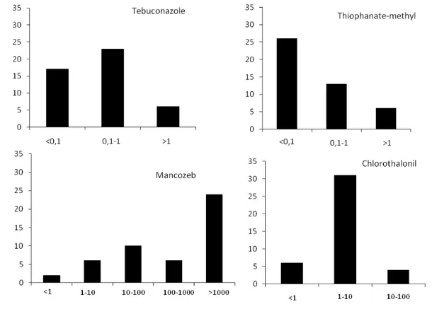
The Sigatoka complex is one of the most serious phytosanitary problem affecting banana crops because of the high yield losses associated to it. Among the species of Mycosphaerella three are frequently associated with Sigatoka diseases: Mycosphaerella musicola, causing yellow Sigatoka; M. fijiensis, causing black Sigatoka, and M. eumusae, causing eumusae leaf spot. The latter has not been reported in Brazil yet. In the State of Minas Gerais, yellow Sigatoka is found in all banana growing areas, while black Sigatoka has been reported only in the South and the Zona da Mata regions. The objectives of this work were to evaluate the distribution of Mycosphaerella spp.; to assess the genetic variability of the most common Mycosphaerella species associated with Sigatoka in Minas Gerais state; and to evaluate the sensitivity of M. fijiensis to different fungicides. A total of 239 isolates were obtained from different regions of Minas Gerais and all belonged to M. musicola. No isolate of M. fijiensis or M. eumusae was found. The isolates of M. musicola were characterized through the use of nine microsatellites markers and mating type. The population reproduces sexually and the isolates of each mating type are distributed at a 1:1 ratio. Additionally high haplotype and genetic diversity were observed, but no population structure was revealed by the Bayesian assignment method. The sensitivity of M. fijiensis to the fungicides thiophanate-methyl, tebuconazole, chlorothalonil and mancozeb was assessed using 45,
45, 41 and 48 isolates respectively, obtained from different regions. No isolate was insensitive to tebuconazole. The highest values of EC50 to thiophanate-methyl and
chlorothalonil were 8.22 μg mL-1 and 53.7 μg mL-1, respectively. Approximately 50% of the isolates grew on culture medium amended with 1000 μg of mancozeb mL-1. There was no correlation between EC50 and geographic region. Yellow Sigatoka disease is the
viii
ix RESUMO
GOMES, Lahyre Izaete Silveira, D.Sc., Universidade Federal de Viçosa, março de 2012. Etiologia de doenças de Sigatoka em Minas Gerais, estrutura genética da população de Mycosphaerella musicola e sensibilidade de isolados de Mycosphaerella fijiensis do Brasil a fungicidas Orientador: Eduardo Seiti Gomide Mizubuti. Coorientadores: Luiz Antonio Maffia e Robert Weingart Barreto.
O complexo Sigatoka é um dos problemas fitossanitários mais graves para a cultura da banana, pois são doenças foliares que resultam em grandes perdas de produção. Dentre as espécies fúngicas do gênero Mycosphaerella, três são frequentemente relatadas estar associadas ao complexo Sigatoka: Mycosphaerella musicola, causadora da Sigatoka Amarela; M. fijiensis, causadora da Sigatoka Negra e M. eumusae que causa a mancha foliar eumusae. Esta última ainda não foi relatada no Brasil. Em Minas Gerais, a Sigatoka Amarela é encontrada em todos os locais onde se cultiva banana, enquanto a ocorrência de Sigatoka Negra foi relatada em alguns municípios das regiões do Sul de Minas e da Zona da Mata. O objetivo deste trabalho foi avaliar a distribuição de Mycosphaerella spp. em Minas Gerais, determinar a espécie predominante e realizar um estudo populacional desta espécie em Minas Gerais; e avaliar a sensibilidade de M. fijiensis a diferentes fungicidas. Os 239 isolados obtidos de diferentes regiões de Minas Gerais, foram identificados com base nos caracteres morfológicos e moleculares, como pertencendo a M. musicola. Nenhum isolado de M. fijiensis ou de M. eumusae foi encontrado. O estudo da população de M. musicola foi realizado com nove marcadores microssatélites e mating type. Os resultados obtidos demonstram que em Minas Gerais
M. musicola reproduz sexuadamente e a distribuição de isolados dos dois mating types
foi de 1:1. Além disso, foi observada alta diversidade haplotípica e genética, além da
ausência de estruturação da população revelada pela análise Bayesiana. Avaliaram-se
45, 45, 41 e 48 isolados de M. fijiensis coletados em bananais de diferentes regiões do
Brasil quanto à sensibilidade aos fungicidas tiofanato metílico, tebuconazole, clorotalonil e mancozebe, respectivamente. Nenhum isolado foi insensível ao fungicida tebuconazole. Os maiores valores de EC50 para tiofanato metílico e clorotalonil foram
8,22 μg mL-1e 53,7 μg mL-1
x
os valores de EC50 e regiãogeográfica. Sigatoka amarela é a doença foliar prevalente em
1
GENERAL INTRODUCTION
The Sigatoka diseases of banana are caused by species of the Mycosphaerella genus which are often considered as destructive plant pathogens (Arzanlou et al., 2007; Arzanlou et al., 2008; Crous, 2009; Crous and Mourichon, 2002; Zapater et al., 2008). Mycosphaerella spp. that infect banana induce necrosis of leaf tissue leading to a reduction in the photosynthetic capacity of the plant and also cause physiological changes that contributes to premature ripening of the fruits (Marin et al., 2003). The main Mycosphaerella species that infect banana are: Mycosphaerella musicola, M. fijiensis and M. eumusae. Yellow Sigatoka is caused by M. musicola R. Leach ex J. L. Mulder (anamorph Pseudocercospora musae) (Zimm.) Deighton, and is present in all continents (Jones, 2003). In Brazil, the disease was detected in 1944 in the state of Amazonas and is currently present in all states (Cordeiro et al., 2005).
Black leaf streak disease (BLSD), also known as Black Sigatoka, is caused by M. fijiensis M. Morelet (anamorph Pseudocercospora fijiensis) (M. Morelet) Deighton
and was first reported in the state of Amazonas in Brazil in 1998 (Pereira et al., 1998). BLSD causes major yield losses in banana, ranging from 20 to 80%, when fungicides are not used (Cordeiro and Matos, 2003). In Minas Gerais the occurrence of BLSD was
first reported in 2005 in the Zona da Mata and South regions (Castro et al., 2005). Mycosphaerella eumusae Crous and Mourichon (anamorph Pseudocercospora eumusae Crous and Mourichon), the most recent species recognized as also involved in the Sigatoka complex, causes the eumusae leaf spot and was first reported in the early 1990s in Southeast Asia (Carlier et al., 2000). To date, M. eumusae is not known to be present in Brazil.
Mycosphaerella musicola was already widely distributed before M. fijiensis was first reported, but when introduced into a new area the latter generally displaces the former (Jones, 2003). However, in many regions where both diseases are known to occur M. musicola remained the major problem, particularly in regions with high altitudes and cooler temperatures. Mycosphaerella fijiensis causes more severe leaf spot at lower and warmer regions (Churchill, 2011; Moulion-Pefoura et al., 1996).
2
transcribed spacer region of rDNA made it possible to differentiate M. musicola from M. fijiensis (Johanson et al., 1994). More recently, a new set of species-specific primers based on the actin gene and the use of real-time PCR TaqMan diagnostic probes based on the sequence of the B-tubulin gene also contributed to sorting M. musicola, M. fijiensis and M. eumusae (Arzanlou et al., 2007). Overall, molecular tools have been helpful in elucidating key issues of the Sigatoka complex.
The study of genetic diversity of fungal populations is important to the development of strategies for plant propagation and management of disease resistance (Churchill, 2011). There are not many studies conducted to investigate the genetic structure of M. musicola, and most used RAPD and RFLP markers (Hayden et al., 2003; Hayden et al., 2005; Moreira et al., 2003). In 2003 isolates of M. musicola from four geographic populations representing Africa, Latin America and Caribbean, Australia and Indonesia were examined using RFLP and moderate levels of genetic diversity were observed for most populations. Genetic differentiation between populations was high when pairwise comparisons were made except for the Africa and Latin America-Caribbean populations, suggesting the occurrence of migration of the pathogen between these regions (Hayden et al., 2003). A total of 363 isolates collected along the Australian coast was studied by Hayden et al. (2005) using RFLP, and the population displayed moderate levels of gene diversity and low gene flow at a continental scale. In Brazil a study with few isolates using RAPD was made and revealed high genetic diversity among isolates (Moreira et al., 2003). Microsatellites markers were also developed to study the genetic diversity of M. musicola (Molina et al., 2001; Zapater et al., 2008), however no thorough population genetics study using these markers were conducted yet.
3
registered for use in the control of BLSD are DMI, QoI and dithiocarbamates (Agrofit, 2012).
Due to its short generation time and high levels of genetic variation, M. fijiensis has a high potential for development of fungicide resistance (Brent and Hollomon, 1998). Since 1987 there is a working group of banana industry representatives participating in the Fungicide Resistance Action Committee (FRAC) that searches for
ways to minimize the risk of the development of resistance. For Sigatoka disease the intensive use of fungicides has imposed selective pressures that have been promoting the development of resistant populations (Ma and Michailides, 2005). This serious issue can be exemplified by several reports of changes in the sensitivity of M. fijiensis populations to fungicides (Amil et al., 2007; Cañas-Gutíerrez et al., 2009; Chin et al., 2001; Knight et al., 2002; Marin et al., 2003; Romero and Sutton, 1997).
The aims of the present study were:
- To identify the predominant species of Mycosphaerella in banana fields in
Minas Gerais state using molecular and traditional tools.
- To describe the genetic structure of M. musicola populations in Minas
Gerais using microsatellites markers.
4
REFERENCES
AGROFIT. 2012. Available at: http://extranet.agricultura.gov.br/agrofit_cons/ principal_agrofit_cons [Assessed January 12, 2012].
Amil, A., Heaney, S., Stanger, C., and Shaw, M. 2007. Dynamics of QoI sensitivity in
Mycosphaerella fijiensis in Costa Rica during 2000 to 2003. Phytopathology, 97: 1451-1457.
Arzanlou, M., Abeln, E., Kema, G., Waalwijk, C., Carlier, J., de Vries, I., Guzman, M., and Crous, P. 2007. Molecular diagnostics for the sigatoka disease complex of banana. Phytopathology, 97: 1112-1118.
Arzanlou, M., Groenewald, J., Fullerton, R., Abeln, E., Carlier, J., Zapater, M., Buddenhagen, I., Viljoen, A., and Crous, P. 2008. Multiple gene genealogies and phenotypic characters differentiate several novel species of Mycosphaerella and related anamorphs on banana. Persoonia, 20: 19-37.
Brent, K. J., and, Hollomon, D.W. 1998. Fungicide resistance: the assessment of risk.
FRAC Monogr. No. 2, Global Prot. Fed. 48 pp.
Canas-Gutierrez, G., Angarita-Velasquez, M., Restrepo-Florez, J., Rodriguez, P., Moreno, C., and Arango, R. 2009. Analysis of the CYP51 gene and encoded protein in propiconazole-resistant isolates of Mycosphaerella fijiensis. Pest Management Science, 65: 892-899.
Carlier, J., Zapater, M. F., Lapeyre, F., Jones, D. R., and Mourichon, X. 2000. Septoria
Leaf Spot of Banana: A newly discovered disease caused by Mycosphaerella eumusae
(Anamorph Septoria eumusae). Phytopathology, 90: 884-890.
5
Chin, K. M., Wirz, M., and Laird, D. 2001. Sensitivity of Mycosphaerella fijiensis from banana to Trifloxystrobin. Plant Disease, 85: 1264-1270.
Churchill, A. C. L. 2011. Mycosphaerella fijiensis, the black leaf streak pathogen of banana: progress towards understanding pathogen biology and detection, disease development, and the challenges of control. Molecular Plant Pathology, 12:307-328.
Cordeiro, Z. J. M., and Matos, A. P. de. 2005. Doenças da banana. Informe Agropecuário. Belo Horizonte, 26: 12-16.
Cordeiro, Z. J. M., and Matos, A. P. de. 2003. Impact of Mycosphaerella spp in Brazil. In: Workshop on Mycosphaerella leaf spot diseases held in San Jose, 2002, Costa Rica. Mycosphaerella leaf spot diseases of bananas: present status and outlook. Montpellier: INIBAP, 2003. p. 91-97.
Crous, P. W. 2009. Taxonomy and phylogeny of the genus Mycosphaerella and its
anamorphs. Fungal Diversity, 38: 1-24.
Crous, P. W., and Mourichon, X. 2002. Mycosphaerella eumusae and its anamorph Pseudocercospora eumusae spp. nov.: causal agent of eumusae leaf spot disease of banana. Sydowia, 54: 35-43.
FRAC. 2010. Fungicide resistance action committee. Available at: http://www.frac.info/frac/index.htm [Assessed September 12, 2011].
Gasparotto, L., Pereira, J. C. R., Hanada, R. E., and Montarroyos, A. V. V. 2006. Sigatoka-negra da bananeira. Manaus: Embrapa Amazônia Ocidental. 177p.
Hayden, H. L., Carlier, J., and Aitken, E. A. B. 2003. Population differentiation in the
banana leaf spot pathogen Mycosphaerella musicola, examined at a global scale. Plant Pathology, 52: 713-719.
6
Johanson, A., Crowhurst, R. N., Rikkerink, E. H. A., Fullerton, R. A., and Templeton, M. D. 1994. The use of species-specific DNA probes for the identification of
Mycosphaerella fijiensis and M. musicola, the causal agents of Sigatoka disease of banana. Plant Pathology, 43: 701-707.
Jones, D. R., 2000. Sigatoka. In Diseases of Banana, Abacá and Enset, D.R. Jones (ed.), pp. 79-92. Wallingford, UK: CABI Publishing.
Jones, D. R. 2003. The distribution and importance of the Mycosphaerella leaf spot diseases of banana. In: Mycosphaerella Leaf Spot Diseases of Bananas: Present Status and Outlook. Proceedings of the Workshop on Mycosphaerella Leaf Spot Diseases, San José, Costa Rica, 20-33 May 2002 (Jacome, L., Lepoivre, P., Marin, D., Oriz, R., Romero, R. and Escalant, J.V., eds), pp. 25-41. Montpellier: The International Network
for the Improvement of Banana and Plantain.
Knight, S., Wirz, M., Amil, A., and Cook, A. 2002. Fungicide resistance in Mycosphaerella fijiensis Morelet: Current status and outlook.In Acorbat. Memorias XV reunion, Realizada en Cartagena de Indias, Colombia. 27 de octubre al 02 Noviembre 2002. Medellín. (COL): Asociacion de Bananeros de Colombia AUGURA, 2002.
Ma, Z., and Michailides, T. J. 2005. Advances in understanding molecular mechanisms of fungicide resistance and molecular detection of resistant genotypes in phytopathogenic fungi. Crop Protection, 24: 853-863.
Marín, D. H., Romero, R. A., Guzmán, M., and Sutton, T. B. 2003. Black Sigatoka: An increasing threat to banana cultivation. Plant Disease, 87: 208-222.
Molina, C., Kaemmer, D., Aponte, S., Weising, K., and Kahl, G. 2001. Microsatellite markers for the fungal banana pathogen Mycosphaerella musicola. Molecular Ecology Notes, 1: 137-139.
7
Mouliom-Pefoura, A., Lassoudière, A., Foko, J., and Fontem, D. A. 1996 Comparison of development of Mycosphaerella fijiensis and Mycosphaerella musicola on banana and
plantain in the various ecological zones in Cameroon. Plant Disease, 80: 950-954.
Pereira, J. C. R., Gasparotto, L., Coelho, A. F. S., and Urben, A. 1998. Ocorrência da sigatoka-negra no Brasil. Fitopatologia Brasileira, 23: 295.
Romero, R. A., and Sutton, T. B. 1997. Sensitivity of Mycosphaerella fijiensis, causal agent of black Sigatoka of banana, to propiconazole. Phytopathology, 87: 96-100.
Zapater, M., Duchemin, M., Dussart, J., Coste, D., Brottier, P., and Carlier, J. 2008. Microsatellite markers for the fungal banana pathogens Mycosphaerella fijiensis,
8
1. Etiology of Sigatoka Diseases in Minas Gerais State, Brazil
1.1 Abstract
A thorough assessment of the distribution of Mycosphaerella spp. in Minas Gerais state,
Brazil, was conducted after the first report of the occurrence of Mycosphaerella
fijiensis, published in 2005. From 2009 to 2011, 80 fields located in 20 municipalities
including the same fields where the disease was first reported were sampled. A total of
800 samples of leaf tissue with symptoms similar to those of yellow or black leaf streak
Sigatoka diseases were examined, and 239 isolates were obtained. The identification of
the fungi was based on morphological characters combined with DNA sequences
obtained after amplification with species-specific primers and phylogeny inferred from
the ITS region of Mycosphaerella strains from banana. All 239 isolates were identified
as M. musicola. The absence of M. fijiensis in the examined samples may have been due
to the no displacement of M. musicola by M. fijiensis or no occurrence of this species in
Minas Gerais. Yellow Sigatoka is the prevailing leaf spot disease of bananas in Minas
9 1.2 Introduction
There are three leaf spots commonly referred to as Sigatoka diseases of banana which are caused by three distinct species of Mycosphaerella. All species cause necrosis of leaf tissues which leads to defoliation that result in yield loss and premature or uneven ripening of the fruit. Yellow Sigatoka (YS) and black Sigatoka, also known as black leaf streak disease (BLSD), are the two major leaf spot diseases that affect banana (Musa spp.) production in many tropical countries (Stover, 1972). Mycosphaerella musicola R. Leach ex J. L. Mulder (anamorph Pseudocercospora musae) (Zimm.)
Deighton is the most frequently reported causal agent of YS and was the first „Sigatoka‟
foliar pathogen to be identified in 1902 (Jones, 2003). This pathogen is globally distributed and causes crop losses wherever bananas are grown. In 1963,
Mycosphaerella fijiensis M. Morelet (anamorph Pseudocercospora fijiensis (M. Morelet) Deighton) was identified as the causal agent of BLSD (Rhodes, 1964). Mycosphaerella fijiensis is not as widely distributed as M. musicola but due to its more aggressive nature has often displaced populations of M. musicola where the diseases co-occur (Romero, 2003) and BLSD requires more intensive fungicide spray schedules to be controlled (Carlier et al., 1999a). When the species co-occur within a region, their distribution appears to be affected by weather conditions, mainly temperature and humidity (Cintra et al., 2008; Mouliom-Pefoura, 1996; Romero and Gauhl, 1988). More recently, Mycosphaerella eumusae Crous & Mourichon (anamorph Pseudocercospora eumusae Crous & Mourichon) was reported in Asia (Carlier et al., 2000) and is currently distributed in South-East Asia and parts of Africa (Arzanlou et al., 2010).
Diagnosis of the fungal species that cause Sigatoka diseases has traditionally been based on disease symptoms and morphology of the fungi (Fortune et al., 2005; Gasparotto et al., 2006; Ploetz, 2004; Stover, 1972). However, all three Mycosphaerella species cause similar symptoms in banana leaves, which makes disease diagnosis very challenging. Due to the conserved teleomorph morphology, there are no differences between perithecia and ascospores among the three species that infect banana (Meredith and Lawrence, 1969). The major morphological differences between M. musicola, M. fijiensis and M. eumusae is in the anamorphic state (Crous and Mourichon, 2002).
Conidiophores of M. musicola produce dense fascicles (sporodochia) on a dark brown or black stroma while the conidiophores of M. fijiensis emerge singly or in small groups
10
morphologically very similar to M. eumusae, and reports suggest that these two pathogens have commonly been misidentified (Crous and Mourichon, 2002). Sporodochia of M. eumusae have a similar shape to those of M. musicola showing epiphyllous sporodochia formed on dark brown substomatal stromata. Moreover, even when the pathogens are isolated from diseased tissues, identification of the various Mycosphaerella species is often difficult due to the overlapping and limited
morphological characters (Arzanlou et al., 2007). Reliable diagnosis is essential since all three diseases require unique management strategies to achieve satisfactory levels of disease control (Johanson and Jeger, 1993). M. fijiensis, for example, infects younger leaves on susceptible banana clones than those affected by M. musicola (Jones, 2003) and symptoms develop faster on banana infected with M. fijiensis and M. eumusae than with M. musicola (Balint-Kurti et al., 2001). Therefore, chemicals are to be applied at different times, perhaps in different targets, and at variable number of applications to control each disease.
To overcome difficulties in disease diagnosis, polymerase chain reaction (PCR) methods have been developed to aid in proper species identification as has been done for many other important plant pathogens (Arzanlou et al., 2007; Frederick et al., 2002; Johanson and Jeger, 1993). Specifically, primers designed to amplify the internal transcribed spacer regions of rDNA have made it possible to differentiate M. musicola from M. fijiensis (Johanson and Jeger, 1993). More recently, species-specific PCR primers were developed based on the actin locus and the development of TaqMan real-time quantitative PCR assay based on the beta-tubulin locus has made it possible to differentiate M. musicola, M. fijiensis and M. eumusae (Arzanlou et al., 2007).
In Brazil, YS occurs in all regions where banana is cultivated and BLSD was
identified as occurring in Amazonas state in 1998 (Pereira et al., 1998). BLSD spread rapidly to other areas throughout the North of Brazil, but has not been reported in the state of Tocantins. Afterwards, the reports of occurrence of BLSD followed a North-South of the country pathway. The disease spread to the central-west regions (Mato Grosso and Mato Grosso do Sul) and was also reported to be present in the states of São Paulo and Minas Gerais, both in the Southeast region of Brazil (Castro et al., 2005; Gasparotto et al., 2006; Hanada et al., 2007) and in other states of the South region (Figure 1A).
11
(Castro et al., 2005; Ferrari et al., 2005). Mycosphaerella fijiensis is a quarantine pathogen in some places in Brazil and regulatory mechanisms prohibiting the movement of fruits and other plant materials are reinforced to avoid the spread of the pathogen. This regulatory measure has resulted in serious economic implications to some banana producing states since fruits or plantlets produced in states where the disease has been recorded cannot be sold in states which are free of BLSD. In October 2006, one year
after the first report of BLSD in Minas Gerais, a phytosanitary survey was conducted across the main banana regions of the state and a regulatory document was issued by the Ministry of Agriculture of Brazil declaring some municipalities in the North, Northwest, Triângulo and Vale do Jequitinhonha regions as free from BLSD (Maciel, 2006). After the abovementioned survey, no change in the intensity of necrotic leaf spots affecting banana leaves was observed by growers in areas where BLSD was originally reported; nor has the usage of fungicide increased to control the disease. The disease is
considered to be “under control” and probably restricted or absent to the areas where it was initially found. Apparently, in the state of Minas Gerais, putative epidemics of BLSD, are not as severe as those recorded in others regions of Brazil. This is a controversial scenario which remains unresolved. A clear response to whether M. fijiensis have been rightfully reported in Minas Gerais must be reached. Therefore, the objectives of this work were to (i) determine the etiological agent of the Sigatoka diseases in the main banana producing regions of Minas Gerais state, including those fields where the BLSD was first reported and (ii) to clarify the present status of the disease in Minas Gerais.
1.3 Materials and methods
Sampling scheme and isolate recovery
Leaf pieces (20 by 20 cm) with symptoms of Sigatoka disease were collected,
packed in plastic bags and taken to the laboratory. After arriving at the laboratory the
samples were kept in the refrigerator until examination and pathogen isolation.
Symptomatic leaves were collected from plants of different ages, but no adjacent plants
were sampled in a field. Leaves of a minimum of 10 symptomatic plants were collected
per field. For each sample, geographic coordinates were obtained using a portable GPS
12
Diseased plant material was collected from 80 fields in 20 municipalities within four regions of Minas Gerais state: South, North, Triângulo Mineiro (located to the West)
and Zona da Mata (located to the East) (Figure 1B and Table 1). The sampling started in
January 2009 in the North region of Minas Gerais. From January 2010 until March 2011
a more intensive sampling was performed at different locations. Fields were chosen
such as to represent the diversity of farming practices within the regions from small
local farms (0.5 ha) with limited input to large (280 ha), well-managed banana
plantations. All samples were collected during the summer, period of more intense
rainfall and higher temperature, favorable for the occurrence of the diseases. In some
fields from Zona da Mata, South and North of Minas Gerais state sampling was
conducted in two different years (Table 1).
All plant samples were first tentatively diagnosed by critically observing the Sigatoka disease symptoms in the field. The sampling procedure was biased towards the
detection of BLSD as leaves with symptoms resembling those of BLSD were sought more carefully and all suspicious plants were sampled. A subset of the samples, mainly comprised of leaf pieces with BLSD-like symptoms, were subjected to more detailed analysis using microscopic characters.
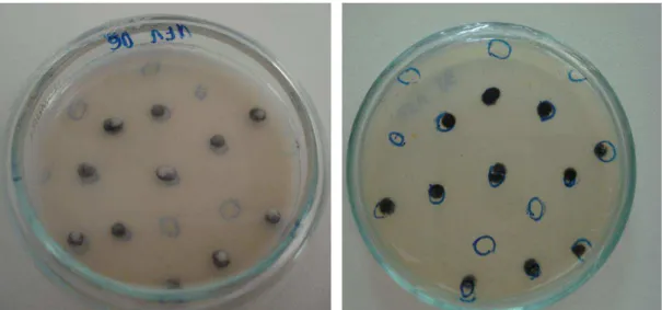
Under a compound microscope and using a sterile fine needle, conidia present on abaxial surface of the symptomatic leaves were picked and transferred to water-agar medium. Single conidium was then taken from the water-agar medium and transferred to be cultured on V8 agar medium (300 mL V8 juice, 3 g of CaCO3 and 20 g of agar per
liter of medium). The single-spore isolates were incubated at 25oC under 12 h of daily white light regime for 10 days prior to examination and DNA extraction (Figure 2).
Discharge of ascospores from necrotic leaf material was used for leaf samples when
13 Species identification
Morphological characters
Due to the difficulties to produce large quantities of asexual spores in vitro, four isolates of each region were randomly chosen for the morphometric assessments and characterization. The conidia were transferred to microscope slides previously prepared with 50% glycerin and then observations were made under a light microscope. The morphological characters examined were: conidium dimension (length and diameter), pattern of conidia, septation, basal hilum and conidial shape and presence of sporodoquia (Stover, 1972). The identification was derived from approximately 50 observations of conidium from each region.
DNA extraction
DNA was extracted from every single-spore culture. Four mycelium plugs from colonies growing on V8 agar were transferred to 50 mL of V8 broth (250 mL of V8 juice and 3 g CaCO3 per liter of medium, pH 6.5) in a 250 mL flask and incubated at
25oC for 10 days at 25°C at 120 rpm in a shaker. The mycelium was then washed with sterile water and the excess water was removed using sterile filter paper. The mycelium was ground to powder using liquid nitrogen in a porcelain mortar and pestle. DNA was extracted using the cetyl trimethyl ammonium bromide (CTAB) method (Doyle and Doyle, 1990). An aliquot of 50 μl of TE buffer (10 mM Tris-HCl and 1 mM EDTA, pH 8.0) was added to each DNA sample. RNA was digested with RNase A for 2h at 37oC before storage at -20°C. The concentration of DNA was quantified in a spectrophotometer (NanoDrop 2000 Thermo Scientific) and working solutions were standardized to 20g/µ L of DNA.
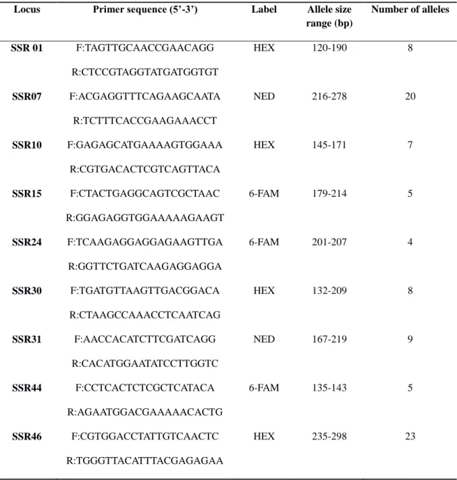
Species specific primers
14
performed in a thermal cycler MJ PTC-100. After the PCR reaction, 5µl of each PCR product was subjected to electrophoresis in 2.0% agarose gel in 1x TBE and viewed under UV on gel stained with GelRed (Biotium). Fragments were compared with a 100bp DNA ladder and scored.
Amplifications controls were made to validate the results. Thus the specific primer used to detect M. fijiensis was tested with DNA from naturally infected banana
leaves collected in Manaus, Amazonas state, where BLSD is known to occur and the expected diagnostic bands were present. The specific primer sets (ACTR/MFactF, MmactF2/MmactRb and MEactR/ACTF) were also used for DNA amplification from healthy banana leaves (controls). The primers MEactR/ACTF were not tested as control due the absence of M. eumusae in Brazil, but were tested with DNA from leaf tissue with symptoms of yellow Sigatoka.
DNA phylogeny
Partial sequences of the internal transcribed spacer (ITS) region was
PCR-amplified using fungal specific primers ITS1F (Gardes and Bruns, 1993) and ITS4 (White et al., 1990). PCR amplifications were done in 20 μl reaction volumes containing 1X PCR buffer (Invitrogen, Carlsbad, California), 2.5 mM MgCl2, 0.2 mM
each dNTP (Invitrogen), 0.375 mM each primer and 0.5 U Taq polymerase (Invitrogen). Thermocycling conditions consisted of an initial melt at 94o C for 3 min, followed by 30 cycles of 94o C (30 s), 55o C (30 s), and 72o C (1 min), and a final hold of 72o C for 8 min. All amplifications were performed in a MyCycler thermocycler (Bio-Rad
Laboratories Inc., Hercules, California). For each PCR amplification, 5 μl were subjected to electrophoresis in 1.8% agarose gels stained with SYBR Green I nucleic acid stain (Invitrogen) and viewed with UV light. PCR products were cleaned with ExoSap-IT (USB, Cleveland, Ohio) following the manufacturer‟s instructions. Sequencing was performed at the Core Instrumentation Facility (CIF) of the University
of California‟s (UC) Institute of Integrative Genome Biology at UC Riverside.
15
EU140340, GU168036), M. musicola (AY646473, AY646466, AY646505, AY646507), Pseudocercospora indonesiana (EU514283, EU514283), P. basiramifera (AF309595), P. paraguayensis (DQ267602), P. assamensis (EU514281), and P. longispora (EU514284, EU514285) which were downloaded from GenBank. The first set was used to estimate the number of haplotypes in the sample and the second set was used for phylogenetic inferences. Sequences of the second set were subjected to Bayesian
phylogenetic analysis using MrBayes v.3 (Ronquist and Huelsenbeck, 2003). Nucleotide substitution model was selected using MrModeltest v. 2.2 (Nylander, 2004) and the HKY+I evolution model was used for the analysis. The Markov Chain Monte Carlo (MCMC) analysis of four chains started from a random tree topology and lasted 10.000.000 generations. Trees were saved at each 100 generations, resulting in 75.000 saved trees. Burn-in of 25% was made and posterior probabilities (PP‟s) were calculated.
Maximum parsimony (MP) analysis was made with the heuristic searches with 1000 random-addition sequence replicates and tree bisection reconnection (TBR) branch swapping using PAUP* (4.0 beta 10) (Swofford, 2002). Gaps were treated as missing data.
1.4 Results
Sampling and fungal isolation
A total of 800 samples of leaf tissue were examined and most had typical symptoms of the yellow Sigatoka: elongated streaks, elliptical in shape and chlorotic areas around the necrotic spots with dark brown borders, gray color center where regular lines of sporodochia could be seen. Symptoms of five samples collected in the Zona da Mata region were similar to those of BLSD (Figure 3), but no conidia of M. fijiensis were found. In total, 239 isolates were obtained from the four regions of Minas Gerais state (Table 1). Twenty-two isolates were obtained from the Triângulo Mineiro, 76 from the North, 80 from the South, and 61 from the Zona da Mata region.
Species identification
16
Two hundred conidia of isolates obtained from the four regions (50 conida/region) were morphologically characterized. Conidia were typically cylindric to obclavate in shape, pale brown to olivaceous in color, 2-7 septa predominating 3-6, straight or curved, indistinct basal hilum and dimensions 30-70 x 3-5 µm (Table 2). Sporodochia were detected in the majority (>90%) of leaf tissue samples with
sporulating lesions, suggesting that most lesions were caused by Pseudocercospora
musae.
Species-specific PCR analysis
DNA of all isolates was screened PCR-amplified with species-specific primers based on the partial sequence of the actin gene. The 200bp amplicons were detected only for M. musicola specific primer. No amplification with M. fijiensis and M. eumusae specific primers was observed.
ITS phylogeny and haplotype diversity
Seven haplotypes were identified based on the ITS sequences of 186 isolates.The haplotype 1 was shared by most isolates (161) and was detected in all sampling regions. Haplotype 2 consisted of seven individuals, three from the North and four from the Triângulo regions. Haplotype 3 consisted of two individuals from Zona da Mata and haplotypes 4 and 5 had one individual from the Zona da Mata and South regions, respectively. Haplotype 6 had two individuals from the North and Triângulo regions. The haplotype 7 was the second most frequently found and consisted of 12 individuals, five from the Triângulo, four from the South and three from the North. Haplotype 1 differs from haplotypes 2, 3, 4, and 5 by a single base pair (bp); it differs by 9 bp and 1 indel from haplotype 6; and by 11 bp and 1 indel from haplotype 7.
Results of BLAST analyses revealed that four out of seven haplotypes found in this study were also identified in other countries: haplotype 1 has been identified in
Saint Lucia, Martinique, Guadeloupe, Guinea, Cameroon and Australia, haplotype 2 in Colombia; haplotype 6 in Australia; haplotype 7 in Australia, Venezuela, Martinique,
17
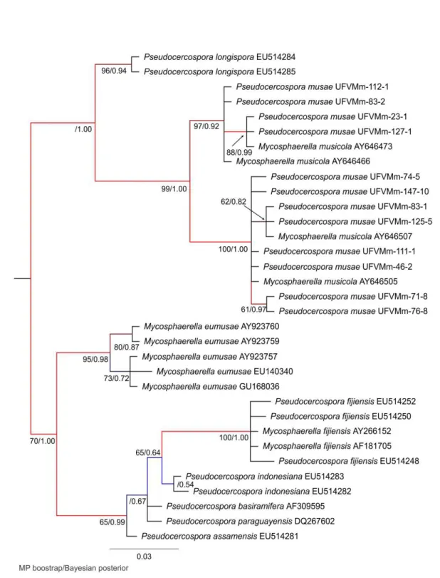
Thirty three sequences were used in the phylogenetic analysis based on ITS which included representative haplotypes (n=7) detected in this study and representative sequences downloaded from GenBank. A total of 486 bp were subjected to the analyses. The maximum parsimony analysis resulted in 1 tree of length 72, consistency index (CI) 0.847, and retention index (RI) 0.971.
The MP and Bayesian analyses based on ITS sequence resulted in a tree with
five well-supported clades. Clade I, IV and V consisted of P. longispora, M. eumusae and M. fijiensis species had bootstrap/posterior probability (PP) values of 96/0.94, 94/0.98 and 100/1.00 respectively. Members of M. musicola were divided into two clades with bootstrap/PP values of 97/0.92 and 100/1.00 (Figure 4).
1.5 Discussion
The establishment of BLSD in banana producing areas located in the tropical regions usually leads to severe crop losses. However, in Minas Gerais State no changes in disease impact or in disease management were recorded as a result of the occurrence
of BLSD as reported in Castro et al. (2005) and Ferrari et al. (2005). Therefore, it was hypothesized that M. fijiensis did not establish in the State; the pathogen is not fully adapted yet to the environmental conditions of these areas or the first report of BLSD
was not correct. Based on classical and molecular diagnostic techniques, only M. musicola was detected in all samples examined in the current study.
It is generally pointed out that once M. fijiensis has been introduced into an area M. musicola is rapidly displaced. This has been observed in the Pacific Islands, Latin America and Africa (Carlier et al., 1999b; Zandjanakou-Tachin et al., 2009). However, in other places such as The Philippines and Vietnam this has not occurred and M. musicola is still found after the appearance of BLSD in 1965 and 1993, respectively (Carlier et al., 1999b; Jones, 2003). Thus, the rate of displacement seems to be influenced by genetic and/or ecological factors (Marin et al., 2003). Displacement of species of plant pathogenic fungi as the result of the introduction of more aggressive genotypes is well documented (Day and Shattock, 1997). Unfortunately, no comparison of aggressiveness between isolates of M. musicola and M. fijiensis was possible due to the fact that isolates of the latter species were not detected in the sampled fields.
18
co-exist (Romero, 1988; Johanson, 1995). The co-existence and distribution of both species is apparently linked with temperature, which in turn is markedly influenced by altitude (Churchill, 2011; Marín et al., 2003). In two studies carried out to investigate the distribution of Mycosphaerella spp., M. musicola was the dominant species at high altitudes and M. fijiensis developed better at low altitudes (Mouliom-Pefoura et al., 1996; Romero, 1988). The analyses of conidia germination, growth of germ tubes,
incubation period and life cycle of both M. fijiensis and M. musicola under different ecological conditions revealed that in low altitude zone (80 m elevation) with temperature around 21 and 33oC both diseases were found in the area, but BLSD was dominant. On the other hand, in highland areas (> 1300 m elevation) with temperatures around 15 and 18 oC, M. musicola had shorter incubation period than M. fijiensis and was more prevalent (Mouliom-Pefoura et al., 1996). Interestingly, studies have also suggested that M. fijiensis may be adapting to higher elevations (Arzanlou et al., 2007; Carlier et al., 2000). Despite these facts, species prevalence according to altitude seems not to occur in all areas where both diseases co-occur. In Colombia, both pathogens are equally severe at altitudes of 1500 m (Marín et al., 2003) and low temperatures and high altitude are not limiting factors for M. fijiensis infections, also in Costa Rica where the pathogen has been detected affecting banana plants at 1360 m of (Arzanlou et al., 2007). Inherent characteristics of the populations need to be investigated in more details, mainly those related to the aggressiveness of the individuals.
The topographic conditions of Minas Gerais State where samples were collected vary considerably regarding altitude and temperature. This seems to be the case for most areas of South and Southeast regions of Brazil. Overall, however, there are favorable conditions for the development of BLST in all regions where it has been reported in
19
Another intriguing aspect of the putative occurrence of BLSD in Minas Gerais is the restriction of the disease to the South and Zona da Mata regions. The North of Minas Gerais is the most important banana producing region and approximately 15,000 ha are cultivated (IBGE, 2010). Based on the reports from other countries, one would expect that BLSD should have easily spread throughout the state. Ascospores of M. fijiensis may not spread more than 100-200 kilometers when there is a combination of strong
winds and heavy clouds, or at night (Jones, 2003). The producing areas in the North region are located approximately 750 kilometers from the South and Zona da Mata regions. Likewise, the Triângulo Mineiro region is located 600 kilometers from the areas where BLSD occur. Aerial dissemination to these areas would be expected, particularly since banana are widely cultivated as a subsistence crop and even in great areas in the state. The availability of susceptible host plants would have contributed to
the spread of the pathogen in “small jumps”, mediated by the dispersal of airborne
ascospores of M. fijiensis that could have spread slowly to the more distant regions. In warmer areas of Brazil such as Rondônia and Mato Grosso states the establishment of BLSD was fast. Within a 3 year-period, the disease spread from 9 to 28 municipalities in Rondônia (Fernandes et al., 2007). In Mato Grosso state, BLSD was reported to occur in more than 18 municipalities in 2010 (Souza and Feguri, 2010).
Other possible but unlikely explanation for the absence of M. fijiensis would be that the emergency control measures adopted when BLSD was reported for the first time were effective in preventing the spread of the recently detected pathogen. When the disease was reported in Minas Gerais, plant pathologists from several institutions were gathered to assess the epidemiology of the disease and provide technical guidance to farmers. Additionally, abandoned banana fields were destroyed as part of prevention to
avoid the dispersal of the pathogen to other areas of the state. These practices might have prevented dissemination of the disease to unaffected areas. Nevertheless, considering the nature of an airborne pathogen having a broad availability of host populations this would be of low likelihood.
20
of septa is higher than those of M. musicola and there are some slight differences in the shape of the conidia and in the basal hilum. However, in addition to the characteristics of the conidia, the presence of sporodoquia, which are not produced by M. fijiensis and were constantly found on the samples examined in the present study support the identification of M. musicola as the sole fungus in the samples.
The symptoms of BLSD and yellow Sigatoka can be confused and vary depending
on the cultivar and whether the plants have been treated with fungicides (Gasparotto et al., 2006). In the municipality of Piau, in the Zona da Mata region, where M. fijiensis was first reported (Castro et al., 2005), banana leaves with symptoms resembling those of BLSD were found, but data from molecular tests did not confirm the presence of M. fijiensis. The combination of morphological and molecular data helped the identification of more than 20 species of Mycosphaerella occurring on banana (Arzanlou et al., 2008). The latter approach was used in the present study and we are confident that M. fijensis was not present in the analyzed samples.
The nucleotide variation between the haplotypes ranged between 1 and 11 bp. Similar results were found by Thomas-Hall et al. in 2004 where phylogenetic analysis of the ITS region from 111 samples of Sigatoka disease revealed 7 distinct clades with nucleotide variation ranging from 4 to 14 bp. The split of M. musicola sequences into two clades found in this study could suggest the occurrence of more than one closely related or cryptic species in Minas Gerais state, but additional analyses did not support any evidence for this. For example, isolates with different haplotypes from both clades were scored for genetic variation using AFLP and all isolates produced variable banding patterns suggesting there was not enough genome-wide variation to support a speciation
event. Additionally, three nuclear loci (β-tubulin, Actin and Elongation factor 1-alpha, EF-1α) and one mitochondrial locus (small subunit rDNA) were also sequenced from representative haplotypes which also did not suggest evidence of cryptic species. The analysis of other genomic regions combined with an assessment of genome-wide variation with microsatellite markers could help to elucidate this and we are currently investing this.
21
important question that remained opened and which is difficult to deal with: could the first report of the occurrence of BLSD have been related to the difficulty in identifying the pathogen? If this is the case, regulatory/legislative control measures need to be revised, because the restrictions imposed by phytosanitary laws have been perverse to
farms and the state‟s economy.
1.6 Acknowledgements
22 1.7 References
Arzanlou, M., Abeln, E., Kema, G., Waalwijk, C., Carlier, J., de Vries, I., Guzman, M.,
and Crous, P. W. 2007. Molecular diagnostics for the sigatoka disease complex of banana. Phytopathology, 97: 1112-1118.
Arzanlou, M., Crous, P. W., and Zwiers, L. H. 2010. Evolutionary dynamics of mating-type loci of Mycosphaerella spp. occurring on banana. Eukaryotic Cell, 9: 164-172.
Arzanlou, M., Groenewald, J., Fullerton, R., Abeln, E., Carlier, J., Zapater, M., Buddenhagen, I., Viljoen, A., and Crous, P. W. 2008. Multiple gene genealogies and phenotypic characters differentiate several novel species of Mycosphaerella and related anamorphs on banana. Persoonia, 20: 19-37.
Balint-Kurti, P. J., May, G. D., and Churchill, A. C. L. 2001. Development of a transformation system for Mycosphaerella pathogens of banana: a tool for the study of
host/pathogen interactions. FEMS Microbiology Letters, 195: 9-15.
Carlier, J., El Hadrami, A., Hayden, H., Zapatar, M. F., and Lapere, F. 1999a. Populations study of Mycosphaerella fijienis and genetic improvement of bananas for resistance to black leaf streak disease. InfoMusa, 8: 16
Carlier, J., Fouré, E., Gauhl, F., Jones, D. R., Lepoivre, P., Mourichon, X., Pasberg-Gauhl, C., and Romero, R. A. 1999b. Black leaf streak. In: Jones, D. R. (Ed.). Diseases of banana, abacá and enset. Wallingford, UK: CAB Publishing, p. 37-79.
Carlier, J., Zapater, M. F., Lapeyre, F., Jones, D. R., and Mourichon, X. 2000. Septoria leaf spot of banana: A newly discovered disease caused by Mycosphaerella eumusae (Anamorph Septoria eumusae). Phytopathology, 90: 884-890.
Castro, M. E. A., Pereira, J. C. R., and Gasparotto, L. 2005. First report of black-sigatoka in the State of Minas Gerais, Brazil. Fitopatologia Brasileira, 30: 668-668.
23
development, and the challenges of control. Molecular Plant Pathology, 12: 307-328.
Cintra, W. J., Valadares, R., Avelino, R. C., Bucker, W. M., Xavier, F. R. V., Ramos, A.
F., and Anderson, P. P. 2008. Worldwide geographical distribution of Black Sigatoka for banana: predictions based on climate change models. Scientia Agricola, 65: 40-53.
Crous, P. W., and Mourichon, X. 2002. Mycosphaerella eumusae and its anamorph Pseudocercospora eumusae spp. nov.: causal agent of eumusae leaf spot disease of banana. Sydowia, 54: 35-43.
Day, J. P., and Shattock, R. C. 1997. Aggressiveness and other factors relating to displacement of populations of Phytophthora infestans in England and Wales. European Journal of Plant Pathology, 103: 379-391.
Doyle, J. J., and Doyle, J. L. 1990. Isolation of plant DNA from fresh tissue. Focus, 12: 13-15.
Fernandes, C. F., Júnior, J. R. V., Silva, D. S. G., Santiago, V., Miranda, S. L. V., Neto, A. F., Costa, J. N. M., Filho, Z. F. H., and Nunes, A. M. L. 2007. Sigatoka-negra em Rondônia. Comunicado Técnico-Embrapa. Porto Velho, RO.
Ferrari, J. T., Harakava, R., Nogueira, E. M. de C., and Castro, M. E. A. 2005. Ocorrência de sigatoka-negra da bananeira no Sul de Minas Gerais. Summa Phytopathologica, 31: 34.
Ferrari, J. T., and Nogueira, E. M. de C. Sigatoka Negra: como identificar e combater a
Sigatoka Negra da bananeira. Available at:
http://www.biologico.sp.gov.br/artigos_ok.php?id_artigo=16. [Accessed September, 22 2011].
24
Frederick, R. D., Snyder, C. L., Peterson, G. L., and Bonde, M. R. 2002. Polymerase chain reaction assays for the detection and discrimination of the soybean rust pathogens Phakopsora pachyrhizi and P. meibomiae. Phytopathology, 92: 217-222.
Gardes, M., and Bruns, T. D. 1993. ITS primers with enhanced specificity for basidiomycetes application to the identification of mycorrhizae and rusts. Molecular Ecology, 2: 113-118.
Gasparotto, L., Pereira, J. C. R., Hanada, R. E., and Montarroyos, A. V. V. 2006. Sigatoka-negra da bananeira. Manaus: Embrapa Amazônia Ocidental.
Hanada, R. E., Gasparotto, L., Resende, J. C. P., and Sedoguchi, E. T. 2007. Panorama atual da sigatoka-negra da bananeira no Brasil. Fitopatologia Brasileira, 32: 159.
IBGE- Cidades- Lavoura Permanente 2010. Available: http://www.ibge.gov.br/cidadesat/topwindow.htm?1. [Accessed: November 29, 2011].
INMET- INSTITUTO NACIONAL DE METEREOLOGIA –. Available: http://www.inmet.gov.br/html/clima/mapas/?mapa=tmax. [Accessed: November 12, 2011].
Johanson, A. 1995. Detection of banana leaf spot pathogens by PCR. Bull. OEPP/EPPO Bull, 25: 99-107.
Johanson, A., and Jeger, M. J. 1993. Use of PCR for detection of Mycosphaerella fijiensis and M. musicola, the causal agents of Sigatoka leaf spots in banana and plantain. Mycological Research, 97: 670-674.
25
Maciel, G. A. 2006. Instrução Normativa No- 289, de 20 de outubro de 2006. Available: http://www.in.gov.br. [Accessed: January 12, 2011].
Marín, D. H., Romero, R. A., Guzmán, M., and Sutton, T. B. 2003. Black Sigatoka: an increasing threat to banana cultivation. Plant Disease, 87: 208-222.
Meredith, D. S., and Lawrence, J. S. 1969. Black leaf streak disease of banana (Mycosphaerella fijiensis): symptoms of disease in Hawaii, and notes on the conidial state of the causal fungus. Transactions of the British Mycological Society, 52: 459-476.
Mouliom-Pefoura, A., Lassoudière, A., Foko, J., and Fontem, D. A. 1996. Comparison of development of Mycosphaerella fijiensis and Mycosphaerella musicola on banana and plantain in the various ecological zones in Cameroon. Plant Disease, 80: 950-954.
Nylander, J. A. A. 2004. MrModeltest v2. Program distributed by the author. Evolutionary Biology Centre, Uppsala University.
Pereira, J. C. R., Gasparotto, L., Coelho, A. F. S., and Urben, A. F. 1998. Ocorrência da Sigatoka negra no Brasil. Fitopatologia Brasileira, 23: 295.
Ploetz, R. C. 2004 . First report of Black Sigatoka of banana caused by Mycosphaerella fijiensis on Grand Bahama Island. Plant Disease, 88: 772.
Rhodes, P. L. 1964. A new banana disease in Fiji. Commonwealth Phytopathological
News, 10: 38-41.
Romero, R. A. 2003. The spread, detection and impact of black leaf streak disease and other Mycosphaerella species in the 1990s. In: Mycosphaerella Leaf Spot Diseases of Bananas: Present Status and Outlook. Proceedings of the Workshop on Mycosphaerella Leaf Spot Diseases, San José, Costa Rica, 20–23 May 2002 (Jacome, L., Lepoivre, P., Marin, D., Ortiz, R., Romero, R. and Escalant, J.V., eds), pp. 21-24. Montpellier: The International Network for the Improvement of Banana and Plantain.
26
Ronquist, F., and Huelsenbeck, J. P. 2003. MrBayes 3: Bayesian phylogenetic inference under mixed models. Bioinformatics, 19: 1572-1574.
Souza, N. S., and Feguri, E. 2010. Ocorrência da Sigatoka Negra em bananeira causada por Mycosphaerella fijiensis no Estado de Mato Grosso. Available at: http://www.mt.gov.br/wps.2010. [Acessed September 23, 2011].
Stover, R. H. 1972. Banana, Plantain and Abaca diseases. Commonwealth Mycological Institute, Kew, Surrey, UK. 316pp.
Swofford, D. L. 2002. paup*: Phylogenetic analysis using parsimony (* and other
methods). Sunderland, MA: Sinauer Associates.
Tamura, K., Peterson, D., Peterson, N., Stecher, G., Nei, M., and Kumar, S. 2011. MEGA5: Molecular evolutionary genetics analysis using maximum likelihood,
evolutionary distance, and maximum parsimony methods. Molecular Biology and Evolution, 28: 2731-2739.
Thomas-Hall, S., Porchum, S., Henderson, J. Pattemore, J., and Aitken, E. 2004. Phylogenectic diversity of Mycosphaerella leaf spot disease, 1st International Musa
Congress, Penang, Malaysia, July 6-9.
Volpato, O. 2005. Sigatoka Negra, Situação atual. Informativo Gedev, 1:3 Available at: http://www.cidasc.sc.gov.br/html/artigos/Volpato_Sigatoka_Negra_Situa%E7%E3o_atual.pdf. [Assessed November 14, 2011].
White, T. J., Bruns, T., Lee, S., and Taylor, J. 1990. Amplification and direct sequencing of fungal ribosomal RNA genes for phylogenetics. In: PCR Protocols: A Guide to Methods and Applications (eds. M.A. Innis, D.H. Gelfand, J. J. Sninsky and T. J. White. Academic Press, San Diego: 315-322.
27
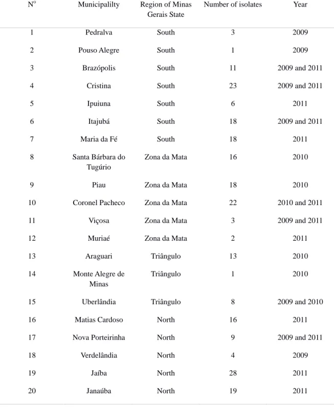
Tables:
Table 1. Origin of the isolates of Mycosphaerella ssp. collected from banana fields located in 20
municipalities of Minas Gerais state, in 2009 to 2011.
No Municipalilty Region of Minas Gerais State
Number of isolates Year
1 Pedralva South 3 2009
2 Pouso Alegre South 1 2009
3 Brazópolis South 11 2009 and 2011
4 Cristina South 23 2009 and 2011
5 Ipuiuna South 6 2011
6 Itajubá South 18 2009 and 2011
7 Maria da Fé South 18 2011
8 Santa Bárbara do Tugúrio
Zona da Mata 16 2010
9 Piau Zona da Mata 18 2010
10 Coronel Pacheco Zona da Mata 22 2010 and 2011
11 Viçosa Zona da Mata 3 2009 and 2011
12 Muriaé Zona da Mata 2 2011
13 Araguari Triângulo 13 2010
14 Monte Alegre de Minas
Triângulo 1 2010
15 Uberlândia Triângulo 8 2009 and 2010
16 Matias Cardoso North 16 2011
17 Nova Porteirinha North 9 2009 and 2011
18 Verdelândia North 4 2009
19 Jaíba North 28 2011
28
Table 2. Dimensions (µm) and characteristics of Mycosphaerella musicola conidia from
different regions of Minas Gerais.
Source Length Diameter Number of
septa
Basal hilum Form
Minas Gerais 30 - 70
(mean 50.45)
3 - 5 (mean 3.30)
2 - 7 (mostly 3 - 6)
absent Straight and curved
Meredith and Lawrence
(1970)
19 - 94 (mean 59)
2.5 - 3.8 (mean 3.0)
1 - 7 (mostly 3 - 6)
indistinct Straight and curved
Figures:
Figure 1. A- A map of Brazil showing the state distribution of Mycosphaerella fijiensis (in gray)
29
Figure 2. Colonies from single-conidium in medium V8 after 10 days of isolation.
Figure 3. Symptoms similar to BLSD from samples collected in the Zona da Mata, Minas
30
Figure 4. The 50% majority rule tree of 75,000 trees from Bayesian phylogeny based on the
31
2.0 Genetic structure of the population of Mycosphaerella musicola in the state of Minas Gerais, Brazil
2.1 Abstract
A total of 215 single-conidium isolates obtained from several regions of Minas Gerais
32 2.2 Introduction
Banana (Musa spp.) is one the most important fruit crops in the world and is cultivated in more than 4.7 million hectares, and approximately 102 million tons of fresh fruit were produced in 2010. It is the second most important fruit crop produced in Brazil, the fifth largest producer in the world after India, China, Philippines and
Ecuador (FAO, 2010). Leaf spot diseases caused by species of Mycosphaerella are major limiting factors for banana production worldwide and can have considerable social and agricultural economic impacts due to lowering crop yields. The leaf spot diseases are particularly threatening in tropical areas where banana is a staple food (Crous et al., 2002; Jones, 1999).
Mycosphaerella musicola R. Leach ex J. L. Mulder (anamorph: Pseudocercospora musae) (Zimm.) Deighton is a fungal (Ascomycete) pathogen that infects banana and plantain worldwide causing Sigatoka leaf spot disease, also known as yellow Sigatoka. Leaf spots can coalesce and lead to premature death of large areas of leaf tissue reducing the photosynthetic capabilities of the plant. The disease can also disturb the physiology of the fruits, resulting in premature ripening (Jones, 1999; Stover, 1972).
Black Sigatoka, also known as black leaf streak disease (BLSD) is another disease that also affects banana and is caused by Mycosphaerella fijiensis M. Morelet (anamorph Pseudocercospora fijiensis) (M. Morelet) Deighton. Usually, after the introduction of BLSD in a region the occurrence the yellow Sigatoka becomes a
“secondary problem” and a subject of lower relevance (Molina et al., 2001). Nevertheless, in many areas, M. musicola remains a destructive pathogen to banana
plants mainly where M. fijiensis has not become established (Jones, 1999). This seems to be the case in Minas Gerais state, located in southeastern Brazil. The BLSD was first reported in the state in 2005 (Castro et al., 2005), but there was no increase in the severity of leaf spot epidemics or in fungicide usage to control the disease. This puzzling outcome raised several questions including one related to the distribution of the disease. Recently, a thorough survey was conducted aimed at assessing the
distribution of „Sigatoka‟ diseases of banana based on 800 samples collected in four
33
The management of banana leaf spots is costly and often requires aerial application of fungicides with aircrafts or big tractor-mounted terrestrial apparatus (Marín et al., 2003). The use of resistant varieties would greatly contribute to disease management, but in Brazil most cultivars that are commercially desirable lack enough level of leaf spot resistance (Dias, 2008). The understanding of the amount and distribution of genetic variation in the population of M. musicola would be useful to
breeders for developing new varieties and for pathologists for setting strategies to better use resistant materials and prevent resistance breakdown. These issues are particularly important for perennial crops cultivated in large areas.
Molecular markers such as restriction fragment length polymorphism (RFLP) and random amplified polymorphic DNA (RAPD) have been used to analyze the genetic structure of M. musicola populations at regional, continental and global scales (Hayden et al., 2003; Hayden et al., 2005; Moreira et al., 2003). Hayden et al. (2003) used RFLP markers to analyze the global population structure of M. musicola and they reported genetic differentiation among the geographic populations from Indonesia, Australia, Africa, Latin America and the Caribbean. The authors also suggested migration of the pathogen between Africa and Latin America. In another study, moderate genetic differentiation and levels of genetic diversity within the Australian population of M. musicola were reported (Hayden et al., 2005). In Brazil, preliminary information was gathered regarding the genetic variability of the population of M. musicola (Moreira et al., 2003). Only 24 isolates of M. musicola from five states were analyzed, but a high level of genetic variability was revealed using RAPD markers (Moreira et al., 2003). In order to make stronger inferences about the genetic structure of the population of M. musicola, it is necessary to implement a better sampling scheme,
a much larger sample size, and to use higher resolution genetic markers.
34
developed to assess genetic variation in M. musicola (Molina et al., 2001; Zapater et al., 2008), but to date no study aimed at elucidating the genetic structure of the pathogen population has been conducted.
Studies aimed to quantify the genetic variability should be complemented with more detailed analysis of the mode of reproduction of the pathogen. Determining the prevalent mode of reproduction can be both genetically and epidemiologically relevant.
M. musicola is a heterothallic fungus, and both the MAT1-1 and MAT1-2 mating idiomorphs have been characterized (Arzanlou et al., 2010; Conde-Ferráez et al., 2010). However, no study using a population approach has been conducted to infer the frequency of mating types in Brazilian isolates of M. musicola. If both idiomorphs are found in approximately similar frequencies, i.e. 1:1 ratio, this could indicate that recombination can take place and a genetically variable population may be established. From the epidemiological perspective, determining the prevalent inoculum associated with epidemics is important for disease management and policy making. If sexual reproduction can occur then two types of spores may contribute to disease development: ascospores and conidia.
The main objective of this study was to determine the genetic structure of M. musicola populations in Minas Gerais State, Southeast Brazil, estimate the level of genetic diversity in each population, gametic disequilibrium, frequency of mating type idiomorphs, genetic differentiation and gene flow based on SSR marker and mating type frequencies.
2.3 Materials and methods
Sampling collection and isolation
35
isolation. Using a sterile fine needle, conidia present on abaxial surface of the leaves with symptoms were picked and put in a water-agar medium on Petri plates using a stereoscopic microscope. Single conidium was taken with a sterile fine needle using a compound microscope and cultured on V8 agar medium (300 mL V8 juice, 3 g of CaCO3 and 20 g of agar per liter of medium). The single-spore isolates were incubated at 25oC under 12 h of daily white light regime for 10 days prior to examination and
DNA extraction.
DNA extraction, quantification and identification
Fungal cultures were grown in flasks containing 50 mL of V8 broth culture medium (250 mL of V8 juice and 3 g CaCO3 per liter of medium, pH 6.5) at 25oC for 10
days at 120 rpm on a shaker. Total genomic DNA was extracted from each isolate using a CTAB protocol as described by Doyle and Doyle (1990). An aliquot of 50 μl of TE buffer (10 mM Tris-HCl and 1 mM EDTA, pH 8.0) was added to each DNA sample. RNA was digested with RNAse A for 2 h at 37oC before storage at –20°C. The concentration of DNA was quantified with a spectrophotometer (NanoDrop 2000 Thermo Scientific) and working solutions at 20 g of DNA/μl were prepared from each
sample for all polymerase chain reaction (PCR) analyses.
The identification of all isolates was based on the product of PCR analysis using species-specific primers for the actin genes MMactF2 (ACGGCCAGGTCATCACT), MMactRb (GCGCATGGAAACATGA) designed by Arzanlou et al. (2007). The PCR products were subjected to electrophoresis in 2.0% agarose gel in 1×TBE buffer and viewed under UV in gels stained with GelRed (Uniscience).
Mating type characterization