Submitted30 September 2016 Accepted 18 November 2016 Published19 January 2017
Corresponding author Juan Martin Leardi, jmleardi@gl.fcen.uba.ar
Academic editor Mark Young
Additional Information and Declarations can be found on page 50
DOI10.7717/peerj.2801
Copyright 2017 Leardi et al.
Distributed under
Creative Commons CC-BY 4.0
OPEN ACCESS
Detailed anatomy of the braincase of
Macelognathus vagans
Marsh, 1884
(Archosauria, Crocodylomorpha) using
high resolution tomography and new
insights on basal crocodylomorph
phylogeny
Juan Martin Leardi1, Diego Pol2and James Matthew Clark3
1CONICET, Instituto de Estudios Andinos ‘‘Don Pablo Groeber’’ (IDEAN), Facultad de Ciencias Exactas y Naturales, Departamento de Ciencias Geológicas, Universidad de Buenos Aires, Buenos Aires, Argentina 2CONICET, Museo Paleontológico Egidio Feruglio, Trelew, Chubut, Argentina
3Department of Biological Sciences, George Washington University, Washington, D.C., United States of America
ABSTRACT
Background.Macelognathus vagansMarsh, 1884from the Late Jurassic Morrison Fm. of Wyoming was originally described as a dinosaur by Marsh and in 1971 Ostrom suggested crocodilian affinities. In 2005, Göhlich and collaborators identified new material of this species from Colorado as a basal crocodylomorph. However, a partial skull found in association with mandibular and postcranial remains was not described. Methods. Due to the small size and delicate structures within the braincase, micro CT studies were performed on this specimen. The new anatomical information was incorporated in a phylogenetic dataset, expanding both character and taxon sampling. Results. This new material reinforces the non-crocodyliform crocodylomorph affinities ofMacelognathusas it bears a large otic aperture, unfused frontals and lacks ornamen-tation on the dorsal cranial bones. The internal structures also support these affinities as this specimen bears traits (i.e., heavily pneumatized and expanded basisphenoid; the presence of additional pneumatic features on the braincase; and the otoccipital-quadrate contact) not present in most basal crocodylomorphs. Furthermore, the presence of a wide supraoccipital and a cranioquadrate passage are traits shared with
Almadasuchusfrom the early Late Jurassic of Argentina.Macelognathuswas recovered
as one of the closest relatives of crocodyliforms, forming a clade (Hallopodidae) with two other Late Jurassic taxa (AlmadasuchusandHallopus).
Discussion. The clade formed byAlmadasuchus+Hallopus+Macelognathus, the Hal-lopodidae, is characterized by a higher degree of suturing of the braincase, posteriorly closed otic aperture (paralleled in mesoeucrocodylians) and cursorial adaptations. Also, the phylogenetic position of this lineage of derived crocodylomorphs as the sister group of Crocodyliformes implies a large amount of unsampled record (ghost lineage), at least 50 million years.
SubjectsEvolutionary Studies, Paleontology, Taxonomy
INTRODUCTION
Macelognathus vagansMarsh, 1884was originally described byMarsh (1884), and since
then it has been surrounded by several controversies. The holotype specimen was recovered in 1880 at Como Bluff (Wyoming, USA), in levels belonging to the Morrison Formation. Based on the isolated mandibular symphysis that bears no teeth,Marsh (1884)assigned this taxon to a chelonian reptile. However, several authors questioned Marsh’s assignment and referred the type specimen ofMacelognathus vagansto Dinosauria, either as a coelurosaur (Theropoda;Baur, 1891) or even as an ornithischian (Simpson, 1926;Von Huenne, 1956). Later,Ostrom (1971)reexamined the holotype specimen (YPM 1415), and based on the anatomical evidence reidentifiedMacelognathus vagansas a crocodilian.
Exploration of other outcrops of the Morrison Formation in Colorado (Brushy Basin Member; Fruita Paleontological Area) during the late 70 s and early 80 s retrieved additional material ofMacelognathus.Göhlich et al. (2005)established that this material belongs to
Macelognathus based on the presence of mandibular remains with the same peculiar
anatomy of the type specimen: a spatulate anterior end that lacks any teeth. In that contribution,Göhlich et al. (2005)described new cranial and postcranial materials of the taxon and concluded, based on that material, thatMacelognathushas non-crocodyliform crocodylomorph affinities. However, given the incomplete nature of the remains, no clear relationship among basal crocodylomorphs was claimed. Later on,Pol et al. (2013) described a new taxon from the Late Jurassic of Argentina (Almadasuchus) and noted some similarities between the femur of this new taxon andMacelognathus, but the large amount of missing data precluded the finding of a stable phylogenetic placement for the latter.
Upon personal examination (J. Leardi and D. Pol, 2012) of the collections of the Los Angeles County Museum (LACM), we were able to find a partial braincase among the material ofMacelognathusretrieved from the Fruita Paleontological Area (Figs. 1A–1D). The objective of the present contribution is to describe this new material and to evaluate its phylogenetic affinities. We use high-resolution computed tomography (CT) to study the internal anatomy of Macelognathus(Figs. 2A–2E). This contribution represents the first study of CT scans from a non-crocodyliform crocodylomorph, a group whose internal braincase anatomy is highly relevant to their evolution (Clark, 1986;Walker, 1990;Wu & Chatterjee, 1993;Pol et al., 2013).
MATERIALS AND METHODS
CT analysis
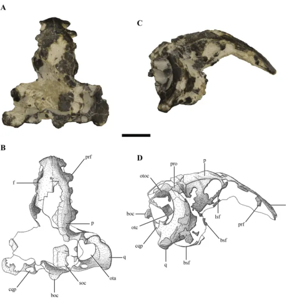
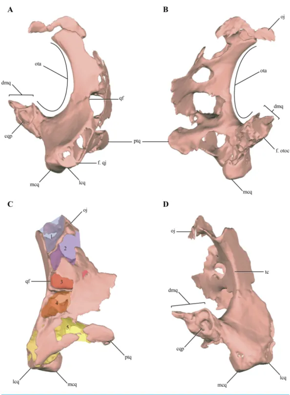
Figure 1 Posterior region of the skull ofMacelognathus vagans(LACM 5572/150148). (A–B), dorsal; and, (C–D), lateral view. Scale bar equals 1 cm. Abbreviations: boc, basioccipital; bsf, basisphenoid; cqp, cranioquadrate passage; f, frontal; lsf, laterosphenoid; ota, otic aperture; otc, otic capsule; otoc, otoccipital; p, parietal; prf, prefrontal; pro, protic; q, quadrate; soc, supraoccipital.
Systematic paleontology
ArchosauriaCope, 1869
CrocodylomorphaHay, 1930,sensuWalker, 1970 HallopodidaeMarsh, 1881
Discussion: This family has been little used since its initial erection, and then only as a monotypic taxon. Here we apply this taxon to the clade found by our phylogenetic analysis comprisingHallopus victor(Marsh, 1890),Macelognathus vagans, andAlmadasuchus figarii figariiPol et al., 2013. We redefine it here as all taxa more closely related toHallopus victor
(Marsh, 1890) than toProtosuchus richardsoniBrown, 1933 or toDibothrosuchus elaphros
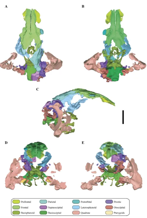
Figure 2 Digital reconstruction of the segmented posterior region of the skull ofMacelognathus (LACM 5572/150148). (A) dorsal; (B) ventral; (C) right lateral; (D) anterior; and, (E) posterior views.
Diagnosis: Hallopodidae can be diagnosed by the following synapomorphies: the presence
paralleled inJunggarsuchus); dorsoventrally large otic appertures (present inAlmadasuchus
andMacelognathus, also paralleled in Junggarsuchus), and, a quadrate-laterosphenoid contact but leaving the prootic exposed within the supratemporal fossa (only known in
Almadasuchus).
Macelognathus vagansMarsh, 1884
Holotype: YPM 1415, articulated anterior portions of both dentaries, which lack teeth on the anterior end.
Referred materials: LACM 5572/150148, left dentary, partial braincase, and postcranial remains including dorsal vertebrae, a partial left ilium, partial femora, and other elements of the hindlimbs; LACM 4684/128271, partial right and left hindlimbs; LACM 4684/128272, a left femur which may belong to LACM 4684/128271; LACM 5572/150211, both calcanea, and metatarsal III; LACM 4684/133772, a portion of the right maxilla, and both dentaries. For a more detailed explanation of the elements included the reader is encouraged to refer toGöhlich et al.’s (2005) paper.
Horizon and locality: The holotype specimen was recovered from Como Bluff, Albany
County, Wyoming (USA); the referred specimens were found in the Fruita Paleontological Area, Mesa County, Colorado (USA). In both of these localities the fossils derive from the Brushy Basin Member in the upper part of the Morrison Formation (Upper Jurassic, Kimmeridgian-early Tithonian;Foster, 2003).
Emended diagnosis: A non-crocodyliform crocodylomorph with dorsoventrally flattened
and anteriorly edentulous dentary*, heterodont dentition, and tooth crowns devoid of mesial and distal serrations; dentary lacking caniniform teeth; maxilla with laterally concave and ventrally sinuous alveolar margin; lateral longitudinal ridge above alveolar margin of maxilla; at least two enlarged anterior maxillary teeth; enlarged maxillary teeth serrated only distally; quadrate with five pneumatic cavities; laterally closed cranioquadrate passage; large otic aperture two-thirds of the height of the quadrate; quadrate contacts the otoccipital; wide supraoccipital in posterior view; posterior bony ring on the basisphenoid, allowing the precarotid recess to open posteriorly*; internal carotids exit anteroventrally from the hypophyseal fossa*; presacral vertebrae with large neural canals (almost as large as the centrum); ilium without supraacetabular crest; ventral margin of preacetabular process of ilium thickened and medially projecting; round femoral head oriented medially and separated from proximal end by a distinct neck; proximal facet of tibia distinctly slanted laterally; longitudinal groove on proximoanterior end of fibula; calcaneum with a completely flat distal surface; strong medioplantar crest on medial base of calcaneal tuber*; overlapping proximal ends of metatarsals. Autapomorphies are marked with an asterisk (*).
RESULTS
General features
specimen, highlighting two details: the presence of a weak sagittal crest and a concave nuchal crest that delimited the occipital surface of the skull. With later mechanical preparation of the specimen, the parietal was disarticulated and is no longer associated with the rest of the skull. No further details were given about the rest of the braincase of
MacelognathusbyGöhlich et al. (2005).
The skull of LACM 5572/150148 measures 53 mm from the occipital condyle to almost the anterior end of the frontals (Figs. 1A–1Dand2A–2E), being anteroposteriorly shorter than the length of the mandibular symphysis of the holotype specimen (YPM 1415, approximately 58 mm (Göhlich et al., 2005)). Thus LACM 5572/150148 probably belonged to a small juvenile individual, an idea further reinforced by the strongly convex skull roof (Figs. 1C–1Dand2C). This fossil preserves a partial posterior region of the skull including partial remains of the braincase. The braincase of this specimen is poorly exposed, so in order to recover more information from this region a high resolution CT scan was performed (see ‘Materials and Methods’).
Other crocodylomorph remains were recovered from the same outcrop, including crocodyliforms (Goniopholis(Göhlich et al., 2005) andFruitachampsa(Clark, 2011)). The ‘‘sphenosuchian’’ affinities of LACM 5572/150148 are clearly evidenced by the very large otic aperture (Figs. 1C–1D), unlike the smaller one present in basal crocodyliforms (e.g.,
Protosuchus richardsoniandFruitachampsa), and by the lack of ornamentation in the cranial roof (Figs. 1A–1B). Also, the unfused frontals might reveal non-mesoeucrocodylian affinities, but this lack of fusion might be due to an early ontogenetic stage.
Description
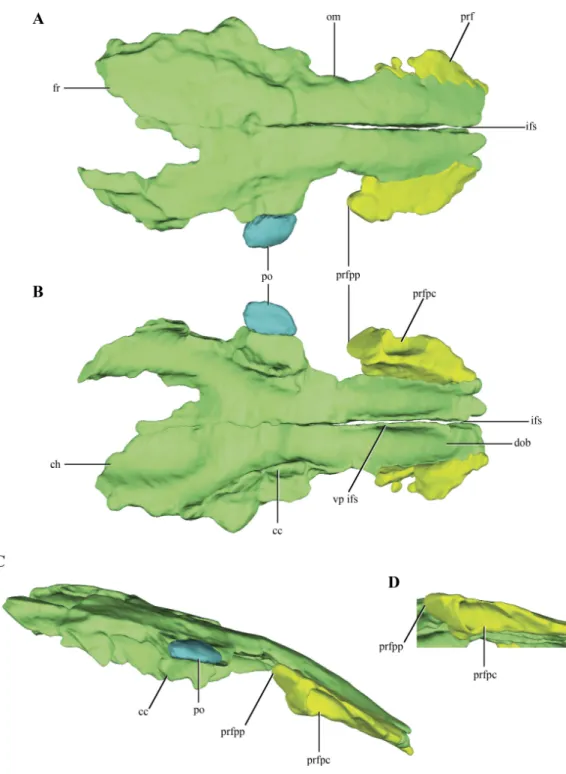
Bothprefrontalsare preserved on LACM 5572/150148, but they are only represented by the dorsal process of this bone (Figs. 3A–3D). The prefrontal contacts medially with the anterolateral edge of the frontal only. This is rare condition among crocodylomorphs, as the frontals usually contact also with the posterolateral edge of the nasals. However, no partial remains of the nasals could be identified on the anteriormost end of LACM 5572/150148, but this possibility should not be discarded as this region is badly preserved, bearing many cracks and fissures. Where this contact is better preserved, on the right side of the skull, the suture is almost straight (Fig. 3A). The prefrontals are anteroposteriorly elongate in dorsal view and they form the anteromedial border of the orbit. Anteriorly, the prefrontals are almost laminar, while posteriorly they have a higher dorsoventral development. In dorsal aspect, the prefrontal has a posterior process projected posterolaterally (Figs. 3A–3B). The posterolateral edge of the prefrontals is broadened in many basal crocodylomorphs (Pseudhesperosuchus,Dromicosuchus,Hesperosuchus agilis(CM 29894),Terrestrisuchus,
Saltoposuchus, Litargosuchus, Sphenosuchus,Dibothrosuchus, Junggarsuchus), but only
in Macelognathus does it form a distinct process. The CT data exposed the internal
Thefrontals are paired and have a clear median suture between them along their entire length (Figs. 3A–3B). The frontals are separated from each other anteriorly (where both frontals barely touch each other) but they are tightly joined to each other along the posteriormost region of the dorsal surface of the frontals. The dorsal surface of the frontals is smooth (Fig. 3A) as in most non-crocodyliform crocodylomorphs (e.g.,Hesperosuchus,
Sphenosuchus, Litargosuchus) with the exception ofAlmadasuchus, which is sculpted, andDibothrosuchus, which has three longitudinal ridges. The frontals are rectangular in dorsal view, although their anterior end is lateromedially narrower than the posterior one, and convex in lateral view, but to a lesser extent than the parietals (see below). The frontals wedge between the prefrontals (see above), displaying a rather anteroposteriorly elongated contact. The prefrontal-frontal contact extends approximately along one third of the preserved length of the frontals. Just anterior to the mid-length of the frontals as preserved, a constriction is present on the left element (damaged on the right one) that is here interpreted as the dorsal margin of the orbits. Unlike the condition of most eusuchians and some close relatives of that clade (Glen Rose Form, Bernissartia,Shamosuchus), the dorsal margin of the orbits is not dorsally projected (Fig. 3C).
The posteroventral margins of the frontals have ventrally directed flanges, the cristae cranii (Figs. 3B–3C). These flanges contact the anterodorsal end of the laterosphenoids, as in most archosauriforms. Even though these crests are only partially preserved, they deeply project ventrally, as in most non-crocodyliform crocodylomorphs. The posteromedial region of both frontals is not preserved. Partial remains of the parietals are preserved on their posterolateral ends allowing the observation of the lateral region of the frontoparietal suture, which has its main axis anterolaterally oriented (Figs. 2A and2C). Anterior to this region a slightly depressed area is delimited anteriorly by a curved crest. This crest is continuous with a crest present on the parietal (see below), so it is interpreted as the anterior border of the supratemporal fossa.
The ventral surface of the frontals bears two wide grooves that extend anteroposteriorly along its anterior half, interpreted here as the depression for the olfactory bulbs (Fig. 3B). These grooves are deep, limited laterally by the cristae cranii, and attenuate approximately at the level of the posterior end of the orbit. The division in two grooves is given by a well-developed and ventrally projected crest located medially at the interfrontal suture. This crest would imply a partially divided (or bilobed) olfactory bulb, and is much more developed than in crocodyliforms (e.g.,Araripesuchus gomesiiPrice, 1959 (AMNH 24450);
Sebecus icaeorhinusSimpson, 1937;Crocodylus acutusCuvier, 1807 (Colbert, 1946)). The frontals ofMacelognathusalso have two elongated depressions on the posterior half of the ventral surface of the frontals, which are interpreted here as the impressions of the cerebral hemispheres (Fig. 3B). These depressions have a considerable anteroposterior development, occupying 25% of the total length of the frontal.
A fragment of bone is preserved articulated to right frontal along its mid-length, interpreted as part of thepostorbital(Figs. 3A–3C). The region preserved corresponds to the postorbital plate, just anterior to the descending process.
Figure 4 Digital reconstructions of the parietal ofMacelognahus.(A) dorsal; and, (B) lateral views. Ab-breviations: alpf, anterolateral process of the frontal; dc, dorsal crest; f. lsf, facet for the laterosphenoid; f. pro, facet for the protic; STF, border of the supretemporal fossa.
if the parietals were fused. The central right half of the parietal was already described previously (Göhlich et al., 2005; see above), and for additional information on that region the reader is referred to that contribution. Thus, most of the description of the parietal will be based on the right half of this element. Most crocodylomorphs have the parietals fused as a single element, with the exception ofHesperosuchus,Pseudhesperosuchus,Dromicosuchus,
TerrestrisuchusandSaltoposuchus(Clark, Sues & Berman, 2000); although in some cases this could be due to the early ontogenetic stage of the specimens. The parietal is strongly convex in lateral view, a condition that is more evident at its contact with the supraoccipital (Figs. 1C–1Dand2C–2E). The dorsal surface of the parietal is smooth, as in most basal crocodylomorphs (Hesperosuchus,Pseudhesperosuchus,Dromicosuchus,Terrestrisuchus,
Saltoposuchus,Litargosuchus,Kayentasuchus,Sphenosuchus,Dibothrosuchus,Junggarsuchus
andAlmadasuchus), contrasting with the ornamented condition present in crocodyliforms
(e.g.,Protosuchus richardsoniBrown, 1933).
surface of the parietal bears a concavity that occupies almost two thirds of the total length of the bone. This region is identified as the supratemporal fossa. Its great anteroposterior development is consistent with the condition of most basal crocodylomorphs, in which the supratemporal fossae are very large and occupy almost the entire dorsal surface of the parietal.
Along its ventral edge, the parietal contacts the laterosphenoid and the prootic. The contact with the former is extended through the anterior 75% of the ventral margin of the parietal along a straight suture (Fig. 4B). The posterior fourth of the ventral border of the parietal contacts the prootic. Posteriorly, the parietal is very fragmentary, thus its contact with the supraoccipital is not preserved. The internal surface of the parietal is concave, which is especially marked on the posterolateral region of the medial surface of the parietal, near the sutural contact with the laterosphenoids. This depressed internal surface, which continues on to the laterosphenoids, is interpreted as the posterolateral expansion of the cerebral hemispheres.
Thequadrate, despite being damaged in many regions, is one of the most complete bones of the skull. Both quadrates are preserved in LACM 5572/150148, although the right one is better preserved than the left one, as with most elements of the skull (Figs. 2A–2E). The quadrate body is curved posteriorly in lateral view, being markedly convex anteriorly. One of the most notable features of the right quadrate is the presence of a large otic aperture on its main body (Figs. 5A–5B). The quadrate only forms the ventral and anterior borders of this fenestra. Despite being incomplete (as the squamosal is not preserved) we can infer its size, which is approximately two thirds of the dorsoventral length of the quadrate. The presence of a large otic aperture, where the quadrate forms the anteroventral borders of a well-delimited fenestra, is a feature shared withAlmadasuchusandJunggarsuchus.
In these taxa the otic aperture is also dorsoventraly large, attaining at least half of the height of the quadrate body. In other known non-crocodyliform crocodylomorphs (Hesperosuchus ‘‘agilis,’’Pseudhesperosuchus,Dromicosuchus,Terrestrisuchus,Sphenosuchus,
Dibothrosuchus), the otic aperture is not such a circumscribed fenestra as the otic aperture lies posterior to the quadrate body and it is not closed posteriorly (see below). However, this condition is unknown inKayentasuchusandLitargosuchusas the posteroventral region of their quadrates are not well preserved.
Anterior to the otic aperture, on the posterior surface of the quadrate body, a crest is present (Fig. 5D). Given its anterior position to the otic aperture, this crest is here identified as the tympanic crest. The lateral surface of the main body of the quadrate ofMacelognathus
margin of the infratemporal fenestra, as the quadratojugal covers all the anterior surface of the quadrate that could be exposed in this fenestra.
Also on the anterior margin of the quadrate body, and dorsal to the facet for the quadratojugal the posterior border of an elongated rounded fenestra is present (Figs. 5A
and5C). This quadrate fenestra is located at the dorsoventral midpoint of the anterolateral surface of the quadrate and appears to be pneumatic, as it connects with the internal space of this bone (see below). Among non-crocodyliform crocodylomorphs, pneumatic quadrate fenestrae are also present in simple form inTerrestrisuchusand more complexly
in JunggarsuchusandAlmadasuchus. These pneumatic fenestrae are always located on
the main body of the quadrate and anterior to the tympanic crest. However, there is a great deal of variation in the shape and size of these fenestrae among different taxa. The borders of the quadrate fenestra ofTerrestrisuchushave at least some participation with the quadratojugal on the anterior end (Crush, 1984), while inDibothrosuchus,Junggarsuchus,
Almadasuchusand crocodyliforms (e.g.,Protosuchus richardsoni,Gobiosuchus) the quadrate fenestrae are completely surrounded by the quadrate. As was previously mentioned, this condition cannot be precisely known inMacelognathusdue to the incomplete preservation of the quadrate fenestra. Furthermore, these fenestrae are variable in size and in most taxa are small, while the quadrate fenestrae of TerrestrisuchusandAlmadasuchusare elongated and quite large, being at least half of the dorsoventral height of the quadrate body.Junggarsuchusis unusual also in this aspect, as at least two fenestrae are identified on its quadrate (M Klein et al., 2016, unpublished data). This condition is similar to the one present in basal crocodyliforms which have multiple quadrate fenestrae (e.g.,Protosuchus richardsoni). Finally, the condition ofDibothrosuchusdeserves mention. This structure was not described in the original paper (Wu & Chatterjee, 1993), but upon personal examination (D. Pol), we identified what could be interpreted as the posterior border of a large quadrate fenestra on the left quadrate. The anterolateral margin of the quadrate of the skull of the referred specimen (IVPP V 7907) bears an elongate, rounded border on its anterior end, and thus can be interpreted as the posterior border of a pneumatic quadrate fenestra. The quadratojugal is very incompletely preserved and its relationships with neighboring elements are unclear (Wu & Chatterjee, 1993).
The orbital process of the quadrate is preserved on the right quadrate on its dorsomedial region (Figs. 5B–5D). The orbital process of the quadrate is a lateromedially thin and medially projected process that contacts the dorsolateral region of the prootic. The articular surface for the prootic is exposed only medially (or slightly dorsomedially). The quadrate-prootic contact is anteroposteriorly long, extending through almost the entire length of the prootic as in most crocodylomorphs (Fig. 2C). The dorsolateral region of the quadrate is not preserved in any of the elements, precluding observation of the primary head of the quadrate.
of the pterygoid ramus of the quadrate is asymmetrical, being more expanded dorsally. The ventral region of the pterygoid ramus of the quadrate seems to be separated from the most dorsal part by an anteromedial notch. A similar condition is present inAlmadasuchus, where the quadrate is greatly expanded medially and this notch represents the lateral border of the passage for the middle cerebral vein (sensuWalker, 1990). The distal end of the left quadrate bears well-preserved distal condyles, which are divided by a shallow intercondylar groove. The lateral condyle is mediolaterally broader than the medial one, while the latter is slightly more distally projected.
The body of the quadrate and the distal condyles are heavily pneumatized, being hollow internally with thin walls. The body of the quadrate is internally divided from dorsal to ventral in a series of individualized interconnected chambers. A total of five (5) internal divisions, which increase in volume towards the distal condyles, can be identified in the quadrate (Fig. 5C). The first chamber, located on the most dorsal region of the quadrate is approximately triangular in dorsal view. The exact size of this chamber cannot be precisely known, as the dorsal region of the quadrate is incomplete. The dorsal chamber is separated from the second one by a thin bony septum only developed on the posterior half of the inner cavity of the quadrate. The second chamber is parallelogram shaped in dorsal view and is dorsoventrally higher than anteroposteriorly long. The second chamber communicates with the central chamber via several passages on its ventral septum. One is located on the posterior border (the largest one) and three on the anteromedial border (the medial most is the largest of these). The third and central cavity is volumetrically smaller than the second, being dorsoventrally flat when it is compared with the other internal divisions of the quadrate. The bony septum dividing the central chamber of the fourth chamber seems to be complete, whereas the incompleteness on the anterolateral region could be attributed to preservational features. The communication with the ventral chamber is across a small foramen located on the anteromedial border of the chamber. The fourth chamber is dorsoventrally higher than the third, even though it is lateromedially wider than its dorsoventral development. The posteromedial border appears to be open, as it is confluent with the cavity formed within the quadrate and limited externally by the otic aperture (middle ear). This fourth chamber is also externally connected via the pneumatic quadrate foramen described above (usually called the quadrate fenestra in most crocodylomorphs). The fourth chamber is separated from the ventralmost one by an incomplete septum which has three communicating foramina, one posteriorly and two anteriorly. Finally, the largest pneumatic chamber in the quadrate is the ventral one. This chamber occupies the whole interior volume of the distal body of the quadrate, including the pterygoid process of the quadrate. Pneumaticity invades the distal condyles via three lateral diverticula. The ventral chamber is open ventrally as in other derived non-crocodyliform crocodylomorphs (e.g.,
AlmadasuchusandDibothrosuchus).
the morphology of this region of the quadrate is unknown in some of the other non-crocodyliform crocodylomorphs. The dorsal surface of the posterodorsal process of the quadrate of Macelognathusbears a dorsomedially oriented furrow (Figs. 5Aand
5D). The position and direction of this groove and the fact that the quadrate forms its ventral border are consistent with the cranioquadrate passage. Such passages are absent in most ‘‘sphenosuchians’’ and even basal crocodyliforms (Protosuchus richardsoniand
Orthosuchus). On the other hand, cranioquadrate passages are present in Almadasuchus
and in the vast majority of crocodyliforms (such asGobiosuchus,Sichuanosuchusand all mesoeucrocodylians).
Theprootic, although damaged, is preserved almost complete on the right side of the skull (Figs. 2A–2Eand6A–6B). The left prootic only preserves part of its ventral region and the ventrolateral flange (see below). The protic is a dorsoventrally high bone when it is compared with its lateromedial development (Figs. 7A–7D). As in most non-crocodyliform crocodylomorphs (e.g.,Hesperosuchus,Sphenosuchus,Dibothrosuchus,Almadasuchus) and thalattosuchians (e.g.,Cricosaurus araucanensis (Gasparini & Dellapé, 1976)) the prootic is exposed on the posterodorsal region of the supratemporal fossa. InMacelognathus
the prootic is verticalized on the supratemporal fossa, with the exposed surface facing anteriorly, a similar condition to the one observed inDibothrosuchus,Sphenosuchusand
Kayentasuchus. On the other hand, the prootic is exposed facing anterodorsally within the supratemporal fossa inAlmadasuchus,Litargosuchusand thalattosuchians. In dorsal view, the prootic contacts the squamosal posteriorly (not preserved) and anteriorly the parietal along its medial margin.
The dorsal region of the prootic is more lateromedially developed, and houses a large mastoid antrum (Figs. 7Aand7C). This pneumatic cavity is subrectangular in shape and is restricted to the prootic, although the squamosal (not preserved) might form the dorsal roof of the mastoid antrum. The mastoid antrum appears to communicate with the middle ear via at least two large foramina on the dorsolateral surface of the prootic: a larger one located on the anterior half and a smaller, more elongated one, located on the posterior half of this surface (Figs. 6C–6D). One or more ventral openings could be present on the anteroventral surface of the mastoid antrum, but these may have been caused by breakage of the specimen. Also, the presence of such ventral apertures of the mastoid antrum would depend on the particular shape of the unpreserved contact with the pterygoids. Multiple lateral openings of the mastoid antrum into the middle ear cavity are present inKayentasuchus,DibothrosuchusandP. haughtoni; while inSphenosuchusthe mastoid antrum opens through a unique undivided foramen.
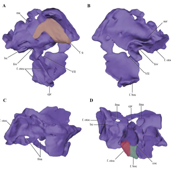
Figure 7 Digital reconstructions of the right protic ofMacelognathus.(A) lateral; (B) medial; (C) dor-sal; and, (D) ventral views. Abbreviations: aur, auricular recess; coc, cochlear recess; cpr, crista prootica; f. boc., facet for the basioccipital; fma, foramina of the lateral wall of the mastoid antrum; f. otoc, facet for the otoccipital; f. q, facet for the quadrate; fov, fenestra ovalis; lsc, groove for the lateral semicircular canal; ma, mastoid antrum; VII, exit for the VII cranial nerve.
just medial to the mastoid antrum. Upon reaching the anterodorsal end of the prootic, it turns posteriorly very abruptly, leaving a groove on the dorsal surface of the prootic. This groove continues posteriorly on the medial aspect of the supraoccipital (see below). These traits are consistent with the identification of this structure as the rostral semicircular canal. Ventral to the region of the mastoid antrum and the groove for the lateral semicircular canal, in the central region of the posterior margin of the prootic, a rounded notch is present (Fig. 7A). The shape of this notch is due to the lack of preservation of the anteriormost area of the otoccipital. The position and the bones that form this notch (i.e., the prootic forms its anterior and dorsal margins) help us to identify it as the fenestra ovalis. Ventral to the fenestra ovalis, the prootic bears a large laterally projected flange (prootic-basisphenoid flange sensuWalker (1990)or crista prootica). This flange extends to the ventral margin of the prootic and continues also on the dorsal region of the basisphenoid, as inSphenosuchus. The otoccipital contacts the prootic-basisphenoid flange along the posterior margin of its prootic part. The prootic-otoccipital contact also involves the posterior surface of the prootic (excluding the region of the fenestra ovalis), although the central region of the prootic is not preserved. This contact is preserved on the dorsal and ventral region of the posterior surface of the prootic. This double contact involving the otoccipital and the prootic encloses a dorsoventrally oriented pyramidal cavity interpreted as the cochlear recess (=lagena). Further ventrally the prootic contacts the basisphenoid through its posteroventral margin, and as a result closes the cochlear recess ventrally.
The medial surface of the prootic is damaged, exposing parts of the bony labyrinth medially. Dorsal to the bony canal of the lateral semicircular canal and its associated ampulla a strongly projected medial bony flange is present. This flange is sigmoidal in medial view and it limits ventrally a deep depression. This depression is capped dorsally by the supraoccipital. A similar depression has been observed in medial view of the braincase of Sphenosuchusand has been interpreted as the auricular (or floccular) recess of the cerebellum (Walker, 1990) (Figs. 6Aand7B). Medial views of the braincases of other basal crocodylomorphs are rare, precluding the observation of this feature.
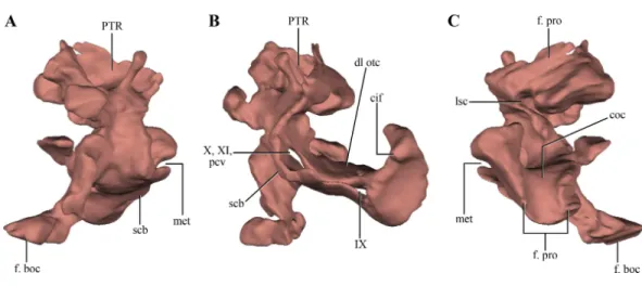
Figure 8 Digital reconstructions of the right otoccipital (excluding the paroccipital processes) ofMacelognathus. (A) posterior; (B) lateral; and, (C) anterior views. Abbreviations: cif, crista interfenestralis; coc, cochlear recess; dl otc, dorsal lamina of the otic capsule; f. boc, facet for the basioccipital; f. pro, facet for the protic; lsc, groove for the lateral semicircular canal; met, metotic fissure; pcv, posterior cerebral vein; PTR, posterior tympanic recess; scb, subcapsular buttress; IX–X, exit for cranial nerves IX. . . X.
not that well exposed in other crocodylomorphs. On the dorsal border of the otic capsule a posteriorly directed groove is observed, the extension of the groove conducting the lateral semicircular canal observed in the prootic (Fig. 8C). This groove continues medially up to the level of the otic capsule, and at the posterior border of the capsule it expands into the ampulla of the caudal semicircular canal. Dorsal to this region an oblique groove runs through the dorsomedial surface of the otoccipital, medial to mastoid antrum. This groove continues anteriorly on the ventral surface of the supraoccipital and represents the caudal semicircular canal.
Dorsal to the otic capsule, the otoccipital is heavily pneumatized (Figs. 8A–8B). This pneumatic cavity is completely separated from the mastoid antrum by a bony wall formed by the posterodorsal region of the prootic. The otoccipital also contributes to this separation, as a thin bony lamina that articulates anterodorsally with posterior bony septum of the mastoid antrum formed by the prootic. The position of this pneumatic cavity, located dorsal to the otic capsule and to the metotic fissure, is consistent with its identification as the posterior tympanic recess (PTR sensuWu & Chatterjee, 1993). A slight posterior tympanic recess is present inSphenosuchus, being represented by a shallow groove between the prootic-opisthotic suture. A heavily pneumatized cavity in this same area is present inDibothrosuchusandP. haughtoni, thus sharing the same condition withMacelognathus. The posterior tympanic recess ofMacelognathusis partially divided anteriorly by a bony pillar located near the suture with the prootic. However, due to the almost complete lack of preservation of the paroccipital processes we are unable to evaluate if this pneumatic cavity penetrates these processes as inDibothrosuchusandP. haughtoni.
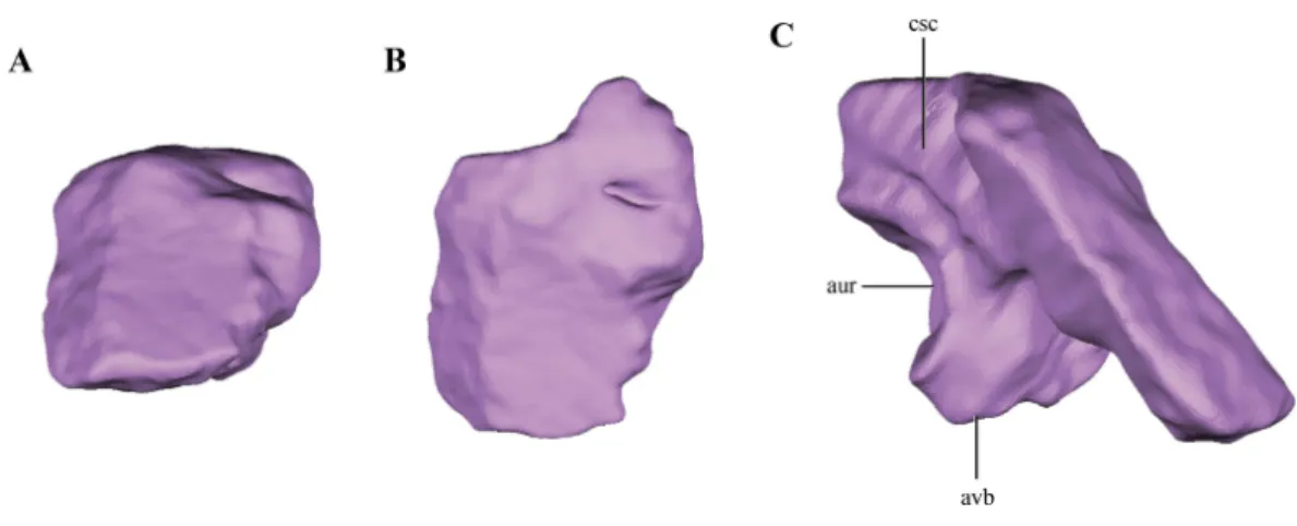
Figure 9 Digital reconstructions of the supraoccipital ofMacelognathus. (A) posterior; (B) dorsal; and, (C) medial view. Abbreviations: aur, auricular recess; avb, anteroventral bulge of the supraoccipital; csc, groove for the caudal semicircular canal.
Therefore, the otoccipital forms the entire lateral margin of this foramen (Fig. 6A). The otoccipital-basioccipital contact reaches the occipital condyle, and as result the otoccipital participates in the dorsolateral margin of it. Finally, only the ventralmost part of the right paroccipital process is preserved. The fragment preserved is exposed posteroventrally and separates the basioccipital from the quadrate in this view. This region of the paroccipital process contacts with the quadrate laterally; anteriorly and medially with the basioccipital.
Thesupraoccipitalis present between the foramen magnum and the parietals. Only the right half of the supraoccipital is preserved (Figs. 2A–2Eand6A–6B). The preserved half is rectangular in external view (Figs. 9A–9B), thus the full element would have a quadrangular shape in occipital view. This condition is similar to the one present in Litargosuchus,
Junggarsuchus,Almadasuchusand crocodyliforms that have a wide supraoccipital, unlike the dorsoventrally elongated supraoccipitals of other crocodylomorphs (Pseudhesperosuchus,
Terrestrisuchus,Kayentasuchus, Sphenosuchus, Dibothrosuchus). Anteroventrally, the supraoccipital bears a ventral bulge that is pyramidal shaped and with its apex oriented ventrally, giving this region a higher dorsoventral development than the rest of the supraoccipital (Fig. 9C). The anteromedial region of the anteroventral bulge bears a very deep concavity, which is the part of the auricular (or floccular) recess that extends into the supraoccipital. Dorsal to this recess a shallow groove can be observed. This groove continues on the dorsal border of the anteroventral bulge, just ventral to the dorsal plate of the supraoccipital. As mentioned above, this groove is interpreted as for the caudal semicircular canal.
As in all ‘‘sphenosuchians’’ where this condition has been properly explored, the supraoccipital of Macelognathusdoes not participate in any pneumatic cavity (i.e., transverse canal communicating the mastoid antra from both sides) (Figs. 6A–6B), unlike the condition seen in crocodyliforms (e.g.,P. richardsoni(Clark, 1986),P. haugthoni
Dibothrosuchus(Wu & Chatterjee, 1993), however this condition cannot be observed in the external view of the skull material known for this taxon (IVPP V 7907; J Clark, pers. obs., 2014). Posterolaterally, the supraoccipital articulates with the otoccipital on its posterior third through an oblique interdigitated suture (Fig. 2A). Anterolaterally, the supraoccipital contacts the prootic on its anterior two thirds through an interdigitated suture on the anteroventral bulge.
Thebasioccipitalforms the floor of the posterior half of the endocranial cavity, while the anterior half is formed by the basisphenoid (Figs. 6A–6B). Anteriorly, the median region of the basioccipital is divided by a short posterodorsal process of the basisphenoid. Lateral to this area, the basioccipital-basisphenoid articulation is straight, through a transverse suture (Figs. 10A–10C). On the anterolateral corner of the endocranial cavity, the basioccipital bears a posteroventrally elongated and obliquely oriented facet for the prootic (Figs. 10B–10C). Posterior to this facet, and along its dorsolateral surface, the basioccipital contacts the subcapsular buttress of the otoccipital. On the central region of the dorsal aspect of the basioccipital, this element has lateral expansions that contact the medial wall of the subcapsular buttress and the otic capsule. These lateral flanges contract before reaching the occipital condyle.
The occipital condyle of the basioccipital is rugose and has the articular surface for the atlas-axis complex oriented posteriorly, but slightly inclined dorsally. The occipital condyle is projected ventrally, delimiting a ventral rim around this area (Figs. 10Band10D). On the ventral surface of the basioccipital, anterior to the occipital condyle, the basioccipital tubera are partially preserved. These are laminar as in most crocodylomorphs (Nesbitt, 2011) and are separated from the occipital condyle by a shallow groove (Fig. 10A). Only the right basioccipital tuber and partial remains of the medial lamina uniting both tubera are preserved. A wide depressed area is present on the ventral surface of the basioccipital, the basioccipital recess. This wide recess is present in almost all non-crocodyliform crocodylomorphs (e.g., Hesperosuchus, Dromicosuchus,Sphenosuchus,Dibothrosuchus,
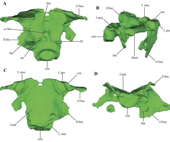
Figure 10 Digital reconstructions of the basioccipital ofMacelognathus.(A) ventral; (B) lateral; (C) dorsal; and, (D) posterior views. Abbreviations: bor, basioccipital recess; bot, basal tubera; bt, blind tubes of the basioccipital; cc bor, central crest of the basioccipital recess; coc, cochlear recess; con, occipital condyle; f end, floor of the endocranial cavity; f. otoc, facet for the otoccipital; f. pro, facet for the protic; lf boc, lateral flanges of the basioccipital; ml, medial lamina of the basal tubera; rhom, rhomboidal sinus; vf boc, ventral flanges of the basioccipital.
well-developed bony passages for Eustachian tubes, which are present in Crocodyliformes (e.g.,P. richardsoni(Clark, 1986)).
In addition to the dorsal contact with the otoccipital along the endocranial cavity, the basioccipital has a contact with the otoccipital. This contact is between the distal end of the anteroventral flanges of the basioccipital and the paroccipital process (Fig. 10A).
Thebasisphenoid, although damaged and incomplete ventrally, is fairly well preserved. The basisphenoid is greatly expanded lateromedially in ventral view (Fig. 2B) as in
Junggarsuchus, Almadasuchusand basal crocodyliforms (e.g., P. richardsoni), unlike the more compressed basisphenoids present in Terrestrisuchus, Sphenosuchus and
Dibothrosuchus. CT data of the central region of the basisphenoid allowed the observation of the highly pneumatized nature of this region (Figs. 11A–11D), a condition also shared
withAlmadasuchus,Junggarsuchusand Crocodyliformes.
Figure 11 Digital reconstructions of the basisphenoid ofMacelognathus.(A) lateral; (B) dorsal; (C) anterior; and, (D) posterior views. Abbreviations: bsfp, basisphenoid plate; bsf ros, rostrum of the ba-sisphenoid; car, passage for the carotid arteries; coc, cochlear recess; cp, carotid pillar; ds, dorsum sellae; f. boc, facet for the basioccipital; f. lsf, facet for the laterosphenoid; f. pro, facet for the protic; pal; passage for the palatine artery; pp bsf, posterior process of the basisphenoid; ppd, posterior depression of the ba-sisphenoid; pr bsf, posterior ‘‘ring’’ of the baba-sisphenoid; st, sella turica; VI, exit for the VI cranial nerve.
of the posterior edge of the basisphenoid articulates with the prootic through a dorsally exposed facet (Figs. 11A–11B). Thus, there is a triple contact involving the basioccipital, basisphenoid, and the prootic (Fig. 6D). As was previously mentioned, this triple contact closes the ventralmost end of the cochlear recess (see prootic). Additional participation in this contact by the otoccipital is possible, as the ventrolateral region of the cochlear recess is not preserved.
Anteriorly, the basisphenoid of Macelognathusis ‘‘W’’-shaped, comprising a dorsal basisphenoid plate and a central carotid pillar, which conveyed the internal carotids (Fig. 11C). The carotid pillar can be divided into the dorsal postcarotid recess and the ventral precarotid recess (Walker, 1990). The basisphenoid plate is directed anterodorsally and continues anteriorly up to the level of the hypophyseal fossa (Fig. 11A). Due to the fragmentary preservation of LACM 5572/150148 it is not possible to establish if the basisphenoid plate formed a continuous bony sheet. On the anterolateral ends of the basisphenoid plate, two elongated oblique facets for the laterosphenoids are present (Fig. 11B). Ventromedial to these facets two anterolaterally directed grooves, which have an associated foramen (only preserved on the left side) can be seen. These represent the exits for the abducens cranial nerve (VI).
Just medial to the grooves for the abducens nerve, the basisphenoid plate forms the dorsum sellae, the dorsal roof of an anteriorly placed hypophyseal fossa (Figs. 11B–11C). The hypophyseal fossa is not bounded anteriorly, as in other basal crocodylomorphs (e.g.,
Sphenosuchus,Almadasuchus) and crocodyliforms. Although not well-preserved (only on
the right side), bulges are present lateral to the hypophyseal fossa. Anteroventrally an anteriorly exposed bony plate is present that is pierced by two foramina, the exit for the internal carotid arteries (Fig. 11C). This condition is very unusual among crocodylomorphs, as in taxa where this trait is known the exit of the internal carotids is located on the ventrolateral surface of the hypophyseal fossa (e.g., Sphenosuchus,Dibothrosuchus, P. haughtoni). The bony plate bears a dorsal bulge and, separated from it, the base of the basisphenoid rostrum is preserved. Thus the base of the basisphenoid rostrum is located well ventral to the base of the hypophyseal fossa, as inSphenosuchus, contrasting with the condition present inAlmadasuchusand Crocodyliformes (e.g.,P. haughtoni,Caiman yacare Daudin, 1802). In these taxa the hypophyseal fossa is bounded anteroventrally by the basisphenoid rostrum, and in crocodyliforms this anterior process expands dorsally forming the anterior wall of the fossa. On the posterior surface of the anteriorly exposed bony plate and ventral to the hypophyseal fossa, an elliptical, lateromedially elongated recess is present. A similar rostral recess is also present inSphenosuchus, but the structure of this region is not known in other basal crocodylomorphs.
posteriorly projected depressions that excavate this surface. Ventral to the carotid pillar the precarotid recess is also partially preserved on its posterior region where it has a bony ring delimiting a circular passage (Fig. 11D). Thus, the precarotid recess seems to be posteriorly open, unlike the condition inSphenosuchuswhere this recess is closed.
Two anterolaterally oriented bony struts arise from the ventral surface of the posterior region of the basisphenoid, ventrolateral to the carotid pillars (Fig. 11A). The lateral surface of the bony struts bears a foramen that divides in two rami, an anteriorly directed ramus and an anteroventrally oriented one. The anterior ramus of this foramen communicates with the carotid pillar, representing the initial passage where the internal carotid enters the basisphenoid. The anteroventral ramus is only formed by a dorsal bony bridge and then continues anteroventrally through a slight groove. The path described by the initial part of the foramina, and then by the groove, is consistent with the identification of this structure as representing the branching of the internal carotid into the palatine artery. The two lateroventral bony struts branch out rapidly towards the ventral surface of the basisphenoid, within the highly pneumatized ventral region of this bone. These branches expand distally to form the ventrally expanded plate of the basisphenoid.
Posteroventral to the posterior edge of the basisphenoid are two isolated bony laminae (Figs. 2Band2D). These posterolateral laminae contact the posteromedial process of the quadrate and the paroccipital processes of the ottocipital. Given their topological position these are interpreted as fragmentary remains of thepterygoids, particularly the quadrate ramus of that bone.
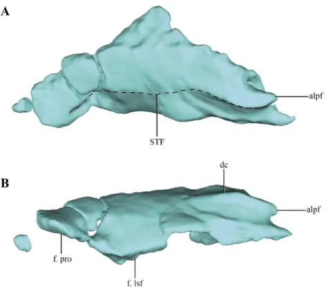
Although elements from both sides are preserved, the laterosphenoids are poorly preserved (Fig. 2). The laterosphenoids articulate posteriorly with the prootics through a straight overlapping suture (Fig. 12A). Posteroventrally the laterosphenoid contacts the basisphenoid dorsolateral to the hypophyseal fossa via an elongated facet (see basisphenoid above) (Fig. 12C). The region in between these sutures is missing, precluding evaluating the shape of the trigeminal foramen. Most of the dorsal edge of the laterosphenoid contacts the parietal through a straight suture. The anterodorsal end of the laterosphenoid articulates with the frontals, having short capitate processes located on its anteriormost end (only preserved on the right side).
The lateral surface does not seem to have any crest on its external surface, as in most basal crocodyliforms (Pol et al., 2014) and ‘‘sphenosuchians’’ where this region is preserved (i.e.,
Sphenosuchus,Dibothrosuchus,JunggarsuchusandAlmadasuchus). The lateral surface of the laterosphenoid is convex in general, as it forms the anterolateral border of the lateral wall of the forebrain. This convexity is particularly developed anteriorly, where it is markedly beveled. On the posterior half of the lateral surface an elongated, foramen leading dorsally into the bone is present (Fig. 12C). However, this foramen does not communicate with the neurocranial cavity as it is closed by a medial lamina.
Phylogenetic relationships
Figure 12 Digital reconstructions of the left and right laterosphenoids ofMacelognathus.(A) lateral; and, (C) ventral views. Left laterosphenoid in (A) lateral; and, (B) posterior view. Abbreviations; cap, cap-itated processes of the laterosphenoid; f. bsf, facet for the basisphenoid; for?, foramen?; f. par, facet for the parietal; f. pro, facet for the protic; i. cav, internal cavity of the laterosphenoid.
crocodyliform sampling when it is compared to other datasets (e.g.,Clark, Sues & Berman, 2000; Clark et al., 2004;Nesbitt, 2011). However, considering recent contributions on crocodylomorph phylogenies (Nesbitt, 2011;Zanno et al., 2015) we expanded this matrix including new characters and new taxa. As a result, our new dataset is composed of 39 taxa and 138 characters.
Taxon sampling
The new taxa incorporated are: the paracrocodylomorphSaurosuchus galileiReig, 1959; and the crocodylomorphs CM 73372 (seeNesbitt, 2011),Carnufex carolinensisZanno et al., 2015,Redondavenator quayensisNesbitt, Irmis & Lucas, 2005,Trialestes romeri(Reig, 1963),Hallopus victor, Macelognathus vagans, andHemiprotosuchus lealiBonaparte, 1969.
Saurosuchusis added to increase the number of outgroups in the analysis, asWilberg (2015) noted that the inclusion of more outgroups might affect the results. Also, Saurosuchus
Among the crocodylomorphs included, CM 73372 was recently found as the one of the basalmost members of the clade (Nesbitt, 2011), and thus is relevant to the following analysis. Other taxa relevant in this matter are two putative crocodylomorphs relatively recently published:Redondavenator quayensis(Nesbitt et al., 2005) andCarnufex carolinensis(Zanno et al., 2015;Drymala & Zanno, 2016). InZanno et al.’s (2015) original analysisCarnufex was recovered at the base of Crocodylomorpha, in a polytomy with CM 73372, whileRedondavenator was depicted as the sister taxon ofSphenosuchus. The sister group relationship betweenSphenosuchusandRedondavenator was supported by a single synapomorphy: an elongated maxillary process on the premaxilla (Nesbitt, 2011; char. 2-1). It is important to consider that this character cannot be scored inSphenosuchus, as Walker stated in his seminal paper that the dorsal end of this process is not preserved (Walker, 1990, p. 12; SAM-PK 3014). However, in a second analysis including both
Carnufex andRedondavenatorDrymala & Zanno (2016)recovered both taxa at the base of Crocodylomorpha together with CM 73372.
On the other hand, TrialestesandHallopus have not been included in any analysis in almost the past 30 years. OnlyTrialesteswas incorporated in the precursor dataset of Benton & Clark (1988). Finally, Macelognathuswas added inPol et al.’s (2013) dataset, but it was recovered as a wildcard taxon in that analysis and was removed from the consensus. However,Pol et al. (2013) mentioned thatMacelognathuswas recovered as the sister group ofAlmadasuchus,Terrestrisuchus+Litargosuchusor Saltoposuchuson the different most parsimonious trees obtained in their analysis. Those topologies were mostly supported by femoral characters, as the braincase of Macelognathuswas not yet described.Hemiprotosuchus lealirepresents the only crocodyliform added in this analysis as it represents the oldest record of the clade (Late Triassic; Los Colorados Formation, La Rioja, Argentina;Bonaparte, 1972a).
Finally, it is relevant to discuss which materials were considered for some of the operational taxonomical units (OTUs) of our analysis. For a full list of the specimens examined the reader is referred to the Supplemental Information. The two most taxonomically controversial taxa areTrialestes romeriandHesperosuchus agilis.Trialestes
has been involved in a controversy since its original description by Reig (1963). Two specimens have been assigned to this taxon (PVL 2561, 3889), but other authors recognized that one specimen could represent a dinosaur (Clark, Sues & Berman, 2000) or even another crocodylomorph (Ezcurra, Lecuona & Irmis, 2008). In consequence, to avoid future problems with our codifications of the taxon, we restrict our scorings ofTrialestesto the type specimen (PVL 2561), which also preserves clear crocodylomorph synapomoporphies (e.g., elongated proximal carpals). The other controversial taxon is Hesperosuchus agilis
have mentioned and even scored some differences between both specimens (Drymala & Zanno, 2016). However, we see little differences between both specimens and many of these morphological differences are indeed present both in the type (AMNH FR 6756) and the referred specimen (CM 29894) (e.g., see characters 137 and 138). Furthermore, both specimens and another specimen assigned toH. ‘‘agilis’’ (UCMP 129470) share the development of sharp postzygodiapophyseal laminae, a trait not present in other North American ‘‘small’’ crocodylomorphs (i.e., not inCarnufex). As a result, we base our scorings
ofHesperosuchus agilison both the type (AMNH FR 6756) and the Carnegie Museum (CM
29894) specimens.
Character sampling
A total of 40 characters were added to Pol et al.’s (2013) dataset of 96 characters scored for 32 taxa. Among these 13 are new and the remaining 27 are from other recent datasets, where the majority of them come fromNesbitt’s (2011) contribution on archosaurian relationships. As new characters were formulated, and many of the characters from other contributions were modified (either in their formulation or scorings), an explanation of them is given below. Our focus on this discussion is on non-crocodyliform taxa, but for more information on the crocodyliform specimens used to evaluate these characters, the reader is referred to theSupplemental Information.
97. Supraoccipital: fused with the exoccipital (0); or, as a separate ossification (1) (NEW) (Figs. 13A,13Cand13D). In basal pseudosuchians (Gracilisuchus, MCZ 4117;Stagonolepis, Walker, 1961) and paracrocodylomorphs (Saurosuchus, PVSJ 32;Postosuchus(Weinbaum, 2011)) the supraoccipital is fused with the paroccipital processes, not being present as a separate ossification. In crocodylomorphs where this region of the skull is known (e.g.,
Terrestrisuchus(Crush, 1984),Kayentasuchus, UCMP 131830,Almadasuchus, MPEF-PV
3838;P. richardsoni, UCMP 131827) the supraoccipital is a separate ossification.
98. Supraoccipital shape: narrow, being dorsoventrally taller than lateromedially wide (0); or, wide, being lateromedially wider than dorsoventrally high (1) (NEW) (Figs. 13A,13C and13D). In most crocodylomorphs the supraoccipital bone is rectangular in posterior view, as it is taller than wide, such as Pseudhesperosuchus (PVL 3830),
Hesperosuchus(CM 29894),Terrestrisuchus(Crush, 1984),Kayentasuchus(UCMP 131830),
Sphenosuchus(SAM-PK 3014),Dibothrosuchus(IVPP V 7907) andJunggarsuchus(IVPP
V 14010). On the other hand,Almadasuchus(MPEF-PV 3838),Macelognathus(LACM 5572/150148, see description), crocodyliforms (e.g.,Protosuchus haughtoni(BP/1/4726, 4746, 4770),GobiosuchusOsmólska, Hua & Buffetaut, 1997) and most thalattosuchians (e.g., Teleosaurus(Jouve, 2009);Pelagosaurus (BSP 1890);Metriorhynchus superciliosus
Blainville, 1853 (AMNH 997);Dakosaurus andiniensisVignaud & Gasparini, 1996 (Pol & Gasparini, 2009);Cricosaurus araucanensis(MLP 72-IV-7-1)) have a wide supraoccipital bone, where this element is wider than tall.
exception of thalattosuchians. Among sphenosuchians this condition is only present in
Almadasuchus(MPEF-PV 3838). See also character 104.
100. Basioccipital recesses: absent (0); or, present as paired foramina located in a median deep depression on the ventral surface of the bone (1) (NEW). In paracrocodylomorphs (e.g., Postosuchus(Weinbaum, 2011);Saurosuchus, PVSJ 32) the basioccipital does not bear any ventral depression anterior to the occipital condyle. In most non-crocodyliform crocodylomorphs the basioccipital bears ventral a median depression, which is divided in two parallel tubes internally. These are observed in Hesperosuchus (CM 29894),
Sphenosuchus (SAM-PK 3014),Dibothrosuchus (IVPP V 7907)Macelognathus(LACM
5572/150148) andAlmadasuchus(MPEF-PV 3838).Crush(1984, p. 138) describes a well-developed basioccipital recess forTerrestrisuchus, although he mentions it is square shaped. The basioccipital is not recessed inJunggarsuchus(IVPP V 14010) and crocodyliforms (e.g.,
P.richardsoni, UCMP 131827), although in the latter forms the posterior margin of the median Eustachian tubes.
101. Length of the posterodorsal process of the postorbital: short, not reaching the midlength of the supratemporal fenestra (0); or, long, exceeding the midlength of the supratemporal fenestra (1) (NEW) (Figs. 13B,13D and13F). As in most basal pseudosuchians (Postosuchus(Weinbaum, 2011);Saurosuchus, PVSJ 32), crocodyliforms generally have a short posterodorsal process of the postorbital, very short and not exceeding the midlength of the supratemopral fenestra (e.g.,P. richardsoni, MCZ 6727;P. haughtoni, BP/1/4726;Orthosuchus, SAM-PK 409,C. yacare). This condition is also present in the ‘‘sphenosuchians’’ Terrestrisuchus (Crush, 1984) and Dibothrosuchus (IVPP V 7907).
In Pseudhesperosuchus (PVL 3830),Hesperosuchus (CM 29894),Sphenosuchus
(SAM-PK 30141), Junggarsuchus (IVPP V 14010) andAlmadasuchus(MPEF-PV 3838) the posterodorsal process of the postorbital is posteriorly elongated, reaching more than half the anteroposterior development of the supratemporal fenestra.
102. Quadrate fenestra: with participation of the quadratojugal (0); or, exclusively bounded by the quadrate (1) (NEW). Pneumatic quadrate fenestrae have been recognized in the crocodylomorphsTerrestrisuchus,Junggarsuchus,Almadasuchus,Macelognathusand
Dibothrosuchus(see Description and char. 46 in theSupplemental Information). However, besides size variation, there is variation in the contribution of the surrounding bones in the different taxa. Terrestrisuchus(Crush, 1984) has at least some participation of the quadratojugal on the anterior end; while inJunggarsuchus(IVPP V 14010),Almadasuchus
(MPEF-PV 3838) and crocodyliforms (e.g., Protosuchus richardsoni, UCMP 131827;
Gobiosuchus (Omolska, 1997)) the quadrate fenestrae are completely surrounded by
the quadrate. As previously mentioned, this condition cannot be precisely known in
Dibothrosuchus (IVPP V 7907) andMacelognathus (LACM 5572/150148) due to the
incomplete preservation of the quadrate fenestra.
one present in basal crocodyliforms that have multiple quadrate fenestrae (e.g.,Protosuchus
richardsoni(UCMP 131827)).
104. Prootic: exposed in dorsal view, on the supratemporal fossa (0); or not exposed in dorsal view (1) (NEW) (Figs. 13B,13Dand13F). In basal pseudosuchians
(Postosuchus(Weinbaum, 2011);Saurosuchus, PVSJ 32;Gracilisuchus, MCZ 4117) and
non-crocodyliform crocodylomorphs (e.g., Hesperosuchus, CM 29894; Litargosuchus, BP/1/5237;Dibothrosuchus, IVPP V 7907;Almadasuchus, MPEF-PV 3838) the prootic is exposed on the posterodorsal region of the supratemporal fossa. Most crocodyliforms lack the prootic exposed dorsally on the supratemporal fossa (e.g., P. richardsoni, UCMP 131827;Orthosuchus, SAM-PK 409) due to the broad contact of the quadrate with the laterosphenoid. However, Almadasuchushas a dorsally exposed prootic on the posterodorsal region of the supratemporal fossa, but has a quadrate-laterosphenoid contact (see char. 99). On the other hand, thalattosuchians (e.g., Cricosaurus araucanensis, MLP 72-IV-7-1;Teleosaurus(Jouve, 2009);Pelagosaurus(Pierce & Benton, 2006)) exhibit the same condition as in non-crocodyliform crocodylomorphs.
105. Exit of cranial nerves IX–XI: exit the braincase ventromedially (0); or, through a common foramen on the ventromedial region of the paroccipital process (vagus foramen) (1) (NEW) (Fig. 13E). The primitive condition for Crocodylomorpha is the exit of the cranial nerves IX–XI through the ventrolateral region of the skull, thus not leaving any distinct foramen (e.g., Gracilisuchus, MCZ 4117;Postosuchus, (Weinbaum, 2011); Saurosuchus, PVSJ 32). This condition is present in most ‘‘sphenosuchian’’ crocodylmorphs, likePseudhesperosuchus(PVL 3810),Hesperosuchus(CM 29894; AMNH FR 6758),Terrestrisuchus(Crush, 1984),Litargosuchus(BP/1/5237),Kayentasuchus(UCMP 131830),Sphenosuchus(SAM-PK 3014),Dibothrosuchus(IVPP V 7907) andJunggarsuchus
(IVPP V 14010).Almadasuchus(MPEF-PV 3838) and crocodyliforms (e.g.,P.richardsoni, UCMP 131827; Protosuchus haughtoni(BP/1/4726, 4746, 4770),Orthosuchus, SAM-PK 409) exhibit a foramen on the ventrolateral region of the paroccipital process that has been interpreted widely as the exit of the nerves IX–XI (i.e., vagus foramen). This condition is also present in thalattosuchians (e.g.,Pelagosaurus(Clark, 1986;Pierce & Benton, 2006);
Teleosaurus(Jouve, 2009);Dakosaurus(Pol & Gasparini, 2009)).
106. Postzygodiapophyseal laminae on the posterior cervical and anterior dorsal vertebrae: absent or low (0); or, present as sharp lamina delimiting a pit posterior to them on the neural arch (1) (NEW) (Figs. 14Band14C). Sharp postzygodiapophyseal laminae are present on the posterior cervical and anterior dorsal vertebrae of paracrocodylomorph archosaurs (Saurosuchus, PVL 2198;Fasolasuchus, PVL 3950;Postosuchus(Long & Murry, 1995)), Carnufex (Drymala & Zanno, 2016) andHesperosuchus(AMNHFR 6576, CM 29894, UCMP 129470). Posterior to these laminae, a well-marked semicircular depression is limited.Dibothrosuchus(IVPP V 7907) has a similar condition on its anterior dorsal vertebrae, although the postzygodiapophyseal lamina is not as sharply projected as in
Hesperosuchus.
Figure 14 Selected phylogenetic characters related toHesperosuchus agilisused in this contribution.
(A) Maxilla of the type specimen ofHesperosuchus agilis(AMNH FR 6758) in lateral view. Posterior cervi-cal vertebrae in lateral view of: (B) the type (AMNH FR 6758); and, (C) the referred (CM 29894) specimen ofHesperosuchus agilis. Proximal radii in anterior view of: (D)Pseudhesperosuchus jachaleri(PVL 3830); (E) the type specimen ofHesperosuchus agilis(AMNH FR 6758); and, the referred (CM 29894) specimens ofHesperosuchus agilis. See characters 106, 137, and 138.
of the radius is more widely distributed than previously reported. Besides Trialestes
(PVL 2561), longer radii than their respective humeri are also present in Litargosuchus
(BP/1/5237),Dibothrosuchus(IVPP V 7907),Dromicosuchus(NCSM 13733) and variably in
Hesperosuchus(present in AMNH FR 6758, while in CM 29894 the humerus is longer). On
12597), Junggarsuchus (IVPP V 14010), Hallopus (YPM 1914) and crocodyliforms (e.g., P. richardsoni, UCMP 131827;Protosuchus haughtoni (BP/1/4726, 4746, 4770),
Orthosuchus, SAM-PK 409) have longer humeri than their radii. The relative length of the anterior zeugopodium seems not to be correlated with the lenghts of the posterior one. Taxa such asDromicosuchus(Sues et al., 2003) andHallopus(Walker, 1970) have femora proximodistally shorter than the tibiae (see char. 32,Supplemental Information), while their radii are longer than the humeri.
108. Proximomedial process of the radiale: absent (0); or, present (1) (NEW). Besides the elongation present in this element (Clark, 1986;Benton & Clark, 1988), crocodylomorphs are also characterized by the presence of a proximomedial process on the radiale. This process articulates with the ulna and the ulnare. As with the elongation on the proximal carpals, this process is also lacking in CM 73372. The presence of the proximomedial process of the radiale might be thought to be correlated with the elongation of the proximal carpals, as these traits have the same distribution among crocodylomophs. However, this can be very useful in taxa where the radiale is not preserved completely, and yet represents a crocodylomorph synapomorphy with the exclusion of the CM 73372.
109. Anterior process of the squamosal: elongated, less than one third of the lateromedial width of the supratemporal fossa (0); transversely broad, more than one third the width of the supratemporal fossa (1); or, very broad, as wide as the width of the supratemporal fossa (modified from Clark, 1986;Sereno & Wild, 1992;Nesbitt, 2011) (Figs. 13B,13D
110. Lateral extent of the paroccipital processes: ends lateral to the lateral border of the supratemporal fenestra (including the fossa) (0); or, ends medial to or at the margin of the border of the supratemporal fenestra (1) (modified fromNesbitt, 2011) (Figs. 13A,
13C and13E). In Nesbitt’s study (2011) the derived condition was found in a group of derived crocodylomorphs, which includedKayentasuchus,Litargosuchus,Protosuchusand
Orthosuchus. However, due to difficulties in scoring this character we also included the lateral limit of the supratemporal fossa, which was not stated in the original character. Without taking into account the supratemporal fossa most crocodylomorphs would be scored as having the lateral end of the paroccipital processes lateral to the border of the supratemporal fenestra/fossa, even some of them scored otherwise byNesbitt (2011). However, when the distribution of this character was revised several differences were found. Paroccipital processes with short lateral projection (i.e., not reaching the lateral border of the supratemporal fossa or fenestra) are present inPseudhesperosuchus (PVL 3810),
Litargosuchus (BP/1/5237) andHesperosuchus(CM 29894), among non-crocodyliforms
crocodylomorphs.Kayentasuchus(UCMP 131830) bears laterally projected paroccipital processes, as these almost reach the lateralmost border of the skull in occipital view (Clark & Sues, 2002). Paroccipital processes that end more laterally than the lateral edge of the supratemporal fossa/fenestra are also present inProtosuchus (P. richardsoni
(UCMP 131827); Protosuchus haughtoni (BP/1/4726, 4746, 4770)), as in this genus the supratemporal fenestrae lack a fossa on their lateral part, thus having little lateral development.Orthosuchusalso has this state of the character, but unlikeProtosuchusthe paroccipital processes have a great lateral projection reaching the lateral end of the skull in occipital view (SAM-PK 409;Nash, 1975). Among crocodyliforms there is not a clear trend as laterally short paroccipital processes are present inZosuchus(Pol & Norell, 2004), some notosuchians (Baurusuchus(Carvalho, Campos & Nobre, 2005);Simosuchus(Kley et al., 2010)) and thalattosuchians.
111. Anterior end of the dentaries: tapering to a point (0); or, dorsally expanded, forming a distinct step (1) (modified fromNesbitt, 2011). This trait was used to refer CM 29894 toHesperosuchusbyClark, Sues & Berman (2000). However this observation was questioned later as an anteriorly expanded dentary was also present in Postosuchus
andDromicosuchus(Nesbitt, 2011). We agree with the presence of such an expansion in
Postosuchus(Weinbaum, 2011), but on the other hand, this condition cannot be observed
inDromicosuchus(NCSM 13733) as the mandibles are closed and articulated in the only
known specimen.Trialestes(PVL 2561) also has an anteriorly expanded dentary, but this condition should be handled with care considering the aggressive mechanical preparation that specimen suffered.
112. Anterior part of the dentary: bears teeth (0); or, edentulous (1) (Parrish, 1994). Most archosauriforms have teeth along their entire tooth row. Exceptions among pseudosuchians are some poposauroids (Effigia(Nesbitt, 2007); Lotosaurus(Nesbitt, 2011)), aetosaurs
(Stagonolepis(Walker, 1961)), and among crocodylomorphsMacelognathus(YPM 1415;
LACM 5572/150148).