Submitted30 September 2016 Accepted 20 December 2016 Published25 January 2017 Corresponding author Graham Rose, [email protected] Academic editor Christine Josenhans Additional Information and Declarations can be found on page 13
DOI10.7717/peerj.2928
Copyright 2017 Rose et al. Distributed under
Creative Commons CC-BY 4.0
OPEN ACCESS
Antibiotic resistance potential of the
healthy preterm infant gut microbiome
Graham Rose1, Alexander G. Shaw2, Kathleen Sim2, David J. Wooldridge1,
Ming-Shi Li2, Saheer Gharbia1, Raju Misra1and John Simon Kroll3
1Genomics Research Unit, Public Health England, London, United Kingdom 2Department of Medicine, Imperial College London, London, United Kingdom
3Section of Paediatrics, Department of Medicine, Imperial College London, London, United Kingdom
ABSTRACT
Background. Few studies have investigated the gut microbiome of infants, fewer still preterm infants. In this study we sought to quantify and interrogate the resistome within a cohort of premature infants using shotgun metagenomic sequencing. We describe the gut microbiomes from preterm but healthy infants, characterising the taxonomic diversity identified and frequency of antibiotic resistance genes detected.
Results. Dominant clinically important species identified within the microbiomes includedC. perfringens,K. pneumoniae and members of theStaphylococciand Enter-obactergenera. Screening at the gene level we identified an average of 13 antimicrobial resistance genes per preterm infant, ranging across eight different antibiotic classes, including aminoglycosides and fluoroquinolones. Some antibiotic resistance genes were associated with clinically relevant bacteria, including the identification of mecAand high levels ofStaphylococciwithin some infants. We were able to demonstrate that in a third of the infants theS. aureusidentified was unrelated using MLST or metagenome assembly, but low abundance prevented such analysis within the remaining samples. Conclusions. We found that the healthy preterm infant gut microbiomes in this study harboured a significant diversity of antibiotic resistance genes. This broad picture of resistances and the wider taxonomic diversity identified raises further caution to the use of antibiotics without consideration of the resident microbial communities.
SubjectsComputational Biology, Genomics, Microbiology, Pediatrics, Public Health
Keywords Preterm infants, Shotgun metagenomics, Antibiotic resistance, Gut microbiome
INTRODUCTION
a single pathogen, with little consideration of the wider microbial communities which reside in the microbiome, leading to a situation in which the use of antibiotics may cause unintentional harm to the host.
As our understanding of the microbiome has developed, the collection of AMR genes within a bacterial population has recently been defined as the resistome (Penders et
al., 2013). Antibiotics have a role in shifting the profile of the resistome within the
population (Jernberg et al., 2007), with low antibiotic-use communities harbouring lower AMR gene frequencies (Walson et al., 2001;Bartoloni et al., 2009). Heavy treatment of bacterial populations with antibiotics can lead to the long term overrepresentation of AMR genes. Such dynamics are evident in the microbiome of preterm neonates, who receive multiple antibiotic courses, and are cared for in an Intensive Care Unit environment potentially contaminated with multi-resistant bacteria. Antibiotic treatments for both term and preterm neonates have demonstrated lasting effects on the microbiota (Tanaka et al.,
2009;Arboleya et al., 2015), with the trajectory of population development diverging from
untreated controls, leading to a potential scenario of prolonged—even life-long—high frequency AMR reservoirs through the selection of bacteria within the population that are most resistant. A wide range of AMR genes have been found in neonatal populations (De Vries et al., 2011;Zhang et al., 2011), some shown to be present from birth ( Alicea-Serrano et al., 2013;Gosalbes et al., 2016), whilst twin pairs have been shown to have GI communities with similar distributions of both organisms and resistance genes (Moore et al., 2015). These observations suggest vertical transmission as a source, with discrepancies between mothers and babies being due to the substantial shifts in the microbiota adapting to the very different environment of a newly born infant’s GI tract (Gosalbes et al., 2016).
The GI tract of a premature neonate is a particularly unusual scenario for observation of AMR genes, due to greatly reduced bacterial immigration as a result of the isolated, sterile environment of incubators and very controlled enteral feeds; donor breast milk may be pasteurised and, whilst unpasteurised maternal milk (which harbours specific bacteria (Beasley & Saris, 2004;Jimenez et al., 2008;Martin et al., 2009)) is given where possible, there is a likelihood of little or no breastfeeding due to extreme prematurity.
In these circumstances, the GI community and the resistance genes present are likely in the main to be derived from the mother, and acquired during birth. Whilst limited bacterial numbers and diversity will initially be transferred, mechanisms are available for the dissemination of AMR through the expanding bacterial population (as reviewed by
Van Hoek et al. (2011)) with transfer having been documented within the gut environment
(Shoemaker et al., 2001;Karami et al., 2007;Trobos et al., 2009). Heavy use of antibiotics in the course of care of premature infants would not only then skew the bacterial population and drive resistance selection, but has been shown to increase the activity of some transposable elements due to stressing of bacterial populations (Beaber, Hochhut & Waldor, 2004).
(Gueimonde, Salminen & Isolauri, 2006;Alicea-Serrano et al., 2013;Von Wintersdorff et al., 2016) or through functional metagenomics (De Vries et al., 2011;Moore et al., 2015), which has the disadvantage of not being able to quantify the antibiotic resistance potential of a community (Forslund et al., 2014). We have used shotgun metagenomic sequencing to describe the resistome in its entirety, moving from species level taxonomic profiling, to characterisation of the resistance landscape, including typing of metagenomes identified as potentially harbouringmecA, conferring resistance to methicillin and otherβ-lactam antibiotics.
MATERIALS & METHODS
Study populationThe study was approved by West London Research Ethics Committee (REC) Two, United Kingdom, under the REC approval reference number 10/H0711/39. Parents gave written informed consent for their infant to participate in the study.
Faecal samples analysed were collected from premature infants, defined as less than 32 completed weeks of gestation. Premature infants were recruited to the study at the Imperial College Healthcare National Health Service Trust neonatal intensive care unit (NICU), at Queen Charlotte’s and Chelsea Hospital, between January 2010 and December 2011.
Sample collection
Almost every faecal sample produced by each participant between recruitment and discharge was collected by nursing staff from diapers using a sterile spatula. Samples were placed in a sterile DNase-, RNase-free Eppendorf tube, stored at−20◦C within two
hours of collection and stored at −80◦C within five days. A single faecal sample from
each of twelve infants who had no diagnosis of necrotising enterocolitis or blood-stream infection during their admission was selected for metagenomic sequencing. DNA from one faecal sample did not complete library preparation (see below); clinical characteristics of the remaining eleven infants and faecal sample metadata are presented inTable S1.
DNA extraction and shotgun library preparation
DNA extractions were performed as described previously (Rose et al., 2015), but with the following modifications: DNA extracts were prepared from approximately 200 mg of faeces, which were re-suspended in 10×volume:weight filtered 1×phosphate-buffered saline (PBS), with addition of 1:1 (volume:volume) 2% 2-mercaptoethanol diluted in 1×filtered PBS. The MolYsis selective lysis kit (Molzym) was used for the selective lysis of eukaryotic cells, incorporating the modifications previously described (Rose et al., 2015). Bacterial lysis was performed by addition of 50µl lysozyme (Sigma), 6µl mutanolysin
(Sigma) and 3µl lysostaphin (Sigma) to 100µl of re-suspended bacterial pellet, and
incubated at 37◦C for 1 h. This was followed by addition of 2
µl proteinase K and 150µl
2×Tissue and Cell lysis buffer (Epicentre) and incubated at 65◦C for 30 min. Lysates were
Extracted DNA was fragmented using the NEBNext dsDNA fragmentase kit (NEB) according to the manufacturer’s instructions. Shotgun DNA libraries were subsequently prepared using the KAPA HyperPrep kit (KAPA Biosystems) according to the manufacturer’s instructions. Ligated libraries were amplified by PCR with the number of cycles being dependant on starting material biomass, varying between two and eight (mean three cycles). A negative extraction control was included consisting of 1 ml filtered 1×PBS and processed alongside the samples. After library amplification, the negative extraction control and one preterm infant faecal sample required >8 PCR cycles owing to very low starting pre-PCR biomass (DNA concentration <0.1 ng/ul), therefore these samples were excluded from downstream analysis, leaving faecal samples from eleven premature infants.
Shotgun metagenomic sequencing
Library insert size and quantity was assessed for each sample by Bioanalyser and qPCR as described previously (Rose et al., 2015). Library insert size ranged from 244 bp to 288 bp with a mean of 261 bp. Libraries were sequenced on either an Illumina NextSeq 500 system or part of replicate runs on an Illumina MiSeq system. Prior to loading, libraries were normalised, pooled and diluted to either 1.6 pM or 18 pM for sequencing on the NextSeq or on the MiSeq, respectively. Paired end (PE) sequencing was performed on the NextSeq using a v2 300 cycle high output reagent kit (Illumina) and on the MiSeq using the v3 600 cycle reagent kit (Illumina).
Prior to sequencing the complete dataset, three libraries from three infant faecal samples were sequenced as part of separate multiplexed MiSeq runs, generating a mean 8.3 million PE reads and 5.0 Gbp sequence yield per sample. Later sequencing on a NextSeq instrument included the complete 11 sample set, and inclusion of a technical replicate (sample Q89). A mean 10.0 million PE reads were generated per sample, yielding 3.0 Gbp.
Sequencing data availability
All sequencing data generated for this study is available from the EBI European Nucleotide Archive, under study accessionPRJEB15257.
Processing of metagenomic sequences
Sequence quality was calculated using FastQC (v0.11.3) (Andrews, 2010). Read filtering was performed using Trimmomatic (v0.32) (Bolger, Lohse & Usadel, 2014). This consisted of adapter sequence removal based on an in house database of Illumina adapters, primers and index sequences using non-default parameters (ILLUMINACLIP 2:30:10), and subsequent read trimming consisting of an initial head crop of the first 15 bp, then iterative removal of leading and trailing bp with phred qualities <20, and internal bases where mean base phred qualities <20 in 4 bp sliding windows (parameters were:LEADING:20 TRAILING:20 SLIDINGWINDOW:4:20). Finally sequences with less than 40 bp remaining were discarded (MINLEN:40).
Salzberg, 2012), reads were mapped against the human genome (GRCh38) and the UniVec (version 8) vector database (non-default parameters were: –aligner bowtie2,–nohits). All unmapped PE reads were output as new fastq files and continued within downstream analysis.
The MiSeq replicate datasets entered an identical workflow, except for an additional step designed to utilise the longer read lengths by joining the read pairs using FLASH (v1.2.11) (Magoč & Salzberg, 2011)(–max-overlap 200), thus generating longer single sequences (mean length 207 bp).
Species identification and relative abundances
Primary metagenomic profiling was performed using DIAMOND (v.0.7.9.8) (Buchfink, Xie & Huson, 2014) and MEGAN (v5.10.6) (Huson et al., 2007). All forward reads, or joined reads in the case of the MiSeq dataset, were aligned against a protein reference database under default parameters. The protein database was built using all 73,055,898 sequences from the NCBI non-redundant (nr) database (downloaded 21-10-15).
Processing and taxonomic analysis of the sequence reads with matches to the nr database was performed within MEGAN and under non-default parameters. Sequences were assigned to the NCBI Taxonomy (1,266,115 individual taxa) using the Lowest Common Ancestor (LCA) and the following thresholds: minimum bit-score: 80; max expectation value: 1.0×10⁻⁶; top percentage of hits considered: 10%; minimum taxon support based on all assigned reads: 0.01%. Relative abundances and extraction of species specific binned reads were calculated within MEGAN.
A secondary taxonomic profiling method, MetaPhlAn (v.2.2.0) (Truong et al., 2015) was used on all samples with the following parameters: –mpa_pkl metaphlan2/db_
v20/mpa_v20_m200.pkl –bowtie2db metaphlan2/db_v20/mpa_v20_m200 –input_type fastq.
Relative abundance tables were combined using the packaged MetaPhlAn script— merge_metaphlan_tables.py.
Identifying antimicrobial resistance genes
Presence/absence testing of AMR genes within the samples was performed on unassembled reads. Reads were mapped using bowtie2 (v.2.2.6) (Langmead & Salzberg, 2012) as part of SRST2 (v0.1.7) (Inouye et al., 2014) under default parameters to a clustered ARG-Annot database of acquired resistance genes (Gupta et al., 2014) and all hits recorded (–gene_db srst2/data/ARGannot.r1.fasta). Default parameters set AMR gene reporting at 90% minimum coverage cutoff. ComputationalS. aureussequence typing (ST) was also performed using SRST2 (Inouye et al., 2014) under default scoring parameters, and using
theS. aureusMLST schema downloaded on 18-04-16 from pubmlst.org. Alleles were
flagged uncertain when below threshold depths (–min_edge_depth 2, –min_depth 5).
Metagenome assembly and S. aureusphylogeny
extracted per sample. As a reference, allS. aureuscomplete genomes were downloaded from PATRIC (Release May 2016), totalling 118 genomes.
An anchor based phylogenetic method, andi (v.0.10) (Haubold, Klötzl & Pfaffelhuber, 2015), was used to estimate the evolutionary distances between the study and globalS. aureus genomes set, using PHYLIP (v.3.696) (Felsenstein, 1989) to infer the neighbour-joining phylogeny. Following assembly, very short binnedS. aureuscontigs (<1 kb) and partial assemblies, in this case those with less than half of the medianS. aureusgenome size (<1.5 Mb), were excluded from andi and phylogenetic tree construction as based on recommended guidelines (Haubold, Klötzl & Pfaffelhuber, 2015).
S. aureus typing
Experimental confirmation ofmecAwas attempted for all eleven samples. Faecal samples were cultivated on theStaphylococcal selective growth media manitol salt agar. Sweeps of the presumptiveStaphylococcuscolonies from each sample were propagated and extracted by the following protocol: half a 10µl loop of overnight growth at 35◦C was inoculated
into 2 ml tubes containing 0.5 mm silica/zirconia beads filled to the 0.25 ml mark and 350µl of Master Pure Tissue Cell Lysis Buffer (EpiCentre). Bead beating was performed
using a Fast Prep (MPBio) at 6 m/s for 20 s. This was repeated three times with a 5 min pause between each pulse. Lysates were centrifuged at 8,000×g for 10 min and 300µl of
supernatant transferred to a new tube. A knownmecApositive strain (NCTC strain 12232) and amecAnegative clinical isolate were used as control strains.
A multiplexed PCR method was used to type the SCCmec element within the samples according to the protocol described previously (Milheiri¸co, Oliveira & De Lencastre, 2007), but with the following exceptions. Each 50µl PCR reaction consisted of 1×HotStart Ready
Mix (KAPA Biosystems), 25 ng genomic DNA, and primers at the described concentration. The cycling conditions were as follows: 95◦C for 3 min followed by 30 cycles of 98◦C for
20 s, 53◦C for 30 s and 72◦C for 30 s followed by a final extension of 72◦C for 4 min. Amplicons were purified using the AgenCourt AMpure XP PCR purification kit (Beckman Coulter) following manufacturer’s instructions. Amplicon sizes were determined measured by BioAnalyser (Agilent) on a high sensitivity DNA chip, with classification of a positive result based on fragment sizes±5 bp of those expected, and peak concentration≥500 pg/ul. In addition to the above controls, extraction and PCR negative controls were included, which substituted input genomic DNA for purified water.
Statistics
Table 1 Metagenomic study dataset.Sequencing results for the eleven preterm samples sequenced and four replicates.
Sample Instrument Read length
Raw PE reads Surviving PE reads*
Surviving PE reads %*
Mean read length (bp)
Yield (Gbps)
Q19 NextSeq 151 10,765,181 9,748,620 90.6 118.5 2.3
Q26 NextSeq 151 10,515,261 9,675,994 92.0 125.3 2.4
Q83 NextSeq 151 10,272,541 9,434,574 91.8 128.1 2.4
Q87 NextSeq 151 9,771,928 9,031,166 92.4 126.3 2.3
Q89 NextSeq 151 10,573,383 9,686,447 91.6 130.5 2.5
Q89 (r) NextSeq 151 10,746,440 9,824,221 91.4 129.1 2.5
Q117 NextSeq 151 9,718,743 8,943,195 92.0 124.4 2.2
Q142 NextSeq 151 9,847,000 9,059,108 92.0 129.0 2.3
Q175 NextSeq 151 11,442,761 10,404,737 90.9 121.1 2.5
Q189 NextSeq 151 9,385,745 8,667,469 92.3 125.2 2.2
Q216 NextSeq 151 11,842,425 10,963,555 92.6 125.8 2.8
Q219 NextSeq 151 5,761,388 5,295,708 91.9 118.4 1.3
Q87 (r) MiSeq 301 8,061,151 5,710,557 70.8 193.0 1.1
Q142 (r) MiSeq 301 4,101,014 3,234,121 78.9 204.1 0.7
Q216 (r) MiSeq 301 12,778,363 10,848,709 84.9 224.0 2.4
Mean – – 9,705,555 8,701,879 – – 2.1
Total – – 145,583,324 130,528,181 89.7 – –
Notes.
*Three MiSeq replicate samples paired reads were merged during QC, therefore read number represent single reads. (r) signifies replicate samples.
RESULTS
The healthy preterm metagenome
Using shotgun metagenomic sequencing we have captured an early snapshot of the antimicrobial resistance landscape within the gut microbiota of eleven premature infants who did not have proven sepsis or necrotizing enterocolitis. Infants were born either vaginally (N =6) or by caesarean section (N=5), with gestational ages ranging 24–31 weeks (mean 26.9 weeks). Ages of the infants at which the samples were taken ranged from 5 to 43 days (mean 25.7 days) (Table S1). A mixture of benchtop to medium throughput Illumina platforms were used to generate a dataset of 145.6 million paired end (PE) reads (51.4 Gbp sequence data) (Table 1), enabling us to characterise taxonomic and antimicrobial resistance profiles.
The eleven sequenced samples and four replicates were analysed using a blastx type analysis with filtering by the Lowest Common Ancestor (Huson et al., 2007;Buchfink,
Xie & Huson, 2014), which enabled assignment of taxonomic labels for 71.5% of the
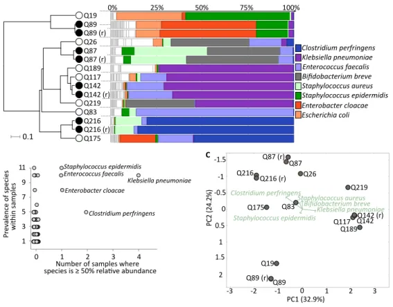
Figure 1 Healthy premature infant gut microbiome.(A) Metagenomic profiles for the eleven preterm samples and four replicates at the species level. Samples clustered by UPGMA using Bray–Curtis distances shown on left, with replicates highlighted by filled nodes. Relative abundances by rank order shown on right, with the top 8 most abundant species coloured and labelled, leaving remaining species in white. (B) Dominant species, based on≥50% abundance, shown onx-axis, with overall prevalence of the species across samples shown ony-axis. Sample number reflects eleven neonates as replicates are averaged. The five labelled species are present in five or more samples with at least one in >50% abundance. (C) Prin-cipal coordinates analysis (PCoA) using Bray–Curtis distances at the species level for all fifteen samples. Separation of the three broad sample groups shown by biplot of the top five species.
Moving to taxonomic composition, each sample was marked by a few highly abundant species, such as sample Q216 with 85.1% Clostridium perfringens, Q189 with 73.1%
Klebsiella pneumoniae, and Q83 with 85.9%Enterococcus faecalis(Fig. 1A). In terms of prevalence, the previous three species, as well asEnterobacter cloacaeand Staphylococcus
epidermidis, were found at over 50% relative abundance in one or more samples.
Furthermore, S.epidermidisandS. aureuswere ubiquitous, ranging from 0.06% to 57.1% abundance in all samples (Fig. 1B). Principal coordinate analysis (PCoA) demonstrated three loose sample groups based on a high abundance ofS. epidermidis,K. pneumoniae, and eitherB. breve,S. aureusorC. perfringens(Fig. 1C). In total we identified a non-redundant set of 172 species across all samples (seeTable S4for complete dataset).
Prevalence of antimicrobial resistance
Depth of coverage (bp)
3 200 400
A P H-St ph A ac3 -I k A ac6 -A ph2 A ac6 -I c A ac6 -I i A adC A n t6 -I a A C T-M IR A M P H_E coli A mpC1 _E coli A mpC2 _E coli A mpH B laZ M e cA O XY P B P _E coli SHV -OK P -LEN F
osB OqxA OqxB
g
b
nor
A
LnuA LnuB LsaA MphC MphD Msr
A M sr C M sr D V gaA
Dha1 FloR Tet-3
8 Te tA -P Te tB -P Te tO Te tW Dfr C DfrSample Q87 Q26 Q175 Q89 Q83 Q219 Q216 Q142 Q189 Q117 Q19 1437
AGly Bla Fos Flq MLS Phe Tet Tmt
Figure 2 Antibiotic resistance genes detected.Heatmap showing distribution of the 39 AMR genes detected within the eleven metagenomic samples. Genes grouped by antibiotic class are demarcated by black lines under gene names: AGly (aminoglycosides), Bla (β-lactam), Fos (Fosfomycin), Flq (fluoro-quinolones), MLS (macrolide-lincosamide- streptogramin), Phe (phenicols), Tet (tetracyclines), Tmt (trimethoprim). Colours show read depth in bp: undetected (grey), 3–199 bp (blue), 200–399 bp (yellow), 400–1,437 (red). Rows clustered by UPGMA method using Euclidean distances.
the known antibiotic exposure of the preterm infants (Fig. S2). Eight of the eleven infants had received a course of prophylactic antibiotic treatment consisting of co-amoxiclav (Table S1), whilst a second course was administered to four infants, consisting of combinations of co-amoxiclav, tazocin or vancomycin. In total, exposure ranged from 2 to 8 days of antibiotics before samples were taken, excluding infants Q87 and Q89 which received no antibiotics. Antibiotics were also administered maternally to three infants (Q26, Q117 & Q189), but this did not include the two above infants with no antibiotic treatment. Sample diversity ranged from 0.9 to 2.9 (SD±0.5), but when compared to cumulative antibiotic exposure expressed in days, no significant difference was found between the taxonomic diversity and amount of antibiotic exposure for untreated and treated infants (unpairedt-test,P=0.17) (Fig. S2). However, it is important to stress that the small and heterogeneous nature of the sample set will have reduced the power to detect differences between antibiotic exposure in this study, and so prevented any meaningful stratification by other clinical variables such as mode of delivery or day of life.
A mapping based approach against a comprehensive collection of acquired antibiotic resistance genes was next used to quantify AMR within the eleven metagenomes (Inouye
et al., 2014). In total 143 AMR genes were identified, consisting of a non-redundant set
Table 2 Antibiotic classes identified.Major antibiotic resistant classes of genes identified within the eleven samples by SRST2. Columns show antibiotic type and number of genes found within class.
Antibiotic type Number of identified genes within class
β-lactam (Bla) 10
Macrolide-lincosamide- streptogramin (MLS) 9
Aminoglycosides (AGly) 7
Tetracyclines (Tet) 5
Fluoroquinolones (Flq) 3
Phenicols (Phe) 2
Trimethoprim (Tmt) 2
Fosfomycin (Fcyn) 1
vancomycin resistance, the latter of which was administered to three preterm infants prior to sample collection (Table S1). The class most frequently detected wereβ-lactamases, comprising ten different genes (Table 2), of which theblaZ gene was present in every infant. Interestingly, within this set ofβ-lactamase genes,mecAwas found in four infants (Q87, Q117, Q175, and Q189), and at a mean depth of coverage ranging from 3.9 to 52.2 bp (Fig. 2).mecAconfers resistance to methicillin as well as otherβ-lactam antibiotics, and is carried on the SCCmec mobile element found across severalStaphylococcispecies. Identification of four infants with potential methicillin resistant S. aureus(MRSA) orS. epidermidis(MRSE) carriage, along with high abundances and prevalence of bothS. aureus
andS. epidermidisspecies across the dataset, could indicate a significant reservoir for AMR transfer between the species, as well as highlight the seeding of the infant gut microbiome from an early stage.
Focus on S. aureusspecies detected
Next, we wanted to understand the relationship of theS. aureusspecies within themecA
positive as well as negative samples, as the premature infants overlapped in time and so could harbour closely related strains. This was undertaken to firstly confirmin silico
prediction of mecAusing an established molecular based typing method, but also to push the metagenomic analysis further on what was known to be a challenging dataset owing to the range of identified S. aureusas described above, with relative abundance ranging from 0.06% to 39.8% (Table S4). We first tested the computational prediction of
mecAexperimentally using a multiplexed PCR typing method (Milheiri¸co, Oliveira & De
Lencastre, 2007), which provides detection of themecAgene, in addition to typing of the
In an attempt to understand strain relatedness directly from the metagenomic data, we undertookin silicoMLST analysis using anS. aureusschema as well as metagenome assembly. The MLST was able to classify four of the eleven samples, all with different ST types—ST8, ST1027, ST22, ST25, although the last two had some degree of uncertainty in their assignment (Table S8). This suggests that for at least these four samples, theS. aureus
strains are unrelated and unlikely a result of transmission. We were interested to know ifde novoassembly of the metagenome could be utilised to resolve these and any of the remaining unclassifiable samples further. Following assembly and identification ofS. aureus
contigs (see ‘Materials & Methods’), we found that it was not possible to capture more than a fifth of the expected genome size for the above unclassified samples, with an abundance of >3% necessary to achieve over 90% estimated capture, which was achieved in four cases (Table S9). Phylogenetic reconstruction of these four genomes alongside a collection of published S. aureusgenomes (Table S10), provided confirmation of the diversity ofS. aureusidentified (Fig. S3), enabling placement across a global collection of strains.
DISCUSSION
It is recognised that one of the most important public health threats worldwide is antimicrobial resistance. Here we report on the gut composition and AMR diversity for eleven healthy but premature infants. Recent studies have shown that the initial seeding of the infant gut microbiome is influenced by the microorganisms in the immediate environment, and whilst colonisation by bacteria with AMR genes has been demonstrated (Brooks et al., 2014), comparatively far fewer studies have investigated the gut microbiome of infants, fewer still preterm healthy infants. Interest has also increased on how the trajectory of the early gut microbiome is influenced to form the ‘stable’ adult microbiome. The preterm infant gut microbiome is very different compared to full-term infants (Groer et al., 2014), displaying a much lower diversity, particularly in anaerobes, with an increase in coagulase-negativeStaphylococciandEnterobacteriaceae(Adlerberth & Wold, 2009); adult microbiomes are characterised by several hundred, mostly anaerobic bacterial species (Adlerberth & Wold, 2009). We found a similarly low level of species diversity across all metagenomes, with each sample dominated by a few highly abundant species, including
C. perfringens,K. pneumoniaeand members of theStaphylococciandEnterobacter genera. Presence of such species are in common with previous studies on the premature gut microbiome (Groer et al., 2014;Gibson et al., 2016).
Merker et al., 2015). It could be that at this very early stage, the microbiota is influenced to a greater extent by seeding during birth from the mother and environment than antibiotic treatment, or that not enough time has passed to detect differences from the antibiotics administered; larger sample numbers would be required, alongside longitudinal studies and parallel maternal sampling to better understand the development of diversity.
A threat to this development is the acquisition of antibiotic resistant bacteria, which can potentially seed the infant microbiome. Coupled with the high rate of horizontal gene transfer within the commensal community (Stecher et al., 2012), the preterm infant gut microbiome has the potential to be a reservoir for AMR. With dominance of the preterm gut by species known to carry clinically relevant antibiotic resistance, we next quantified the burden of antibiotic resistance genes within the infant’s faecal flora, which identified an average of 13 genes per infant. Previous targeted or functional studies based on infants have found some of the AMR genes also identified here, including those for Tetracycline (tet) (Gueimonde, Salminen & Isolauri, 2006;Alicea-Serrano et al., 2013) and β-lactam (bla) (Fouhy et al., 2014). In a wider context, it is known that AMR genes are a common feature of bacterial populations, found in communities inhabiting the soil, rivers and even deep-sea sediment (Knapp et al., 2010;Qin et al., 2011;Kittinger et al., 2016). Therefore, whilst their presence in the human gut microbiome should be of little surprise (Bailey et al., 2010), identification of genes such asmecAdemonstrates the prevalence of some clinically significant resistant bacteria from birth.
One of the advantages of the method used in this study is the utility of the results generated, enabling multiple avenues of questions to be addressed. However, short read sequencing remains a challenge when applied to the linkage of resistance elements, such
mecA, to specific genome sequences (strains), which is made difficult by the nature of metagenomic samples containing multiple alleles from different closely related species, as well as potentially multiple strains of the same species. Secondly, the methods used here were inherently restricted to identification of known AMR genes found within the ARGannot database used in this study, which contains those genes involved in acquired resistance only, therefore chromosomal mutations, such as those conferring resistance to rifampicin as well as novel resistance genes would have been missed, leading to potential underrepresentation of resistance in this study.
CONCLUSIONS
questions such as how this resistance potential contributes to later clinical intervention or disease onset, and if antibiotic treatment without knowledge of prior AMR burden could lead to unintentional harm. More broadly, this and other studies show the great promise that shotgun metagenomics holds for clinical microbiology.
Abbreviations
AMR antimicrobial resistance
GI gastrointestinal
PCR polymerase chain reaction
qPCR quantitative polymerase chain reaction BLAST basic local alignment search tool
SCCmec staphylococcal chromosome cassettemec
LCA lowest common ancestor
MLST multilocus sequence typing
MALDI-TOF matrix-assisted laser desorption/ionization –time of flight PCoA principal coordinates analysis.
ADDITIONAL INFORMATION AND DECLARATIONS
Funding
This study was supported by a programme grant (to J.Simon Kroll) from The Winnicott Foundation and generous additional funding from Micropathology Ltd., Meningitis Now and the National Institute for Health Research (NIHR) Biomedical Research Centre based at Imperial Healthcare NHS Trust and Imperial College London. The funders had no role in study design, data collection and analysis, decision to publish, or preparation of the manuscript.
Grant Disclosures
The following grant information was disclosed by the authors: The Winnicott Foundation.
Micropathology Ltd.
National Institute for Health Research (NIHR) Biomedical Research Centre. Imperial Healthcare NHS Trust and Imperial College London.
Competing Interests
The authors declare there are no competing interests.
Author Contributions
• Graham Rose conceived and designed the experiments, analyzed the data, contributed reagents/materials/analysis tools, wrote the paper, prepared figures and/or tables, reviewed drafts of the paper.
• Alexander G. Shaw conceived and designed the experiments, performed the experiments, analyzed the data, wrote the paper, reviewed drafts of the paper.
• Kathleen Sim conceived and designed the experiments, performed the experiments,
• David J. Wooldridge and Ming-Shi Li performed the experiments, contributed
reagents/materials/analysis tools, reviewed drafts of the paper. • Saheer Gharbia reviewed drafts of the paper.
• Raju Misra and John Simon Kroll conceived and designed the experiments, reviewed drafts of the paper.
Human Ethics
The following information was supplied relating to ethical approvals (i.e., approving body and any reference numbers):
The study was approved by West London Research Ethics Committee (REC) Two, United Kingdom, under the REC approval reference number 10/H0711/39. Parents gave written informed consent for their infant to participate in the study.
DNA Deposition
The following information was supplied regarding the deposition of DNA sequences: Database: EBI European Nucleotide Archive.
Accession number:PRJEB15257.
Data Availability
The following information was supplied regarding data availability: The raw data has been supplied as aSupplementary File.
Supplemental Information
Supplemental information for this article can be found online athttp://dx.doi.org/10.7717/ peerj.2928#supplemental-information.
REFERENCES
Adlerberth I, Wold AE. 2009.Establishment of the gut microbiota in Western infants.Acta Paediatrica, International Journal of Paediatrics98(2):229–238 DOI 10.1111/j.1651-2227.2008.01060.x.
Alicea-Serrano AM, Contreras M, Magris M, Hidalgo G, Dominguez-Bello MG. 2013.Tetracycline resistance genes acquired at birth.Archives of Microbiology 195(6):447–451DOI 10.1007/s00203-012-0864-4.
Andrews S. 2010.FastQC: a quality control tool for high throughput sequence data.
Available athttp:// www.bioinformatics.babraham.ac.uk/ projects/ fastqc. Arboleya S, Sanchez B, Milani C, Duranti S, Solis G, Fernandez N, De los
Reyes-Gavilan CG, Ventura M, Margolles A, Gueimonde M. 2015.Intestinal microbiota development in preterm neonates and effect of perinatal antibiotics.Journal of Pediatrics166(3):538–544DOI 10.1016/j.jpeds.2014.09.041.
Bailey JK, Pinyon JL, Anantham S, Hall RM. 2010.CommensalEscherichia coliof
Bartoloni A, Pallecchi L, Rodríguez H, Fernandez C, Mantella A, Bartalesi F, Strohmeyer M, Kristiansson C, Gotuzzo E, Paradisi F, Rossolini GM. 2009. Antibiotic resistance in a very remote Amazonas community.International Journal of Antimicrobial Agents33(2):125–129DOI 10.1016/j.ijantimicag.2008.07.029. Beaber JW, Hochhut B, Waldor MK. 2004.SOS response promotes horizontal
dissemi-nation of antibiotic resistance genes.Nature427:72–74DOI 10.1038/nature02241. Beasley SS, Saris P.EJ. 2004.Nisin-producing lactococcus lactis strains isolated
from human milk.Applied and Environmental Microbiology 70(8):5051–5053 DOI 10.1128/AEM.70.8.5051-5053.2004.
Bolger AM, Lohse M, Usadel B. 2014.Trimmomatic: a flexible trimmer for Illumina se-quence data.Bioinformatics30(15):2114–2120DOI 10.1093/bioinformatics/btu170. Brooks B, Firek BA, Miller CS, Sharon I, Thomas BC, Baker R, Morowitz MJ, Banfield JF. 2014.Microbes in the neonatal intensive care unit resemble those found in the gut of premature infants.Microbiome2:Article 1DOI 10.1186/2049-2618-2-1. Buchfink B, Xie C, Huson DH. 2014.Fast and sensitive protein alignment using
DIAMOND.Nature Methods12:59–60DOI 10.1038/nmeth.3176.
De Vries LE, Valles Y, Agerso Y, Vaishampayan PA, Garcia-Montaner A, Kuehl JV, Christensen H, Barlow M, Francino MP. 2011.The gut as reservoir of antibiotic resistance: Microbial diversity of tetracycline resistance in mother and infant.PLOS ONE6(6):e21644DOI 10.1371/journal.pone.0021644.
Felsenstein J. 1989.PHYLIP—phylogeny inference package (version 3.2).Cladistics 5:164–166DOI 10.1111/j.1096-0031.1989.tb00562.x.
Forslund K, Sunagawa S, Coelho LP, Bork P. 2014.Metagenomic insights into the human gut resistome and the forces that shape it.BioEssays36(3):316–329 DOI 10.1002/bies.201300143.
Fouhy F, Ogilvie LA, Jones BV, Ross RP, Ryan AC, Dempsey EM, Fitzgerald GF, Stanton C, Cotter PD. 2014.Identification of aminoglycoside andβ-lactam resistance genes from within an infant gut functional metagenomic library.PLOS ONE9:e108016DOI 10.1371/journal.pone.0108016.
Gibson MK, Wang B, Ahmadi S, Burnham C-AD, Tarr PI, Warner BB, Dantas G. 2016.Developmental dynamics of the preterm infant gut microbiota and antibiotic resistome.Nature Microbiology1: Article 16024DOI 10.1038/nmicrobiol.2016.24. Gosalbes MJ, Vallès Y, Jiménez-Hernández N, Balle C, Riva P, Miravet-Verde S,
De Vries LE, Llop S, Agersø Y, Sørensen SJ, Ballester F, Francino MP. 2016. High frequencies of antibiotic resistance genes in infants’ meconium and early fecal samples.Journal of Developmental Origins of Health and Disease7:35–44 DOI 10.1017/S2040174415001506.
Greenwood C, Morrow AL, Lagomarcino AJ, Altaye M, Taft DH, Yu Z, Newburg DS, Ward DV, Schibler KR. 2014.Early empiric antibiotic use in preterm infants is asso-ciated with lower bacterial diversity and higher relative abundance of enterobacter.
Groer MW, Luciano AA, Dishaw LJ, Ashmeade TL, Miller E, Gilbert JA. 2014. Develop-ment of the preterm infant gut microbiome: a research priority.Microbiome2:Article 38DOI 10.1186/2049-2618-2-38.
Gueimonde M, Salminen S, Isolauri E. 2006.Presence of specific antibiotic (tet) resis-tance genes in infant faecal microbiota.FEMS Immunology and Medical Microbiology 48:21–25DOI 10.1111/j.1574-695X.2006.00112.x.
Gupta SK, Padmanabhan BR, Diene SM, Lopez-Rojas R, Kempf M, Landraud L, Rolain J-M. 2014.ARG-ANNOT, a new bioinformatic tool to discover antibiotic resistance genes in bacterial genomes.Antimicrobial Agents and Chemotherapy 58(1):212–220 DOI 10.1128/AAC.01310-13.
Haubold B, Klötzl F, Pfaffelhuber P. 2015.Andi: fast and accurate estimation of evo-lutionary distances between closely related genomes.Bioinformatics31:1169–1175 DOI 10.1093/bioinformatics/btu815.
Huson DH, Auch AF, Qi J, Schuster SC. 2007.MEGAN analysis of metagenomic data.
Genome Research17:377–386DOI 10.1101/gr.5969107.
Inouye M, Dashnow H, Raven L-A, Schultz MB, Pope BJ, Tomita T, Zobel J, Holt KE. 2014.SRST2: rapid genomic surveillance for public health and hospital microbiology labs.Genome Medicine6:Article 90DOI 10.1186/s13073-014-0090-6.
Jernberg C, Lofmark S, Edlund C, Jansson JK. 2007.Long-term ecological impacts of antibiotic administration on the human intestinal microbiota.ISME Journal 1:56–66DOI 10.1038/ismej.2007.3.
Jimenez E, Delgado S, Fernandez L, Garcia N, Albujar M, Gomez A, Rodriguez JM. 2008.Assessment of the bacterial diversity of human colostrum and screening of staphylococcal and enterococcal populations for potential virulence factors.Research in Microbiology 159:595–601DOI 10.1016/j.resmic.2008.09.001.
Karami N, Martner A, Enne VI, Swerkersson S, Adlerberth I, Wold AE. 2007.Transfer of an ampicillin resistance gene between twoEscherichia colistrains in the bowel mi-crobiota of an infant treated with antibiotics.Journal of Antimicrobial Chemotherapy 60(5):1142–1145DOI 10.1093/jac/dkm327.
Kittinger C, Lipp M, Baumert R, Folli B, Koraimann G, Toplitsch D, Liebmann A, Grisold AJ, Farnleitner AH, Kirschner A, Zarfel G. 2016.Antibiotic resistance pat-terns of pseudomonas spp. Isolated from the River Danube.Frontiers in Microbiology 7:1–8DOI 10.3389/fmicb.2016.00586.
Knapp CW, Dolfing J, Ehlert P.AI, Graham DW. 2010.Evidence of increasing antibiotic resistance gene abundances in archived soils since 1940.Environmental Science and Technology44(2):580–587DOI 10.1021/es901221x.
Langmead B, Salzberg SL. 2012.Fast gapped-read alignment with Bowtie 2.Nature Methods9:357–359DOI 10.1038/nmeth.1923.
Magoč T, Salzberg SL. 2011.FLASH: fast length adjustment of short reads to improve genome assemblies.Bioinformatics27(21):2957–2963
DOI 10.1093/bioinformatics/btr507.
bifidobacterial population by PCR-denaturing gradient gel electrophoresis and quantitative real-time PCR.Applied and Environmental Microbiology75:965–969 DOI 10.1128/AEM.02063-08.
Merker M, Blin C, Mona S, Duforet-Frebourg N, Lecher S, Willery E, Blum M.GB, Rüsch-Gerdes S, Mokrousov I, Aleksic E, Allix-Béguec C, Antierens A, Augustynowicz-Kopeć E, Ballif M, Barletta F, Beck HP, Barry CE, Bonnet M, Borroni E, Campos-Herrero I, Cirillo D, Cox H, Crowe S, Crudu V, Diel R, Drobniewski F, Fauville-Dufaux M, Gagneux S, Ghebremichael S, Hanekom M, Hoffner S, Jiao W, Kalon S. 2015.Evolutionary history and global spread of the Mycobacterium tuberculosis Beijing lineage.Nature Genetics47:242–249 DOI 10.1038/ng.3195.
Milheiri¸co C, Oliveira DC, De Lencastre H. 2007.Update to the multiplex PCR strategy for assignment ofmecelement types inStaphylococcus aureus.Antimicrobial Agents
and Chemotherapy 51:3374–3377DOI 10.1128/AAC.00275-07.
Moore AM, Ahmadi S, Patel S, Gibson MK, Wang B, Ndao MI, Deych E, Shannon W, Tarr PI, Warner BB, Dantas G. 2015.Gut resistome development in healthy twin pairs in the first year of life.Microbiome3:Article 27 DOI 10.1186/s40168-015-0090-9. Penders J, Stobberingh EE, Savelkoul P.HM, Wolffs P.FG. 2013.The human
micro-biome as a reservoir of antimicrobial resistance.Frontiers in Microbiology4:Article 87DOI 10.3389/fmicb.2013.00087.
Qin Q-L, Li Y, Zhang Y-J, Zhou Z-M, Zhang W-X, Chen X-L, Zhang X-Y, Zhou B-C, Wang L, Zhang Y-Z. 2011.Comparative genomics reveals a deep-sea sediment-adapted life style of Pseudoalteromonas sp. SM9913.The ISME Journal5:274–284 DOI 10.1038/ismej.2010.103.
R Developement Core Team. 2015.R: a language and environment for statistical computing. R Foundation for Statistical Computing.Available athttp:// www.R-project.org/.
Rose G, Wooldridge DJ, Anscombe C, Mee ET, Misra RV, Gharbia S. 2015.Challenges of the unknown: clinical application of microbial metagenomics.International Journal of Genomics2015:1–10DOI 10.1155/2015/292950.
Shoemaker NB, Vlamakis H, Hayes K, Salyers AA. 2001.Evidence for extensive resistance gene transfer among bacteroides spp. and among bacteroides and other genera in the human colon evidence for extensive resistance gene transfer among bacteroides spp. and among bacteroides and other genera in the human C.Applied and Environmental Microbiology 67:561–8DOI 10.1128/AEM.67.2.561.
Stecher B, Denzler R, Maier L, Bernet F, Sanders MJ, Pickard DJ, Barthel M, Wes-tendorf AM, Krogfelt KA, Walker AW, Ackermann M, Dobrindt U, Thomson NR, Hardt W-D. 2012.Gut inflammation can boost horizontal gene transfer between pathogenic and commensalEnterobacteriaceae.Proceedings of the Na-tional Academy of Sciences of the United States of America109(4):1269–1274 DOI 10.1073/pnas.1113246109.
early postnatal period on the development of intestinal microbiota.FEMS Immunol-ogy and Medical MicrobiolImmunol-ogy 56:80–87DOI 10.1111/j.1574-695X.2009.00553.x. Trobos M, Lester CH, Olsen JE, Frimodt-Møller N, Hammerum AM. 2009.Natural transfer of sulphonamide and ampicillin resistance betweenEscherichia coli re-siding in the human intestine.Journal of Antimicrobial Chemotherapy63:80–86 DOI 10.1093/jac/dkn437.
Truong DT, Franzosa EA, Tickle TL, Scholz M, Weingart G, Pasolli E, Tett A, Hut-tenhower C, Segata N. 2015.MetaPhlAn2 for enhanced metagenomic taxonomic profiling.Nature Methods12:902–903DOI 10.1038/nmeth.3589.
Van Hoek AHAM, Mevius D, Guerra B, Mullany P, Roberts AP, Aarts HJM. 2011. Acquired antibiotic resistance genes: an overview.Frontiers in Microbiology2:Article 203DOI 10.3389/fmicb.2011.00203.
Von Wintersdorff CJ, Wolffs PF, Savelkoul PH, Nijsen RR, Lau S, Gerhold K, Hamel-mann E, Penders J. 2016.The gut resistome is highly dynamic during the first months of life.Future Microbiology11(4):501–510DOI 10.2217/fmb.15.154. Walson JL, Marshall B, Pokhrel BM, Kafle KK, Levy SB. 2001.Carriage of
antibiotic-resistant fecal bacteria in Nepal reflects proximity to Kathmandu.The Journal of Infectious Diseases184:1163–1169DOI 10.1086/323647.
Zhang L, Kinkelaar D, Huang Y, Li Y, Li X, Wang HH. 2011.Acquired antibi-otic resistance: are we born with it?.Applied and Environmental Microbiology 77(20):7134–7141